HAL Id: inserm-00380032
https://www.hal.inserm.fr/inserm-00380032
Submitted on 5 May 2009HAL is a multi-disciplinary open access archive for the deposit and dissemination of sci-entific research documents, whether they are pub-lished or not. The documents may come from teaching and research institutions in France or abroad, or from public or private research centers.
L’archive ouverte pluridisciplinaire HAL, est destinée au dépôt et à la diffusion de documents scientifiques de niveau recherche, publiés ou non, émanant des établissements d’enseignement et de recherche français ou étrangers, des laboratoires publics ou privés.
Sharon Eastwood, Louisa Lyon, Lydia George, Annie Andrieux, Didier Job,
Paul Harrison
To cite this version:
Sharon Eastwood, Louisa Lyon, Lydia George, Annie Andrieux, Didier Job, et al.. Altered expression of synaptic protein mRNAs in STOP (MAP6) mutant mice.. Journal of Psychopharmacology, SAGE Publications, 2007, 21 (6), pp.635-44. �10.1177/0269881106068825�. �inserm-00380032�
Altered Expression of Synaptic Protein mRNAs in STOP (MAP6)
Mutant Mice
Sharon L. Eastwooda, Louisa Lyona, Lydia Georgea, Annie Andrieuxb, Didier Jobb and
Paul J. Harrisona
a
University of Oxford, Department of Psychiatry, Neurosciences Building, Warneford
Hospital, Oxford OX3 7JX, U.K. b
INSERM U366, Laboratoire du Cytosquelette, CEA-Grenoble, 38054 Grenoble Cedex 9
France.
Corresponding author:
Sharon L. Eastwood, Neurosciences Building, University Department of Psychiatry, Warneford Hospital, Oxford OX3 7JX, United Kingdom.
Telephone 01865 223620 Fax 01865 251076
Abstract
Stable tubule-only polypeptide (STOP) proteins are a family of microtubule associated proteins (MAPs) important in microtubule stabilization. Data indicating a role for microtubules in synaptic function has come from studies of the STOP null mouse, which exhibits synaptic deficits, in association with behavioural changes that are alleviated by antipsychotic treatment. These findings suggested that STOP mutant mice may be useful in studies of synaptic function, and could be especially relevant to schizophrenia, postulated to be a disorder of the synapse. Moreover, a genetic association between STOP and schizophrenia has been reported. This study aimed to further characterise synaptic alterations in STOP null and heterozygous mice. Using in situ hybridization histochemistry, the mRNA expression of three pre-synaptic (synaptophysin; growth associated protein-43: GAP-43; vesicular glutamate transporter-1: VGlut1) and two post-synaptic (spinophilin; MAP2) proteins, was quantified in female STOP null (n=7), heterozygous (n=5) and wild type (n=6) mice. For STOP null and heterozygous mice, synaptophysin, VGlut1, GAP-43 and spinophilin mRNAs were decreased in the hippocampus, whilst in addition in the null mice, synaptophysin, VGlut1 and spinophilin mRNAs were decreased in the cerebellum. Alterations in synaptic protein mRNA expression were also detected in the frontal and occipital cortex. MAP2 mRNA expression was unchanged in all brain regions. The profile of mRNA changes is broadly similar to that observed in schizophrenia. Together the data provide supporting evidence for a role for microtubules in synaptic function, and suggest that STOP, or other microtubule proteins, may contribute to the synaptic pathology of schizophrenia.
Key words: in situ hybridization histochemistry; MAP6; microtubules; mRNA; schizophrenia; synapse.
Introduction
STOP (stable tubule-only polypeptide) proteins are a family of calmodulin binding and regulated microtubule associated proteins (MAPs), encoded by a single gene (mouse: Mtap6, Denarier et al., 1998b; human: MAP6; Bosc et al., 2003), which play a role in microtubule stabilization in several cell types (Denarier et al., 1998a; Aguezzoul et al., 2003). The major STOP isoforms, N-STOP and E-STOP, are expressed by neurons (Bosc et al., 1999; Guillaud et al., 1998), whereas fibroblasts express F-STOP (Denarier et al., 1998a) and astrocytes and oligodendrocytes express A-STOP and O-STOP respectively (Galiano et al., 2004). In neurons, microtubule stabilization has been demonstrated to be important in neuronal migration (Schaar and McConnell, 2005; Tsai and Gleeson, 2005), morphology and function (Baas and Heidermann, 1986; Guillaud et al., 1998). Hence, like other MAPs (Mack et al., 2000; Takei et al., 2000; Feng and Walsh, 2001; Moores et al., 2004), STOP proteins are thought to play a role in normal brain development and synaptic connectivity.
To evaluate the role of STOP proteins, gene targeting was used to create the STOP null mouse (Andrieux et al., 2002). In association with impaired synaptic plasticity and decreased size of synaptic vesicle pools, STOP null mice exhibit behavioural changes, including disorganized activity, social withdrawal and nurturing defects, the latter of which were alleviated with long term typical antipsychotic drug treatment. These initial findings suggested that STOP null mice may be useful in studies of synaptic function. They could be of particular interest with respect to schizophrenia, which is proposed to be a disorder of the synapse (see Mirnics et al., 2001; Moises et al., 2002; Frankle et al., 2003) and in which the expression of several synaptic proteins is decreased (see Honer et al., 2000), and perhaps with regard to mood disorders, in which microtubule dysfunction has been hypothesised (Bianchi et al., 2005). Subsequent studies of STOP null mice have demonstrated that both sexes exhibit behavioural changes relevant to schizophrenia, including sensorimotor gating deficits
(Fradley et al., 2005) and hyper-locomotor activity (Brun et al., 2005; Fradley et al., 2005), which is reversed by antipsychotic drugs (Brun et al., 2005; Fradley et al., 2005). In addition, dopaminergic changes are evident in STOP null mice. Amphetamine exacerbates the hyper-locomotor activity (Brun et al., 2005), whilst evoked dopamine efflux is increased in the nucleus accumbens (Brun et al., 2005). Based in part upon post mortem findings of decreased hippocampal MAP2 protein in schizophrenia (Arnold et al., 1991), a role for microtubules in the pathophysiology of schizophrenia has been hypothesised (Kerwin, 1993). The finding of synaptic changes in STOP null mice (Andrieux et al., 2002) suggests that altered microtubule and synaptic function may be related, and contribute to the proposed pathogenic role of microtubules in schizophrenia. The aim of the current study was to further characterise synaptic alterations noted in the STOP null mouse in terms of the expression of pre- and post-synaptic protein mRNAs, markers of synapses and post-synaptic function (see Discussion and Honer et al., 2000; Honer and Young, 2003; Law et al., 2004b; Eastwood and Harrison, 2005), and to extend previous studies of STOP null mice by including heterozygous as well as wild type littermates. The pre-synaptic proteins, synaptophysin, growth-associated protein-43 (GAP-43) and vesicular glutamate transporter-1 (VGlut1), and the post-synaptic protein spinophilin, were quantified as their expression has been reported to be altered in post mortem studies of schizophrenia (see Table 1). In addition, MAP2 mRNA was examined, firstly as a dendritic marker (see Discussion), and also to determine if deficits in STOP expression are compensated for by the increased expression of another MAP. We examined synaptic protein mRNAs, not only in the hippocampal CA1 subfield where synaptic vesicle pools in the STOP mice have been quantified, but extended it to include other hippocampal subfields, and additional brain regions, including the prefrontal cortex and cerebellum, all implicated in the pathophysiology of schizophrenia (Katsetos et al., 1997; Eastwood et al., 2001; Weinberger et al., 2001; Harrison, 2004).
Methods and Materials
Animals
STOP mice were generated on a 50:50 BALBc/129 SvPas background as previously described (Andrieux et al., 2002), with gene targeting being used to replace exon 1 of the STOP gene with a non-functional construct. As the mRNAs of all of the STOP proteins characterised to date contain this exon (Denarier et al., 1998a, b), the expression of all STOP isoforms is suppressed in the null mice. Brains from 12 week old STOP null female mice (n=7), and their wild type (n=5) and heterozygote (n=6) littermates were snap frozen by immersion in isopentane chilled on dry ice, and stored at -80ºC until use.
In situ hybridization histochemistry
Frozen coronal sections (15 µm) were cut at the level of the dorsal hippocampus (approximately at Bregma -2.30 mm; Paxinos and Franklin, 2004), the caudate putamen (approximately at Bregma 0.86 mm), and cerebellum, and collected onto Superfrost Plus slides (VWR, Lutterworth, UK). Slides were pretreated for in situ hybridization histochemistry (ISHH) as described (Eastwood et al., 2000a), and stored at -20ºC. Oligonucleotide probes complementary to mouse STOP (bases 975-1009, Genbank accession number NM010837), synaptophysin (bases 846-884, NM009305), VGlut1 (bases 1562-1606, NM182993), GAP-43 (bases 599-628, NM008083), spinophilin (bases 1480-1519, AY508450), MAP2 (bases 698-739, BC052446) and glyceraldehyde-3-phosphate dehydrogenase (GAPDH; bases 201-239, BC095932) mRNAs were 3’ end labelled with [35S]dATP (Perkin Elmer, UK) using terminal deoxynucleotidyl transferase (Promega, Southampton, UK) and established protocols (Eastwood et al., 2001). Sections were incubated overnight (GAP-43, spinophilin, MAP2 and GAPDH: 33ºC; STOP: 35ºC; synaptophysin: 34ºC; VGlut1: 42ºC) in hybridization buffer (Eastwood et al., 2000a) containing 1 million
counts per minute labelled probe and dithiothreitol (STOP, spinophilin, MAP2: 50mM; synaptophysin, GAP-43 and GAPDH: 20mM). For all transcripts except VGlut1 and GAP-43, post-hybridization washes were carried out with 1 X standard sodium citrate (SSC) at either 55ºC (MAP2), 58ºC (spinophilin and GAPDH) or 60ºC (synaptophysin) for 3 X 20 minutes, followed by 2 X 60 minute washes at room temperature. For GAP-43, washes were carried out in 0.5 X SSC at 58ºC for 3 X 20 minutes followed by 2 X 60 minutes at room temperature. For VGlut1, post-incubation washes were as previously published (Miyazaki et al., 2003), and consisted of 2 X 40 minute washes at 55ºC in 0.1 X SSC. Triplicate sections at each anatomical level were run concurrently for each transcript, and the hybridized sections placed against Kodak Biomax MR film (GE Healthcare, Little Chalfont, UK) alongside 14C microscales (GE Healthcare) for the following times: GAP-43, 1 day; synaptophysin, 2 days; VGlut1, 3 days; GAPDH, 4 days; MAP2 and STOP, 7 days; spinophilin, 14 days. Negative controls consisted of incubation in the presence of 50 fold excess cold unlabelled probe and ISHH with sense orientation probes.
Image and statistical analysis
Autoradiographs were measured using an MCID Elite v7.0 image analysis system (Interfocus, Haverhill, UK). Optical density values obtained were calibrated to 35SnCi/g tissue equivalents using the 14C microscales and a conversion factor of 3, and corrected for non-specific background signal as represented by either sense strand hybridization or incubation with excess unlabelled probe.
Measurements were taken over the granule cell layer of the dentate gyrus (DG), the pyramidal layer of CA3 and CA1, and through the depth of the overlying occipital cortex for sections taken at the level of the dorsal hippocampus. For the sections taken at the level of the caudate putatmen, measurements were taken over the dorsolateral quadrant of the caudate
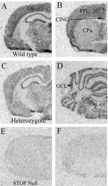
putamen and through the depth of the fronto-parietal and cingulate cortex. Finally, signal was measured in the cerebellum over the granule cell layer. These areas are illustrated in Figure 1 A, B and D.
All statistical analyses were performed using SPSS v 13 software. For each transcript, analysis of variance (ANOVA) was performed to determine if there was an overall effect of genotype or an area by genotype interaction. If either were significant (P<0.05), subsequent ANOVAs were conducted for each individual brain area, with significant differences between each genotype explored using least significant difference (LSD).
Results
STOP mRNA
In order to confirm the genotype of each animal and also examine the distribution of STOP mRNA in the mouse brain (which has not been previously described), the oligonucleotide probe used was designed against exon 1 of STOP mRNA, missing in the mutant mice. STOP mRNA was detectable in all areas examined in wild type mice, with a weaker signal observed in heterozygous mice, but was not detected in the nulls (Fig. 1). Of the areas examined here, STOP mRNA signal was highest over the hippocampus, moderate over the occipital, fronto-parietal and cingulate cortex and cerebellum, with the weakest signal observed over the caudate putamen.
Synaptophysin mRNA
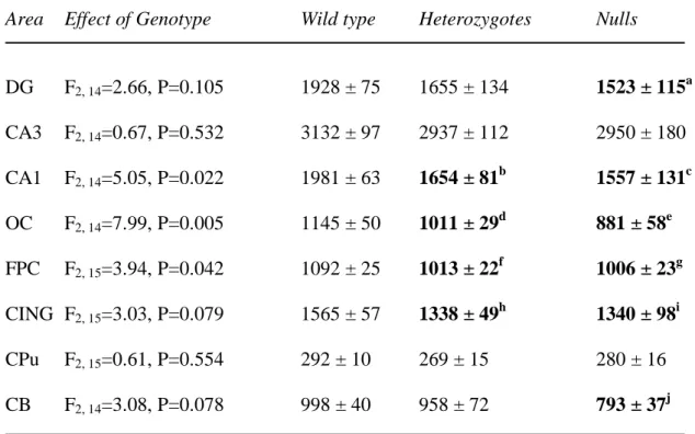
Synaptophysin mRNA distribution was as previously reported in rodents (Marquèze Pouey et al., 1991; Eastwood et al., 1997). In the overall ANOVA, a significant effect of genotype was detected (F2, 115=15.55, P<0.001), but not a genotype by area interaction (F14, 115=0.95, P=0.51). There were significant effects of genotype upon synaptophysin mRNA in
CA1, occipital and fronto-parietal cortex, and a trend towards significance in the cerebellum and cingulate cortex (Table 2). The significant effects of genotype were due to decreases in synaptophysin mRNA expression in STOP null and heterozygous mice as compared to the wild type mice (Table 2).
VGlut1 mRNA
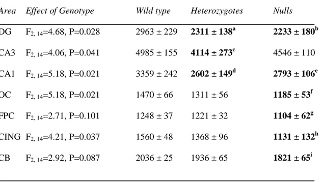
VGlut1 mRNA was robustly detected in every area examined except the caudate putamen, in accordance with previous reports (Miyazaki et al., 2003). Genotype (F2, 98=18.61,
P<0.001) had a significant effect upon VGlut1 mRNA in the overall ANOVA, and there was a significant genotype by area interaction (F12, 98=2.03, P=0.029). Individual ANOVAs
detected effects of genotype upon VGlut1 mRNA in the DG, CA3, CA1, occipital and cingulate cortex, with a trend towards significance in the cerebellum (Table 3). These effects of genotype were due to decreases in VGlut1 mRNA expression in STOP null and heterozygotes as compared to wild type mice.
GAP-43 mRNA
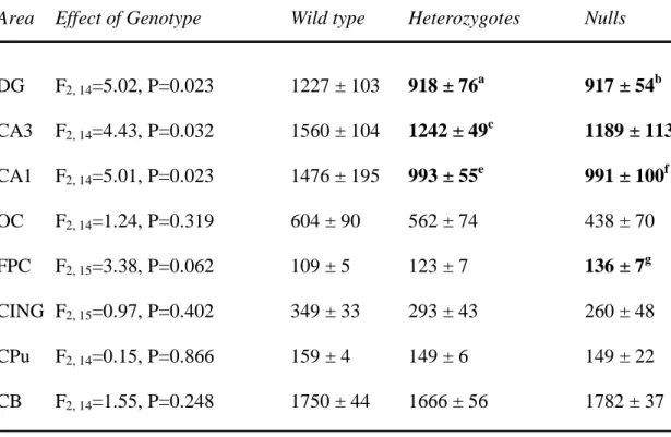
The distribution of GAP-43 mRNA was as previously described (Cantallops and Routtenberg, 1999). As seen in humans, but not in rat, GAP-43 mRNA was robustly detected in the DG (see Eastwood and Harrison, 1998). Genotype (F2, 115=14.30, P<0.001) had a
significant effect upon GAP-43 mRNA in the overall ANOVA, and there was a significant genotype by area interaction (F14, 115=2.48, P=0.004). Individual ANOVAs detected effects of
genotype upon GAP-43 mRNA in the DG, CA3 and CA1, with a trend towards significance for genotype upon GAP-43 mRNA in the fronto-parietal cortex (Table 4). For DG, CA3 and CA1, the effects of genotype were due to decreases in expression as compared to wild type
mice, whilst for the fronto-parietal cortex, GAP-43 mRNA was increased in the null mice as compared to wild type mice.
Spinophilin mRNA
In agreement with previous reports in rats (Law et al., 2004a) and humans (Law et al., 2004b), the distribution of spinophilin mRNA in mutant and wild type mice was consistent with a dendritic localization, with labelling observed not only over the pyramidal cell layer, but also adjacent strata of the DG and Ammon’s horn. Genotype (F2, 110=22.28, P<0.001) had
a significant effect upon spinophilin mRNA in the overall ANOVA, and there was a significant genotype by area interaction (F14, 110=4.03, P<0.001). Subsequent individual
ANOVAs found that genotype had a significant effect upon spinophilin mRNA in the DG, CA3, CA1 and cerebellum, with trends in the occipital and fronto-parietal cortex (Table 5). For the DG, CA3, CA1, occipital cortex and cerebellum, these were due to decreases in expression as compared to wild type mice, whilst for the fronto-parietal cortex, spinophilin mRNA was increased in the STOP null mice.
MAP2 mRNA
As reported above for spinophilin mRNA, and as observed in rats and humans (Tucker et al., 1989; Law et al., 2004a, b), MAP2 mRNA distribution in the STOP mutant and wild type mice is consistent with a dendritic localization. For MAP2 mRNA in the overall ANOVA, neither a significant effect of genotype (F2, 110=2.15, P=0.122), nor a genotype by
GAPDH mRNA
ISHH against the house keeping gene GAPDH was carried out, as an index of overall gene expression. No significant effects of genotype (F2, 102=2.51, P=0.09) nor genotype by
area interactions (F14, 102=0.90, P=0.56) were detected (Table 7).
Discussion
The results of this study demonstrate that the expression of pre- and post-synaptic protein mRNAs is altered in STOP mutant mice. We will focus our discussion on how deficits in STOP and stabilized microtubules may lead to synaptic alterations. Similarities (and differences) between changes in synaptic protein expression exhibited by STOP null and heterozygous mice to those reported in schizophrenia will be discussed. Finally, we will comment upon the role which STOP or other cytoskeletal proteins may play in the origins of synaptic pathology in schizophrenia, including mediation of the potential influence of susceptibility genes.
Synaptic protein mRNA expression as markers of synaptic pathology and their altered
expression in STOP null and heterozygous mice
The synaptic protein mRNAs examined in this study were chosen because they inform on different aspects of synapses, and on the basis of reports of their altered expression in post mortem studies of schizophrenia (see Table 1). The utilization of synaptic proteins as markers of synapses, and their mRNAs as markers of synaptic changes in the regions to which neurons project, has been reviewed elsewhere in depth (see Harrison and Eastwood, 2001; Honer et al., 2000; Honer and Young, 2003; Eastwood and Harrison, 2005) and will only be briefly mentioned here. Of the three pre-synaptic proteins studied, synaptophysin has been the mostly widely utilised, with alterations in its expression often being interpreted as indicative of
changes in overall synaptic density (or size), thereby denoting synaptic pathology (see Masliah et al., 1990; Eastwood et al., 1994). To provide an indication of any preferential involvement of excitatory or inhibitory neurons, pre-synaptic proteins selectively expressed by subpopulations of neurons have been used (see Harrison and Eastwood, 1998; Eastwood and Harrison, 2005). Of these, the vesicular glutamate transporter, VGlut1, which loads synaptic vesicles with glutamate, is exclusive to glutamatergic terminals (Bellochio et al., 1998, 2000; Fremau et al., 2001). The last pre-synaptic protein examined in this study, GAP-43, is a phosphoprotein involved in neurodevelopment, injury response and synaptic plasticity (see Benowitz and Routtenberg, 1997; Eastwood, 2003). Post-synaptic proteins have also been utilised as synaptic markers (see Law et al., 2004b). MAP2 is a general dendritic marker (Pollard et al., 1994), whilst spinophilin is specifically involved in dendritic spine formation and function (Feng et al., 2000). As most cortical glutamatergic synapses terminate on spines, altered spinophilin expression in the absence of any change of the expression of MAP2 is interpreted to indicate particular structural or functional involvement of spines and glutamatergic synapses.
Table 8 summarizes the differences in pre- and post-synaptic protein mRNA expression detected in STOP mutant mice as compared to their wild type littermates. Note that synaptic protein mRNA expression was decreased in both STOP null and heterozygous mice, whilst the mRNA for the housekeeping gene GAPDH was unchanged, indicating that altered synaptic protein mRNA expression is unlikely to be due to a generalized decrease in gene expression. As outlined above, the pattern of change in synaptic protein mRNA is indicative of synaptic alterations in STOP mutant mice. The finding that spinophilin (but not MAP2) and VGlut1 mRNAs are altered suggests that glutamatergic synapses are involved, though as markers of inhibitory synapses were not examined, it remains to be determined whether GABAergic synapses may be similarly affected.
As schizophrenia is considered a disorder of aberrant neurodevelopment, it will be interesting to determine when the synaptic changes noted in these adult STOP mutant mice first occur. Furthermore, as all behavioural studies conducted to date have focussed on the null mouse, it will also be interesting to determine whether heterozygous STOP mice also exhibit altered sensorimotor gating and locomotor activity (Fradley et al., 2005). If they do, the use of heterozygous mice in place of nulls would overcome practical limitations of generating sufficient numbers of mice (see Fradley et al., 2005). Finally, as the present study only examined females, examination of males will be necessary to establish whether there are sex differences in the molecular profile of STOP mutant mice.
How may lack of STOP lead to synaptic deficits?
Decreases of cold-stable microtubules exhibited by STOP null and heterozygous mice (Andrieux et al., 2002) are likely to influence neurodevelopmental processes, and ongoing adult synaptic plasticity, by perturbing microtubule dynamics and/or their interactions with molecular motors. Growth cone turning (Tanaka and Kirschner, 1995; Williamson et al., 1996), axon pathfinding (Suter and Forscher, 2000; Schaefer et al., 2002), and axon branching (Dent et al., 2004; Kalil and Dent, 2005), are all dependent on interactions between microfilaments and microtubules, and changes in the dynamics of microtubules could potentially lead to abnormalities in the formation and maintenance of synaptic connections. Our data demonstrating that STOP null and heterozygous mice exhibit changes in the expression of pre- and post-synaptic proteins provides some molecular evidence in support of this. How such altered connectivity may occur will be discussed.
Microtubules function as rails along which kinesin superfamily proteins (KIFs) and dyneins act as molecular motors to transport intracellular cargoes such as mRNAs, protein complexes and organelles (Hirokawa and Takemura, 2004). Two KIFs, KIF1A and KIF1B,
transport precursors of synaptic vesicles to axon terminals (Okada et al., 1995; Yonekawa et al., 1998; Zhao et al., 2001). Of particular relevance for the current study, knock out mice for KIF1A and KIF1B have reduced numbers of synaptic vesicles, indicating that our finding of decreased synaptophysin mRNA expression, and reductions in the number of synaptic vesicles in STOP mutant mice (Andrieux et al., 2002), may be caused by altered KIF function and diminished transport (and thence synthesis) of synaptic vesicles. The finding that treatment of STOP null mice with the microtubule stabilizing drug epothilone D partially returns to normal synaptic vesicle density (Andrieux et al., 2006) supports a role for microtubule stabilization in synaptic vesicle transport, and it will be interesting to determine if synaptophysin mRNA expression is likewise increased
Given the importance of microtubules and their associated proteins in the formation and maintenance of dendrites (Liu et al., 2000; Yu et al., 2000; Scott and Luo, 2001; Jan and Jan, 2003), it might be predicted that deficits in cold stabilized microtubules in STOP mutant mice would lead to dendritic abnormalities. However, our data demonstrating decrements in spinophilin mRNA in the absence of alterations in that for MAP2, and the normal dendritic arborization observed in STOP null mice (Andrieux et al., 2002) suggest that changes in microtubule dynamics may instead preferentially affect spines. The presence of STOP (Andrieux et al., 2002) and microtubules (see van Rossum and Hanisch, 1999) in dendritic spines has been documented and stabilized microtubules are important in the transport and function of glutamate receptors, both during synaptogenesis and in adult synaptic plasiticity (Sergé et al., 2003; Washbourne et al., 2002, 2004; Yuen et al., 2005). Of note, the translocation of the RNA binding protein TLS (translocated in liposarcoma) into dendritic spines is dependent on stabilized microtubules, and hippocampal pyramidal neurons from TLS null mice exhibit decreased spine density and abnormal spine morphology (Fujii et al., 2005). Together these data indicate that changes in microtubule dynamics in STOP deficient
mice may contribute to altered spine formation and/or function, and may underlie our findings of decreased spinophilin mRNA in STOP null and heterozygous mice.
Are the present findings relevant to the understanding of schizophrenia?
The profile of change in expression of synaptic protein mRNAs seen here is quite similar to that observed in schizophrenia. For example, as in the hippocampus of STOP null and heterozygous mice (Table 8), post mortem studies mostly agree that, with the exception of MAP2 (see Table 1), synaptic protein mRNAs are reduced in the disorder. Although fewer studies have examined the cerebellum or occipital cortex in schizophrenia, synaptophysin mRNA is reportedly decreased in these areas too, consistent with the reduction seen in the STOP null mice. Concordance in findings between the STOP mutant mice and schizophrenia for the other brain areas examined here is difficult to determine, either because there are no comparable schizophrenia data (e.g. in cingulate cortex or caudate putamen) or because the rodent homologue of human dorsolateral prefrontal cortex is unclear.
The above similarities, together with the previously described behavioural phenotype of the STOP mutant mice (Brun et al., 2005; Fradley et al., 2005), suggests that STOP is a candidate to contribute to the role which microtubules have been proposed to play in the pathophysiology of schizophrenia (Kerwin, 1993). Indeed, this candidacy recently received empirical support with the report of genetic association between STOP and schizophrenia (Shimizu et al., 2006). On the other hand, Shimizu et al. (2006) also found that one isoform of STOP mRNA was increased (and the other unchanged) in the prefrontal cortex in schizophrenia, arguing against a role for reduced STOP expression in the disorder. The situation remains unclear, however, since we have found STOP mRNA to be decreased in the hippocampus in schizophrenia (S.L.E. and P.J.H., unpublished observations). Thus, it remains to be determined whether STOP expression is altered in schizophrenia and, if so, whether this
is regionally specific. Clearly, the face validity of STOP null or heterozygous mice as a ‘model’ of schizophrenia is affected by this issue. Equally, even if STOP expression is not found to be consistently reduced in schizophrenia, it does not negate the potential value of these mice, or the present findings, with regard to the disorder. Firstly, since STOP expression may have been reduced (and played its pathogenic role) in subjects with schizophrenia earlier in life, and subsequently normalised, perhaps due to antipsychotic medication. Secondly, the main significance of the present data is that they emphasise that alterations in synaptic protein gene expression, and synaptic functioning, can arise from “non-synaptic” mechanisms involving microtubules. As such, synaptic pathology in schizophrenia may have its origin elsewhere in the neuron (or even in non-neuronal cells). This is particularly relevant to our understanding of how susceptibility genes for schizophrenia may converge to alter synaptic function, given that most do not encode established structural components of the synapse or proteins known to be part of the synaptic proteome (see Grant et al., 2005; Harrison and Weinberger, 2005). Of note, one of the leading susceptibility genes, DISC1 (Disrupted in Schizophrenia 1), forms a complex with several proteins including NUDEL (nuclear distribution element-like) and Lis1 (lissencephaly gene 1 product), which binds to microtubules and is involved in neuronal migration and dynein-mediated motor transport (see Brandon et al., 2004; Kamiya et al., 2005.). The importance of this protein complex in axonal transport and dendritic morphogenesis has been demonstrated in Lis1 null Drosophila (Liu et al., 2000), whilst depletion of DISC1 or expression of a mutated form (mutDISC1) results in aberrant neuronal migration in vivo and impaired neurite outgrowth in vitro (Kamiya et al., 2005). Interestingly, although DISC1 mRNA expression is not altered in schizophrenia, there are decrements in NUDEL and Lis1 mRNAs, associated with the DISC1 risk single nucleotide polymorphisms (Lipska et al., 2006), suggesting that DISC1 may exert its pathogenic effect in schizophrenia by impacting on the expression of its binding partners and
thereby alter microtubule dynamics and function. One can speculate on a comparable role for STOP and its interacting proteins in schizophrenia.
Conclusions
The results of the current study complement data implicating microtubules in synaptic formation, maintenance and function, and give a precedent for altered microtubule dynamics producing changes in the molecular composition as well as functioning of synapses. Although serendipitously discovered, the STOP mutant mouse may be useful for studying aspects of the genetic pathophysiology of schizophrenia, especially its synaptic pathology and the roles that microtubules play in this process.
Acknowledgements
Work supported by the Stanley Medical Research Institute and the Medical Research Council. We thank Mary Walker and Annie Schweitzer for technical assistance.
References
Aguezzoul M, Andrieux A, Denarier E (2003) Overlap of promoter and coding sequences in the mouse STOP gene (Mtap6). Genomics 81: 623-627
Andrieux A, Salin PA, Vernet M, Kujala P, Baratier J, Gory Faure S, Bosc C, Pointu H, Proietto D, Schweitzer A, Denarier E, Klumperman J, Job D (2002) The suppression of brain cold-stable microtubules in mice induces synaptic defects associated with neuroleptic-sensitive behavioral disorders. Genes Dev 16: 2350-2364
Andrieux A, Salin P, Schweitzer A, Bégou M, Pachoud B, Brun P, Gory-Fauré S, Kujala P, Suaud-Chagny M, Höfle G, Job D (2006) Microtubule stabilizer ameliorates synaptic function and behaviour in a mouse model for schizophrenia. Biol Psychiatry. In press
Arnold SE, Lee VM, Gur RE, Trojanowski JQ (1991) Abnormal expression of two microtubule-associated proteins (MAP2 and MAP5) in specific subfields of the hippocampal formation in schizophrenia. Proc Natl Acad Sci USA 88: 10850-10854
Baas PW, Heidemann SR (1986) Microtubule reassembly from nucleating fragments during the regrowth of amputated neurites. J Cell Biol 103: 917-927
Bellochio EE, Hu H, Pohorille A, Chan J, Pickel VM, Edwards RH (1998) The localization of the brain-specific inorganic phosphate transporter suggests a presynaptic role in glutamatergic neurotransmission. J Neurosci 18: 638-8659
Bellochio EE, Reimer RJ, Fremeau RT Jr, Edwards RH (2000) Uptake of glutamate into synaptic vesicles by an inorganic phosphate transporter. Science 289: 957-960
Benowitz LI, Routtenberg A (1997) GAP-43: an intrinsic determinant of neuronal development and plasticity. Trends Neurosci. 20: 84-91
Bianchi M, Hagan JJ, Heidbreder CA (2005) Neuronal plasticity, stress and depression: involvement of the cytoskeletal microtubular system? Curr Drug Targets-CNS Neurol Disord 4: 597-611.
Bosc C, Oenarier E, Andrieux A, Job D (1999) STOP proteins. Cell Struct Funct 24: 393-399
Bosc C, Andrieux A, Job D (2003) STOP proteins. Biochemistry 42: 12125-12132
Brandon NJ, Handford EJ, Schurov I, Rain JC, Pelling M, Duran Jimeniz B, Camargo LM, Oliver KR, Beher D, Shearman MS, Whiting PJ (2004) Disrupted in Schizophrenia 1 and Nudel form a neurodevelopmentally regulated protein complex: implications for schizophrenia and other major neurological disorders. Mol Cell Neurosci 25: 42-55
Brun P, Begou M, Andrieux A, Mouly Badina L, Clerget M, Schweitzer A, Scarna H, Renaud B, Job D, Suaud Chagny MF (2005) Dopaminergic transmission in STOP null mice. J Neurochem 94: 63-73
Cantallops I, Routtenberg A (1999) Activity-dependent regulation of axonal growth: posttranscriptional control of the GAP-43 gene by the NMDA receptor in developing hippocampus. J Neurobiol 41: 208-220
Denarier E, Fourest Lieuvin A, Bosc C, Pirollet F, Chapel A, Margolis RL, Job D (1998a) Nonneuronal isoforms of STOP protein are responsible for microtubule cold stability in mammalian fibroblasts. Proc Natl Acad Sci USA 95: 6055-6060
Denarier E, Aguezzoul M, Jolly C, Vourc'h C, Roure A, Andrieux A, Bosc C, Job D (1998b) Genomic structure and chromosomal mapping of the mouse STOP gene (Mtap6). Biochem Biophys Res Commun 243: 791-796
Dent EW, Barnes AM, Tang F, Kalil K (2004) Netrin-1 and semaphorin 3A promote or inhibit cortical axon branching, respectively, by reorganization of the cytoskeleton. J Neurosci 24: 3002-3012
Eastwood, SL (2003) The synaptic pathology of schizophrenia: Is aberrant neurodevelopment and plasticity to blame? In: Smythies, J (ed), Disorders of Synaptic Plasticity and Schizophrenia. International Reviews of Neurobiology, volume 59. Elsevier, Amsterdam, pp 47-72.
Eastwood SL, Burnet PW, McDonald B, Clinton J, Harrison PJ (1994) Synaptophysin gene expression in human brain: a quantitative in situ hybridization and immunocytochemical study. Neuroscience 59: 881-892
Eastwood SL, Burnet PW, Harrison PJ (1995) Altered synaptophysin expression as a marker of synaptic pathology in schizophrenia. Neuroscience 66: 309-319
Eastwood SL, Heffernan J, Harrison PJ (1997) Chronic haloperidol treatment differentially affects the expression of synaptic and neuronal plasticity-associated genes. Mol Psychiatry 2: 322-329
Eastwood, SL, Burnet, PWJ, Harrison, PJ (2000a) Expression of complexin I and II mRNAs and their regulation by antipsychotic drugs in the rat forebrain. Synapse 36:167-177.
Eastwood SL, Cairns NJ, Harrison PJ (2000b) Synaptophysin gene expression in schizophrenia. Investigation of synaptic pathology in the cerebral cortex. Br J Psychiatry 176: 236-242
Eastwood SL, Cotter D, Harrison PJ (2001) Cerebellar synaptic protein expression in schizophrenia. Neuroscience 105: 219-229
Eastwood SL, Harrison PJ (1998) Hippocampal and cortical growth-associated protein-43 messenger RNA in schizophrenia. Neuroscience 86: 437-448
Eastwood SL, Harrison PJ (1999) Detection and quantification of hippocampal synaptophysin messenger RNA in schizophrenia using autoclaved, formalin-fixed, paraffin wax-embedded sections. Neuroscience 93: 99-106
Eastwood SL, Harrison PJ (2005) Decreased expression of vesicular glutamate transporter 1 and complexin II mRNAs in schizophrenia: further evidence for a synaptic pathology affecting glutamate neurons. Schizophr Res 73: 159-172
Feng J, Yan Z, Ferreira A, Tomizawa K, Liauw JA, Zhuo M, Allen PB, Ouimet CC, Greengard P (2000) Spinophilin regulates the formation and function of dendritic spines. Proc Natl Acad Sci USA 97: 9287-9292
Feng Y, Walsh CA (2001) Protein-protein interactions, cytoskeletal regulation and neuronal migration. Nat Rev Neurosci 2: 408-416
Fradley RL, O'Meara GF, Newman RJ, Andrieux A, Job D, Reynolds DS (2005) STOP knockout and NMDA NR1 hypomorphic mice exhibit deficits in sensorimotor gating. Behav Brain Res 163: 257-264
Frankle WG, Lerma J, Laruelle M (2003) The synaptic hypothesis of schizophrenia. Neuron 39: 205-216
Fremeau RT Jr, Troyer MD, Pahner I, Nygaard GO, Tran CH, Reimer RJ, Bellochio EE, Fortin D, Storm-Mathisen J, Edwards RH (2001) The expression of vesicular glutamate transporters defines two classes of excitatory synapse. Neuron 31: 247-260
Fujii R, Okabe S, Urushido T, Inoue K, Yoshimura A, Tachibana T, Nishikawa T, Hicks GG, Takumi T (2005) The RNA binding protein TLS is translocated to dendritic spines by mGluR5 activation and regulates spine morphology. Curr Biol 15: 587-593
Galiano MR, Bosc C, Schweitzer A, Andrieux A, Job D, Hallak ME (2004) Astrocytes and oligodendrocytes express different STOP protein isoforms. J Neurosci Res 78: 329-337
Glantz LA, Austin MC, Lewis DA (2000) Normal cellular levels of synaptophysin mRNA expression in the prefrontal cortex of subjects with schizophrenia. Biol Psychiatry 48: 389-397
Grant SGN, Marshall MC, Page K-L, Cumiskey MA, Armstrong JD (2005) Synapse proteomics of multiprotein complexes: en route from genes to nerous system diseases. Hum Mol Genet 14: R225-234
Guillaud L, Bosc C, Fourest Lieuvin A, Denarier E, Pirollet F, Lafanechere L, Job D (1998) STOP proteins are responsible for the high degree of microtubule stabilization observed in neuronal cells. J Cell Biol 142: 167-179
Harrison PJ, Eastwood SL (1998) Preferential involvement of excitatory neurons in medial temporal lobe in schizophrenia. Lancet 352: 1669-1673
Harrison PJ, Eastwood SL (2001) Neuropathological studies of synaptic connectivity in the hippocampal formation in schizophrenia. Hippocampus 11: 508-519
Harrison PJ (2004) The hippocampus in schizophrenia: a review of the neuropathological evidence and its pathophysiological implications. Psychopharmacology 174: 151-162
Harrison PJ, Weinberger DR (2005) Schizophrenia genes, gene expression, and neuropathology: on the matter of their convergence. Mol Psychiatry 10: 420
Hirokawa N, Takemura R (2004) Kinesin superfamily proteins and their various functions and dynamics. Exp Cell Res 301: 50-59
Honer WG, Young CE (2003) Presynaptic proteins and schizophrenia. Int Rev Neurobiol 59: 175-199
Honer WG, Young C. Falkai P (2000). Synaptic pathology, in: Harrison PJ, Roberts GW (eds.), The Neuropathology of Schizophrenia. Progress and Interpretation. Oxford University Press, Oxford, pp. 105-136.
Kalil K, Dent EW (2005) Touch and go: guidance cues signal to the growth cone cytoskeleton. Curr Opin Neurobiol 15: 521-526
Kamiya A, Kubo K, Tomoda T, Takaki M, Youn R, Ozeki Y, Sawamura N, Park U, Kudo C, Okawa M, Ross CA, Hatten ME, Nakajima K, Sawa A (2005) A schizophrenia-associated mutation of DISC1 perturbs cerebral cortex development. Nat Cell Biol 7: 1067-1078
Karson CN, Mrak RE, Schluterman KO, Sturner WQ, Sheng JG, Griffin WS (1999) Alterations in synaptic proteins and their encoding mRNAs in prefrontal cortex in schizophrenia: a possible neurochemical basis for 'hypofrontality'. Mol Psychiatry 4: 39-45
Katsetos CD, Hyde TM, Herman MM (1997) Neuropathology of the cerebellum in schizophrenia--an update: 1996 and future directions. Biol Psychiatry 42: 213-224
Kerwin RW (1993) Glutamate receptors, microtubule associated proteins and developmental anomaly in schizophrenia: an hypothesis. Psychol Med 23: 547-551
Law AJ, Hutchinson LJ, Burnet PW, Harrison PJ (2004a) Antipsychotics increase microtubule-associated protein 2 mRNA but not spinophilin mRNA in rat hippocampus and cortex. J Neurosci Res 76: 376-382
Law AJ, Weickert CS, Hyde TM, Kleinman JE, Harrison PJ (2004b) Reduced spinophilin but not microtubule-associated protein 2 expression in the hippocampal formation in schizophrenia and mood disorders: molecular evidence for a pathology of dendritic spines. Am J Psychiatry 161: 1848-1855
Lipska BK, Peters T, Hyde TM, Halim N, Horowitz C, Mitkus S, Weickert CS, Matsumoto M, Sawa A, Straub RE, Vakkalanka R, Herman MM, Weinberger DR, Kleinman JE (2006) Expression of DISC1 binding partners is reduced in schizophrenia and associated with DISC1 SNPs. Hum Mol Genet 15: 1245-1258
Liu Z, Steward R, Luo L (2000) Drosophila Lis1 is required for neuroblast proliferation, dendritic elaboration and axonal transport. Nat Cell Biol 2: 776-783
Mack TG, Koester MP, Pollerberg GE (2000) The microtubule-associated protein MAP1B is involved in local stabilization of turning growth cones. Mol Cell Neurosci 15: 51-65
Marquèze Pouey B, Wisden W, Malosio ML, Betz H (1991) Differential expression of synaptophysin and synaptoporin mRNAs in the postnatal rat central nervous system. J Neurosci 11: 3388-3397
Masliah E, Terry RD, Alford M, DeTeresa R (1990) Quantitative immunohistochemistry of synaptophysin in human neocortex: an alternative method to estimate density of presynaptic terminals in paraffin sections. J Histochem Cytochem 38: 837-844
Mirnics K, Middleton FA, Lewis DA, Levitt P (2001) Analysis of complex brain disorders with gene expression microarrays: schizophrenia as a disease of the synapse. Trends Neurosci 24: 479-486
Miyazaki T, Fukaya M, Shimizu H, Watanabe M (2003) Subtype switching of vesicular glutamate transporters at parallel fibre-Purkinje cell synapses in developing mouse cerebellum. Eur J Neurosci 17: 2563-2572
Moises HW, Zoega T, Gottesman II (2002) The glial growth factors deficiency and synaptic destabilization hypothesis of schizophrenia. BMC Psychiatry 2: 8
Moores CA, Perderiset M, Francis F, Chelly J, Houdusse A, Milligan RA (2004) Mechanism of microtubule stabilization by doublecortin. Mol Cell 14: 833-839
Okada Y, Yamazaki H, Sekine Aizawa Y, Hirokawa N (1995) The neuron-specific kinesin superfamily protein KIF1A is a unique monomeric motor for anterograde axonal transport of synaptic vesicle precursors. Cell 81: 769-780
Paxinos G, Franklin KBJ (2004) The mouse brain in stereotaxic coordinates. Academic Press, San Diego, CA
Pollard H, Khrestchatisky M, Moreau J, Ben Ari Y, Represa A (1994) Correlation between reactive sprouting and microtubule protein expression in epileptic hippocampus. Neuroscience 61: 773-787
Schaar BT, McConnell SK (2005) Cytoskeletal coordination during neuronal migration. Proc Natl Acad Sci USA 102: 13652-13657
Schaefer AW, Kabir N, Forscher P (2002) Filopodia and actin arcs guide the assembly and transport of two populations of microtubules with unique dynamic parameters in neuronal growth cones. J Cell Biol 158: 139-152
Scott EK, Luo L (2001) How do dendrites take their shape? Nat Neurosci 4: 359-365
Sergé A, Fourgeaud L, Hemar A, Choquet D (2003) Active surface transport of metabotropic glutamate receptors through binding to microtubules and actin flow. J Cell Sci 116: 5015-5022
Shimizu H, Iwayama Y, Yamada K, Toyota T, Minabe Y, Nakamura K, Nakajima M, Hattori E, Mori N, Osumi N, Yoshikawa T (2006) Genetic and expression analyses of the STOP (MAP6) gene in schizophrenia. Schizophr Res 84: 244-252
Suter DM, Forscher P (2000) Substrate-cytoskeletal coupling as a mechanism for the regulation of growth cone motility and guidance. J Neurobiol 44: 97-113
Takei Y, Teng J, Harada A, Hirokawa N (2000) Defects in axonal elongation and neuronal migration in mice with disrupted tau and map1b genes. J Cell Biol 150: 989-1000
Tanaka E, Kirschner MW (1995) The role of microtubules in growth cone turning at substrate boundaries. J Cell Biol 128: 127-137
Tsai LH, Gleeson JG (2005) Nucleokinesis in neuronal migration. Neuron 46: 383-388
Tucker RP, Garner CC, Matus A (1989) In situ localization of microtubule-associated protein mRNA in the developing and adult rat brain. Neuron 2: 1245-1256
van Rossum D, Hanisch UK (1999) Cytoskeletal dynamics in dendritic spines: direct modulation by glutamate receptors? Trends Neurosci 22: 290-295
Washbourne P, Bennett JE, McAllister AK (2002) Rapid recruitment of NMDA receptor transport packets to nascent synapses. Nat Neurosci 5: 751-759
Washbourne P, Liu XB, Jones EG, McAllister AK (2004) Cycling of NMDA receptors during trafficking in neurons before synapse formation. J Neurosci 24: 8253-8264
Webster MJ, Shannon Weickert C, Herman MM, Hyde TM, Kleinman JE (2001) Synaptophysin and GAP-43 mRNA levels in the hippocampus of subjects with schizophrenia. Schizophr Res 49: 89-98
Weickert CS, Webster MJ, Hyde TM, Herman MM, Bachus SE, Bali G, Weinberger DR, Kleinman JE (2001) Reduced GAP-43 mRNA in dorsolateral prefrontal cortex of patients with schizophrenia. Cereb Cortex 11: 136-147
Weickert CS, Straub RE, McClintock BW, Matsumoto M, Hashimoto R, Hyde TM, Herman MM, Weinberger DR, Kleinman JE (2004) Human dysbindin (DTNBP1) gene expression in normal brain and in schizophrenic prefrontal cortex and midbrain. Arch Gen Psychiatry 61: 544-555
Weinberger DR, Egan MF, Bertolino A, Callicott JH, Mattay VS, Lipska BK, Berman KF, Goldberg TE (2001) Prefrontal neurons and the genetics of schizophrenia. Biol Psychiatry 50: 825-844
Williamson T, Gordon Weeks PR, Schachner M, Taylor J (1996) Microtubule reorganization is obligatory for growth cone turning. Proc Natl Acad Sci USA 93: 15221-15226
Yonekawa Y, Harada A, Okada Y, Funakoshi T, Kanai Y, Takei Y, Terada S, Noda T, Hirokawa N (1998) Defect in synaptic vesicle precursor transport and neuronal cell death in KIF1A motor protein-deficient mice. J Cell Biol 141: 431-441
Yu W, Cook C, Sauter C, Kuriyama R, Kaplan PL, Baas PW (2000) Depletion of a microtubule-associated motor protein induces the loss of dendritic identity. J Neurosci 20: 5782-5791
Yuen EY, Jiang Q, Feng J, Yan Z (2005) Microtubule regulation of N-methyl-D-aspartate receptor channels in neurons. J Biol Chem 280: 29420-29427
Zhao C, Takita J, Tanaka Y, Setou M, Nakagawa T, Takeda S, Yang HW, Terada S, Nakata T, Takei Y, Saito M, Tsuji S, Hayashi Y, Hirokawa N (2001) Charcot-Marie-Tooth disease type 2A caused by mutation in a microtubule motor KIF1Bbeta. Cell 105: 587-597
Figure 1. Regional distribution of STOP mRNA. Representative autoradiographic images showing the distribution of STOP mRNA at the level of the dorsal hippocampus in wild type (A), heterozygous (C) and STOP null (E) mice, and at the level of the caudate putamen (B) and cerebellum (D) of wild type mice. F: Incubation with excess cold unlabelled probe, showing minimal background signal. CA: cornu Ammonis; CING: cingulate cortex; CPu: caudate putamen; DG: dentate gyrus; GCL: granule cell layer; FPC: fronto-parietal cortex; OC: occipital cortex.
schizophrenia.
Brain Region mRNA Main Result Study
Hippocampus Synaptophysin ↓ Eastwood et al., 1995.
Eastwood and Harrison, 1999. Webster et al., 2001.
VGlut1 ↓ Eastwood and Harrison, 2005.
GAP-43 ↓ Eastwood and Harrison, 1998. ↔ Webster et al., 2001.
Spinophilin ↓ Law et al., 2004a.
MAP-2 ↔ Law et al., 2004a.
DLPFC Synaptophysin ↔ Karson et al., 1999.
Eastwood et al., 2000. Glantz et al., 2000. Weickert et al., 2004.
VGlut1 ↓ Eastwood and Harrison, 2005.
GAP-43 ↔ Eastwood and Harrison, 1998. ↓ Weickert et al., 2001.
Spinophilin ↔ Weickert et al., 2004.
Occiptal Cortex Synaptophysin ↓ Eastwood et al., 2000b.
GAP-43 ↓ Eastwood and Harrison, 1998.
Cerebellum Synaptophysin ↓ Eastwood et al., 2001.
Summary is limited to published papers in brain regions relevant to those examined in mice in the current study. DLPFC: dorsolateral prefrontal cortex.
Table 2. Synaptophysin mRNA expression in wild type, heterozygous and STOP null mice.
Area Effect of Genotype Wild type Heterozygotes Nulls
DG F2, 14=2.66, P=0.105 1928 ± 75 1655 ± 134 1523 ± 115a CA3 F2, 14=0.67, P=0.532 3132 ± 97 2937 ± 112 2950 ± 180 CA1 F2, 14=5.05, P=0.022 1981 ± 63 1654 ± 81b 1557 ± 131c OC F2, 14=7.99, P=0.005 1145 ± 50 1011 ± 29d 881 ± 58e FPC F2, 15=3.94, P=0.042 1092 ± 25 1013 ± 22f 1006 ± 23g CING F2, 15=3.03, P=0.079 1565 ± 57 1338 ± 49h 1340 ± 98i CPu F2, 15=0.61, P=0.554 292 ± 10 269 ± 15 280 ± 16 CB F2, 14=3.08, P=0.078 998 ± 40 958 ± 72 793 ± 37j
Values are mean 35SnCi/g tissue equivalents ± SEM. a P=0.041, b P=0.025, c P=0.010, d P=0.045, e P=0.001, f P=0.029, g P=0.022, h P=0.042, i P=0.050, j P=0.037, as compared to wild type. No significant differences were found between heterozygous and null mice. CA: cornu Ammonis; CB: cerebellum; CING: cingulate cortex; CPu: caudate putamen; DG: dentate gyrus; FPC: fronto-parietal cortex; OC: occipital cortex.
Table 3. VGlut1 mRNA expression in wild type, heterozygous and STOP null mice.
Area Effect of Genotype Wild type Heterozygotes Nulls
DG F2, 14=4.68, P=0.028 2963 ± 229 2311 ± 138a 2233 ± 180b CA3 F2, 14=4.06, P=0.041 4985 ± 155 4114 ± 273c 4546 ± 110 CA1 F2, 14=5.18, P=0.021 3359 ± 242 2602 ± 149d 2793 ± 106e OC F2, 14=5.18, P=0.021 1470 ± 66 1311 ± 56 1185 ± 53f FPC F2, 14=2.71, P=0.101 1248 ± 37 1221 ± 32 1104 ± 62g CING F2, 14=4.21, P=0.037 1560 ± 48 1368 ± 96 1131 ± 132h CB F2, 14=2.92, P=0.087 2036 ± 25 1936 ± 65 1821 ± 65i
Values are mean 35SnCi/g tissue equivalents ± SEM . a P=0.020, b P=0.016, c P=0.013, d P=0.007, e P=0.046, f P=0.006, g P=0.050, h P=0.012, i P=0.030, as compared to wild types. No significant differences were found between heterozygous and null mice. Abbreviations as in Table 2.
Table 4. GAP-43 mRNA expression in wild type, heterozygous and STOP null mice.
Area Effect of Genotype Wild type Heterozygotes Nulls
DG F2, 14=5.02, P=0.023 1227 ± 103 918 ± 76a 917 ± 54b CA3 F2, 14=4.43, P=0.032 1560 ± 104 1242 ± 49c 1189 ± 113d CA1 F2, 14=5.01, P=0.023 1476 ± 195 993 ± 55e 991 ± 100f OC F2, 14=1.24, P=0.319 604 ± 90 562 ± 74 438 ± 70 FPC F2, 15=3.38, P=0.062 109 ± 5 123 ± 7 136 ± 7g CING F2, 15=0.97, P=0.402 349 ± 33 293 ± 43 260 ± 48 CPu F2, 14=0.15, P=0.866 159 ± 4 149 ± 6 149 ± 22 CB F2, 14=1.55, P=0.248 1750 ± 44 1666 ± 56 1782 ± 37
Values are mean 35SnCi/g tissue equivalents ± SEM. a P=0.015, b P=0.015, c P=0.031, d P=0.015, e P=0.015, f P=0.014, g P=0.020, compared to wild types. No significant differences were found between heterozygous and null mice. Abbreviations as in Table 2.
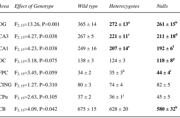
Table 5. Spinophilin mRNA expression in wild type, heterozygous and STOP null mice.
Area Effect of Genotype Wild type Heterozygotes Nulls
DG F2, 13=13.26, P=0.001 365 ± 14 272 ± 13a 261 ± 15b CA3 F2, 13=4.27, P=0.038 267 ± 5 221 ± 11c 211 ± 18d CA1 F2, 13=4.23, P=0.038 249 ± 16 207 ± 14e 192 ± 6f OC F2, 13=3.18, P=0.075 138 ± 3 124 ± 3 118 ± 8g FPC F2, 15=3.45, P=0.059 34 ± 2 35 ± 3h 44 ± 4i CING F2, 15=1.27, P=0.310 80 ± 3 74 ± 4 82 ± 5 CPu F2, 15=2.63, P=0.105 37 ± 2 36 ± 1j 45 ± 5 CB F2, 13=4.09, P=0.042 675 ± 15 628 ± 20 580 ± 32k
Values are mean 35SnCi/g tissue equivalents ± SEM. a P=0.001, b P<0.001, c P=0.033, d P=0.016, e P=0.043, f P=0.014, g P=0.027, i P=0.037, k P=0.013, as compared to wild types.
h
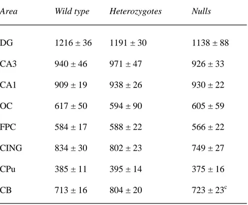
Table 6. MAP2 mRNA expression in wild type, heterozygous and STOP null mice.
Area Wild type Heterozygotes Nulls
DG 1216 ± 36 1191 ± 30 1138 ± 88 CA3 940 ± 46 971 ± 47 926 ± 33 CA1 909 ± 19 938 ± 26 930 ± 22 OC 617 ± 50 594 ± 90 605 ± 59 FPC 584 ± 17 588 ± 22 566 ± 22 CING 834 ± 30 802 ± 23 749 ± 27 CPu 385 ± 11 395 ± 14 375 ± 16 CB 713 ± 16 804 ± 20 723 ± 23c
Values are mean 35SnCi/g tissue equivalents ± SEM. No overall effect of genotype, or genotype by area interactions were detected. Abbreviations as in Table 2.
Area Wild type Heterozygotes Nulls DG 1105 ± 22 1169 ± 68 1157 ± 35 CA3 1793 ± 104 1871 ± 123 2102 ± 168 CA1 1350 ± 87 1440 ± 59 1562 ± 56 OC 677 ± 5 675 ± 5 670 ± 6 FPC 371 ± 24 353 ± 14 397 ± 27 CING 501 ± 40 470 ± 40 542 ± 44 CPu 248 ± 15 238 ± 17 281 ± 19 CB 863 ± 15 956 ± 102 847 ± 6.5
Values are mean 35SnCi/g tissue equivalents ± SEM . No overall effect of genotype, or genotype by area interactions were detected. Abbreviations as in Table 2.
Area STOP null mice Heterozygous mice
SYN VGlut1 GAP-43 SPINO SYN VGlut1 GAP-43 SPINO
DG ↓ ↓ ↓ ↓ ↔ ↓ ↓ ↓ CA3 ↔ ↔ ↓ ↓ ↔ ↓ ↓ ↓ CA1 ↓ ↓ ↓ ↓ ↓ ↓ ↓ ↓ OC ↓ ↓ ↔ ↓ ↓ ↔ ↔ ↔ FPC ↓ ↓ ↑ ↑ ↓ ↔ ↔ ↔ CING ↓ ↓ ↔ ↔ ↓ ↔ ↔ ↔ CPu ↔ ND ↔ ↔ ↔ ND ↔ ↔ CB ↓ ↓ ↔ ↓ ↔ ↔ ↔ ↔