HAL Id: hal-01668977
https://hal-amu.archives-ouvertes.fr/hal-01668977
Submitted on 6 Feb 2018
HAL is a multi-disciplinary open access
archive for the deposit and dissemination of
sci-entific research documents, whether they are
pub-lished or not. The documents may come from
teaching and research institutions in France or
abroad, or from public or private research centers.
L’archive ouverte pluridisciplinaire HAL, est
destinée au dépôt et à la diffusion de documents
scientifiques de niveau recherche, publiés ou non,
émanant des établissements d’enseignement et de
recherche français ou étrangers, des laboratoires
publics ou privés.
Lamin A and ZMPSTE24 (FACE-1) defects cause
nuclear disorganization and identify restrictive
dermopathy as a lethal neonatal laminopathy
Claire Navarro, Annachiara de Sandre-Giovannoli, Rafaelle Bernard, Irène
Boccaccio, Amandine Boyer, David Geneviève, Smail Hadj-Rabia, Caroline
Gaudy-Marqueste, Henk Smitt, Pierre Vabres, et al.
To cite this version:
Claire Navarro, Annachiara de Sandre-Giovannoli, Rafaelle Bernard, Irène Boccaccio, Amandine
Boyer, et al.. Lamin A and ZMPSTE24 (FACE-1) defects cause nuclear disorganization and
iden-tify restrictive dermopathy as a lethal neonatal laminopathy. Human Molecular Genetics, Oxford
University Press (OUP), 2004, 13 (20), pp.2493 - 2503. �10.1093/hmg/ddh265�. �hal-01668977�
Lamin A and ZMPSTE24 (FACE-1) defects cause
nuclear disorganization and identify restrictive
dermopathy as a lethal neonatal laminopathy
Claire L. Navarro
1,{, Annachiara De Sandre-Giovannoli
1,2,{, Rafae¨lle Bernard
1,2,{,
Ire`ne Boccaccio
1, Amandine Boyer
2, David Genevie`ve
4, Smail Hadj-Rabia
5,
Caroline Gaudy-Marqueste
2, Henk Sillevis Smitt
6, Pierre Vabres
8, Laurence Faivre
9,
Alain Verloes
11, Ton Van Essen
12, Elisabeth Flori
13, Raoul Hennekam
7, Frits A. Beemer
14,
Nicole Laurent
10, Martine Le Merrer
4, Pierre Cau
1,3and Nicolas Le´vy
1,2,*
1
Inserm U491, Ge´ne´tique Me´dicale et De´veloppement, Faculte´ de Me´decine de Marseille,2De´partement de Ge´ne´tique Me´dicale, Hoˆpital d’enfants de la Timone, Marseille, France,3Laboratoire de Biologie Cellulaire, Hoˆpital Conception, Institut Fe´de´ratif de Physiopathologie Humaine de Marseille, IFR 125, Marseille, France,4Inserm U393 and5Service de dermatologie pe´diatrique, Hoˆpital Necker enfants malades, Paris, France,6Department of
Dermatology and7Department of Pediatrics and Clinical Genetics, Academic Medical Center, Amsterdam, The Netherlands,8Service de Dermatologie, CHU Poitiers, France,9Centre de Ge´ne´tique and10Service d’Anatomie-Pathologique, CHU de Dijon, France,11Service de Ge´ne´tique, Hoˆpital Robert Debre´, Paris, France,12Department of Clinical Genetics, University Hospital, Groningen, The Netherlands,13Service de Cytoge´ne´tique, Hoˆpital de
Hautepierre, Strasbourg, France and14Clinical Genetics Center, University Medical Center, Utrecht, The Netherlands
DDBJ/EMBL/GenBank accession nos X03444.1, Y13834.
Restrictive dermopathy (RD), also called tight skin contracture syndrome (OMIM 275210), is a rare disorder mainly characterized by intrauterine growth retardation, tight and rigid skin with erosions, prominent superficial vasculature and epidermal hyperkeratosis, facial features (small mouth, small pinched nose and micrognathia), sparse/absent eyelashes and eyebrows, mineralization defects of the skull, thin dysplastic clavicles, pulmonary hypoplasia, multiple joint contractures and an early neonatal lethal course. Liveborn children usually die within the first week of life. The overall prevalence of consanguineous cases suggested an autosomal recessive inheritance. We explored nine fetuses/newborns children with RD. Two were found to have an heterozygous splicing mutation in the LMNA gene, leading to the complete or partial loss of exon 11 in mRNAs encoding Lamin A and resulting in a truncated Prelamin A protein. Lamins are major constituents of the nuclear lamina, a filamentous meshwork underlying the inner nuclear envelope. In the other seven patients, a unique heterozygous insertion leading to the creation of a premature termination codon was identified in the gene ZMPSTE24, also known as FACE-1 in human. This gene encodes a metalloproteinase specifically involved in the post-translational processing of Lamin A precursor. In all patients carrying a ZMPSTE24 mutation, loss of expression of Lamin A as well as abnormal patterns of nuclear sizes and shapes and mislocalization of Lamin-associated proteins was evidenced. Our results indicate that a common pathogenetic pathway, involving defects of the nuclear lamina and matrix, is involved in all RD cases. RD is thus one of the most deleterious laminopathies identified so far in humans caused by (primary or secondary) A-type Lamin defects and nuclear structural and functional alterations.
*To whom correspondence should be addressed at: Inserm U491: ‘Ge´n´etique Me´dicale et De´veloppement’, Faculte´ de M´edecine de Marseille, 13385 Marseille Cedex 05, France. Tel: þ33 491786894; Fax: þ33 491804319; Email: nicolas.levy@medecine.univ-mrs.fr
{
INTRODUCTION
Restrictive dermopathy (RD) is a lethal genodermatosis (MIM no. 275210) in which tautness of the skin causes fetal akinesia or hypokinesia deformation sequence (FADS). Polyhydramnios with reduced fetal movements is followed by premature delivery at about 31 weeks of gestation. It was first described by Witt et al. in 1986 (1). Other manifestations include a tightly adherent, thin, translucent skin with prominent vessels, typical facial changes, general-ized joint contractures, enlarged fontanels, dysplasia of clavi-cles, respiratory insufficiency and an enlarged placenta with short umbilical cord (2 – 4). Histological abnormalities of the skin include thin dermis with paucity and hypoplasia of the appendages and abnormally dense collagen bundles. Elastic fibres are nearly missing and the subcutaneous fat is slightly increased (5). These abnormalities usually appear after 22 – 24 weeks of gestation, which is the reason why prenatal diagnosis may fail. Clinically at birth, several RD features recall premature ageing disorders: hypoplastic clavi-cles, bone-density reduction, sparse eyebrows and eyelashes, micrognathism and joint contractures are reminiscent of the Hutchinson – Gilford syndrome, the classical Progeria that we and others recently identified as being due to LMNA mutations (HGPS, OMIM no. 176670) (6,7).
The LMNA gene encodes, by alternative splicing, four A-type Lamin isoforms: Lamins A and C—two major products expressed in all vertebrates’ differentiated cells, and Lamins AD10 and C2—minor isoforms expressed in particular cell lines (8). Together with B-type Lamins (Lamins B1 and B2, encoded by two genes, LMNB1 and LMNB2 ) (9), A-type Lamins constitute the intermediate filaments type V subgroup (10). A- and B-type Lamins assemble to form the nuclear lamina, a filamentous meshwork forming an interface between the inner nuclear membrane and chromatin (11). Lamins A and C are also located throughout the nucleo-plasm (12,13), where they seem to play fundamental roles in DNA replication and RNA transcription (reviewed in 14).
Like B-type Lamins or several other proteins including Ras, mature Lamin A isoforms are produced through a series of post-translational modifications, including prenyla-tion (15), performed on a precursor. The modificaprenyla-tions oper-ated on Prelamin A include successively: (i) farnesylation at the cystein of the CaaX (C, cystein; a, aliphatic; X, any aminoacid) prenylation motif located at the Carboxy-terminal (C-terminal); (ii) proteolytic cleavage of the aaX terminal tripeptide (aminoacids 662 – 664); (iii) methylation of the prenylated cystein; and (iv) cleavage of the last C-terminal 15 residues (aminoacids 647 – 661). The enzyme involved in the first proteolytic cleavage (step ii) and possibly also in the second (step iv) is the zinc metalloproteinase ZMPSTE24. To our knowledge, Lamin A is the only substrate for ZMPSTE24 in mammals; consistent with this, skeletal abnormalities and lipodystrophy were reported in mice inactivated for Zmpste24 (16,17), and mutations of the human orthologue were recently identified in patients affected with Mandibuloacral Dysplasia (MAD) (18).
To date, LMNA mutations have been identified in at least nine distinct disorders involving different tissues in isolated or combined fashions (striated muscle, peripheral nerve,
adipose tissue, bone, skin). Two of these diseases are mainly characterized by features recalling premature ageing, i.e. MAD and HGPS (reviewed in 19,20).
On the basis of common clinical phenotypes presented by patients affected by these disorders and RD, we screened LMNA coding sequence and exon – intron boundaries in nine children affected with RD. In one case, we identified the most common heterozygous mutation responsible of HGPS (G608G) (6,7), which causes the partial deletion of exon 11 in transcripts encoding Lamin A, whereas another case carried a novel splicing mutation specifically affecting Lamin A. Both mutations were predicted to produce truncated Lamin A precursors that were not post-translationally modified into mature Lamin A (7,21). On these bases and because the LMNA mutations we identified in the first two cases were predicted to affect Lamin A post-translational processing, we analyzed ZMPSTE24 coding sequence and exon – intron boundaries in all other seven patients. Subsequently, functional explorations including western blot and immunocytochemistry were performed to search for potential alterations in nuclear envelope proteins’ expression and localizations, together with nuclear morphological and structural defects.
RESULTS
Nine affected patients from eight families were included in this study. None of the cases were from consanguineous families. Patients P4 and P9 were the second and third affected cases of their sibship, respectively, and P5 and P6 belonged to the same sibship. This was consistent with the previously reported autosomal recessive mode of inheritance.
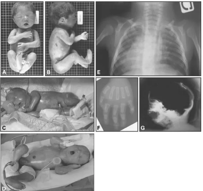
P1, a sporadic case, was a female fetus issued from parents originating from Corsica (France) after five cycles of in vitro fertilization. Delivery occurred at 29 weeks by cesarean section for fetal distress and intra Uterine growth retardation: birth weight was 980 g, length 36 cm and OFC 24 cm. Feeding difficulties, dysmorphism and abnormal skin were present at birth. The skin was tight and sclerotic, especially on the abdomen with visible superficial vessels. There were joint retraction of the knees, hips, elbows and digits; microretrog-nathism and a beaked nose with proptosis. X-rays showed shortened hypoplastic clavicles, acro-osteolyses of terminal phalanges (Fig. 1), kyphosis of the spine, small facial bones and overtubulated long bones and ribs. At 4 months, rectal prolapsus and bilateral hernia occurred. She died at 6 months due to respiratory distress. The diagnosis of RD was retained after the skin biopsy showed a thickened skin with a flat dermis depleted of elastic fibers, hypertrophic endo-plasmic reticulum in keratinocytes, and numerous collagen fibers at the dermo – epidermal junction.
Patient P2, is the first child of healthy unrelated parents of Moroccan and Algerian origin. Pregnancy was uneventful, with the exception of reduced fetal movements, and delivery was at term. At birth, the child’s skin was edematous, rapidly developing erosions and scleroderma-like lesions. Blood vessels were apparent under the skin of the tibias, and on the trunk, skin was taut, thick and slightly desquamating, whereas the venous bed was not prominent in this area.
Nipples were prominent and microretrognathism with exoph-talmia was present. Mouth movements were not limited; weeping caused gastroesophageal reflux and cyanosis, secon-dary to thoracic and abdominal restriction. Clavicular hypoplasia was observed at X-ray. Patient P2 is still alive at age of 5 months and her clinical condition evolves severely. All the other patients (P3 – P9), of French and Dutch origin, were born from healthy non-consanguineous parents and showed typical RD signs both during pregnancy and at birth. These included reduced fetal movements, intrauterine growth retardation and pre-term delivery (30 – 32 weeks) by
cesarean section in most cases. At birth, patients showed reduced weights, lengths and occipitofrontal circumferences (between third and tenth centiles), and died within a few hours in all cases. Their clinical phenotype included tight, translucent, thin skin with prominent superficial vessels on the trunk, erosions of the skin at flexure or pressure sites, multiple joint ankylosis, a characteristic expressionless ‘Asian porcelain doll’ face with small fixed open mouth, retromicrognathism, beaked nose, slight antimongoloid slant with apparent hypertelorism, sparse eyebrows and eyelashes, a large anterior fontanella, clavicular hypoplasia and long
Figure 1. Clinical and radiological aspects of different patients affected with RD. Typical fixed facial expression, micrognathism and mouth in the ‘o’ position are observed in these three cases. (A, B) Frontal and lateral view of patient P3. Note the ‘Asian porcelain doll’ expression due to skin tautness, multiple joint ankylosis and skin erosion at flexure sites. (C, D) Patient P4 and P8, respectively: translucent and tight skin with prominent superficial vessels; artrho-gryposis multiplex and skin fissuring are evident, as in patient P3. (E) Clavicular hypoplasia in patient P1. (F) Acro-osteolysis of distal phalanges, evident in particular at the 2nd and 5th fingers, in patient P1. (G) Cranium ossification defects with a wide anterior fontanel and retromicrognathism can be observed in patient P8.
nails. Hair was present in patient P3 only (Fig. 1). Detailed clinical and histological descriptions of the Dutch cases included in this study have been published elsewhere (22,23). In all cases, there was no history of exposure to prenatal environmental toxins or other noxious agents. Clinical and radiological findings from five patients are reported in Figure 1.
Owing to clinical similarities with patients affected with HGPS or MAD (OMIM no. 248370), we searched for muta-tions in the LMNA gene in all patients. Subsequently, because Zmpste24 inactivation causes skeletal, skin and adipose tissue abnormalities in mice (16,17) and ZMPSTE24 (FACE-1) mutations in absence of LMNA defect have been recently identified in one patient affected with MAD (18), we analyzed the coding region and intron – exon boundaries of this gene, encoding a metalloproteinase involved in Lamin A post-translational processing.
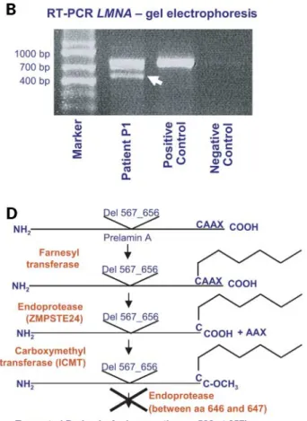
Sequencing of LMNA exons and intron – exon bounda-ries allowed us to observe, in patient P1, an unreported heterozygous mutation at intron 11 donor splice site (IVS11 þ 1G . A) (Fig. 2A). We have further explored the effects of this mutation at the mRNA level by reverse tran-scription – PCR (RT – PCR); transcript amplification between exons 7 and 12 evidenced an aberrant smaller amplicon of around 450 bp, together with the 712 bp wild-type allele (Fig. 2B). Sequencing of the smaller transcript allowed us to observe the in-frame skipping of the entire exon 11 (270 bp) (Fig. 2C), indicating a reduced efficiency of the mutated splicing site. These changes at the RNA level are described as follows: [r. ¼ , r.1699_1968del]. The deletion of exon 11 is predicted to remove 90 aminoacids from the C-terminal end of the protein precursor, corresponding to a large part of the globular domain (p.G567_Q656del). The truncated mRNA was stable and translated into the truncated precursor as observed by the western blot (Fig. 3A). The latter precursor thus lacks the region in which the second post-translational cleavage is performed (located between wild-type aminoacids Y646 and L647) (Fig. 2D), consisting thus of a 571 amino-acid-long immature precursor, ending with the CaaX domain. DNAs from parents of patients P1 and P2 were not available for genetic analysis.
In patient P2, the c.1824C.T mutation in LMNA was found in exon 11 (Fig. 2E). This apparently synonymous variation (G608G), the most common pathogenic mutation identified in patients affected with HGPS (6,7), activates an upstream cryptic splice site and causes the production of a truncated mRNA, lacking the last 150 bp of exon 11, as already described (6). The deletion preserves the reading frame, causing the appearance of a truncated Prelamin A precursor, called ‘Progerin’ in patients’ cells (24,21).
In all the remaining patients, no genomic sequence varia-tion was identified in LMNA. Conversely, exploravaria-tion of the ZMPSTE24 gene allowed us to identify, in all the other seven patients (P3 – P9), the same heterozygous 1 bp insertion in exon 9 (c.1085_1086insT) (Fig. 2F). This insertion causes a frameshift and leads to a premature termination codon, located 18 codons downstream, predicted to result in a truncated protein, L362fsX18. For patients P4 – P6, P8 and P9, DNAs from parents were available for molecular studies, which further evidenced that the L362fsX18 was inherited either
from the father (patients P4 and P8) or from the mother (patients P5, P6 and P9).
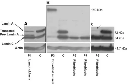
On the basis of protein-domain predictions, the mutated protein lacks the seventh transmembrane domain as well as the C-terminal cytoplasmic tail involved in the protease’s catalytic action (18). ZMPSTE24 coding sequence and intron – exon boundaries did not show any variation in pati-ents P1 and P2. Furthermore, no second mutation could be identified in LMNA or ZMPSTE24 in any of the patients. These two genes were analyzed at both the genomic and transcriptional level by RT – PCR in patients P2 – P4 and P6; the coding parts of the transcripts did not reveal any variation and no altered splice fragment was observed (data not shown). By western blotting (Fig. 3), we explored the overall expression levels of Lamins A/C on lymphoblasts or fibroblast cells issued from different RD patients, as well as from skeletal muscle cells issued from patient P3.
Patient P1 had both Lamins A and C and also the truncated Lamin A precursor was detected (Fig. 3A). In Patients P3, P6 and P7, a complete loss of Lamin A expression was observed (Fig. 3B). Moreover, very low levels of wild-type Prelamin A were detected (Fig. 3C). Lamin C expression seemed to be maintained at normal levels, except in patient P3. In protein extracts from skeletal muscle of patient P3 (the only patient for whom this tissue was available), we observed a strong reduction of Lamin C expression levels, undetectable Lamin A and a supplementary band, specifically recognized by the antibody directed against Lamins A/C, with a molecular weight around 150 kDa (Fig. 3B). This structure’s composition, unknown to date, will be determined with further analyses.
Cell and tissue immunocytochemical and immunohisto-chemical explorations were performed on four fibroblast cell lines issued from patients P4 – P7, on a lymphoblastoid cell line issued from patient P1 and on skin biopsy samples issued from patient P3 (Figs 4 and 5). Only DNA was avail-able for patients P2 and P9, and fibroblasts from patient P8 failed to grow in culture.
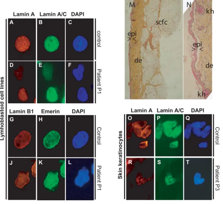
All the examined cells showed several nuclear abnormal-ities with an average of 50% of abnormal nuclei from each patient when explored within the first three divisions. This ratio dramatically increased with the number of divisions (data not shown). Patient P1 lymphoblastoid cells showed altered shapes (Fig. 4D – F and J – L) with the different anti-bodies used. Overall expression levels of lamins A and C were reduced, with delocalization to nucleoplasmic dense aggregates in some cells. Abnormal distribution patterns of Lamin B1 and emerin were observed in the same patient: the two proteins were lacking at one nuclear pole (Fig. 4J – L). Histopathological and immunohistochemical analyses were performed on a skin biopsy from patient P3 (Fig. 4M – T). They showed an atrophic epidermis with ortho-keratotic and focally paraortho-keratotic hyperkeratosis, kerato-hyalin granules were also observed (Fig. 4N). The dermis was compacted with collagen fibers that were parallel to the dermo – epidermic basal lamina; dermal vessels were apparent around the rare and hypoplastic pilosebaceous annexes. Elastic fibers were almost totally lacking. By immunocyto-chemistry, keratinocytes showed an overall strong reduction of Lamins A and C nuclear expression levels with inter-ruptions of nuclear envelope boundaries and mis-distribution
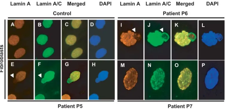
in nucleoplasm, absence of staining in one pole of the nucleus (Fig. 4R – S). In fibroblasts, herniations of nucleo-plasm lacking Lamin A staining but expressing Lamin C (Fig. 5E – G and I – K, arrowheads) could be observed.
Nuclear envelope irregularities with blebs (Fig. 5I – L) and heterogeneous deposits of Lamins A and C in nucleoplasmic foci (Fig. 5M – O) were present. Nuclear sizes were larger than in controls in 30% of patients’ cells (data not shown).
Figure 2. Molecular exploration of patients and predicted consequences on patient P1 Prelamin A product. (A) LMNA heterozygous IVS11 þ 1G . A mutation at the DNA level on patient P1. (B) Agarose gel electrophoresis of wild-type and mutant (arrowhead) transcripts amplified by RT – PCR between exons 7 and 12; the marker’s sizes are indicated on the left. (C) Sequence of the mutant transcript, revealing an aberrant junction fragment between exons 10 and 12, with the in-frame skipping of exon 11. (D) Predicted alteration of Prelamin A post-translational processing: the absence of aminoacids 567 – 656, encoded by exon 11, eliminates the second cleavage site (crossover the arrow in the scheme), preventing the achievement of Prelamin A processing. (E) LMNA heterozygous c.1824C . T transversion in patient P2. (F) Heterozygous one base insertion in ZMPSTE24 exon 9 (c.1085_1086insT), identified in patients P3 – P9, the reverse sequence is shown.
DISCUSSION
Restrictive dermopathy is a lethal condition usually diagnosed during perinatal stages. On the basis of clinical and radio-logical similarities between RD and HGPS caused by LMNA mutations, we searched for mutations in LMNA as possibly causing RD. This disorder is suspected to be recessively inherited since several families have been reported that included at least two affected sibs, without any phenotype in parents. Thus, although none of the families included in this study was consanguineous, we expected to find either homozygous or compound heterozygous mutations in patients. The recurrent heterozygous G608G de novo mutation causing the vast majority of HGPSs was identified in patient P2, who presented a phenotype of intermediate severity between classical progeria and RD. This mutation is known to remove only the 30 end of exon 11 and produces a truncated Prelamin A precursor in classical HGPS patients. No other mutation was detected in this patient, neither in LMNA nor in ZMPSTE24.
In patient P1, an unreported heterozygous LMNA muta-tion was identified at the donor splicing site of intron 11. At the transcript level, this mutation removes the entire exon 11, encoding a major part of the C-terminal globular domain. Indeed, this deletion removes 90 aminoacids from the C-terminal tail domain of the Prelamin A precursor. This patient presented a much more severe phenotype than the one presented by patient P2 and died at 6 months. In all the other patients studied (P3 – P9), we identified a unique heterozygous 1 bp insertion in ZMPSTE24 exon 9, leading to a premature termination codon and a truncated protein.
On the basis of domain function predictions (18), this mutation is very likely to suppress the protease activity of the protein. The LMNA or ZMPSTE24 mutations identified were thus heterozygous in each patient and, although RD is considered as being an autosomal recessive inherited disorder, none of the patients was issued from a consanguineous union. None-theless, different considerations can be formulated concerning each mutation.
Concerning the LMNA mutation identified in patient P2 (c.1824C . T, p.G608G), although its pathogenic effect at the molecular level is undoubted (6,7,17), the phenotype presented by the patient is much more severe than the one (HGPS) presented by patients carrying the same mutation. This observation suggests that, in this patient, other genetic or epigenetic factors might contribute to the clinical picture. Genetic factors could be represented either by a major mutated second gene, which still remains to be ident-ified, or a series of pre-disposing polymorphisms in genes encoding proteins correlated with Lamin A processing, func-tion or expression levels. Further funcfunc-tional and genetic analyses on this patient will help to elucidate these aspects. However, due to the atypical RD observed in this particular patient, we suggest that his phenotype bridges the gap between HGPS and RD, thus representing an overlapping phenotype between these two disorders. Nonetheless, LMNA heterozygous dominant mutations cause a spectrum of allelic disorders (25), the severest of which known to date is HGPS (6,7). Indeed, patient P1 expresses a Prelamin A precursor, which contains a very large C-terminal deletion, encom-passing the deleted part of Progerin. In this case, as it is specu-lated for Progerin, the truncated and unprocessed Prelamin A
Figure 3. Lamins A/C expression analysis in patients affected with RD. Protein extracts from lymphoblastoid cell lines (patient P1), fibroblasts cells (patients P6 and P7) or skeletal muscle (patient P3) were submitted to electrophoresis, western blotted and probed using anti-human Lamins A/C antibody and anti-pan-actin antibody as a loading control. C ¼ fibroblast control cells. (A) Patient P1 lymphoblastoid cells, respectively, show conserved Lamin A and normal Lamin C expression, together with the truncated Lamin A precursor encoded by the mutant allele, weighing around 65 kDa. (B) Patient P3 skeletal muscle shows reduced Lamin C levels, undetectable Lamin A and a supplementary band of undetermined composition weighing around 150 kDa. Patients P6 and P7 show conserved Lamin C levels and a loss of Lamin A expression. (C) On western blots loaded with a higher amount of proteins, a weakly expressed band at the level to which the Prelamin A unprocessed precursor is expected to migrate (74 kDa) was detected in P6 (arrow).
precursor could have dominant negative deleterious effects on wild-type Lamin A levels, function and distribution, which, could be even worse than those exerted by Progerin due to the larger deletion. The RD truncated protein could thus be sufficient by itself, to cause the reduction of wild-type Lamin A protein levels and ultimately an RD phenotype in this patient.
Other transcript splicing defects have been involved in less severe laminopathies; Todorova and collegues (26) reported an LGMD1B affected patient in which Lamins A and C
were predicted to have a 15 aminoacids insertion at the N-terminal end. Another heterozygous mutation generating an aberrant transcript missing the entire tail domain was ident-ified in a patient affected with EDMD (27). The much milder phenotypes described reinforce the hypothesis of a dominant negative effect taking place in HGPS as well as RD patients and possibly due to the expression of truncated proteins specifically missing aminoacids encoded by exon 11. Several fundamental interactions taking place at Lamin A C-terminal tail [with DNA (28), actin (29), emerin (30,31), LAP2a
Figure 4. Immunohistochemical and histological phenotypes of patients P1 and P3. (A – L): EBV immortalized lymphoblastoid cells from control (A – C, G – I) and patient P1 (D – F, J – L). Nuclear shape irregularities (D – F, J – L). Lamins A and C staining has low intensity and is largely delocalized to the nucleoplasm (D). (J, K) Mis-localization of Lamin B1 and Emerin, which are absent at one nuclear pole. (M, N) Cutaneous histopathology (trichrome staining) of patient P3. (M) Low magnification: atrophic epidermis (epi) with hyperkeratosis, densely packed dermic (de) collagen bundles parallel to the linear dermo – epidermic junction with rarefaction of papillae and pilosebaceous appendages, relatively preserved subcutaneous fat cells (scfc). (N) High magnification: hyperkeratosic epidermis with keratohyalin granules (kh). (O – T): Keratinocytes from skin frozen sections. Five nuclei of a control patient (O – Q) and one nucleus of patient P3 (R – T) can be observed, showing a strong reduction of both Lamin A and Lamin C expression levels, with absent expression at one nuclear pole in an overall Lamins A/C inhomogeneous localization pattern.
(32), Narf (33), PKCa, (34)], could be dominantly perturbed by the truncated proteins. Moreover, in patient P1, the Lamin A C-terminal tail deletion is larger than the one observed in HGPS patients (90 versus 50 aminoacids), suggesting an even worse deleterious effect. As an additional argument to suggest a dominant negative effect of the trun-cated Prelamin A, the immunocytochemical staining observed in patient P1, in particular the absence of Lamins B1 from a nuclear pole, highly resembles the pattern observed in homo-zygous Lmna KO mice (35). Conversely, in one case, it has been shown that homozygous nonsense LMNA mutations trun-cating Lamins A and C in their rod domain lead to premature delivery and neonatal death at 30 weeks. The effect of the mutation was the complete absence of both Lamins A and C (36).
Regarding the ZMPSTE24 mutation, although it was ident-ified in seven typical RD patients, it is clearly not sufficient by itself to cause RD since, in four families, it was inherited from one of the parents. In addition, the same heterozygous mutation has been already observed in unaffected related individuals of an MAD patient. Indeed, this mutation has recently been reported (as Phe361fsX379) in one patient affected with severe MAD (18). This patient, reported by Agarwal and collegues, was compound heterozygous for the c.1085_1086insT (Phe361fsX379) mutation and the 1018 C.T missense mutation (W340R) located in close vicinity of the active catalytic site, whereas his healthy parents carried each mutation at the heterozygous state. It is thus very likely that we identified the first mutation of the (at least) two ones that cause the RD phenotype in seven patients from six families. The fact that two ZMPSTE24 mutations
cause an MAD phenotype makes it not surprising that we did not find the second mutation in the same gene: RD phenotype is much more severe and is thus possibly due to the digenic inheritance of mutations in ZMPSTE24 and another gene that must have a key role in skin and bone development. Indeed, we have performed genomic analysis in all patients carrying the ZMPSTE24 mutation, and transcrip-tional analysis in four of them, by RT – PCR. No other mutations or differences in bands’ size have been brought to light after migration on agarose gels thus eliminating potential splicing defects due to intronic variations that would not have been detected in the genomic screening. In this respect, the c.1085_1086insT seems to be a ‘hotspot’ mutation and might result from a slippage effect of the DNA polymerase due to the presence of a repeated thymine (T)9 at this position in the normal sequence. Paradoxically, it is very unlikely that the mutations remaining to be identified are at the same position in all patients, since they are from diverse ethnic backgrounds. Conversely, it has been demon-strated in mice inactivated for Zmpste24 that only the complete loss of expression of this enzyme makes it unable to correctly process Lamin A precursor to normal mature Lamin A and leads to an accumulation of Prelamin A (16,17). Since normal Prelamin A is evidenced by western blotting, even at low levels, this suggests that either the two copies of ZMPSTE24 are inactivated, or, more likely, that a mutation lying in a not-yet-identified causing gene is respon-sible for the absence of Lamin A processing. Additionally, anti-Lamin A antibodies detected significant levels of proteins by fluorescent immunocytochemistry, a much more sensitive method than western blot. This is consistent with the fact
Figure 5. Immunocytochemical characterization of fibroblast cell lines issued from patients affected with RD. Fibroblasts from three differents patients (E – P) are compared to control fibroblasts (A – D). Detection of Lamin A (A, E, I, M) and Lamin A/C (B, F, J, N). Merged images (C, G, K, O). DAPI staining (D, H, L, P). RD patients’ fibroblast nuclei show several abnormalities: nucleoplasm herniation (arrowheads) is delineated by Lamin C, with a non-homogeneous staining of the nuclear periphery, but not by Lamin A; nuclear envelope irregularities (E – H, I – L) can be observed; heterogeneous focal deposits of Lamins A and C (M – P) in the nucleoplasm are frequently observed. Magnification is the same for all images, image width: 3.5 mm.
that only Prelamin A accumulates in the RD nuclei, rather than the normally processed Lamin A. Immunocytochemistry experiments have also allowed to evidence dramatic nuclear damages that increase proportionally to the number of cell divisions as recently shown in HGPS fibroblasts (37,21).
However, according to our results and the fact that two-copy LMNA missense mutations or heterozygous compound mutations in ZMPSTE24 cause the less severe progeroid disease MAD (38,18), we speculate that digenism could be involved in RD pathogenesis in all patients, but more likely in patients carrying a single ZMPSTE24 mutation, which would be a necessary but not sufficient genetic defect to lead to RD.
All together, our data orientate our hunt for a yet unidenti-fied mutation, primarily in genes encoding proteins whose function is essential for the correct Lamin A processing pathway. Other genes could also be proposed as candidates, and, in this respect, FATP4 gene sequencing is currently being performed in patients carrying the heterozygous 1 bp insertion in ZMPSTE24. Indeed, Fatp4 homozygous targeted inactivation has been identified recently in mice displaying features reminiscent of neonatal lethal RD (39,40).
In summary, RD is a novel direct or indirect laminopathy, alternatively involving LMNA or ZMPSTE24, both associated in the same processing pathway. To our knowledge, this dis-order represents one extremity in the severity’s spectrum of already known laminopathies, and the presence of overlapp-ing phenotypes indicates that laminopathies are a continuum of related disorders rather than separated clinical entities. In this respect, RD as well as Lamin-associated premature ageing disorders are likely to display a great genetic hetero-geneity, whose actors still have to be identified. In future, our study will find important applications in genetic counsel-ling. Indeed, in RD cases carrying a ZMPSTE24 mutation, we have demonstrated a complete loss of Lamin A protein expres-sion, whereas in case of LMNA truncating mutations, Lamin A as well as the corresponding truncated Prelamin may be identified. Since amniocytes are differentiated cells expressing Lamin A at normal levels (41), a combined genetic and proteic approach for prenatal diagnosis might be proposed to couples having faced this devastating disorder in the past.
MATERIALS AND METHODS
Patients and samples
We collected samples from nine patients affected with RD (OMIM no. 275210) from eight families. The patients origi-nated from France (P1, P3, P8), The Netherlands (P4 – 7, P9) and North-Africa (P2). Informed consent was obtained from the parents of all patients included in this study, which complies with the ethic guidelines of the institutions involved. Figure 1A and B are reproduced with the parents’ authorization. Blood samples were used to establish EBV immortalized lymphoblastoid cell lines in patient P1. Skin biopsy and skeletal muscle samples from patient P3 were available for histopathological and immunohistochemical analyses or protein extraction and immunoblot, respectively. Fibroblasts were obtained by skin biopsy and were cultured in a DMEM medium containing 10% fetal calf serum,
2 mm/ml L-glutamine and 100 U/ml penicillin – streptomycin
(GIBCO BRL).
Genomic and transcriptional analysis
Genomic DNA was extracted from peripheral blood lympho-cytes by standard procedures. PCR conditions and primers used for LMNA coding sequence amplification were described elsewhere (42). RNA extraction from patient P1 EBV immor-talized lymphoblastoid cell lines was performed with TRIZOL (Invitrogen-Life Technologies), following the manufacturer recommendations. Reverse transcription was performed with superscript II reverse transcriptase (Invitrogen-Life Techno-logies) following company recommendations. A panel of over-lapping primer pairs was used to sequence the full length Lamin A/C cDNA. Primers located in exons 7 (50-GATGA GGAGGGCAAGTTTGT-30) and 12 (50-GTGAGGAGGAC GCAGGAA-30) were used to amplify patient P1’s truncated transcript junction fragment. The LMNA RT – PCR primers and those used for ZMPSTE24 amplification are available upon request. LMNA and ZMPSTE24 coding sequences are available at http://genome.ucsc.edu/. Sequencing reactions were performed with a dye terminator procedure and loaded on to a capillary automatic sequencer CEQTM 8000 (Beckman Coulter) according to the manufacturer0s rec-ommendations. Direct sequencing was performed in both orientations in order to exclude sequencing artefacts. All sequence variations are described according to den Dunnen recommendations at http://www.hgvs.org/mutnomen.
Western blots
Protein extraction was performed from patients0EBV immor-talized lymphoblastoid cell lines, skeletal muscle frozen tissue or fibroblasts as already described (43). Total proteins of 40 mg were loaded and separated on a 7% SDS/Page gel and transferred on a PVDF membrane overnight at 48C. Non-specific epitopes were blocked with 10% casein solution/ TBST. The membrane was incubated for 1 h with the follow-ing primary antibodies: anti-Lamin A/C (JOL2, Serotec, Oxford, UK) diluted at 1 : 10 and an anti-pan-actin as control (MAB1501R, Chemicon, CA, USA), diluted at 1 : 2000, both in a blocking solution. After several washes in TBST, the membranes were incubated with peroxidase conjugated secondary antibodies (Jackson Immunoresearch, Baltimore, MD, USA) diluted at 1 : 5000 in blocking solution. For visualization, ECL and western blot detection systems were used.
Histopathological analysis
The skin sample from patient P3 was fixed in paraformalde-hyde and embedded in paraffin. Sections were stained using Gomori trichrome.
Immunocytochemistry and immunohistochemistry
Fibroblasts grown on Lab Tek Chamber Slides (Nalge Nunc International) were washed twice with PBS then fixed with 1% formaldehyde for 10 min. They were then washed for 10 min in PBS, dehydrated in ethanol (70%, 90% and twice
100%), and permeabilized with 0.5% Triton X-100 in PBS for 5 min at room temperature. Frozen cytospin slides (Shandon, Pittsburgh, PA, USA) were spread with lymphoblastoid cells, fixed in methanol for 6 min at 2208C and rinsed in PBS. The slides were then pre-incubated for 15 min (three times) at room temperature with a solution containing PBS, 5% BSA, 4% normal serum (goat or rabbit). Incubation with primary antibodies diluted in the incubation solution for 1 h at room temperature was next performed. Primary antibodies were anti-Lamin A/C monoclonal antibody (Clone 4A7), anti-Emerin (MANEM 8), a courtesy from Dr G. Morris, anti-Lamin B1 (Sc-6217, Santa Cruz Biotechnologies, CA, USA) and anti-Lamin A polyclonal antibody (M52-1), a cour-tesy from Dr Y. Hayashi. All primary antibodies were diluted at 1 : 100. After several washes in PBS, the slides were incu-bated with secondary antibodies diluted in the incubation sol-ution for 1 h at room temperature. Secondary antibodies were obtained from Jackson company (Jackson ImmunoResearch, Westgrove, PA, USA): (FITC)-conjugated rabbit anti-mouse (1 : 100), (TRITC)-conjugated rabbit anti-goat (1 : 100 or 1 : 200) or (FITC)-conjugated donkey anti-mouse (1 : 100) and (TRITC)-conjugated donkey anti-rabbit (1 : 100). The cells were then washed three times for 10 min in PBS, incu-bated with DAPI (Sigma-Aldrich) at 100 ng/mL for 15 min, and finally washed three times for 5 min in Tween 20 at 0.1% in PBS. The slides were mounted in Vectashield mount-ing medium (Vector), coverslipped and sealed. Digitized microphotographs were recorded using a Leica DMR micro-scope (Leica Microsystems, Wetzlar, Germany) equipped with a CoolSNAP camera (Princeton, Trenton, NJ, USA). Sec-tions of 15 mm were made from frozen skin samples (Patient P3) using a cryostat (Microm, France). They were fixed in 4% paraformaldehyde during 45 min, then washed three times. The slides were incubated overnight (48C) with primary antibodies. The protocol as described for cultured cells was then applied to skin sections.
ACKNOWLEDGEMENTS
We thank the members of the families for their invaluable contribution to this study. Drs G. Morris and Y. Hayashi are acknowledged for providing us with antibodies. We are extremely grateful to C. Badens and J. C. Courvalin criti-cal reading of this manuscript, helpful comments and discus-sions. This study was supported by a grant from the ‘Association Franc¸aise contre les Myopathies’ (AFM), the Institut National de la Sante´ et de la Recherche Me´dicale (INSERM), the Assistance Publique des Hoˆpitaux de Mar-seille (AP-HM) and the Association Me´diterrane´enne pour la Recherche en Ge´ne´tique (AMRG). A.D.G. was supported by a fellowship grant from the AFM and C.N. by a fellowship grant from the Association pour le De´veloppement de la Recherche en Ge´ne´tique.
REFERENCES
1. Witt, D.R., Hayden, M.R., Holbrook, K.A., Dale, B.A., Baldwin, V.J. and Taylor, G.P. (1986) Restrictive dermopathy: a newly recognized autosomal recessive skin dysplasia. Am. J. Med. Genet., 24, 631 – 648.
2. Verloes, A., Mulliez, N., Gonzales, M., Laloux, F., Hermanns-Le, T., Pierard, G.E. and Koulischer, L. (1992) Restrictive dermopathy, a lethal form of arthrogryposis multiplex with skin and bone dysplasias: three new cases and review of the literature. Am. J. Med. Genet., 43, 539 – 547. 3. Lenz, W. and Meschede, D. (1993) Historical note on restrictive
dermopathy and report of two new cases. Am. J. Med. Genet., 47, 1235 – 1237.
4. Mau, U., Kendziorra, H., Kaiser, P. and Enders, H. (1997) Restrictive dermopathy: report and review. Am. J. Med. Genet., 71, 179 – 185. 5. Pierard-Franchimont, C., Pierard, G.E., Hermanns-Le, T., Estrada, J.A.,
Verloes, A. and Mulliez, N. (1992) Dermatopathological aspects of restrictive dermopathy. J. Pathol., 167, 223 – 228.
6. De Sandre-Giovannoli, A., Bernard, R., Cau, P., Navarro, C., Amiel, J., Boccaccio, I., Lyonnet, S., Stewart, C.L., Munnich, A., Le Merrer, M. et al. (2003) Lamin A truncation in Hutchinson– Gilford progeria. Science, 300, 2055.
7. Eriksson, M., Brown, W.T., Gordon, L.B., Glynn, M.W., Singer, J., Scott, L., Erdos, M.R., Robbins, C.M., Moses, T.Y., Berglund, P. et al. (2003) Recurrent de novo point mutations in lamin A cause Hutchinson– Gilford progeria syndrome. Nature, 423, 293 – 298.
8. Mounkes, L., Kozlov, S., Burke, B. and Stewart, C.L. (2003) The laminopathies: nuclear structure meets disease. Curr. Opin. Genet. Dev., 13, 223 – 230.
9. Moir, R.D., Spann, T.P., Lopez-Soler, R.I., Yoon, M., Goldman, A.E., Khuon, S. and Goldman, R.D. (2000) Review: the dynamics of the nuclear lamins during the cell cycle-relationship between structure and function. J. Struct. Biol., 129, 324 – 334.
10. Aebi, U., Cohn, J., Buhle, L. and Gerace, L. (1986) The nuclear lamina is a meshwork of intermediate-type filaments. Nature, 323, 560 – 564. 11. Fawcett, D. (1966) On the occurrence of a fibrous lamina on the inner
aspect of the nuclear envelope in certain cells of vertebrates. Am. J. Anat., 119, 129 – 145.
12. Bridger, J.M., Kill, I.R., O’Farrell, M. and Hutchison, C.J. (1993) Internal lamin structures within G1 nuclei of human dermal fibroblasts. J. Cell. Sci., 104, 297 – 306.
13. Hozak, P., Sasseville, A.M., Raymond, Y. and Cook, P.R. (1995) Lamin proteins form an internal nucleoskeleton as well as a peripheral lamina in human cells. J. Cell. Sci., 108, 635 – 644.
14. Shumaker, D.K., Kuczmarski, E.R. and Goldman, R.D. (2003) The nucleoskeleton: lamins and actin are major players in essential nuclear functions. Curr. Opin. Cell. Biol., 15, 358 – 366.
15. Roskoski, R., Jr (2003) Protein prenylation: a pivotal posttranslational process. Biochem. Biophys. Res. Commun., 303, 1 – 7.
16. Pendas, A.M., Zhou, Z., Cadinanos, J., Freije, J.M., Wang, J., Hultenby, K., Astudillo, A., Wernerson, A., Rodriguez, F., Tryggvason, K. et al. (2002) Defective prelamin A processing and muscular and adipocyte alterations in Zmpste24 metalloproteinase-deficient mice. Nat. Genet., 31, 94 – 99.
17. Bergo, M.O., Gavino, B., Ross, J., Schmidt, W.K., Hong, C., Kendall, L.V., Mohr, A., Meta, M., Genant, H., Jiang, Y. et al. (2002) Zmpste24 deficiency in mice causes spontaneous bone fractures, muscle weakness, and a prelamin A processing defect. Proc. Natl Acad. Sci. USA, 99, 13049– 13054.
18. Agarwal, A.K., Fryns, J.P., Auchus, R.J. and Garg, A. (2003) Zinc metalloproteinase, ZMPSTE24, is mutated in mandibuloacral dysplasia. Hum. Mol. Genet., 12, 1995 – 2001.
19. Novelli, G. and D’Apice, M.R. (2003) The strange case of the ‘lumper’ lamin A/C gene and human premature ageing. Trends Mol. Med., 9, 370 – 375.
20. Mounkes, L.C. and Stewart, C.L. (2004) Aging and nuclear organization: lamins and progeria. Curr. Opin. Cell. Biol., 16, 322 – 327.
21. Goldman, R.D., Shumaker, D.K., Erdos, M.R., Eriksson, M., Goldman, A.E., Gordon, L.B., Gruenbaum, Y., Khuon, S., Mendez, M., Varga, R. et al. (2004) Accumulation of mutant lamin A causes progressive changes in nuclear architecture in Hutchinson – Gilford progeria syndrome. Proc. Natl Acad. Sci. USA, 101, 8963 – 8968.
22. Happle, R., Stekhoven, J.H., Hamel, B.C., Kollee, L.A., Nijhuis, J.G., Anton-Lamprecht, I. and Steijlen, P.M. (1992) Restrictive dermopathy in two brothers. Arch. Dermatol., 128, 232 – 235.
23. Smitt, J.H., van Asperen, C.J., Niessen, C.M., Beemer, F.A., van Essen, A.J., Hulsmans, R.F., Oranje, A.P., Steijlen, P.M., Wesby-van Swaay, E., Tamminga, P. et al. (1998) Restrictive dermopathy. Report of 12 cases. Dutch Task Force on Genodermatology. Arch. Dermatol., 134, 577 – 579.
24. Csoka, A.B., Cao, H., Sammak, P.J., Constantinescu, D., Schatten, G.P. and Hegele, R.A. (2004) Novel lamin A/C gene (LMNA) mutations in atypical progeroid syndromes. J. Med. Genet., 41, 304 – 308.
25. Worman, H.J. and Courvalin, J.C. (2004) How do mutations in lamins A and C cause disease? J. Clin. Invest., 113, 349 – 351.
26. Todorova A., Hallinger-Keller, B., Walter, M.C., Debauvalle, M.C., Lochmu¨ller, H. and Mu¨ller, C.R. (2003) A synonymous codon change in the LMNA gene alters mRNA splicing and causes limb girdle muscular dystrophy type 1B. J. Med. Genet., 43, e115.
27. Bonne, G., Mercuri, E., Muchir, A., Urtizberea, A., Becane, H.M., Recan, D., Merlini, L., Wehnert, M., Boor, R., Reuner, U. et al. (2000) Clinical and molecular genetic spectrum of autosomal dominant Emery-Dreifuss muscular dystrophy due to mutations of the lamin A/C gene. Ann. Neurol., 48, 170 – 180.
28. Stierle, V., Couprie, J., Ostlund, C., Krimm, I., Zinn-Justin, S., Hossenlopp, P., Worman, H.J., Courvalin, J.C. and Duband-Goulet, I. (2003) The carboxyl-terminal region common to lamins A and C contains a DNA binding domain. Biochemistry, 42, 4819 – 4828.
29. Sasseville, A.M. and Langelier, Y. (1998) In vitro interaction of the carboxy-terminal domain of lamin A with actin. FEBS Lett., 425, 485 – 489.
30. Vaughan, A., Alvarez-Reyes, M., Bridger, J.M., Broers, J.L., Ramaekers, F.C., Wehnert, M., Morris, G.E., Whitfield, W.G.F. and Hutchison, C.J. (2001) Both emerin and lamin C depend on lamin A for localization at the nuclear envelope. J. Cell. Sci., 114, 2577 – 2590.
31. Sakaki, M., Koike, H., Takahashi, N., Sasagawa, N., Tomioka, S., Arahata, K. and Ishiura, S. (2001) Interaction between emerin and nuclear lamins. J. Biochem. (Tokyo), 129, 321 – 327.
32. Dechat, T., Korbei, B., Vaughan, O.A., Vlcek, S., Hutchison, C.J. and Foisner, R. (2000) Lamina-associated polypeptide 2alpha binds intranuclear A-type lamins. J. Cell. Sci., 113, 3473 – 3484.
33. Barton, R.M. and Worman, H.J. (1999) Prenylated prelamin A interacts with Narf, a novel nuclear protein. J. Biol. Chem., 274, 30008– 30018. 34. Martelli, A.M., Bortul, R., Tabellini, G., Faenza, I., Cappellini, A.,
Bareggi, R., Manzoli, L. and Cocco, L. (2002) Molecular characterization of protein kinase C-alpha binding to lamin A. J. Cell. Biochem., 86, 320 – 330.
35. Sullivan, T., Escalante-Alcalde, D., Bhatt, H., Anver, M., Bhat, N., Nagashima, K., Stewart, C.L. and Burke, B. (1999) Loss of A-type lamin expression compromises nuclear envelope integrity leading to muscular dystrophy. J. Cell. Biol., 147, 913 – 920.
36. Muchir, A., van Engelen, B.G., Lammens, M., Mislow, J.M., McNally, E., Schwartz, K. and Bonne, G. (2003) Nuclear envelope alterations in fibroblasts from LGMD1B patients carrying nonsense Y259X
heterozygous or homozygous mutation in lamin A/C gene. Exp. Cell. Res., 291, 352 – 362.
37. Bridger, J.M. and Kill, I.R. (2004) Aging of Hutchinson– Gilford progeria syndrome fibroblasts is characterised by hyperproliferation and increased apoptosis. Exp. Gerontol., 39, 717 – 724.
38. Novelli, G., Muchir, A., Sangiuolo, F., Helbling-Leclerc, A., D’Apice, M.R., Massart, C., Capon, F., Sbraccia, P., Federici, M., Lauro, R. et al. (2002) Mandibuloacral dysplasia is caused by a mutation in
LMNA-encoding lamin A/C. Am. J. Hum. Genet., 71, 426 – 431. 39. Moulson, C.L., Martin, D.R., Lugus, J.J., Schaffer, J.E., Lind, A.C. and
Miner, J.H. (2003) Cloning of wrinkle-free, a previously uncharacterized mouse mutation, reveals crucial roles for fatty acid transport protein 4 in skin and hair development. Proc. Natl Acad. Sci. USA, 100, 5274 – 5279. 40. Herrmann, T., van der Hoeven, F., Grone, H.J., Stewart, A.F., Langbein, L., Kaiser, I., Liebisch, G., Gosch, I., Buchkremer, F., Drobnik, W. et al. (2003) Mice with targeted disruption of the fatty acid transport protein 4 (Fatp 4, Slc27a4) gene show features of lethal restrictive dermopathy. J. Cell. Biol., 161, 1105 – 1115.
41. Gerner, C., Holzmann, K., Grimm, R. and Sauermann, G. (1998) Similarity between nuclear matrix proteins of various cells revealed by an improved isolation method. J. Cell. Biochem., 71, 363 – 374. 42. De Sandre-Giovannoli, A., Chaouch, M., Kozlov, S., Vallat, J.M., Tazir,
M., Kassouri, N., Szepetowski, P., Hammadouche, T., Vandenberghe, A., Stewart, C.L. et al. (2002) Homozygous defects in LMNA, encoding lamin A/C nuclear – envelope proteins, cause autosomal recessive axonal neuropathy in human (Charcot-Marie-Tooth disorder type 2) and mouse. Am. J. Hum. Genet., 70, 726 – 736.
43. Cardoso, C., Lutz, Y., Mignon, C., Compe, E., Depetris, D., Mattei, M.G., Fontes, M. and Colleaux, L. (2000) ATR-X mutations cause impaired nuclear location and altered DNA binding properties of the XNP/ATR-X protein. J. Med. Genet., 37, 746 – 751.