HAL Id: inserm-00696112
https://www.hal.inserm.fr/inserm-00696112
Submitted on 10 May 2012
HAL is a multi-disciplinary open access
archive for the deposit and dissemination of
sci-entific research documents, whether they are
pub-lished or not. The documents may come from
teaching and research institutions in France or
abroad, or from public or private research centers.
L’archive ouverte pluridisciplinaire HAL, est
destinée au dépôt et à la diffusion de documents
scientifiques de niveau recherche, publiés ou non,
émanant des établissements d’enseignement et de
recherche français ou étrangers, des laboratoires
publics ou privés.
A common X-linked inborn error of carnitine
biosynthesis may be a risk factor for nondysmorphic
autism.
Patrícia Celestino-Soper, Sara Violante, Emily Crawford, Rui Luo, Anath
Lionel, Elsa Delaby, Guiqing Cai, Bekim Sadikovic, Kwanghyuk Lee, Charlene
Lo, et al.
To cite this version:
Patrícia Celestino-Soper, Sara Violante, Emily Crawford, Rui Luo, Anath Lionel, et al.. A common
X-linked inborn error of carnitine biosynthesis may be a risk factor for nondysmorphic autism..
Pro-ceedings of the National Academy of Sciences of the United States of America , National Academy of
Sciences, 2012, 109 (21), pp.7974-7981. �10.1073/pnas.1120210109�. �inserm-00696112�
A common X-linked inborn error of carnitine biosynthesis
may be a risk factor for nondysmorphic autism
Patrícia B. S. Celestino-Sopera,1, Sara Violanteb,c,1, Emily L. Crawfordd, Rui Luoe, Anath C. Lionelf, Elsa Delabyg, Guiqing Caih, Bekim Sadikovica, Kwanghyuk Leea, Charlene Loa, Kun Gaoe, Richard E. Persona, Timothy J. Mossa, Jennifer R. Germana, Ni Huangi, Marwan Shinawia,j,2, Diane Treadwell-Deeringj,k, Peter Szatmaril, Wendy Robertsm,
Bridget Fernandezn, Richard J. Schroero, Roger E. Stevensono, Joseph D. Buxbaumh, Catalina Betancurg, Stephen W. Schererf,m, Stephan J. Sandersp, Daniel H. Geschwinde, James S. Sutcliffed, Matthew E. Hurlesi,
Ronald J. A. Wandersb, Chad A. Shawa, Suzanne M. Leala, Edwin H. Cook, Jr.q, Robin P. Goin-Kochela,j,r, Frédéric M. Vazb,1, and Arthur L. Beaudeta,j,r,1,3
Departments ofaMolecular and Human Genetics,kPsychiatry, andrPediatrics, Baylor College of Medicine, Houston, TX 77030;jTexas Children’s Hospital,
Houston, TX 77030;bLaboratory Genetic Metabolic Disease, Departments of Clinical Chemistry and Pediatrics, Academic Medical Center, University of
Amsterdam, 1105 AZ, Amsterdam, The Netherlands;cMetabolism and Genetics Group, Research Institute for Medicines and Pharmaceutical Sciences
(iMed.UL), Faculdade de Farmácia, Universidade de Lisboa, 1649-003 Lisbon, Portugal;dDepartment of Molecular Physiology and Biophysics, Center for
Molecular Neuroscience, Vanderbilt University, Nashville, TN 37232;eDepartment of Human Genetics, David Geffen School of Medicine, University of
California, Los Angeles, CA 90095;fCentre for Applied Genomics and Program in Genetics and Genome Biology,mHospital for Sick Children, Toronto, ON,
Canada M5G 1X8;gInstitut National de la Santé et de la Recherche Médicale U952, Centre National de la Recherche Scientifique Unité Mixte de Recherche
7224, and Université Pierre et Marie Curie, University of Paris 6, Paris 94010, France;hSeaver Autism Center for Research and Treatment, Department of
Psychiatry, and Friedman Brain Institute, Mount Sinai School of Medicine, New York, NY 10029;iWellcome Trust Sanger Institute, Hinxton, Cambridge CB10
1SA, United Kingdom;lThe Offord Centre for Child Studies, McMaster Children’s Hospital and Department of Psychiatry and Behavioural Neurosciences,
McMaster University, Hamilton, ON, Canada L8S 4L8;nDisciplines of Genetics and Medicine, Memorial University of Newfoundland, St. John’s, NF, Canada A1B
3V6;oGreenwood Genetic Center, Greenwood, SC 29646;pProgram on Neurogenetics, Child Study Center and Departments of Psychiatry and Genetics, Yale
University School of Medicine, New Haven, CT 06520; andqInstitute for Juvenile Research, Department of Psychiatry, University of Illinois, Chicago, IL 60608
This article is part of the special series of Inaugural Articles by members of the National Academy of Sciences elected in 2011.
Edited by Helen H. Hobbs, University of Texas Southwestern Medical Center, Dallas, TX, and approved March 27, 2012 (received for review December 20, 2011)
We recently reported a deletion of exon 2 of the trimethyllysine hydroxylase epsilon (TMLHE) gene in a proband with autism. TMLHE maps to the X chromosome and encodes the first enzyme in carnitine biosynthesis, 6-N-trimethyllysine dioxygenase. Deletion of exon 2 ofTMLHE causes enzyme deficiency, resulting in increased substrate concentration (6-N-trimethyllysine) and decreased product levels (3-hydroxy-6-N-trimethyllysine and γ-butyrobetaine) in plasma and urine.TMLHE deficiency is common in control males (24 in 8,787 or 1 in 366) and was not significantly increased in frequency in probands from simplex autism families (9 in 2,904 or 1 in 323). However, it was 2.82-fold more frequent in probands from male-male multiplex autism families compared with controls (7 in 909 or 1 in 130;P = 0.023). Additionally, six of seven autistic male siblings of probands in male-male multiplex families had the deletion, sug-gesting thatTMLHE deficiency is a risk factor for autism (metaa-nalysis Z-score = 2.90 andP = 0.0037), although with low pene-trance (2–4%). These data suggest that dysregulation of carnitine metabolism may be important in nondysmorphic autism; that ab-normalities of carnitine intake, loss, transport, or synthesis may be important in a larger fraction of nondysmorphic autism cases; and that the carnitine pathway may provide a novel target for therapy or prevention of autism.
T
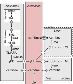
he role of carnitine in biology and disease has been studied for decades (1, 2). Carnitine has been proposed to be a con-ditionally essential nutrient, and even termed vitamin BT.Car-nitine content of foods varies widely, being very low in fruits, vegetables, and grains; intermediate in milk products, eggs, chicken, andfish; and very high in red meats. The proportion of carnitine derived from the diet varies widely in humans, being quite low in vegetarians and especially low with a vegan diet that excludes dairy products and eggs. In contrast, about 75% of carnitine is derived from the diet in meat eaters. Carnitine ho-meostasis in humans (Fig. 1) is maintained by a modest rate of endogenous synthesis, absorption from dietary sources, and efficient tubular reabsorption by the kidney. Apart from the dietary intake, carnitine is synthesized in humans in kidney, liver, and brain from protein-derived 6-N-trimethyllysine (TML) via
3-hydroxy-6-N-trimethyllysine (HTML), 4-N-trimethylaminobutyr-aldehyde (TMABA), and 4-N-trimethylaminobutyric acid [γ-butyrobetaine (γBB)] (3). Renal resorption plays an important role in carnitine metabolism, with considerable excretion if car-nitine intake is abundant, but there is extremely efficient re-sorption if body stores of carnitine are low. Carnitine is present as free carnitine and as acylcarnitines, of which the latter reflect the cellular acyl-CoA ester pool. Up to 99% of carnitine is in-tracellular and is essential for mitochondrial function, where its role is to enable transport of fatty acids into mitochondria, where β-oxidation takes place (3); it is also involved in the transport of peroxisomal oxidation products to the mitochondria.
Carnitine deficiency can develop secondary to dietary inade-quacy or as an adverse effect of medical treatments. Although humans can synthesize carnitine, nutritional deficiency can occur, as when infants were fed early preparations of soy formulas that were deficient in carnitine (4). Similarly, deficiency can arise with
Author contributions: P.B.S.C.-S., S.V., S.J.S., R.J.A.W., C.A.S., F.M.V., and A.L.B. designed research; P.B.S.C.-S., S.V., E.L.C., R.L., A.C.L., B.S., K.L., C.L., R.E.P., T.J.M., J.R.G., N.H., S.J.S., C.A.S., F.M.V., and A.L.B. performed research; K.G., C.A.S., and S.M.L. contributed new reagents/analytic tools; P.B.S.C.-S., S.V., E.L.C., R.L., A.C.L., E.D., G.C., B.S., K.L., R.E.P., T.J.M., N.H., M.S., D.T.-D., P.S., W.R., B.F., R.J.S., R.E.S., J.D.B., C.B., S.W.S., S.J.S., D.H.G., J.S.S., M.E.H., R.J.A.W., C.A.S., S.M.L., E.H.C., R.P.G.-K., F.M.V., and A.L.B. analyzed data; and P.B.S.C.-S., S.V., F.M.V., and A.L.B. wrote the paper.
Conflict of interest statement: Multiple authors are based in the Department of Molecular and Human Genetics at Baylor College of Medicine, which offers extensive genetic labo-ratory testing, and Baylor College of Medicine derives revenue from this activity. The department offers biochemical and molecular diagnostic testing for trimethyllysine hy-droxylase, epsilon gene deficiency. P.B.S.C.-S., S.V., F.M.V., and A.L.B. have filed a patent related to some of the work reported.
This article is a PNAS Direct Submission.
Freely available online through the PNAS open access option. 1P.B.S.C.-S., S.V., F.M.V., and A.L.B. contributed equally to this work.
2Present address: Department of Pediatrics, Washington University School of Medicine, St. Louis, MO 63110.
3To whom correspondence should be addressed. E-mail: abeaudet@bcm.edu. This article contains supporting information online atwww.pnas.org/lookup/suppl/doi:10. 1073/pnas.1120210109/-/DCSupplemental. GENET ICS INAUGU RAL ARTICLE
parenteral alimentation in neonates (5). Carnitine deficiency can also occur secondary to administration of pivalate-conjugated antibiotics or valproic acid (2). Various disease processes and medical interventions, such as renal tubular disorders and chronic hemodialysis, respectively, can also be associated with carnitine deficiency.
There are primary and secondary genetic forms of carnitine deficiency (6). Secondary deficiency is caused by various fatty oxidation defects and organic acidemias that lead to carnitine deficiency through urinary loss of acylcarnitines that accumulate related to the enzyme deficiency. Primary systemic carnitine de-ficiency is caused by biallelic loss-of-function mutations in the SLC22A5 gene that encodes the plasma membrane organic cation transporter-2 (OCTN2). OCTN2 deficiency is characterized by excessive urinary loss of carnitine, leading to systemic deficiency with associated skeletal myopathy, cardiomyopathy, fatty liver, and hypoglycemia. Although the possibility of a primary systemic carnitine deficiency caused by a defect in carnitine biosynthesis was postulated long ago, no primary disorders of carnitine bio-synthesis have been described until now (7).
Administration of carnitine is the centerpiece of therapy for systemic carnitine deficiency, and it is beneficial in some genetic forms of secondary carnitine deficiency. Administration of car-nitine and acetylcarcar-nitine has been explored as an antioxidant and for treatment of many disorders, including diabetic peripheral neuropathy (8), heart failure (9), and mitochondrial disorders.
dioxygenase (TMLD) gene [also known as trimethyllysine hydrox-ylase, epsilon (TMLHE)] while studying probands with autism, raising the question of whether there might be an association of autism withTMLHE mutations (10). TMLHE maps to the long arm of the X chromosome near the boundary of the pseudoautosomal region and encodes TMLD. TMLD is thefirst enzyme of the car-nitine biosynthesis pathway (3) and is localized in mitochondria (11). The etiology of severe, dysmorphic autism with a male/female ratio of 3.2:1 (12) has become increasingly well defined as often attributable to de novo mutations or recent mutations trans-mitted for a few generations. These mutations include large copy number variants (CNVs), which are detectable by chromosomal microarray analysis in up to 25% of the most severe cases with phenotypes that include major intellectual disability, which re-stricts reproduction (13). De novo point mutations are also being discovered as causes of autism using next-generation sequencing of genomic DNA (14). Disease-causing CNVs are found in ∼10% of patients with intermediate phenotypes, often with less severe intellectual disability (15, 16). In these cases, penetrance may be incomplete and the phenotype can be highly variable, with diagnoses of intellectual disability, autism, schizophrenia, and idiopathic epilepsy seen with the same CNV (17–19). These examples typify single-locus conditions, perhaps with genetic and nongenetic modifier effects. Autism spectrum disorders and related neurocognitive phenotypes blend into even more com-plex genotype-phenotype relationships with evidence for two-hit or two-locus pathogenesis (20). At the milder end of the autism spectrum are patients who often have speech, have an in-telligence quotient (IQ) ranging from low to the normal range, and are nondysmorphic. This milder population, some of whom meet diagnostic criteria for Asperger syndrome, can display up to an 8:1 male/female ratio (21, 22), and will be referred to herein as having nondysmorphic autism (NDA). This includes the milder portion of the autism spectrum, but patients who have NDA can have severe cognitive and behavioral phenotypes. The etiology of NDA remains almost completely unknown, but the extreme sex ratio may provide a clue as to its etiology.
In this paper, we show that TMLHE deficiency is a very common inborn error of metabolism in males and suggest that it may be significantly more frequent in autistic male-male sib pairs than in controls.
Results
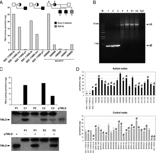
Deletions of Exon 2 Are Heterogeneous and Common in Autistic and Healthy Males. Given the discovery of a deletion of exon 2 in TMLHE in a male simplex proband with autism (10), we exam-ined the frequency ofTMLHE mutations in autism and control populations. We studied simplex families primarily from the Simons Simplex Collection (SSC) and multiplex families pri-marily from the Autism Genetic Resource Exchange (AGRE) collection, and we recruited multiple collaborators to study ad-ditional families (Table 1). We have now identified a total of 16 male autism probands, six affected male siblings of probands, and 24 healthy adult males with deletions of exon 2, indicating that this is a relatively common CNV (Table 1). For 28 of the 29 deletions characterized more thoroughly, size ranged from 5.7 to 15.9 kb and only exon 2 was deleted; one additional deletion of 59.6 kb removed exons 2–6 (Fig. 2 A and B andTable S1). Based on position, size, and sequence, there was a minimum of 14 dif-ferent deletion junctions among 29 unrelated families. Sequenc-ing of the breakpoints of many deletions showed that almost all junctions occurred in long interspersed elements and short in-terspersed elements in the intronsflanking exon 2 (Fig. 2B and
Table S2), as has been seen in other loci (23). For all SSC samples
(probands, heterozygous mothers, and healthy fathers), the de-letions were present in both DNA extracted directly from blood and DNA extracted from lymphoblastoid cell lines (LCLs).
Fig. 1. Carnitine biosynthesis and homeostasis in humans. Carnitine is syn-thesized in four enzymatic steps. After release of TML by lysosomal protein degradation, this compound is hydroxylated by TMLD, producing HTML. HTML is cleaved by HTML aldolase (HTMLA) into TMABA and glycine. Sub-sequently, TMABA is oxidized by TMABA-dehydroxygenase (TMABA-DH) to form 4-N-trimethylaminobutyrate, also namedγ-butyrobetaine (γBB). Fi-nally,γBB is hydroxylated by γBBD, yieldingL-carnitine. Because TMLD is
located in mitochondria, TML needs to be transported out of the lysosome and across the inner mitochondrial membrane into the mitochondrial matrix by means of transporters, which are unknown at this time. Depending on the subcellular localization of the HTLMA (also uncertain, likely the cytosol), HTML orγBB needs to be transported back to the cytosol (where γBBD is located). In cells that do not containγBBD, γBB is exported from the cell and imported into tissues (liver, kidney, and brain in humans) that do express γBBD by means of at least one specific transporter, presumably SLC6A13. Carnitine is transported by OCTN2 and other lower affinity transporters (not shown).
In addition, we identified an extremely common intronic de-letion (Fig. 2A) that appeared on the basis of comparative ge-nomic hybridization (CGH) array to be indistinguishable in all cases. This is equivalent to Database of Genomic Variants num-bers 115349 and 104572, which appear to be identical, and/or to number 97130, which is very similar. The intronic deletion was present in 74% of 93 autism male probands and 71% of 48 control males examined, and it should not be misinterpreted as causing enzyme deficiency. This intronic deletion was present on 24 of 29 chromosomes from unrelated families with exon 2 deletion of TMLHE (Table S1).
Genomic sequencing of exons forTMLHE is complicated by the presence of two pseudoexons (7aP and 8aP) that are highly homologous to exons 7a and 8a, and are imbedded in a large inverted repeat downstream ofTMLHE (24) (Fig. 2C). In addi-tion, there are two alternative exons 7b and 8b, which are located between the two inverted repeats and have sequences unrelated to 7a and 8a. Sequencing of exons 1–8 of TMLHE from genomic DNA in 536 SSC autism male probands, 98 AGRE probands, and 443 National Institute of Mental Health (NIMH) controls iden-tified very few point mutations (Table S3). In addition to exon 2 deletion, sequencing in a multiplex AGRE family (AU 0177) identified an arginine-to-glutamine change in codon 241 (R241Q) in the mother and unaffected half-brother of the two autistic males (Fig. S1). More recently, we have been able to study plasma from 156 male SSC probands for carnitine biosynthesis metabo-lites. We identified one male (Table S3) with biochemical ab-normalities similar to those described below, and sequencing identified an R70H mutation likely causing TMLHE deficiency.
Exon 2 Deletion Results in Loss of TMLD Activity and Absence of TMLD Protein.The functional effect of deletion of exon 2 ofTMLHE was examined as TMLD enzyme activity based on its role in carnitine biosynthesis. Cultured LCLs from males with deletion of exon 2 had low or undetectable TMLD enzyme activity, and heterozygous mothers had reduced activity compared with healthy males (Fig. 3A). Results from family AU 0177 were complex, with the two affected brothers with deletion of exon 2 having very low or undetectable enzyme activity but the un-affected mother and half-brother showing low activity but higher than that of the affected brothers (Fig. 3A and B). The mother is a compound heterozygote and transmitted the R241Q mutation to the unaffected half-brother of the siblings with autism. Cells with exon 2 deletion also lacked immunodetectable protein by Western blot analysis (Fig. 3C). Many of the control and autism cell lines in Fig. 3D have the intron 1 deletion, and many do not. Analysis of RNA from LCLs using RT-PCR revealed low levels of skipping of exon 2 in most samples and a stable transcript with complete absence of exon 2 in cells from males with deletion of this exon (Fig. S2A). Thus, nonsense-mediated decay is not prominent for the exon 2 deletion transcript. This was confirmed using a quantitative RT-PCR assay, which showed normal levels of transcript for the exon 5/6 junction in all samples but complete absence of the exon 1/2 junction in deletion samples (Fig. S2B).
Diagnostic Metabolite Abnormalities in Plasma and Urine.The two affected brothers from AGRE family AU 0177 had a normal facial appearance in childhood and were otherwise nondysmorphic. They both had normal plasma free carnitine levels (33 and 34μmol/L, normal = 22–65 μmol/L) at the recent ages of 15 and 17 y. In urine of the affected brothers, HTML and γBB were undetectable and the excretion of TML was threefold that of controls (Fig. 4A). Plasma from the two brothers and from 5 SSC probands showed a significant increase in TML, complete absence of HTML, and severely reduced levels ofγBB, except for one case (Fig. 4B). The (HTML + γBB)/TML ratio was very low in patients compared with control plasma and may be an excellent index of TMLD activity (Fig. 4C). These data indicate that TMLHE de-ficiency represents a unique inborn error of carnitine biosynthesis. To search for other evidence of a commonTMLHE deficiency and for other defects in carnitine biosynthesis, urine from 29 SSC probands who did not have any known TMLHE mutations was studied and did not reveal any abnormal carnitine metabolites.
Sex Ratio in NDA Is Not Caused by a Common Inherited Mutation in TMLHE.It was important to determine whether there was a com-mon mutation or epigenetic mechanism causing TMLD enzyme deficiency, and perhaps explaining the male predominance in some forms of autism. To address the possibility of a common but difficult to detect inherited mutation, we analyzed SNP data from Illumina arrays on 411 AGRE families; this revealed no evidence of linkage forTMLHE, which is the most telomeric gene on Xq that is not on Y, or forVAMP7, which is nearby but across the pseudoautosomal boundary and present on both the X and Y chromosomes. For the TMLHE region, a maximum nonpara-metric linkage score of 1.25 and logarithm of odds (LOD) score of 0.34 were observed for markers located within andflanking the TMLHE gene. For the VAMP7 gene region, a maximum non-parametric linkage score of 0.76 and LOD score of 0.15 were observed. Thus, there was no evidence for linkage at either locus. This is not surprising, given the extensive genetic heterogeneity in autism.
TMLHE Deficiency Likely Is a Risk Factor for Autism.Because deletion of exon 2 was more common than any other mutations detect-able by genomic sequencing, and because it was associated with loss of enzyme activity, it was expedient to analyze a large series of autism cases and controls for exon 2 deletion. A PCR assay
Table 1. Sources of male autism and control samples and methods of testing No. Deletion Simplex autism SSC 1,887 6* SCAP 80 0 Houston 24 0 Toronto 328 3 Paris 333 0 New York 252 0 Totals 2,904 9
Male probands from male-male sibling pairs†
AGRE + NIMH 752 7
Toronto 93 0
Paris 53 0
New York 11 0
Totals 909 7
Male probands from male-female sibling pairs†
Paris 38 0
New York 5 0
Controls
SSC, NIMH, and AGRE autism fathers 2,197 7 NIMH controls 897 3 BPR 49 1 Houston fathers 36 0 Multiplex fathers 615 0 WTCCC 3,018 9 Toronto 1,975 4 Totals 8,787 24
SCAP, South Carolina Autism Project; WTCCC, Wellcome Trust Case –Con-trol Consortium.
*Screening for the deletion and confirmation were performed as described in Materials and Methods.
†Excludes affected sibling.
GENET
ICS
INAUGU
RAL
was designed with primers slightly outside the boundaries of exon 2 to give a product of 538 bp in normal males but no product for males with deletion of exon 2. An example of the PCR assay with an internal control product is shown inFig. S3. If a sample failed to give a PCR product for exon 2, the presence or absence of the deletion was then confirmed using CGH custom array with densely spaced oligonucleotides interrogating the TMLHE region, as shown in Fig. 2A. For PCR analysis, we fo-cused entirely on males because the assay did not reliably detect the deletion in heterozygous females.
Using the PCR assay, we tested simplex male probands pri-marily from the SSC with lesser numbers of probands from the South Carolina Autism Project and from probands from Houston, TX. We also tested male controls, including SSC fathers of autism probands, NIMH controls, and Baylor Polymorphism Resource (BPR) controls. With the collaboration of the laboratories of two of the authors (J.S.S. and D.H.G.), we tested multiplex probands (here, multiplex refers to male-male sibling pairs both affected with autism) from the AGRE, NIMH, and Nashville collections. We subsequently developed collaborations with the laboratories of four of the authors (S.W.S., C.B., J.D.B., and M.E.H.) to expand the data for exon 2 deletion in autism male probands and control males. Some collaborating laboratories used quantitative PCR (qPCR) or existing Affymetrix 6.0 array data as the primary test for deletion of exon 2, as specified in Table 1. All deletion probands were validated, and approximate coordinates were determined using the customTMLHE array. Deletions in control males from the laboratories of S.W.S. and M.E.H. were not validated.
Comparison of the data for male probands from simplex families (9 in 2,904 or 1 in 323 deleted) with all controls (24 in 8,787 or 1 in 366 deleted) did not provide evidence for an as-sociation (P = 0.44) (Table 1). One SSC proband (11680.p1) also had deletion of chromosome 16p11.2 and was eliminated from
these calculations and from the phenotypic data. We hypothe-sized for numerous reasons reviewed in the discussion that the frequency of exon 2 deletion might be substantially higher in probands from male-male affected sib-pairs. The frequency of exon 2 deletion was 2.85-fold higher in these multiplex probands (7 in 909 or 1 in 130) compared with all male controls, with aP value of 0.023 (Table 1). For each multiplex family, only one affected male (identified as the proband) was tested initially. All genotypes were consistent with the X-linked inheritance; all 17 mothers of probands with deletion of exon 2 were tested and were heterozygous for the deletion, confirming that the deletions were not cell culture artifacts.
We next examined the affected male siblings of the multiplex male probands and found that six of the seven had the same de-letion as the proband. Based on analysis using the Transmission Disequilibrium Test (TDT), the probability of obtaining this re-sult, if there were no association, is 0.012. Using metaanalysis, we calculated a Stouffer’s z-statistic to combine the data for multi-plex probands compared with control males and data from the TDT. We obtained a Z-score of 2.90 and aP value of 0.0037 using Stouffer’s method, suggesting that TMLHE deficiency is a risk factor for autism. If the data from the simplex families are in-cluded in the metaanalysis, the Z-score is−2.81 and the P value is 0.0051, which is only slightly higher than theP value of 0.0037.
Cognitive Function of TMLHE-Deficient Males with Autism Varies Widely. Significant phenotypic information was available for
seven SSC probands, two of three Canadian Genetic probands, seven SSC unaffected fathers, all seven multiplex probands and six affected brothers, and 3 NIMH control males with deletion of exon 2 ofTMLHE (Tables S4andS5). The levels of cognitive and language functioning varied considerably across patients. The full-scale IQ of autistic males with deletion of exon 2 ranged from
found in TMLHE and structure of TMLHE. (A) Array CGH showing eight exonic deletions in TMLHE, with the relative location of TMLHE exons 2–6 aligned above the array CGH plots (GRCh37/hg19 assembly;
http://genome.ucsc.edu). The hori-zontal axis shows chromosome po-sition, and the vertical axis shows the log2ratio of array signal.
Semi-transparent filled boxes on CGH plots highlight the regions of aber-ration; all samples have deletions involving exon 2, and most have a separate deletion in intron 1 (red circle). All samples are autism pro-bands with identifiers found in
Tables S1,S4, andS5. (B) Twenty-four unrelated individuals with deletions involving exon 2 of TMLHE are mapped. Deletion coordinates were determined by array CGH un-less specified by PCR assay. White arrowheads indicate the continua-tion of the delecontinua-tion for SSC 13928. p1. NA 12003 is an unaffected in-dividual whose deletion is published (38) and was better characterized in this study. BPR 664 is an unaffected individual. (C) Diagram of gene structure. Large open arrows repre-sent near-identical inverted repeats. Fa, father; P1, proband; HI, AGRE individuals; #, individual first de-scribed by Celestino-Soper et al. (10).
38 to 143; 5 of 21 with available data were in the range of in-tellectual disability, and 3 of 21 were reported as untestable. One proband had seizures. For six of six cases in which information was available, patients were described as nondysmorphic. With respect to the controls, two of the seven SSC fathers had at least one domain with an elevated broader autism phenotype score based on the self-report Broad Autism Phenotype Questionaire, but the Social Responsiveness Scale rating by significant other and Family History Interview-Interviewee Impression scores were not consistent with the broader autism phenotype.
Assuming a True Association, the Penetrance for Autism inTMLHE Deficiency Would Be Very Low. The majority of males with an exon 2 deletion in the US and UK populations are expected to
be phenotypically “normal” as adults. TMLHE deficiency was present in slightly less than 1% of probands from male-male affected sibling pairs; thus, it would be present in substantially less than 1% of all cases of autism. If we assume an overall frequency of 1 in 100 for autism, with a 4:1 male/female ratio, a frequency of 1 in 350 forTMLHE deficiency in normal males, and a frequency ofTMLHE deficiency of 1 in 250 or 1 in 150 in males with autism, the penetrance would calculate at 2.2% or 3.6%, respectively (SI Materials and MethodsandTable S6). Discussion
TMLHE deficiency is a previously undescribed inborn error of metabolism discovered about 100 y after Garrod described such conditions in his 1908 Croonian Lectures to the Royal College of
Fig. 3. Genetic and enzymatic characterization of hemizygous deletion of exon 2. (A) TMLD activity measured in lymphoblast homogenates of three families with exon 2 deletion. (B) PCR assay results for the AU 0177 family showing the deletion in the two affected brothers (1, 2) and in the mother (3) but not in the father (4), unaffected maternal half-brother (5), or unaffected controls (C1 and C2). There is bias of amplification in the mother, such that the normal band is faint. dl, deletion; nl, normal. (C) (Upper) TMLD activity and Western blot analysis of 2 individuals with exon 2 deletion (P1, HI0690; P2, BPR664) and three controls (C1–C3). Purified TMLD (pTMLD) is used as a positive control. (Lower) Western blot analysis of 2 individuals (P1 and P2) with (+) or without (−) addition of pTMLD, showing the complete absence of protein in cases of exon 2 deletion and confirmation of the identity of the immunoreactive material as TMLD. The upper band in the Western blot is an irrelevant protein. (D) (Upper) TMLD activity measured in lymphoblast homogenates from several autism males. *,TMLHE exon 2 deletion; #, E287K; d, deletion in intron 1 in 13 individuals;−, no deletion in intron 1 in 9 individuals. SSC 12353.p1 was not tested for the presence of intron 1 deletion. (Lower) TMLD activity measured in lymphoblast homogenates from male controls. BPR indicates local unaffected controls, and NA 12003 is an unaffected individual. SSC 12353.fa was not tested for presence of intron 1 deletion. There was no apparent correlation of the level of enzyme activity with the presence or absence of the intronic deletion. For A, C, and D, assays were run in duplicate and the average is plotted without error bars. fa, father; mo, mother; p1, proband.
GENET
ICS
INAUGU
RAL
Surgeons. The frequency ofTMLHE deficiency is startling, at ∼1 in 350 control males of European descent, making it at least 20-fold more frequent than phenylketonuria in males. The enzyme deficiency and metabolite changes in plasma and urine are typ-ical for an inborn error of metabolism.TMLHE appears to be a gene in which deletions are much more common than point mutations. There is precedent for this at the DMD, PMP22, UBE3A, and other loci causing Duchenne muscular dystrophy, hereditary neuropathy with liability to pressure palsies, Angel-man syndrome, and other phenotypes, respectively. These biases are usually explained, in part, by genome architecture, as is likely the case forTMLHE.
It might be of some concern that we did not detect any in-dication thatTMLHE deficiency is a risk factor for simplex au-tism. However, simplex and multiplex groups of autism families are significantly different, with an expectation of higher rates of de novo mutations in simplex compared with multiplex families and a higher rate of inherited mutations in multiplex vs. simplex families. In addition, shared genetic modifiers and shared envi-ronment are potential factors in multiplex families. Given these differences between simplex and multiplex families, the apparent low penetrance ofTMLHE deficiency for autism, and the modest sample size, the negative result for simplex families is not sur-prising. Assuming that the association with multiplex autism is replicated, we would expect that there would be a significant association with simplex autism males with a much larger sample size, because all multiplex families are initially simplex before the birth of a second affected sibling. We would argue that it is not appropriate in this circumstance of low penetrance to combine the probabilities from simplex and multiplex families for an as-sociation of TMLHE deficiency with autism, because they are significantly different samples. However, if one were to do so, the failure to detect an association in a sample of simplex families of this size, given the necessarily low penetrance, has relatively weak statistical significance and does not detract substantially from the P value of 0.0037 observed with male-male multiplex families. We conclude thatTMLHE deficiency is likely to be a weak risk factor for autism, but replication studies are needed, particularly those focusing on male-male multiplex families. The data make it clear thatTMLHE deficiency is neither necessary nor sufficient to cause autism. With roughly 4 million births per year in the United States, this would equate to about 5,600 deficient males born per year, which, in turn, would equate to 168 males with TMLHE deficiency and autism assuming a 3% penetrance.
One might ask whether carnitine metabolism plays a broader role in the etiology of NDA. One possibility is that TMLHE deficiency is entirely benign, as is generally believed to be the case for pentosuria and histidinemia. Alternatively,TMLHE deficiency could mediate harmful effects either through toxic accumulation of
TML or through deficiency of downstream metabolites, including HTML, TMABA,γBB, or carnitine. All these are possible, but we believe that the most attractive hypothesis at this time is that there is an increased risk for autism, and that this risk is modified by dietary intake of carnitine from birth through thefirst few years of life. Carnitine intake of the pregnant or nursing mother could also be important. There are extensive reports of mitochondrial ab-normalities in autism, as reviewed recently (25), and some of the mitochondrial dysfunction could be secondary to carnitine de-ficiency. There are reports of low plasma carnitine in autism (26– 29), but these reports have not prompted intensive investigations into a possible role of carnitine deficiency in autism and further studies are needed.
Another hypothesis could be that other genetic abnormalities involving the carnitine pathway might confer a risk for autism. Features of autism generally are not reported in children with systemic carnitine deficiency, although cases of autism with carnitine deficiency have been reported (26). Given the neuro-logical basis of autism and the prominent expression ofTMLHE in hippocampal neurons and Purkinje cells, one possibility would be that symptoms of autism might be secondary to carnitine deficiency in the brain. If that were the case, the pathophysiology of systemic carnitine deficiency would be very different from TMLHE deficiency. The former has low plasma carnitine, but ability to synthesize carnitine in the brain and elsewhere is intact. In the latter, plasma carnitine may be normal or low-normal based on dietary intake, but neurons are unable to synthesize carnitine and become completely dependent on transfer across the blood–brain barrier. If carnitine deficiency in the brain was deleterious, dietary deficiency, excess renal losses, disorders of transport (especially across the blood–brain barrier), and defects in synthesis might be risk factors for autism. Relatively little is known about transport across the blood–brain barrier, but this transport may be a limiting factor, because the concentration of carnitine in cerebrospinalfluid is 10- to 15-fold lower than in plasma (30, 31). As shown in Fig. 1, not all tissues are capable of complete carnitine biosynthesis because of the differential expression of the last en-zyme,γBB dioxygenase (γBBD), which is only expressed in kidney, liver, and brain in humans. After degradation of proteins that contain TML residues, TML is converted toγBB, which is then transported to the tissues that expressγBBD and converted into carnitine. The plasma membraneγBB transporter likely is enco-ded by theSLC6A13 gene, which is known as a betaine/GABA transporter and has recently been suggested to function in carni-tine biosynthesis as the liverγBB transporter (32). Transport of either or both carnitine andγBB across the blood–brain barrier could be important.
One important question is whether the association with autism is valid and can be replicated in future studies. Although
biosynthesis intermediates in urine of two patients with exon 2 deletion (1, HI0690; 2, HI0691) compared with controls. (B) Box and whisker plot of carnitine biosynthesis intermediates in plasma of seven patients with exon 2 deletion (HI0690, HI0691, SSC 13928.p1, SSC 13489.p1, SSC 11000.p1, SSC 11229.p1, and SSC 11680.p1) compared with controls. (C) Box and whisker plot showing the diagnostic potential of the (HTML +γBB)/HTML ratio as an indicator of TMLHE deficiency. All seven patients have a very low ratio. The black square represents the mean of the controls, and whiskers show the minimum and maximum values of the control group.
TMLHE deficiency was discovered by a genome-wide molecular analysis, aP value of genome-wide significance is not needed here, because this is a simple test of the hypothesis that a newly discovered inborn error of metabolism is a risk factor for autism or not. The metaanalysisP value of 0.0037, indicating an asso-ciation with multiplex autism, suggests that the data indicating an association are unlikely to have occurred by chance. If pene-trance for autism is influenced by carnitine intake during infancy, the risk for autism associated with TMLHE deficiency may be greater in countries with a high frequency of vegetarian diets and lower meat or beef intake. China, India, and South Korea are all countries where some studies of the incidence of autism are available (22, 33, 34) and there is a more vegetarian diet and/or much lower beef intake.
Two clinical investigations are of immediate interest and are being initiated. One is studying carnitine metabolites in cere-brospinalfluid of infants with autism with and without TMLHE deficiency near the age of onset, and the other is treating very young infants with autism with and withoutTMLHE deficiency with carnitine or γBB supplementation. Whether increased carnitine intake before onset of autism might prevent the de-velopment of symptoms would require a more complex study. There is a recent report of a trial of carnitine supplementation in autism suggesting clinical improvement (35), but the study included some patients up to 10 y of age who might be unlikely to respond; it would be desirable to have data from very young patients, preferably nondysmorphic, with and withoutTMLHE deficiency. The data reported here suggest that TMLHE de-ficiency is a risk factor for NDA and that carnitine metabolism could be a target for therapeutic intervention in this and related disorders.
Materials and Methods
Human Subjects and DNA. All work with direct involvement of human subjects was approved by the relevant institutional review boards or equivalents, and informed consent was obtained from all subjects. For SSC, AGRE, and NIMH samples, DNA derived from LCLs was obtained from the Rutgers University Cell and DNA Repository. Additional information is provided inSI Materials and Methods. The numbers of simplex probands, multiplex male-male sib pairs, and controls from various sources are specified in Table 1. Detailed information for cell culture and for identification of deleted probands and controls is given inSI Materials and Methods. For the screening of samples included in Table 1, we used PCR assays (Fig. S2), except for SSC samples, where Illumina arrays (36) were also used; for Toronto and the Wellcome Trust Case–Control Consortium, where Affymetrix 6.0 arrays were used; and for Paris and New York, where qPCR assays were used. All deletions in patients with autism were confirmed using custom arrays for the TMLHE region (Fig. 2A).
PCR and Sanger dideoxy-sequencing of TMLHE exons 1–8 was performed for 536 SSC male probands, 98 affected AGRE males from male-male mul-tiplex families (brothers or half-brothers with the same mother), and 443 NIMH male controls (primers provided inTable S7).
CGH Array. All arrays used in this study were designed and analyzed based on University of California, Santa Cruz (UCSC) Genome Browser hg18 (National Center for Biotechnology Information Build 36, March 2006). The coor-dinates found in tables andfigures are converted to hg19 (Genome Refer-ence Consortium: human GRCh Build 37, February 2009). An Agilent CGH custom array of design ID 028249 was used to confirm TMLHE deletions originally found by PCR assay or those that were detected by the 1M Illu-mina SNP array through a collaborative study of SSC families (36). The cus-tom array design is available on the Agilent’s eArray website (www.agilent. com/genomics/earray). Analysis of CNVs was done using Agilent’s DNA Analytics software (version 4.0.76) with the following settings: aberration algorithm ADM-2, a minimum of three consecutive probes per region, and a minimum absolute average log2ratio of 0.25 for any given region.
The protocol for DNA digestion, labeling, purification, and hybridization to the arrays followed the manufacturer’s instructions with some mod-ifications, as described previously (37). Genomic DNA (800 ng) from the SSC individual and from a single male reference was used in the digestion. Each slide was scanned into an imagefile using the Agilent G2565 DNA
Microarray Scanner at a 3-μm scan resolution. Each image file was quantified using Agilent Feature Extraction software (version 10.7.3.1). The Agilent custom-focused validation files were uploaded into the DNA Analytics software for analysis.
Enzyme Assays and Metabolite Determinations. All individuals tested for TMLD enzyme activity were assayed for the presence or absence of exon 2 of TMLHE by PCR or CGH array. These included BPR controls, AGRE and SSC individuals, and a Centre d’Étude du Polymorphisme Humain control (NA12003) (38). TML was obtained from Sigma–Aldrich. [2H
9]TML and [2H3]γ
BB were synthesized as described previously (11). [2H
9]HTML was prepared
enzymatically by incubating [2H
9]TML with Neurospora crassa TLMD,
het-erologously expressed in Saccharomyces cerevisiae as described previously (39). The resulting mixture of [2H
9]HTML and [2H9]TML was applied to
Amicon Ultra 30-kDafilters (Millipore), and the deproteinized filtrate was used as an internal standard for TML and HTML. All other reagents were of analytical grade.
Lymphoblast pellets were homogenized in 10 mM Mops buffer containing 0.9% (wt/vol) NaCl, 10% (wt/vol) glycerol, and 5 mM DTT (pH 7.4). The protein concentration was determined by the method of Bradford (40) using human serum albumin as a standard. For measurement of TMLD andγBBD activities, the reaction mixture consisted of 20 mM potassium phosphate buffer con-taining 50 mM KCl, 3 mM 2-oxoglutarate, 10 mM sodium ascorbate, 0.5 mM DTT, 0.5 mM ammonium iron sulfate, 2.5 mg/mL BSA, 2 mM TML, and 0.2 mM [2H
3]γBB at pH 7.4, with a final volume of 250 μL. The reaction was
started by adding 50μL of homogenate (target final protein concentration of 0.2 mg/mL for lymphoblast homogenates) to the reaction mixture and was incubated at 37 °C for 30 min. The reaction was terminated by the addition of ZnCl2to afinal concentration of 1 mM, and the reaction
mix-tures were placed on ice. The ZnCl2solution also contained the following
internal standards: 50 pmol of [2H
9]HTML, 140 pmol of [2H9]TML, 140 pmol
of [2H
3]γ-BB, and 550 pmol of [2H3]carnitine. Subsequently, the reaction
mixture was loaded onto an Amicon Ultra 30-kDafilter and centrifuged at 14,000× g for 20 min to separate the metabolites (TML, HTML, γ-BB, and carnitine) from the enzymes and remove most of the proteins. Thefiltrate (100μL) was derivatized with methylchloroformate, and the produced HTML was quantified using ion-pair ultra performance liquid chromatography (UPLC)-tandem MS essentially as previously described (11).
For determination of carnitine biosynthesis metabolites in plasma and urine, internal standards were added to each homogenate and derivatization was performed as described above. Plasma samples were deproteinized using an Amicon Ultra 30-kDafilter. Urine samples were directly derivatized, and TML, HTML, carnitine, andγ-BB were quantified using ion-pair UPLC-tandem MS as previously described (11). For immunoblot analysis, a Multiphor II Nova Blot electrophoretic transfer unit (Amersham Pharmacia Biotech) was used to transfer proteins onto a Protran nitrocellulose membrane (What-man) as described by the manufacturer. After blocking of nonspecific binding sites with 50 g/L Protifar (Nutricia) and 10 g/L BSA in PBS with Tween 20 (1 g/L) for 1 h, the membrane was incubated for 2 h in the same buffer without Protifar with 1:3,000 dilution of rabbit polyclonal antibodies raised against human recombinant TMLD fused to maltose-binding protein (41). Detection was performed with IRDye 800-conjugated goat rabbit anti-body (LI-COR Biosciences) according to the manufacturer’s instructions. Membranes were then dried and scanned using the Odyssey Infrared Im-aging System (LI-COR Biosciences).
TDT, Metaanalysis, and Penetrance Calculations. After 7 of 909 probands from multiplex male-male families were identified, the P value favoring a risk re-lationship of TMLHE deficiency to autism was 0.022 based on a one-sided Fisher’s exact test. This outcome could have occurred by chance or could have occurred because there is indeed a risk relationship. If the result occurred by chance, 3.5 of the seven siblings would be expected statistically to have the deletion, if all mothers are assumed to be carriers. If the result reflects a true risk relationship, a higher proportion, but not necessarily all, of the autistic siblings should have the deletion. Seven of the eight siblings carried the de-letion. Statistical analysis of the sibling data was performed by implementing the TDT (42–45). Because the X chromosome is being analyzed, only trans-missions from the mother are informative. We examined whether or not the deletion had been transmitted to the affected male sibling from his mother and applied McNemar’s χ2(46) to the resulting 2× 2 table to guard against
significant results attributable to population substructure/admixture. To combine the results from the comparison of multiplex probands to controls (P = 0.023) and from the TDT analysis of affected male siblings (P = 0.012), metaanalysis was performed using Stouffer’s method (47). The metaanalysis resulted in a Z-score of 2.90 and a P value of 0.0037.
GENET
ICS
INAUGU
RAL
individuals at risk with an equal number of males and females, as shown in
Table S6. We assumed a frequency of autism of 1 in 100 with a 4:1 male/ female ratio. We assumed that 1 in 350 normal males carried deletion of exon 2 of TMLHE. We then calculated the penetrance assuming that either 1 in 250 or 1 in 150 males with autism carries the deletion.
ACKNOWLEDGMENTS. We thank all the families and the clinicians at the participating Simons Foundation Autism Research Initiative (SFARI) SSC sites and collaborators within the SSC Genetic Consortium; the principal investi-gators of the SSC Genetic Consortium are listed elsewhere (36). We also thank many other clinicians and families from around the world for making samples available. We are grateful for the participating AGRE Consortium families and all the resources provided by AGRE. We especially acknowledge access to the Illumina AGRE genotype data submitted by Dr. Hakon Hako-narson at the Center for Applied Genomics at The Children’s Hospital of Philadelphia. We thank Dr. Joachim Hallmayer for help in recruiting Autism Genome Project investigators. This study makes use of data generated by the Wellcome Trust Case–Control Consortium. A full list of the investigators who contributed to the generation of the data is available atwww.wtccc.org.uk. J.S.S. acknowledges the assistance of Melissa Potter in the Computational Genomics Core for managing family, sample, genetic, and phenotypic data for samples examined at Vanderbilt University. A.L.B. thanks Dr. Karen
Zoghbi for helpful discussions and critical reading of the manuscript, and Dr. John Belmont and Gladys Zapata for providing LCLs and DNA from the BPR collection. F.M.V. acknowledges assistance from the Metabolite/Mass Spec-troscopy section of the laboratory Genetic Metabolic Diseases for technical assistance and L. IJlst for helpful discussions. C.B. acknowledges all the fam-ilies and the clinicians participating in the Paris Autism Research Interna-tional Sibpair study, particularly Christopher Gillberg, Marion Leboyer, Mary Coleman, Maria Rastam, Gudrun Nygren, and Richard Delorme. The AGRE is a program of Autism Speaks and is supported, in part, by Grant 1U24MH081810 from the National Institute of Mental Health (to Clara M. Lajonchere). Part of this work was supported by Grant SFARI 124827 from the Simons Foundation (to the investigators of the SSC Genetic Consor-tium) and Grant HD-37283 (to A.L.B) and Grant P30HD-0240640 from the National Institutes of Health. Part of this work wasfinancially supported by the Fundação para a Ciência e Tecnologia, Lisbon, Portugal, by Grant SFRH/BD/38074/2007 (to. S.V.). Part of this work was supported by National Institutes of Health Grants R01 MH061009 and R01 NS049261 (to J.S.S.). Funding for part of this work was provided by the Wellcome Trust under Award 076113 and by Grant 077014/Z/05/Z. Funding for the Paris Autism Research International Sibpair study was provided, in part, by the Institut National de la Santé et de la Recherche Médicale, Fondation de France, Fondation Orange, Fondation pour la Recherche Médicale, Assistance Pub-lique–Hôpitaux de Paris, and the Swedish Science Council.
1. Conference Proceedings (2004) Carnitine. The science behind a conditionally essential nutrient. Proceedings of a conference. March 25–26, 2004. Bethesda, MD. Ann N Y Acad Sci 1033:1–197.
2. Flanagan JL, Simmons PA, Vehige J, Willcox MD, Garrett Q (2010) Role of carnitine in disease. Nutr Metab (Lond) 7:30.
3. Vaz FM, Wanders RJ (2002) Carnitine biosynthesis in mammals. Biochem J 361: 417–429.
4. Slonim AE, et al. (1981) Dietary-dependent carnitine deficiency as a cause of non-ketotic hypoglycemia in an infant. J Pediatr 99:551–555.
5. Schmidt-Sommerfeld E, Penn D (1990) Carnitine and total parenteral nutrition of the neonate. Biol Neonate 58(Suppl 1):81–88.
6. Longo N, Amat di San Filippo C, Pasquali M (2006) Disorders of carnitine transport and the carnitine cycle. Am J Med Genet C Semin Med Genet 142C(2):77–85. 7. Strijbis K, Vaz FM, Distel B (2010) Enzymology of the carnitine biosynthesis pathway.
IUBMB Life 62:357–362.
8. Evans JD, Jacobs TF, Evans EW (2008) Role of acetyl-L-carnitine in the treatment of diabetic peripheral neuropathy. Ann Pharmacother 42:1686–1691.
9. Sarma S, Gheorghiade M (2010) Nutritional assessment and support of the patient with acute heart failure. Curr Opin Crit Care 16:413–418.
10. Celestino-Soper PBS, et al. (2011) Use of array CGH to detect exonic copy number variants throughout the genome in autism families detects a novel deletion in TMLHE. Hum Mol Genet 20:4360–4370.
11. Vaz FM, et al. (2002) Analysis of carnitine biosynthesis metabolites in urine by HPLC-electrospray tandem mass spectrometry. Clin Chem 48:826–834.
12. Miles JH, et al. (2008) Development and validation of a measure of dysmorphology: Useful for autism subgroup classification. Am J Med Genet A 146A:1101–1116. 13. Jacquemont M-L, et al. (2006) Array-based comparative genomic hybridisation
iden-tifies high frequency of cryptic chromosomal rearrangements in patients with syn-dromic autism spectrum disorders. J Med Genet 43:843–849.
14. O’Roak BJ, et al. (2011) Exome sequencing in sporadic autism spectrum disorders identifies severe de novo mutations. Nat Genet 43:585–589.
15. Sebat J, et al. (2007) Strong association of de novo copy number mutations with autism. Science 316:445–449.
16. Marshall CR, et al. (2008) Structural variation of chromosomes in autism spectrum disorder. Am J Hum Genet 82:477–488.
17. van Bon BW, et al. (2009) Further delineation of the 15q13 microdeletion and du-plication syndromes: A clinical spectrum varying from non-pathogenic to a severe outcome. J Med Genet 46:511–523.
18. Sebat J, Levy DL, McCarthy SE (2009) Rare structural variants in schizophrenia: One disorder, multiple mutations; one mutation, multiple disorders. Trends Genet 25: 528–535.
19. Shinawi M, et al. (2009) A small recurrent deletion within 15q13.3 is associated with a range of neurodevelopmental phenotypes. Nat Genet 41:1269–1271.
20. Girirajan S, et al. (2010) A recurrent 16p12.1 microdeletion supports a two-hit model for severe developmental delay. Nat Genet 42:203–209.
21. Scott FJ, Baron-Cohen S, Bolton P, Brayne C (2002) Brief report: Prevalence of autism spectrum conditions in children aged 5-11 years in Cambridgeshire, UK. Autism 6: 231–237.
22. Kalra V, Seth R, Sapra S (2005) Autism—Experiences in a tertiary care hospital. Indian J Pediatr 72:227–230.
23. Boone PM, et al. (2011) Alu-specific microhomology-mediated deletion of the final exon of SPAST in three unrelated subjects with hereditary spastic paraplegia. Genet Med 13:582–592.
24. Monfregola J, et al. (2007) Functional characterization of the TMLH gene: Promoter analysis, in situ hybridization, identification and mapping of alternative splicing variants. Gene 395(1–2):86–97.
25. Rossignol DA, Frye RE (2012) Mitochondrial dysfunction in autism spectrum disorders: A systematic review and meta-analysis. Mol Psychiatry 17:290–314.
26. Gargus JJ, Imtiaz F (2008) Mitochondrial energy-deficient endophenotype in autism. American Journal of Biochemistry and Biotechnology 4(2):198–207.
27. Mostafa GA, El-Gamal HA, El-Wakkad ASE, El-Shorbagy OE, Hamza MM (2005) Polyunsaturated fatty acids, carnitine and lactate as biological markers of brain en-ergy in autistic children. International Journal of Child Neuropsychiatry 2(2):179–188. 28. Lombard J (1998) Autism: A mitochondrial disorder? Med Hypotheses 50:497–500. 29. Filipek PA, Juranek J, Nguyen MT, Cummings C, Gargus JJ (2004) Relative carnitine
deficiency in autism. J Autism Dev Disord 34:615–623.
30. Rubio JC, et al. (1998) Cerebrospinalfluid carnitine levels in patients with Alzheimer’s disease. J Neurol Sci 155(2):192–195.
31. Shinawi M, Gruener N, Lerner A (1998) CSF levels of carnitine in children with men-ingitis, neurologic disorders, acute gastroenteritis, and seizure. Neurology 50: 1869–1871.
32. Fujita M, et al. (2009) Hepatic uptake of gamma-butyrobetaine, a precursor of car-nitine biosynthesis, in rats. Am J Physiol Gastrointest Liver Physiol 297:G681–G686. 33. Wong VC, Hui SL (2008) Epidemiological study of autism spectrum disorder in China.
J Child Neurol 23(1):67–72.
34. Kim YS, et al. (2011) Prevalence of autism spectrum disorders in a total population sample. Am J Psychiatry 168:904–912.
35. Geier DA, et al. (2011) A prospective double-blind, randomized clinical trial of levo-carnitine to treat autism spectrum disorders. Med Sci Monit 17:PI15–PI23. 36. Sanders SJ, et al. (2011) Multiple recurrent de novo copy number variations (CNVs),
including duplications of the 7q11.23 Williams-Beuren syndrome region, are strongly associated with autism. Neuron 70:863–885.
37. Ou Z, et al. (2008) Bacterial artificial chromosome-emulation oligonucleotide arrays for targeted clinical array-comparative genomic hybridization analyses. Genet Med 10:278–289.
38. McCarroll SA, et al.; International HapMap Consortium (2006) Common deletion polymorphisms in the human genome. Nat Genet 38:86–92.
39. Swiegers JH, Vaz FM, Pretorius IS, Wanders RJ, Bauer FF (2002) Carnitine biosynthesis in Neurospora crassa: Identification of a cDNA coding for epsilon-N-trimethyllysine hydroxylase and its functional expression in Saccharomyces cerevisiae. FEMS Micro-biol Lett 210(1):19–23.
40. Bradford MM (1976) A rapid and sensitive method for the quantitation of microgram quantities of protein utilizing the principle of protein-dye binding. Anal Biochem 72: 248–254.
41. Vaz FM, Ofman R, Westinga K, Back JW, Wanders RJ (2001) Molecular and Bio-chemical Characterization of Rat epsilon-N-Trimethyllysine Hydroxylase, the First Enzyme of Carnitine Biosynthesis. J Biol Chem 276:33512–33517.
42. Spielman RS, McGinnis RE, Ewens WJ (1993) Transmission test for linkage disequi-librium: The insulin gene region and insulin-dependent diabetes mellitus (IDDM). Am J Hum Genet 52:506–516.
43. Ewens WJ, Spielman RS (1995) The transmission/disequilibrium test: History, sub-division, and admixture. Am J Hum Genet 57:455–464.
44. McGinnis RE, Ewens WJ, Spielman RS (1995) The TDT reveals linkage and linkage disequilibrium in a rare disease. Genet Epidemiol 12:637–640.
45. Spielman RS, Ewens WJ (1996) The TDT and other family-based tests for linkage disequilibrium and association. Am J Hum Genet 59:983–989.
46. McNemar Q (1947) Note on the sampling error of the difference between correlated proportions or percentages. Psychometrika 12(2):153–157.
47. Stouffer SA, Suchman EA, DeVinney LC, Star SA, Williams RM, Jr. (1949) Adjustment During Army Life (Princeton Univ Press, Princeton).