REVIEW
Molecular and cellular basis for pathogenicity
of autoantibodies: lessons from murine
monoclonal autoantibodies
Lucie Baudino&Samareh Azeredo da Silveira&Munehiro Nakata&Shozo Izui
Received: 22 May 2006 / Accepted: 30 May 2006 / Published online: 5 September 2006
#Springer-Verlag 2006
Abstract The pathogenesis of autoantibody-mediated cel-lular and tissue lesions in autoimmune diseases is most straightforwardly attributable to the combined action of self-antigen binding properties and effector functions associated with the Fc regions of the different immuno-globulin (Ig) isotypes. The analysis of two different sets of monoclonal autoantibodies derived from lupus-prone mice revealed remarkable differences in the pathogenic potentials of different IgG subclasses: (1) the IgG2a and IgG2b subclasses of anti-red blood cell (RBC) autoantibodies are the most pathogenic and efficiently activate two classes of activating IgG Fc receptors (FcγRIII and FcγRIV) and complement; (2) the IgG3 subclass is less pathogenic and activate only complement; and (3) the IgG1 subclass is the least pathogenic and interact only with FcγRIII. In addition, because of the unique property of IgG3 to form self-associating complexes and generate cryoglobulins, this subclass of rheumatoid factor and anti-DNA autoantibodies became highly pathogenic and induced lupus-like nephritis and/or vasculitis. Since the switch to IgG2a and IgG3 is promoted by Th1 cytokine interferon γ, these results strongly suggest that Th1 autoimmune responses could be critically involved in the generation of more pathogenic autoantibodies in systemic lupus erythematosus. This finding is consistent with the observation that the progres-sion of murine lupus nephritis is correlated with the relative dominance of Th1 autoimmune responses. Finally, the
analysis of IgG glycosylation pattern revealed that more sialylated IgG autoantibodies remained poorly pathogenic because of limited Fc-associated effector functions and loss of cryoglobulin activity. This suggests that the terminal sialylation of the oligosaccharide side chains of IgG could be a significant factor determining the pathogenic potential of autoantibodies. Our results thus underline the importance of subpopulations of autoantibodies, induced by the help of Th1 cells, in the pathogenesis of autoantibody-mediated cellular and tissue injuries.
Keywords SLE . Autoimmune hemolytic anemia . IgG Fc receptor . Complement . Cryoglobulin . IgG glycosylation
Introduction
Autoantibodies are the essential factors for several clinical manifestations associated with systemic lupus erythemato-sus (SLE). Because of the occasional lack of correlation between elevated serum levels of autoantibodies and clinical manifestations, it has long been suggested that only a subset of autoantibodies generated during the course of SLE is indeed pathogenic and that the qualitative aspects of autoantibodies are important for the pathogenesis of autoan-tibody-mediated cellular and tissue injuries. Immunoglobulin (Ig) G autoantibodies such as anti-DNA autoantibodies can provoke tissue lesions as a result of their deposition as a form of immune complexes (ICs) in renal glomeruli and small vessels, resulting in glomerulonephritis and vasculi-tis. Although it is unique in murine SLE, a major pathogenic role for autoantibodies to the endogenous retroviral glycoprotein gp70 has been demonstrated, as gp70–anti-gp70 ICs become apparent in the circulation
L. Baudino
:
S. Azeredo da Silveira:
S. Izui (*)Department of Pathology and Immunology, University of Geneva, 1211 Geneva 4, Switzerland
e-mail: [email protected]
M. Nakata
Department of Applied Biochemistry, Tokai University, Hiratsuka, Kanagawa, Japan
close to the onset of disease and their concentrations rise with the progression of lupus nephritis [25]. Cross-reactivity of anti-DNA antibodies with glomerular autoan-tigens, such as alpha actinin-4 (a protein within the glomerular epithelial slit pore complex), and direct reactiv-ity of a fraction of autoantibodies with glomerular matrix autoantigens, such as heparin sulfate and laminin, have also been postulated as mechanisms of immune deposition [10, 41, 44]. The binding of certain autoantibodies such as antierythrocyte and antiplatelet autoantibodies to their targets can lead to direct cellular damage, causing hemo-lytic anemia and thrombocytopenia. Moreover, a separate group of autoantibodies are directed against phospholipids complexed to β2-glycoprotein 1 and associated with thrombotic complications called antiphospholipid syn-drome, which is a major clinical problem in some lupus patients [28,38].
The structure of the Fab region defines the specificity and affinity with which autoantibodies bind to self-antigen. In addition, the Fc region plays a critical role in autoantibody pathogenicity by activating IgG Fc receptor (FcγR)-bearing effector cells, by activating the complement cascade, and by inducing IgM multivalency-dependent agglutination of target cells. Thus, the pathogenic potential of autoantibodies is attributable to the combined action of the self-antigen binding properties of the Fab region and the effector functions associated with the Fc region of the different Ig isotypes. Therefore, it is conceivable that a change of IgG subclass can remarkably modify the pathogenicity of autoantibodies because of alterations in Fc-dependent effector functions. Notably, the spontaneous development of murine lupus nephritis is dependent on the activation of complement [67] and FcγR-bearing effector cells [8]. Murine immune effector cells express three different classes of activating FcγRs: the high-affinity FcγRI, the recently identified intermediate-affinity FcγRIV, and the low-affinity FcγRIII [46]. These three FcγRs are hetero-oligomeric complexes in which the respective ligand-binding α-chains are associated with the common γ-chain (FcRγ). FcRγ is critical for the cell surface expression of these three activating receptors and for the triggering of their various effector functions, including phagocytosis by macrophages, degranulation by mast cells, and antibody-dependent cell-mediated cytotoxicity by NK cells.
In an attempt to better define the importance of certain subpopulations of autoantibodies in the pathogenesis of autoantibody-mediated cellular and tissue injuries, we have assessed the pathogenic activity of two different sets of autoantibodies: anti-red blood cell (RBC) monoclonal autoantibodies derived from lupus-prone NZB mice and anti-IgG2a rheumatoid factors (RFs) derived from lupus-prone MRL-Faslpr mice. In this review, we discuss the
molecular and cellular basis of the pathogenic activities of anti-RBC autoantibodies and anti-IgG2a RFs in relation to the effector functions associated with the Fc region of the different Ig isotypes. In addition, we also highlight the role of IgG glycosylation for effector functions and hence pathogenic potential.
Pathogenicity of anti-RBC monoclonal autoantibodies The experimental model of autoimmune hemolytic anemia induced by a single injection of Coombs’ antierythrocyte autoantibodies derived from autoimmune-prone NZB mice presents remarkable advantages for the assessment of the pathogenic activity of autoantibodies. First, the binding of the RBC autoantibodies in vivo after injection of anti-RBC autoantibodies into mice can easily be monitored by flow cytometric analysis of circulating, opsonized RBCs. Second, the pathogenic effect of the anti-RBC autoanti-bodies, i.e., the development of anemia, can be followed by a simple measurement of hematocrit values in mice. Finally, the pathogenic mechanisms of anemia and its severity can be determined through histological analysis of spleen and liver, including evaluation of the extent of erythrophago-cytosis by hepatic Kupffer cells.
Although the molecular nature of the target autoantigens responsible for the induction of anti-RBC autoimmune response has not been well characterized, the analysis of a panel of anti-RBC monoclonal antibodies (mAb) derived from NZB mice has shown that the pathogenic autoanti-bodies (i.e., capable of inducing anemia) recognize only species-specific antigens on mouse RBCs, whereas non-pathogenic autoantibodies cross-react with determinants present on RBCs from many different species [7, 57]. Significantly, comparative analysis between young and aged RBCs revealed that the expression of the murine autoantigen implicated in autoimmune hemolytic anemia progressively increased during the course of RBC aging in the circulating blood [16]. Aged RBCs were preferentially eliminated from the circulating blood following the injec-tion of anti-RBC autoantibodies, despite relatively small differences of anti-RBC binding to young and aged RBCs (less than twofold differences). This emphasizes the biological significance of twofold or even less than twofold differences in the expression levels of autoantigens on target cells in the process of antibody-dependent immune clearance. In marked contrast to the autoimmune determi-nant, the expression level of the CD44, CD47, CD147, and TER-119 epitopes, which are recognized by mAb deliber-ately raised in non-autoimmune animals, rather decreased or remained unchanged with RBC aging. These data unravel the unique molecular feature of RBC epitopes involved in autoimmune hemolytic anemia, suggesting that
age-dependent alterations in cellular membrane proteins might have a critical importance in the development of autoantibody responses to RBCs.
Marked differences in IgG-subclass-dependent pathogenicity of anti-RBC autoantibodies
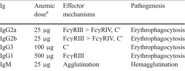
In view of differences in Fc-associated effector functions among the four different IgG subclasses in mice, we have generated IgG subclass switch variants of a high-affinity 34-3C IgG2a anti-RBC autoantibody derived from NZB mice. We then determined the differences in the pathogenic mechanisms between the IgG subclasses by assessing their respective capacities to induce anemia in vivo in mice deficient in the common γ-chains of FcγR (i.e., no functional expression of activating FcγRI, FcγRIII and FcγRIV) or in C3 [3]. The four IgG subclasses of the 34-3C antibody displayed striking differences in their pathogenic activities, which depended on the capacity of the individual IgG subclasses to activate FcγR and/or complement (Tables 1 and 2). IgG2a and IgG2b, which both efficiently interacted with FcγRIII and FcγRIV and activated complement, exhibited the highest pathogenic activity. They were followed by the IgG3 subclass, which activated only complement (but was unable to interact with any FcγR), and then by the IgG1 subclass, interacting only with FcγRIII but failing to activate complement. Overall, IgG2a and IgG2b were 20-fold more potent than IgG1. Role of complement and its inhibitory proteins
Early studies have shown that the development of anemia with a single dose of 34-3C IgG2a or polyclonal rabbit IgG antimouse RBC antibodies was not affected in C3-depleted or C3-deficient mice [57, 58], thus suggesting that complement played no role in the development of autoim-mune hemolytic anemia. However, the analysis of IgG switch variants of the 34-3C mAb with different doses has provided new insight into this issue (Table2): complement activation was indeed involved in the development of anemia through complement receptor (CR)-mediated eryth-rophagocytosis in a dose-dependent and
IgG-subclass-specific manner [3]. For example, C3 deficiency only minimally affected the development of mild anemia at a low dose (50 μg) of the IgG2a subclass, whereas CR-dependent erythrophagocytosis significantly contributed to the development of severe anemia induced by a high dose (200μg) of this subclass in normal mice. This indicates that CR-mediated erythrophagocytosis requires an extensive opsonization of RBCs by C3 fragments. In contrast, a limited opsonization with IgG2a antibodies was sufficient to trigger FcγR-dependent erythrophagocytosis. This is consistent with the finding that despite a lack of appreciable opsonization of RBCs, the low-affinity 4C8 IgG2a mAb was able to induce severe anemia as a result of FcγR-mediated phagocytosis by Kupffer cells [15]. Notably, the IgG3 subclass, which fails to interact with phagocytic FcγR, was able to induce anemia as a result of CR-mediated erythrophagocytosis. Furthermore, the active involvement of complement in autoantibody-mediated autoimmune pathology has also been documented in experimental models of antiglomerular basement membrane nephritis [64], antiphospholipid syndrome [21], and auto-immune vitiligo [65].
It is somehow unexpected that none of the anti-RBC monoclonal autoantibodies, even of the IgM isotype, was able to induce complement-mediated hemolysis. Indeed, the development of anemia after the injection of IgM anti-RBC autoantibodies at any dose was not prevented in mice deficient in C3 or C5 [57]. The lack of involvement of complement-mediated intravascular hemolysis is likely due to the activity of a number of complement inhibitory proteins present on RBC membranes. This idea was supported by the demonstration that 34-3C IgG2a-opson-ized RBCs deficient in the membrane-bound complement regulatory proteins Crry (CR1-related gene/protein y) and DAF (decay accelerating factor) were eliminated more rapidly by complement-dependent clearance than by the FcγR-dependent pathway [42]. Although this study did not directly demonstrate that deficiency in Crry and DAF results in intravascular hemolysis of 34-3C IgG2a-opson-ized RBCs, it has been shown that DAF-deficient RBCs
Table 1 Effector functions of 34-3C anti-RBC Ig class switch variants
Ig FcγRI FcγRIII FcγRIV FcγRIIB C′
IgG2a + ++ ++ + ++
IgG2b − ++ ++ ++ ++
IgG3 − − − − +
IgG1 − + − ++ −
IgM − − − − ++
Table 2 Pathogenic activities of 34-3C anti-RBC Ig class switch
variants and respective contributions of FcγRIII, FcγRIV, and
complement to the development of autoimmune hemolytic anemia
Ig Anemic
dosea
Effector mechanisms
Pathogenesis
IgG2a 25μg FcγRIII > FcγRIV, C′ Erythrophagocytosis
IgG2b 25μg FcγRIII > FcγRIV, C′ Erythrophagocytosis
IgG3 100μg C′ Erythrophagocytosis
IgG1 500μg FcγRIII Erythrophagocytosis
IgM 25μg Agglutination Hemagglutination
were highly sensitive to antibody-induced complement-dependent hemolysis in vitro [39].
It should be stressed that despite strong activation of complement, as documented by C3 opsonization of RBCs in mice injected with IgM anti-RBC mAb, no sign of CR-mediated erythrophagocytosis by Kupffer cells was detect-able. In fact, the pathogenic mechanism responsible for the induction of anemia by IgM is a sequestration of RBCs due to a massive hemagglutination in spleen and occasionally in liver (Table2), which likely interferes with the process of CR-mediated erythrophagocytosis. It should also be em-phasized that 4C8 IgM anti-RBC mAb is as pathogenic as the 34-3C IgM variant, although its affinity is more than 1,000 times weaker than that of 34-3C [15]. This indicates that the affinity of IgM anti-RBC autoantibodies is not of primary importance for their pathogenic activity because its pentameric form dramatically enhances the binding avidity to autoantigens expressed on RBCs. Significantly, the monomeric form of 34-3C IgM variant completely fails to induce anemia, despite its high-affinity binding to circulat-ing RBCs in vivo (manuscript in preparation). The lack of pathogenicity of the monomeric form of IgM anti-RBC mAb can be explained by its inability to induce hemagglu-tination, by its poor activation of complement, and by the absence of phagocytic receptors specific for IgM.
Differential contributions of FcγRI, FcγRIII,
and FcγRIV to IgG-subclass-dependent pathogenicity of anti-RBC autoantibodies
It has been well established that activating FcγRs are critically involved in the pathogenesis of various autoanti-body- and IC-mediated inflammatory disorders [52, 62]. However, the respective contributions of the three different activating FcγRs (FcγRI, FcγRIII, and FcγRIV) have not yet been well defined, except for IgG1 IC-mediated inflammation which is exclusively mediated by FcγRIII [19, 20]. We have recently assessed the development of mild or severe anemia by injecting low or high doses of 34-3C IgG2a or IgG2b mAb in mice deficient in FcγRI, FcγRIII, and/or C3 to define respective contributions of different FcγRs to the development of IgG2a- and IgG2b-mediated autoimmune hemolytic anemia. We have ob-served that the development of mild anemia induced by a low dose of these mAb (25μg) was completely prevented in FcγRIII-deficient mice, whereas the injection of a high dose (200 μg) still induced anemia in mice deficient in FcγRI, FcγRIII, and C3 (manuscript in preparation). This indicates not only a critical role for FcγRIII in the initiation of autoimmune hemolytic anemia, but also a secondary role for FcγRIV in the progression and development of severe anemia (Table2). The limited use of FcγRIV in vivo could
be due to competition with excessive amounts of unbound monomeric IgG2a and IgG2b due to higher affinity of FcγRIV for IgG2a and IgG2b compared with FcγRIII [47]. Thus, only higher densities of 34-3C mAb bound on RBCs may efficiently compete with circulating monomeric IgG2a and IgG2b for FcγRIV binding on phagocytes, while a low density of 34-3C mAb on RBCs can still trigger FcγRIII-dependent erythrophagocytosis because of the absence of the competition. However, it should be stressed that FcγRIV has been shown to play a remarkable role in the development of autoimmune thrombocytopenia induced by the IgG2a and IgG2b subclasses of an antiplatelet mAb [47] and in the development of experimental nephrotoxic glomerulonephritis [29]. This apparent discrepancy could be explained by differential expression levels of FcγRIII and FcγRIV on distinct immune effector cells such as macrophages, Kupffer cells, mast cells, and mesangial cells implicated in triggering IC-mediated inflammatory cascade. Consequently, the respective contributions of both types of FcγRs may be dramatically different, depending on the experimental models studied. In contrast to FcγRIII and FcγRIV, it is expected that the role of FcγRI is relatively limited because of the competition with circulating, unbound, monomeric IgG2a. Again, we cannot exclude the possibility that increased avidity due to extensive opsonization may result in a more efficient binding to FcγRI and contribute to the development of autoimmune hemolytic anemia. Notably, a significant contribution of FcγRI to the development of different IgG2a IC-mediated inflammatory lesions has been reported [4, 22]. Clearly, further studies in mice deficient in different activating FcγRs will provide a more definitive perspective of the respective roles of individual FcγRs in the pathogenesis of autoantibody- and IC-mediated immune disorders.
Murine phagocytic effector cells express an additional low-affinity FcγR, FcγRIIB, which is a single α-chain receptor, in which the cytoplasmic domain contains the inhibitory ITIM (immunoreceptor tyrosine-based inhibition motif) sequence [63]. Upon co-ligation of the inhibitory FcγRIIB with activating FcγR or B-cell antigen receptor, cellular activation is inhibited by the recruitment of an inositol polyphosphate phosphatase, SHIP, which mediates the inhibition of calcium flux. The importance of FcγRIIB for the negative regulation of FcγRI-, FcγRIII-, and FcγRIV-dependent effector functions in vivo has been demonstrated by markedly enhanced anaphylactic and inflammatory immune responses in FcγRIIB-deficient mice [9,45,56,63,66]. However, we have shown that FcγRIIB is unable to down-regulate activating FcγR-mediated phagocytosis of RBCs opsonized with 105-2H IgG1 anti-RBC mAb [56] or 34-3C IgG2a in vivo (unpublished data). These results are in marked contrast with the substantial inhibitory role of FcγRIIB against the development of
immune thrombocytopenia induced by IgG2a and IgG2b antiplatelet mAb [45, 54]. It remains to be determined whether the development of thrombocytopenia following injection of IgG antiplatelet autoantibodies is mediated not only by FcγR-dependent phagocytosis but also by another mechanism (release of cytotoxic agents by activated macro-phages) and whether the latter may efficiently be counter-regulated by the co-engagement of FcγRIIB.
More recent surface plasmon resonance analysis using recombinant proteins of different classes of FcγR α-chains revealed that the binding affinity of FcγRIII for IgG2a and IgG2b subclasses is higher than that for IgG1, whereas the binding affinity of FcγRIIB for IgG2a is lower than that for IgG1 and IgG2b [45]. Notably, FcγRIV displays a comparable affinity for IgG2a and IgG2b, but no affinity for IgG1. These results, together with the fact that IgG2a is the only subclass interacting with FcγRI, confirm that the IgG2a subclass of autoantibodies is the most pathogenic one since it most efficiently interacts with three activating FcγRs (and complement), but less efficiently with inhibi-tory FcγRIIB (Table1).
Pathogenicity of IgG3 monoclonal autoantibodies Analysis of a panel of murine mAb revealed that the IgG3 subclass has a unique physicochemical property, which allows it to self-associate via Fc–Fc interactions and exhibit cryoglobulin activity independently of their specificities [1]. The self-associating property of the IgG3 subclass is necessary, but not sufficient in itself to confer cryoglobulin activity since not all monoclonal IgG3 proteins exhibit cryoglobulin activity. MRL-Faslpr autoimmune mice
defi-cient in Fas apoptosis receptor spontaneously develop a lupus-like syndrome characterized by unique immunopath-ological manifestations such as arthritic-like lesions, necro-tizing vascular lesions of the skin of ears and footpads, and severe glomerulonephritis [2]. In parallel, they produce the highest amounts of cryoglobulins among several lupus-prone mice, including (NZB×NZW)F1 and BXSB mice [1]. Cryoglobulins from MRL-Faslprmice are composed almost
exclusively of IgG of polyclonal origin, in which the IgG3 content is markedly enriched as compared with other IgG subclasses.
Most significantly, a fraction of cryoprecipitating IgG3 monoclonal anti-IgG2a RF and anti-DNA autoantibodies derived from lupus-prone mice are highly pathogenic, generating glomerular and vascular lesions [5,24,34,60]. For example, the implantation of hybridoma cells secreting IgG3 anti-IgG2a RF derived from lupus-prone MRL-Faslpr mice rapidly induced acute glomerulonephritis character-ized by“wire-loop”-like glomerular lesions and cutaneous leukocytoclastic vasculitis [5,34]. The pathogenic potential
of IgG3 autoantibodies has been confirmed by the analysis of Ig class switch variants of 6-19 anti-IgG2a RF mAb [5,17,43]. Furthermore, we have observed that 6-19 IgG3 anti-IgG2a RF mAb induced the development of glomeru-lar but not cutaneous vascuglomeru-lar lesions in Ig-deficient mice lacking the corresponding IgG2a autoantigens [26, 53]. Thus, both RF and cryoglobulin activities are required for the development of skin vasculitis, whereas renal pathoge-nicity is not dependent on the anti-IgG2a RF property. However, studies with 6-19 transgenic mice clearly showed that the nephritogenicity of the 6-19 RF mAb was determined by a unique combination of 6-19 heavy and light chains: 6-19 heavy-chain transgenic mice completely failed to develop any glomerular lesions, in combination with endogenous diverse light chains, despite the presence of substantial amounts of cryoglobulins [30]. However, it remains to be determined whether a cross-reactivity of the 6-19 RF mAb with glomerular antigen is a possible mechanism for the nephritogenicity of the 6-19 RF mAb.
Polymorphonuclear neutrophils (PMN) are the principal inflammatory cells in skin vascular lesions caused by the 6-19 RF mAb since no skin purpuric lesions developed in mice depleted of PMN [18]. Strikingly, mice deficient in C3 still developed typical cutaneous vascular lesions [26]. Thus, complement activation does not play a major role in the pathogenesis of PMN-dependent skin vascular lesions induced by 6-19 IgG3 RF. In contrast, mice deficient in FcγR were unable to develop skin vasculitis following the implantation of 6-19 RF hybridoma cells, but were capable of doing so after the transfer of wild-type FcγR-expressing mast cells [68]. We speculate that the activation of mast cells, through the interaction of FcγRIII with IgG2a-IgG3 IC, may result in the release of inflammatory mediators that recruit and activate PMN, thereby generating leukocyto-clastic vascular lesions. Further analysis with mice deficient in tumor necrosis factor (TNF) has demonstrated that TNF released from activated mast cells was one of the mediators responsible for triggering leukocytoclastic vasculitis [68], thus illustrating the clinical significance of TNF in IC-mediated autoimmune vascular syndrome.
In a marked contrast to the pathogenetic mechanisms responsible for cutaneous leukocytoclastic vascular lesions, the development of glomerulonephritis induced by 6-19 IgG3 RF was not dependent on the activation of FcγR-expressing cells [68]. The lack of any inhibition of glomerular lesions in FcγR-deficient mice is consistent with the fact that the development of these glomerular lesions is independent of the formation of IgG3-IgG2a IC [26,53]. It has recently been reported that FcγR-mediated inflammatory responses play an important role in the pathogenesis of lupus nephritis occurring in (NZB×NZW) F1, but not in MRL-Faslpr mice [8,37]. Considering that
amounts of IgG3 cryoglobulins [1], these data suggest that lupus nephritis occurring in MRL-Faslpr mice might be
more dependent on the production of IgG3 autoantibodies, which fail to activate FcγR [3], and thus less dependent on IC formation and FcγR. This is consistent with the observation that the progression of lupus nephritis in MRL-Faslpr mice was well correlated with the level of
circulating IgG3 autoantibodies [59,61].
To better define the immunopathological consequences associated with the chronic presence of pathogenic 6-19 autoantibodies in sera, as opposed to acute and rapid elevation following implantation of 6-19 hybridoma, we have recently established a transgenic mouse line express-ing IgG3 6-19 RF mAb [30]. These transgenic mice developed severe chronic glomerulonephritis, which were highly heterogeneous, as observed in lupus-prone mice, within 8–10 months of age. In addition, about one third of aged transgenic mice developed necrotizing arteritis in small- and medium-sized arteries of the kidney and skeletal muscle, characterized by fibrinoid necrosis and PMN infiltration in association with a perivascular mononuclear cell infiltration. These changes contrasted markedly with the renal and vascular lesions resulting from the rapid induction of high serum levels of 6-19 RF mAb, induced within 8–10 days after implantation of 6-19 hybridoma in normal mice. In the latter situation, mice developed less heterogeneous glomerular lesions, consisting of massive endothelial wire-loop and intracapillary deposits, as well as a cutaneous leukocytoclastic vasculitis in arterioles [5,34]. These results demonstrate that a single autoantibody can induce remarkably different types of glomerular and vascular lesions, depending on its production levels and kinetics.
The demonstration of the pathogenic potential of monoclonal autoantibodies of the IgG3 subclass with cryoglobulin activity is significant because cryoglobulins have long been suggested to be a potential source of tissue injuries in SLE, rheumatoid arthritis, and related autoim-mune diseases [6]. Clearly, the presence of cryoprecipitable
autoantibodies should be reassessed in relation to clinical manifestations to determine whether this subset of autoanti-bodies is a useful and predictive marker of human rheumatic diseases.
Inhibitory effect of sialylation on the pathogenicity of IgG autoantibodies
The CH2 domain of each IgG heavy chain bears a biantennary-complex-type oligosaccharide chain linked to asparagines at position 297, which has been thought to stabilize the IgG molecule and to contribute to Fc-associated effector functions. Most of these oligosaccharide side chains are nonsialylated and end with either two N-acetylglucosamines, one N-acetylglucosamine and one galactose, or two galactose residues (Fig. 1). However, a significant fraction of galactosylated oligosaccharide chains bear one or two terminal sialic acids and hence become more negatively charged. Since the level of galactosylation is highly heterogeneous and substantially diminished in patients with rheumatoid arthritis and in lupus-prone MRL-Faslpr mice [40, 50], differences in the structure of
carbohydrate side chains may have an important role in modulating the pathogenicity of autoantibodies. Since it has been shown that a mutation at position 265 (from aspartic acid to alanine; D265A) leads to a change in the pattern of IgG galactosylation and sialylation [35], we introduced this D265A mutation into the 34-3C IgG2a heavy-chain gene. The activation of complement and interaction with FcγR on phagocytes were dramatically reduced as a result of the introduction of this mutation, and IgG2a D265A mutant became poorly pathogenic (manuscript in preparation). Surface plasmon resonance analysis confirmed that the IgG2a D265A mutant failed to interact with C1q and with all four types of FcγR.
The structural analysis of oligosaccharide side chains isolated from the 34-3C IgG2a D265A mutant has indeed shown increased galactosylation and sialylation as
com-Man
β
1 4GlcNAc
β
1 4GlcNAc
6
3
±Fuc
α
1
± Sia
α
2 6Gal
β
1 4GlcNAc
β
1 2Man
α
1
±
± Sia
α
2 6Gal
±
β
1 4GlcNAc
β
1 2Man
α
1
6
Man
β
1 4GlcNAc
β
1 4GlcNAc
6
3
±Fuc
α
1
± Sia
α
2 6Gal
β
1 4GlcNAc
β
1 2Man
α
1
±
± Sia
α
2 6Gal
±
β
1 4GlcNAc
β
1 2Man
α
1
6
Fig. 1 Structure of authentic biantennary-complex-type oligosaccha-ride chains of IgG. The extent of the terminal sialylation and galactosylation of oligosaccharide side chains is highly variable
(indicated as ±), and more than 95% of oligosaccharides are
fucosylated.Gal galactose, GlcNAc N-acetylglucosamine, Man
pared with its wild-type counterpart. It has been speculated that aspartic acid at position 265 contacts the primary N-acetylglucosamine of oligosaccharide side chains and that this interaction may lead to the sequestration of the nascent oligosaccharide from glycosylation enzymes [35]. Thus, the reduced interaction of alanine with N-acetylglu-cosamine in the D265A mutant could increase the acces-sibility of the nascent oligosaccharide to glycosylation enzymes, thereby increasing galactosylation and sialylation. At present, it is not clear whether the loss of effector functions of the D265A mutant is due to an increased galactosylation and/or an increased sialylation. A previous publication on agalactosylated IgG from patients with rheumatoid arthritis has claimed that exposure of N-acetylgucosamine by agalactosylated IgG could result in an interaction with mannose-binding lectin, thereby acti-vating complement cascade [36]. However, our analysis of glycosylation of 34-3C IgG subclass switch variants showed that the IgG1 variant carries an increased content of agalactosylated glycoforms as compared with other IgG subclasses (unpublished data), but is unable to activate complement. Thus, this result rather argues against the idea that increased complement activation by agalactosylated IgG is mediated through the interaction of N-acetylglucos-amine with mannose-binding lectin.
In addition to the change in galactosylation, we also noted an increased sialylation of the IgG1 class switch variant, the extent of which was comparable with that of IgG2a D265A mutant. These results raise the attractive hypothesis that poor IgG effector functions of the IgG1 subclass may be related to an increased sialylation. An increased sialylation may result in conformational changes of the Fc structure, which may be responsible for the lack of complement activation and for relatively poor interaction with activating FcγR. Alternatively, increases in negative charges due to sialylation may interfere with the interaction of IgG1 with C1q and FcγR. Indeed, charged amino acid residues present at positions 318, 320, and 322 (glutamic acid, lysine, and lysine, respectively), which are also present in IgG1, are involved in C1q binding [11]. In addition, the introduction of negatively charged glutamic acid into the FcγRI-binding motif at positions 234–238 is known to result in a complete loss of FcγRI interaction of IgG2a [12].
In view of the implication in IgG3 cryoglobulin activity of more positively charged amino acid residues at positions 6 and 23 of the heavy-chain variable (VH) regions [32,49], we have recently explored the inhibitory effect of terminal sialic acids on the cryogenic activity of IgG3 mAb. Comparative oligosaccharide structural analysis of different cryogenic and noncryogenic IgG3 mAb showed an inverse correlation between the extent of sialylation and cryogenic activity [31]. The inhibitory role of sialylation was further
confirmed by the demonstration of enrichment of less and more sialylated IgG3 in cryoprecipitated and noncryopre-cipitated fractions, respectively, separated from different cryogenic IgG3 mAb. Interestingly, one of the mAb, 9-106, which is supposed to be cryogenic because of the presence of more positively charged amino acid residues at positions 6 and 23 of the VH region, failed to display cryoglobulin activity, and its content of sialic acids was abnormally elevated due to the presence of additional oligosaccharide chains in its VH region [31]. Furthermore, we have recently observed that the introduction of a mutation in the CH2 domain or in a potential glycosylation site in the VH region of the cryogenic 6-19 IgG3 mAb resulted in an increased sialylation and in the loss of its cryoglobulin activity (unpublished data). Taken together, these results suggest that the content of negatively charged sialic acids in oligosaccharide side chains is a critical factor in determin-ing IgG3 cryoglobulin activity.
Our demonstration of poor pathogenic activity of more sialylated IgG autoantibodies indicates that the level of IgG sialylation could be an important factor in determining the pathogenic potential of autoantibodies. Furthermore, this would be in line with the increased levels of agalactosylated (i.e., asialylated) IgG in patients with rheumatoid arthritis and in lupus-prone MRL-Faslprmice [40,50]. The level of galactosylation and sialylation is likely to be regulated by one of the six isoforms ofβ-1,4-galactosyltransferases and by α2,6-sialyltransferase, respectively. Dysregulated ex-pression of β-1,4-galactosyltransferase and α2,6-sialyl-transferase could be a significant genetic factor predisposing to the development of autoantibody-mediated tissue lesions. Notably, recognition of sialic acid by factor H in the alternative complement pathway is used to dampen complement activation through the inactivation of C3b deposited on self-membranes [13]. Furthermore, α2, 6-linked sialic acid, which is a common structure on asparagine-linked glycans and is abundantly expressed on the surface of many cells, can be recognized by CD22 expressed on B cells. Since CD22 is a negative regulator of B-cell activation, this could be a mechanism to prevent activation of potentially autoreactive B cells [33]. All these data are consistent with the idea that sialic acids play a protective role against self-destruction in higher organisms.
Critical role of the Th1 subset for the development of SLE
The assessment of the pathogenic potential of different monoclonal autoantibodies derived from lupus-prone mice revealed that the IgG2a subclass of anti-RBC autoanti-bodies is the most pathogenic because of the strongest Fc-associated effector functions. Moreover, the IgG3 subclass
of autoantibodies has a unique pathogenic potential to induce development of glomerulonephritis and vasculitis because of the formation of self-associating complexes and cryoglobulins. Interferon (IFN)-γ, the prototype Th1 cytokine, promotes the production of IgG2a and IgG3, and therefore, Th1-type autoimmune responses may be critically important in autoantibody-mediated autoimmune diseases, such as SLE. In contrast, interleukin (IL)-4, the Th2 cytokine, promotes the switching to IgG1, which is the least pathogenic IgG subclass.
Several findings have underscored the importance of Th1 autoimmune responses in the progression of murine lupus nephritis. First, an accelerated development of SLE was observed in MRL-Faslprand MRL.Yaa mice (Yaa is an
as-yet-unidentified mutation able to accelerate the develop-ment of SLE [27]). In these mice, it has been shown that the progression of disease was correlated with an enhanced expression of IFN-γ vs IL-4 mRNA in CD4+
T cells and an increase in spontaneous production of IgG2a and IgG3 vs IgG1 anti-DNA autoantibodies [61]. Second, the develop-ment of lupus nephritis in lupus-prone (NZW×C57BL/6)F1 males bearing the Yaa mutation was almost completely prevented by transgenic expression of IL-4 in B cells; the protected mice had a lack of IgG3, a decrease in IgG2a, and an increase in IgG1 anti-DNA autoantibody production [55]. The protective effect of the IL-4 transgene has also been confirmed in the classical (NZB×NZW)F1 mouse model of SLE (unpublished data). Third, MRL-Faslprmice deficient in the T-box transcription factor T-bet, which plays a critical role in Th1 lineage commitment, were protected from lupus nephritis in association with a decrease in serum levels of IgG2a and IgG3 vs an increase in IgG1 [51].
These results provide evidence for the hypothesis that the Th1 responses determine the development and progres-sion of SLE by promoting the switch towards more pathogenic IgG subclasses of autoantibodies. However, this does not necessarily exclude an active role of Th2 responses in SLE since Th2 cytokines such as IL-5, IL-6, and IL-10 have been suggested to be involved in the induction of autoimmune manifestations in SLE [14, 23, 48]. These effects may be related to the ability of Th2 cytokines to augment overall production of pathogenic autoantibodies by promoting the terminal differentiation of activated B cells to plasma cells. Clearly, further under-standing of the respective roles of Th1 and Th2 in SLE would help establish new strategies for the development of more specific treatments of SLE and related autoimmune diseases.
Acknowledgment The studies from the authors’ laboratory
dis-cussed in this review were supported by a grant from the Swiss National Foundation for Scientific Research.
References
1. Abdelmoula M, Spertini F, Shibata T, Gyotoku Y, Luzuy S, Lambert PH, Izui S (1989) IgG3 is the major source of
cryoglobulins in mice. J Immunol 143:526–532
2. Andrews BS, Eisenberg RA, Theofilopoulos AN, Izui S, Wilson CB, McConahey PJ, Murphy ED, Roths JB, Dixon FJ (1978) Spontaneous murine lupus-like syndromes. Clinical and immuno-pathological manifestations in several strains. J Exp Med 148:1198–1215
3. Azeredo da Silveira S, Kikuchi S, Fossati-Jimack L, Moll T, Saito T, Verbeek JS, Botto M, Walport MJ, Carroll M, Izui S (2002) Complement activation selectively potentiates the pathogenicity of the IgG2b and IgG3 isotypes of a high affinity anti-erythrocyte
autoantibody. J Exp Med 195:665–672
4. Barnes N, Gavin AL, Tan PS, Mottram P, Koentgen F, Hogarth
PM (2002) FcγRI-deficient mice show multiple alterations to
inflammatory and immune responses. Immunity 16:379–389
5. Berney T, Fulpius T, Shibata T, Reininger L, Van Snick J, Shan H, Weigert M, Marshak-Rothstein A, Izui S (1992) Selective pathogenicity of murine rheumatoid factors of the cryoprecipitable
IgG3 subclass. Int Immunol 4:93–99
6. Brouet JC, Clauvel JP, Danon F, Klein M, Seligmann M (1974) Biological and clinical significance of cryoglobulins. A report of
86 cases. Am J Med 57:775–788
7. Caulfield MJ, Stanko D, Calkins C (1989) Characterization of the spontaneous autoimmune (anti-erythrocyte) response in NZB mice using a pathogenic monoclonal autoantibody and its
anti-idiotype. Immunology 66:233–237
8. Clynes R, Dumitru C, Ravetch JV (1998) Uncoupling of immune complex formation and kidney damage in autoimmune
glomeru-lonephritis. Science 279:1052–1054
9. Clynes R, Maizes JS, Guinamard R, Ono M, Takai T, Ravetch JV (1999) Modulation of immune complex-induced inflammation in vivo by the coordinate expressison of activation and inhibitory Fc
receptors. J Exp Med 189:179–185
10. Deocharan B, Qing X, Lichauco J, Putterman C (2002)α-Actinin
is a cross-reactive renal target for pathogenic DNA anti-bodies. J Immunol 168:3072–3078
11. Duncan AR, Winter G (1988) The binding site for C1q on IgG. Nature 332:738–740
12. Duncan AR, Woof JM, Partridge LJ, Burton DR, Winter G (1988) Localization of the binding site for the human high-affinity Fc
receptor on IgG. Nature 332:563–564
13. Fearon DT (1979) Regulation of the amplification C3 convertase of human complement by an inhibitory protein isolated from
human erythrocyte membrane. Proc Natl Acad Sci USA 76:5867–
5871
14. Finck BK, Chan B, Wofsy D (1994) Interleukin 6 promotes
murine lupus in NZB/NZW F1 mice. J Clin Invest 94:585–591
15. Fossati-Jimack L, Reininger L, Chicheportiche Y, Clynes R, Ravetch JV, Honjo T, Izui S (1999) High pathogenic potential of low-affinity autoantibodies in experimental autoimmune
hemolyt-ic anemia. J Exp Med 190:1689–1696
16. Fossati-Jimack L, Azeredo da Silveira S, Moll M, Kina T, Kuypers FA, Oldenborg P-A, Reininger L, Izui S (2002) Selective increase of autoimmune epitope expression on aged erythrocytes in mice: implications in anti-erythrocyte
autoim-mune responses. J Autoimmun 18:17–25
17. Fulpius T, Spertini F, Reininger L, Izui S (1993) Immunoglobulin heavy chain constant region determines the pathogenicity and the antigen-binding activity of rheumatoid factor. Proc Natl Acad Sci USA 90:2345–2349
18. Fulpius T, Lemoine R, Berney T, Pastore Y, Moll S, Izui S (1996) Polymorphonuclear leukocytes play a key role in the generation of
“wire-loop” lesions induced by a murine IgG3 rheumatoid factor.
Kidney Int 49:647–655
19. Hazenbos WLW, Gessner JE, Hofhuis FMA, Kuipers H, Meyer D, Heijnen IAFM, Schmidt RE, Sandor M, Capel PJA, Daëron M, Van de Winkel JGJ, Verbeek JS (1996) Impaired IgG-dependent
anaphylaxis and Arthus reaction in FcγRIII (CD16) deficient
mice. Immunity 5:181–188
20. Hazenbos WLW, Heijnen IAFM, Meyer D, Hofhuis FMA, de Lavalette CR, Schmidt RE, Capel PJA, Van de Winkel JGJ, Gessner JE, van den Berg TK, Verbeek JS (1998) Murine IgG1 complexes trigger immune effector functions predominantly via FcγRIII (CD16). J Immunol 161:3026–3032
21. Holers VM, Girardi G, Mo L, Guthridge JM, Molina H, Pierangeli SS, Espinola R, Xiaowei LE, Mao D, Vialpando CG, Salmon JE (2002) Complement C3 activation is required for antiphospholipid
antibody-induced fetal loss. J Exp Med 195:211–220
22. Ioan-Facsinay A, de Kimpe SJ, Hellwig SMM, van Lent PL, Hofhuis FMA, van Ojik HH, Sedlik C, da Silveira SA, Gerber J, de Jong YF, Roozendaal R, Arden LA, van den Berg WB, Saito T, Mosser D, Amigorena S, Izui S, van Ommen GJB, van
Vugt M, van de Winkel JGJ, Verbeek JS (2002) FcγRI (CD64)
contributes substantially to severity of arthritis, hypersensitivity responses, and protection from bacterial infection. Immunity
16:391–402
23. Ishida H, Muchamuel T, Sakaguchi S, Andrade S, Menon S, Howard M (1994) Continuous administration of anti-interleukin 10 antibodies delays onset of autoimmunity in NZB/W F1 mice. J Exp Med 179:305–310
24. Itoh J, Nose M, Takahashi S, Ono M, Terasaki S, Kondoh E, Kyogoku M (1993) Induction of different types of
glomerulone-phritis by monoclonal antibodies derived from an MRL/lpr lupus
mouse. Am J Pathol 143:1436–1443
25. Izui S, McConahey PJ, Clark JP, Hang LM, Hara I, Dixon FJ (1981) Retroviral gp70 immune complexes in NZB×NZW F2
mice with murine lupus nephritis. J Exp Med 154:517–528
26. Izui S, Fulpius T, Reininger L, Pastore Y, Kobayakawa T (1998) Role of neutrophils in murine cryoglobulinemia. Inflamm Res 47:
S145–S150
27. Izui S, Ibnou-Zekri N, Fossati-Jimack L, Iwamoto M (2000) Lessons from BXSB and related mouse models. Int Rev Immunol
19:447–472
28. Kandiah DA, Sali A, Sheng Y, Victoria EJ, Marquis DM, Coutts
SM, Krilis SA (1998) Current insights into the“antiphospholipid”
syndrome: clinical, immunological, and molecular aspects. Adv Immunol 70:507–563
29. Kaneko Y, Nimmerjahn F, Madaio MP, Ravetch JV (2006) Pathology and protection in nephrotoxic nephritis is determined by selective engagement of specific Fc receptors. J Exp Med
203:789–797
30. Kikuchi S, Pastore Y, Fossati-Jimack L, Kuroki A, Yoshida H, Fulpius T, Araki K, Takahashi S, Lemoine R, Reininger L, Izui S (2002) A transgenic mouse model of autoimmune glomerulone-phritis and necrotizing arteritis associated with cryoglobulinemia.
J Immunol 169:4644–4650
31. Kuroda Y, Kuroki A, Kikuchi S, Funase T, Nakata M, Izui S (2005) A critical role for sialylation in cryoglobulin activity of
murine IgG3 monoclonal antibodies. J Immunol 175:1056–1061
32. Kuroki A, Kuroda Y, Kikuchi S, Lajaunias F, Fulpius T, Pastore Y, Fossati-Jimack L, Reininger L, Toda T, Nakata M, Kojima N, Mizuochi T, Izui S (2002) Level of galactosylation determines cryoglobulin activity of murine IgG3 monoclonal rheumatoid
factor. Blood 99:2922–2928
33. Lanoue A, Batista FD, Stewart M, Neuberger MS (2002)
Interaction of CD22 with α2,6-linked sialoglycoconjugates:
innate recognition of self to dampen B cell autoreactivity? Eur J Immunol 32:348–355
34. Lemoine R, Berney T, Shibata T, Fulpius T, Gyotoku Y,
Shimada H, Sawada S, Izui S (1992) Induction of “wire-loop”
lesions by murine monoclonal IgG3 cryoglobulins. Kidney Int
41:65–72
35. Lund J, Takahashi N, Pound JD, Goodall M, Jefferis R (1996) Multiple interactions of IgG with its core oligosaccharide can
modulate recognition by complement and human Fcγ receptor I and
influence the synthesis of its oligosaccharide chains. J Immunol 157:4963–4969
36. Malhotra R, Wormald MR, Rudd PM, Fischer PB, Dwek RA, Sim RB (1995) Glycosylation changes of IgG associated with rheumatoid arthritis can activate complement via the mannose-binding protein. Nature Med 1:237–243
37. Matsumoto K, Watanabe N, Akikusa B, Kurasawa K, Matsumura R, Saito Y, Iwamoto I, Saito T (2003) Fc receptor-independent development of autoimmune glomerulonephritis in lupus-prone
MRL/lpr mice. Arthritis Rheum 48:486–494
38. Matsuura E, Igarashi Y, Yasuda T, Triplett DA, Koike T (1994)
Anticardiolipin antibodies recognize β2-glycoprotein I structure
altered by interacting with an oxygen modified solid phase
surface. J Exp Med 179:457–462
39. Miwa T, Zhou L, Hilliard B, Molina H, Song WC (2002) Crry, but not CD59 and DAF, is indispensable for murine erythrocyte protection in vivo from spontaneous complement attack. Blood
99:3707–3716
40. Mizuochi T, Hamako J, Nose M, Titani K (1990) Structural changes in the oligosaccharide chains of IgG in autoimmune MRL/Mp-lpr/lpr mice. J Immunol 145:1794–1798
41. Mohan C, Morel L, Yang P, Watanabe H, Croker B, Gilkeson G, Wakeland EK (1999) Genetic dissection of lupus pathogenesis: a
recipe for nephrophilic autoantibodies. J Clin Invest 103:1685–
1695
42. Molina H, Miwa T, Zhou L, Hilliard B, Mastellos D, Maldonado MA, Lambris JD, Song WC (2002) Complement-mediated clearance of erythrocytes: mechanism and delineation of the regulatory roles of Crry and DAF. Decay-accelerating factor.
Blood 100:4544–4549
43. Moll S, Menoud PA, Fulpius T, Pastore Y, Takahashi S, Fossati L, Vassalli JD, Sappino AP, Schifferli JA, Izui S (1995) Induction of plasminogen activator inhibitor type 1 in murine lupus-like
glomerulonephritis. Kidney Int 48:1459–1468
44. Mostoslavsky G, Fischel R, Yachimovich N, Yarkoni Y, Rosenmann E, Monestier M, Baniyash M, Eilat D (2001) Lupus anti-DNA autoantibodies cross-react with a glomerular structural protein: a case for tissue injury by molecular mimicry. Eur J Immunol 31:1221–1227
45. Nimmerjahn F, Ravetch JV (2005) Divergent immunoglobulin-G subclass activity through selective Fc receptor binding. Science
310:1510–1512
46. Nimmerjahn F, Ravetch JV (2006) Fcγ receptors: old friends and
new family members. Immunity 24:19–28
47. Nimmerjahn F, Bruhns P, Horiuchi K, Ravetch JV (2005)
FcγRIV: a novel FcR with distinct IgG subclass specificity.
Immunity 23:41–51
48. Nisitani S, Tsubata T, Murakami M, Honjo T (1995) Administra-tion of interleukin-5 or -10 activates B-1 cells and induces autoimmune hemolytic anemia in anti-erythrocyte
autoantibody-transgenic mice. Eur J Immunol 25:3047–3052
49. Panka DJ, Salant DJ, Jacobson BA, Minto AW, Marshak-Rothstein A (1995) The effect of VH residues 6 and 23 on IgG3 cryoprecipitation and glomerular deposition. Eur J Immunol
25:279–284
50. Parekh RB, Dwek RA, Sutton BJ, Fernandes DL, Leung A, Stanworth D, Rademacher TW, Mizuochi T, Taniguchi T, Matsuta K, Takeuchi F, Nagano Y, Miyamoto T, Kobata A (1985) Association of rheumatoid arthritis and primary osteoarthritis with
changes in the glycosylation pattern of total serum IgG. Nature
316:452–457
51. Peng SL, Szabo SJ, Glimcher LH (2002) T-bet regulates IgG class switching and pathogenic autoantibody production. Proc Natl Acad Sci USA 99:5545–5550
52. Ravetch JV, Bolland S (2001) IgG Fc receptors. Annu Rev Immunol 19:275–290
53. Reininger L, Berney T, Shibata T, Spertini F, Merino R, Izui S (1990) Cryoglobulinemia induced by a murine IgG3 rheumatoid factor: Skin vasculitis and glomerulonephritis arise from distinct
pathogenic mechanisms. Proc Natl Acad Sci USA 87:10038–
10042
54. Samuelsson A, Towers TL, Ravetch JV (2001) Anti-inflammatory activity of IVIG mediated through the inhibitory receptor. Science
291:484–486
55. Santiago ML, Fossati L, Jacquet C, Müller W, Izui S, Reininger L (1997) Interleukin-4 protects against a genetically linked
lupus-like autoimmune syndrome. J Exp Med 185:65–70
56. Schiller C, Janssen-Graalfs I, Baumann U, Schwerter-Strumpf K,
Izui S, Takai T, Schmidt RE, Gessner JE (1999) Mouse FcγRII is
a negative regulator of FcγRIII in IgG immune complex triggered
inflammation but not in autoantibody induced hemolysis. Eur J Immunol 30:481–490
57. Shibata T, Berney T, Reininger L, Chicheportiche Y, Ozaki S, Shirai T, Izui S (1990) Monoclonal anti-erythrocyte autoanti-bodies derived from NZB mice cause autoimmune hemolytic anemia by two distinct pathogenic mechanisms. Int Immunol
2:1133–1141
58. Sylvestre DL, Clynes R, Ma M, Warren H, Carroll MC, Ravetch JV (1996) Immunoglobulin G-mediated inflammatory responses develop normally in complement deficient mice. J Exp Med
184:2385–2392
59. Takahashi S, Nose M, Sasaki J, Yamamoto T, Kyogoku M (1991)
IgG3 production in MRL/lpr mice is responsible for development
of lupus nephritis. J Immunol 147:515–519
60. Takahashi S, Itoh J, Nose M, Ono M, Yamamoto T, Kyogoku M (1993) Cloning and cDNA sequence analysis of nephritogenic
monoclonal antibodies derived from an MRL/lpr lupus mouse.
Mol Immunol 30:177–182
61. Takahashi S, Fossati L, Iwamoto M, Merino R, Motta R, Kobayakawa T, Izui S (1996) Imbalance towards Th1 predomi-nance is associated with acceleration of lupus-like autoimmune syndrome in MRL mice. J Clin Invest 97:1597–1604
62. Takai T (2002) Roles of Fc receptors in autoimmunity. Nat Rev Immunol 2:580–592
63. Takai T, Ono M, Hikida M, Ohmori H, Ravetch JV (1996)
Augmented humoral and anaphylactic responses in Fc
γRII-deficient mice. Nature 379:346–349
64. Tang T, Rosenkranz A, Assmann KJM, Goodman MJ, Gutierrez-Ramos JC, Carroll MC, Cotran RS, Mayadas TN (1997) A role of Mac-1(CD11b/CD18) in immune complex-stimulated neutrophil
function in vivo: Mac-1 deficiency abrogates sustained Fcγ
receptor-dependent neutrophil adhesion and complement-depen-dent proteinuria in acute glomerulonephritis. J Exp Med
186:1853–1863
65. Trcka J, Moroi Y, Clynes RA, Goldberg SM, Bergtold A, Perales MA, Ma M, Ferrone CR, Carroll MC, Ravetch JV, Houghton AN (2002) Redundant and alternative roles for activating Fc receptors and complement in an antibody-dependent model of autoimmune vitiligo. Immunity 16:861–868
66. Ujike A, Ishikawa Y, Ono M, Yuasa T, Yoshino T, Fukumoto M, Ravetch JV, Takai T (1999) Modulation of IgE-mediated systemic anaphylaxis by low-affinity Fc receptors for IgG. J Exp Med
189:1573–1579
67. Wang Y, Hu Q, Madri JA, Rollins SA, Chodera A, Matis LA (1996) Amelioration of lupus-like autoimmune disease in NZB/W F1 mice after treatment with a blocking monoclonal antibody specific for complement component C5. Proc Natl Acad Sci USA
93:8563–8568
68. Watanabe N, Akikusa B, Park SY, Ohno H, Fossati L, Vecchietti G, Gessner JE, Schmidt RE, Verbeek JS, Ryffel B, Iwamoto I, Izui S, Saito T (1999) Mast cells induce autoantibody-mediated vasculitis syndrome through tumor necrosis factor production