Clinical and molecular features of methicillin-resistant,
coagulase-negative staphylococci of pets and horses
Andrea Kern and Vincent Perreten*
Institute of Veterinary Bacteriology, Vetsuisse Faculty, University of Bern, Bern, Switzerland *Corresponding author. Tel:+41-31-631-2430; Fax: +41-31-631-2634; E-mail: [email protected] Received 10 October 2012; returned 11 November 2012; revised 24 December 2012; accepted 12 January 2013
Objectives: To determine the antibiotic resistance and fingerprint profiles of methicillin-resistant coagulase-negative staphylococci (MRCoNS) from animal infections among different practices and examine the history of antibiotic treatment.
Methods: Isolates were identified by mass spectrometry and tested for antimicrobial resistance by broth dilu-tion, microarrays and sequence analysis of the topoisomerases. Diversity was assessed by PFGE, icaA PCR and staphylococcal cassette chromosome mec (SCCmec), arginine catabolic mobile element (ACME) and multilocus sequence typing. Clinical records were examined retrospectively.
Results: MRCoNS were identified as Staphylococcus epidermidis (n¼ 20), Staphylococcus haemolyticus (n¼ 17), Staphylococcus hominis (n¼ 3), Staphylococcus capitis (n¼ 1), Staphylococcus cohnii (n¼ 1) and Staphylococcus warneri (n¼1). PFGE identified one clonal lineage in S. hominis isolates and several in S. haemolyticus and S. epidermidis. Fourteen sequence types were identified in S. epidermidis, with sequence type 2 (ST2) and ST5 being predominant. Ten isolates contained SCCmec IV, seven contained SCCmec V and the others were non-typeable. ACMEs were detected in 11 S. epidermidis isolates. One S. hominis and 10 S. epidermidis isolates were icaA positive. In addition to mecA-mediated b-lactam resistance, the most frequent resistance was to gentamicin/kanamycin [aac(6′)-Ie– aph(2′)-Ia, aph(3′)-III] (n¼ 34), macrolides/lincosamides [erm(C), erm(A), msr, lnu(A)] (n¼ 31), tetracycline [tet(K)] (n¼ 22), streptomycin [str, ant(6)-Ia] (n¼ 20), trimethoprim [dfr(A), dfr(G)] (n¼ 17), sulfamethoxazole (n¼ 34) and fluoroquinolones [amino acid substitutions in GyrA and GrlA] (n¼ 30). Clinical data suggest selection through multiple antibiotic courses and emphasize the importance of accurate diagnosis and antibiograms.
Conclusions: MRCoNS from animal infection sites are genetically heterogeneous multidrug-resistant strains that represent a new challenge in the prevention and therapy of infections in veterinary clinics.
Keywords: animals, infections, antimicrobial resistance, genotyping, mecA, CoNS, ACME, MLST
Introduction
Coagulase-negative staphylococci (CoNS) are frequently found on the skin and mucous membranes of humans and animals.1 They are opportunistic pathogens and are one of the most fre-quent causes of nosocomial infections in humans, which are mainly associated with immune-compromised patients or with the implantation of medical devices.2–6Staphylococcus epider-midis is the most frequent CoNS causing infection in humans, and 70% of the S. epidermidis strains circulating in the human hospital environment have been estimated to be resistant to methicillin and most of them display additional resistance to other classes of antibiotics.7 The acquisition of methicillin
resistance in staphylococci results from the recombinase-mediated insertion of the staphylococcal chromosomal cassette mec (SCCmec), the mobile genetic element that carries mecA.8,9 The mecA gene encodes the binding protein PBP2a, which med-iates resistance to all b-lactam antibiotics in staphylococci.10 Other methicillin-resistant CoNS (MRCoNS), such as Staphylococcus haemolyticus, Staphylococcus hominis, Staphylococcus capitis, Staphylococcus sciuri, Staphylococcus warneri and Staphylo-coccus saprophyticus, have also been described as causes of clinical human infections.11–13In some S. epidermidis strains, the SCCmec elements have been found to be associated with the arginine catabolic mobile element (ACME), enhancing fitness and the ability to colonize the host.14–16These characteristics
#The Author 2013. Published by Oxford University Press on behalf of the British Society for Antimicrobial Chemotherapy. All rights reserved. For Permissions, please e-mail: [email protected]
J Antimicrob Chemother
doi:10.1093/jac/dkt020
associated with the ability to produce a biofilm are important factors for establishing CoNS, especially S. epidermidis, as noso-comial pathogens.2,17,18
In veterinary medicine, many different classes of antibiotics are used for the treatment of infections. The use of such antibio-tics has likely selected for an antibiotic-resistant commensal flora, as healthy pets and horses have been found to be colo-nized with MRCoNS.4,19–22However, very few reports describe cases of infections caused by MRCoNS in these animals,23–25 although several studies have reported infections with methicillin-susceptible CoNS.26–30In the past 4 years, MRCoNS have been iso-lated from the infection sites of pets and horses in Switzerland. The genetic backgrounds of these multidrug-resistant clinical isolates and their clonal relationships remained to be elucidated. This study provides the first substantial molecular characterization of MRCoNS associated with infections in pets and horses and determines whether specific clones are becoming established in veterinary settings. The history of antibiotic usage as well as the treatment and outcome of the infections are also provided to support the hypothesis that several courses of different antibiotics may have selected for multidrug-resistant CoNS. This study may also serve as a basis for future epidemiological and prevalence studies of MRCoNS circulating in veterinary clinics and other animal environments.
Materials and methods
Sample collection, isolation and identification
Samples were taken by veterinarians from different infection sites of pets and horses that did not respond to antibiotic therapy and sent for iden-tification of the causative agents and antibiograms to the Centre for Zoo-noses, Bacterial Animal Diseases and Antibiotic Resistance (ZOBA) of the Institute of Veterinary Bacteriology, University of Bern, Bern, Switzerland, the IDEXX Diavet Laboratory, Ba¨ch, Switzerland, or the Laboratory Laupe-neck AG, Bern, Switzerland. Isolates of MRCoNS that appeared to be the primary pathogens [either as single pathogenic agent (n¼40) or together with a second pathogen (n¼3)] were kept at 2808C and made available for this study. A total of 43 isolates were collected between 2005 and 2011 (seeTables 1and2). They were routinely cultivated on Tryptone soy agar containing 5% sheep blood (TSA-SB) (Oxoid Ltd, Basingstoke, England) and incubated aerobically for 18 h at 378C. Species identifica-tion was determined phenotypically using Vitek2 (bioMe´rieux, Marcy l’E´toile, France) and matrix-assisted laser desorption/ionization time of flight mass spectrometry (MALDITOF-MS) (Microflex LT, Bruker Daltonik, Bremen, Germany).
Genotyping
PFGE was performed on DNA digested with SmaI as described previous-ly.23PFGE was run on a CHEF DRIII apparatus (Bio-Rad, Hercules, CA, USA)
for 21 h at 6 V/cm and with pulse time ramping from 5 to 40 s at 128C. The PFGE profiles were defined on the basis of DNA banding patterns in compliance with the criteria of Tenover et al.31 for bacterial strain
typing using the BioNumerics software (version 6.6, Applied Maths, Saint-Martens-Latem, Belgium).
SCCmec typing was determined by multiplex PCR.32 SCCmec types were defined by the combination of the type of ccr complex and the class of mec complex: SCCmec type I (mec complex B, ccrAB1), SCCmec type II (mec complex A, ccrAB2), SCCmec type III (mec complex A, ccrAB3), SCCmec type IV (mec complex B, ccrAB2) and SCCmec type V
(mec complex C, ccrC). SCCmec was classified as non-typeable when the ccr complex, the mec complex or both could not be amplified by PCR. The presence and type of ACMEs were determined by PCR using the primer pairs AIPS.27-AIPS.28 (arcA) and AIPS.45-AIPS.46 (opp3A) as described previously.33ACMEs were classified into three allotypes: ACME
type I containing both the arc and opp-3 gene clusters, ACME type II con-taining arc but not opp-3 and ACME type 3 concon-taining opp-3 but not arc.14
The presence of the biofilm-formation operon ica was determined by amplification of the icaA gene by PCR.34 S. epidermidis samples were
characterized by multilocus sequence typing (MLST).35PCR amplifications
were routinely performed using FIREPol DNA polymerase (Solis BioDyne, Tartu, Estonia), except for SCCmec typing, which was performed with the Expand Long Template PCR System (Roche Applied Science, Rotkreuz, Switzerland).
Determination of the antibiotic resistance profile
MICs were determined in Mueller– Hinton broth by use of custom Sensi-titre NLEUST plates (Trek Diagnostics Systems, East Grinstead, UK; MCS diagnostics BV, JL Swalmen, the Netherlands). The MIC breakpoints deter-mining resistance were those recommended for staphylococci by EUCAST (www.eucast.org), except for streptomycin and kanamycin, for which breakpoints came from the French Society for Microbiology (www. sfm-microbiologie.org), and sulfamethoxazole, for which they came from the CLSI.36No breakpoint was available for tiamulin and resistancewas attributed after the detection of a tiamulin resistance gene. The antimicrobial agents tested and breakpoints used consisted of chloramphenicol (.8 mg/L), ciprofloxacin (.1 mg/L), clindamycin (.0.5 mg/L), erythromycin (.2 mg/L), fusidic acid (.1 mg/L), gentamicin (.1 mg/L), kanamycin (.16 mg/L), linezolid (.4 mg/L), mupirocin (.256 mg/L), oxacillin (.0.25 mg/L), penicillin (.0.125 mg/L), quinupris-tin/dalfopristin (.4 mg/L), rifampicin (.0.5 mg/L), streptomycin (.16 mg/ L), tetracycline (.2 mg/L), tiamulin (resistance breakpoint not available), trimethoprim (.4 mg/L), sulfamethoxazole (.256 mg/L) and vancomycin (.2 mg/L). Antibiotic resistance genes were detected using a custom-made microarray (AMR+ ve-2 array tubes, Alere Technologies GmbH, Jena, Germany).37The microarray results were analysed using the
Icono-Clust program (Alere) and the signals obtained were interpreted visually. The acquired trimethoprim resistance dihydrofolate reductase gene dfr(A) in S. epidermidis was distinguished from the chromosomal dfr(A) ( ¼folA) by PCR using one primer specific to dfr(A) and one primer specific to IS431, which is only situated downstream of the acquirable dfr(A) gene and not downstream of the chromosomal dfr(A) of S. epidermidis (Table S1, available as Supplementary data at JAC Online).
Mutations in the fluoroquinolone resistance coding region of the topo-isomerase II (GyrA and GyrB) and IV (GrlA and GrlB) genes were deter-mined by sequence analysis of PCR products obtained using the primers listed in Table S1 (available as Supplementary data at JAC Online). Mutations were detected by comparison of the amino acid sequences of GyrA, GyrB, GrlA and GrlB of fluoroquinolone-susceptible S. epidermidis ATCC12228 (GenBank accession number AE015929), S. haemolyticus JCSC1435 (GenBank accession number NC_007168) and S. hominis SN-013-2010-6-23-5 (GenBank accession numbers HE820118 and HE856265).
Clinical data and statistical analysis
Clinical records of animals that developed an infection containing MRCoNS were examined retrospectively when available. The following data were recorded: underlying diseases, history of antibiotic treatments, specific antibiotic treatment of the infection and outcome (see Table3). PASS 2008 software (NCCS, Kaysville, UT, USA) was used to conduct a Fisher’s exact test (two-tailed) with the level of significance set at a P value ,0.05.
Methicillin-resistant CoNS in animal infections
1257
Table 1. Origin and resistance profile of methicillin-resistant S. epidermidis isolated from infection sites of animals
Isolate (n ¼ 20)
Year of
isolation Animal Infection
Sequence type
Antibiotic resistance properties and resistance breakpoints (mg/L)
OXA PEN GEN/KAN KAN STR STH ERY CLI TMP TET CHL TIA MUP FUS SMX
CIP
.0.25 .0.125 .1/.16 .16 .16 ND .2 .0.5 .4 .2 .8 NA .256 .1 .256
.1
GyrA GrlA
CSNO38 2005 horse dermis ST446 mecA blaZ aac(6′)-Ie – aph(2′)-Ia aph(3′)-III ant(6)-Ia, str
sat4 erm(C) erm(C) dfr(A) tet(K) R
KM794-06 2006 horse abscess ST89 mecA blaZ aac(6′)-Ie – aph(2′)-Ia tet(K)
KM1527-07 2007 cat joint ST22 mecA blaZ aac(6′)-Ie – aph(2′)-Ia dfr(A) S84Y S80F
KM827-09 2009 cat respiratory
tract
ST59 mecA blaZ aac(6′)-Ie – aph(2′)-Ia aph(3′)-III str sat4 tet(K) R
KM505-09 2009 cat urinary
tract
ST22 mecA blaZ aac(6′)-Ie – aph(2′)-Ia erm(C) erm(C) dfr(A) tet(K) catpC223 R S84Y D84Y
KM1077-09 2009 dog abscess ST2 mecA blaZ dfr(A) mupR R R S84Y S80Y/D84Y
KM92-09 2009 dog abscess ST2 mecA blaZ aac(6′)-Ie – aph(2′)-Ia dfr(A) R
KM825-09 2009 horse abscess ST451 mecA blaZ aac(6′)-Ie – aph(2′)-Ia R S84F D84Y
IMD1265-11 2011 horse dermis ST69 mecA aac(6′)-Ie – aph(2′)-Ia erm(C) erm(C) tet(K) mupR
IMD1274-11 2011 cat dermis ST5 mecA blaZ erm(C) erm(C) tet(K) S84F S80Y
IMD1763-11 2011 cat urinary
tract
ST81 mecA blaZ aac(6′)-Ie – aph(2′)-Ia tet(K)
IMD1270-11 2011 cat abscess ST2 mecA blaZ aac(6′)-Ie – aph(2′)-Ia aph(3′)-III ant(6)-Ia sat4 erm(C) erm(C) dfr(A) tet(K) vga(A) R S84F S80F/D84Y
IMD1269-11 2011 cat urinary
tract
ST445 mecA blaZ aac(6′)-Ie – aph(2′)-Ia erm(C),
msr, mph(C)
erm(C) R R S84F S80Y
IMD1528-11 2011 cat dermis ST448 mecA blaZ aph(3′)-III erm(C),
mph(C)
erm(C) dfr(G)
IMD1776-11 2011 cat eye ST286 mecA blaZ R R erm(C) erm(C) dfr(G)
IMD1766-11 2011 dog ear ST5 mecA blaZ erm(C) erm(C) R S84F S80F/D84Y
IMD1778-11 2011 dog respiratory
tract
ST5 mecA blaZ R S84F S80Y
KM1385-1972 2011 dog joint ST450 mecA blaZ R R
IMD1764-11 2011 dog respiratory
tract
ST449 mecA blaZ erm(C),
mph(C)
erm(C) R
IMD1765-11 2011 dog respiratory
tract
ST2 mecA blaZ aac(6′)-Ie – aph(2′)-Ia str sat4 erm(C) erm(C) dfr(A) catpC221 R R S84F S80F/D84Y
CHL, chloramphenicol; CIP, ciprofloxacin; CLI, clindamycin; ERY, erythromycin; FUS, fusidic acid; GEN, gentamicin; KAN, kanamycin; MUP, mupirocin; OXA, oxacillin; PEN, penicillin; STR, streptomycin; STH, streptothricin; TET, tetracycline; TIA, tiamulin; TMP, trimethoprim, SMX, sulfamethoxazole; ND, not defined, susceptibility to streptothricin was not measured, only the gene was detected; NA, no resistance breakpoint available for tiamulin [resistance to tiamulin was attributed in the presence of the vga(A) gene (MIC .4 mg/L)]; R, resistant phenotype, no resistance genes were determined; blank spaces indicate either no resistance or no mutations.
The MIC breakpoints (in mg/L) that determine resistance were those recommended by EUCAST for staphylococci (www.eucast.org). Resistance breakpoints for streptomycin and kanamy-cin were those recommended by the French Society for Microbiology (www.sfm-microbiologie.org) and the resistance breakpoint for sulfamethoxazole was that recommended by the CLSI.36
Antibiotic resistance genes and their functions are indicated as follows: mecA, methicillin-resistance gene encoding PBP2a for resistance to all b-lactam antibiotics; blaZ, b-lactamase gene; aac(6′)-Ie–aph(2′)-Ia, aminoglycoside acetyltransferase and phosphotransferase tandem genes; aph(3′)-III, kanamycin phosphotransferase; ant(6)-Ia, streptomycin adenylnucleotidyl-transferase gene; str, streptomycin adenyladenylnucleotidyl-transferase gene; sat4, strepthothricin acetyladenylnucleotidyl-transferase gene; erm(C), macrolide, lincosamide and streptogramin B 23S rRNA methylase gene; msr, macrolide and streptogramin ATP binding transporter gene; mph(C), macrolide phosphotransferase gene; mupR, isoleucyl-tRNA synthetase gene; dfr(A), dfr(G), trimethoprim resistance dihydrofolate reductase gene; tet(K), tetracycline efflux resistance gene; catpC221,catpC223, chloramphenicol acetyltransferase gene; vga(A), pleuromutilin and streptogramin A ATP binding transporter gene.
Kern
and
Perr
eten
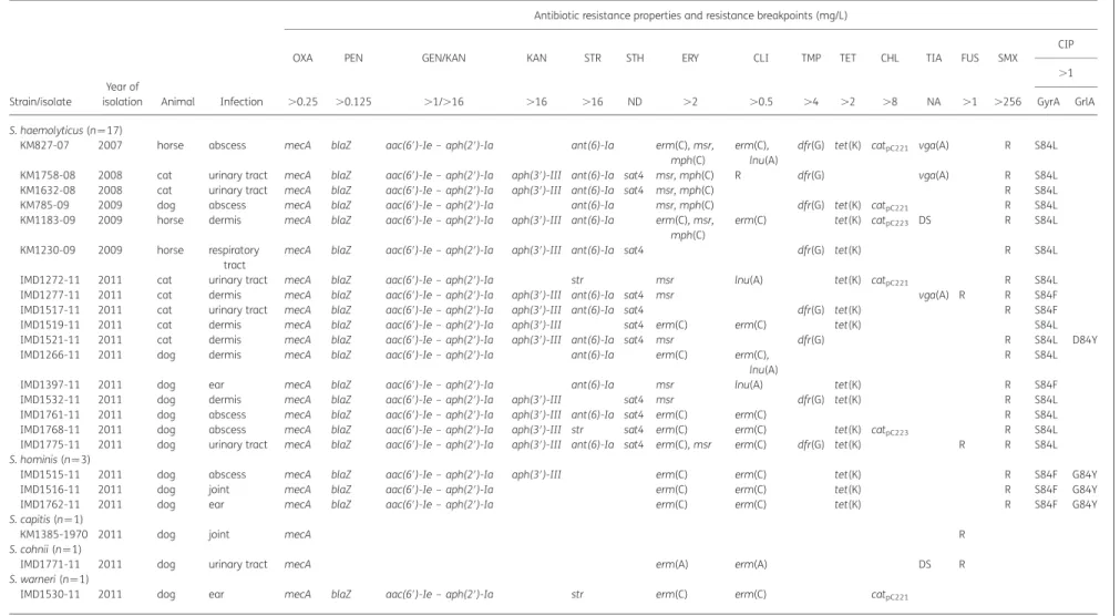
Table 2. Origin and resistance profile of methicillin-resistant S. haemolyticus, S. hominis, S. capitis, S. cohnii and S. warneri isolated from infection sites of animals
Strain/isolate
Year of
isolation Animal Infection
Antibiotic resistance properties and resistance breakpoints (mg/L)
OXA PEN GEN/KAN KAN STR STH ERY CLI TMP TET CHL TIA FUS SMX
CIP
.0.25 .0.125 .1/.16 .16 .16 ND .2 .0.5 .4 .2 .8 NA .1 .256
.1 GyrA GrlA S. haemolyticus (n ¼17)
KM827-07 2007 horse abscess mecA blaZ aac(6′)-Ie – aph(2′)-Ia ant(6)-Ia erm(C), msr,
mph(C)
erm(C), lnu(A)
dfr(G) tet(K) catpC221 vga(A) R S84L
KM1758-08 2008 cat urinary tract mecA blaZ aac(6′)-Ie – aph(2′)-Ia aph(3′)-III ant(6)-Ia sat4 msr, mph(C) R dfr(G) vga(A) R S84L
KM1632-08 2008 cat urinary tract mecA blaZ aac(6′)-Ie – aph(2′)-Ia aph(3′)-III ant(6)-Ia sat4 msr, mph(C) R S84L
KM785-09 2009 dog abscess mecA blaZ aac(6′)-Ie – aph(2′)-Ia ant(6)-Ia msr, mph(C) dfr(G) tet(K) catpC221 R S84L
KM1183-09 2009 horse dermis mecA blaZ aac(6′)-Ie – aph(2′)-Ia aph(3′)-III ant(6)-Ia erm(C), msr, mph(C)
erm(C) tet(K) catpC223 DS R S84L
KM1230-09 2009 horse respiratory
tract
mecA blaZ aac(6′)-Ie – aph(2′)-Ia aph(3′)-III ant(6)-Ia sat4 dfr(G) tet(K) R S84L
IMD1272-11 2011 cat urinary tract mecA blaZ aac(6′)-Ie – aph(2′)-Ia str msr lnu(A) tet(K) catpC221 R S84L
IMD1277-11 2011 cat dermis mecA blaZ aac(6′)-Ie – aph(2′)-Ia aph(3′)-III ant(6)-Ia sat4 msr vga(A) R R S84F
IMD1517-11 2011 cat urinary tract mecA blaZ aac(6′)-Ie – aph(2′)-Ia aph(3′)-III ant(6)-Ia sat4 dfr(G) tet(K) R S84F
IMD1519-11 2011 cat dermis mecA blaZ aac(6′)-Ie – aph(2′)-Ia aph(3′)-III sat4 erm(C) erm(C) tet(K) S84L
IMD1521-11 2011 cat dermis mecA blaZ aac(6′)-Ie – aph(2′)-Ia aph(3′)-III ant(6)-Ia sat4 msr dfr(G) R S84L D84Y
IMD1266-11 2011 dog dermis mecA blaZ aac(6′)-Ie – aph(2′)-Ia ant(6)-Ia erm(C) erm(C),
lnu(A)
R S84L
IMD1397-11 2011 dog ear mecA blaZ aac(6′)-Ie – aph(2′)-Ia ant(6)-Ia msr lnu(A) tet(K) R S84F
IMD1532-11 2011 dog dermis mecA blaZ aac(6′)-Ie – aph(2′)-Ia aph(3′)-III sat4 msr dfr(G) tet(K) R S84L
IMD1761-11 2011 dog abscess mecA blaZ aac(6′)-Ie – aph(2′)-Ia aph(3′)-III ant(6)-Ia sat4 erm(C) erm(C) R S84L
IMD1768-11 2011 dog abscess mecA blaZ aac(6′)-Ie – aph(2′)-Ia aph(3′)-III str sat4 erm(C) erm(C) tet(K) catpC223 R S84L
IMD1775-11 2011 dog urinary tract mecA blaZ aac(6′)-Ie – aph(2′)-Ia aph(3′)-III ant(6)-Ia sat4 erm(C), msr erm(C) dfr(G) tet(K) R R S84L
S. hominis (n ¼3)
IMD1515-11 2011 dog abscess mecA blaZ aac(6′)-Ie – aph(2′)-Ia aph(3′)-III erm(C) erm(C) tet(K) R S84F G84Y
IMD1516-11 2011 dog joint mecA blaZ aac(6′)-Ie – aph(2′)-Ia erm(C) erm(C) tet(K) R S84F G84Y
IMD1762-11 2011 dog ear mecA blaZ aac(6′)-Ie – aph(2′)-Ia erm(C) erm(C) tet(K) R S84F G84Y
S. capitis (n ¼1)
KM1385-1970 2011 dog joint mecA R
S. cohnii (n¼1)
IMD1771-11 2011 dog urinary tract mecA erm(A) erm(A) DS R
S. warneri (n ¼1)
IMD1530-11 2011 dog ear mecA blaZ aac(6′)-Ie – aph(2′)-Ia str erm(C) erm(C) catpC221
CHL, chloramphenicol; CIP, ciprofloxacin; CLI, clindamycin; ERY, erythromycin; FUS, fusidic acid; GEN, gentamicin; KAN, kanamycin; OXA, oxacillin; PEN, penicillin; STR, streptomycin; STH, streptothricin; TET, tetracycline; TIA, tiamulin; TMP, trimethoprim; SMX, sulfamethoxazole; ND, not defined, susceptibility to streptothricin was not measured, only the gene was detected; NA, no resistance breakpoint available for tiamulin [resistance to tiamulin was attributed in the presence of the vga(A) gene (MIC .4 mg/L)]; DS, decreased susceptibility to tiamulin with MIC .4 mg/L); R, resistant phenotype, no resistance genes were determined; blank spaces indicate either no resistance or no mutations.
The MIC breakpoints (in mg/L) that determine resistance were those recommended by EUCAST for staphylococci (www.eucast.org). Resistance breakpoints for streptomycin and kana-mycin were those recommended by the French Society for Microbiology (www.sfm-microbiologie.org) and the resistance breakpoint for sulfamethoxazole was that recommended by the CLSI.36
Antibiotic resistance genes and their functions are indicated as follows: mecA, methicillin-resistance gene encoding PBP2a for resistance to all b-lactam antibiotics; blaZ, b-lactamase gene; aac(6′)-Ie–aph(2′)-Ia, aminoglycoside acetyltransferase and phosphotransferase tandem genes; aph(3′)-III, kanamycin phosphotransferase; ant(6)-Ia, streptomycin adenylnucleo-tidyltransferase gene; str, streptomycin adenyltransferase gene; sat4, strepthotricin acetyltransferase gene; erm(C), macrolide, lincosamide and streptogramin B 23S rRNA methylase gene; msr, macrolide and streptogramin ATP binding transporter gene; mph(C), macrolide phosphotransferase gene; dfr(A), dfr(G), trimethoprim resistance dihydrofolate reductase gene; lnu(A), lincosamide nucleotidyltransferase gene; tet(K), tetracycline efflux resistance gene; catpC221,catpC223, chloramphenicol acetyl transferase gene; vga(A), pleuromutilin and streptogramin A ATP binding transporter gene.
Methicil
lin-r
esistant
C
oNS
in
animal
infec
tions
1259JA
C
Table 3. Clinical data, therapy and outcome of treatment of infections associated with MRCoNS in animals (dogs, cats and horses)
Animals (n¼27) and
CoNS Strains Type of infection
History of antibiotic treatment before identification of the Staphylococcus (no. of courses)
Resistance profile of isolated Staphylococcus from
infection side
Antibiotics used for treatment of the Staphylococcus infection Incompatibility with resistance mechanism Outcome Dogs (n¼11) S. haemolyticus KM 785-09 abscess (granuloma)
amox-clav (1), clindamycin (2) PEN, OXA, KAN, GEN, STR, ERY, TET, TMP, CHL
clindamycin no recovery
S. capitis KM1385-1970 joint (surgery) amox-clav (1), clindamycin (2) PEN, OXA amox-clav,
clindamycin
no unknown
S. epidermidis KM1385-1972
S. haemolyticus IMD1266-11 eye (chronic conjunctivitis)
cefovecin (1), neomycin/polymyxin B (2)
PEN, OXA, STR, ERY, CLI, CIP tetracycline no relapse if treatment with tetracycline stops S. epidermidis IMD1765-11 respiratory tract
(chronic cough)
amox-clav (1), amox-clav (2), amox-clav (3),
PEN, OXA, KAN, GEN, STR, ERY, TMP, CLI, CHL marbofloxacin, tetracycline no recovery after a 4 week therapy
S. hominis IMD1762-11 ear (otitis
externa)
no antibiotics PEN, OXA, KAN, GEN,
ERY, CLI, TET, CIP
framycetin yes [aac(6′)-Ie – aph(2′)-Ia]
recovery
S. warneri IMD1530-11 ear (chronic
otitis externa, relapse)
polymyxin B (1), marbofloxacin (2), cefalexin (3), marbofloxacin (4)
PEN, OXA, KAN, GEN, STR, ERY, CLI, CHL
marbofloxacin no relapse
S. epidermidis IMD1766-11 ear (chronic otitis externa)
metronidazole (1), fusidic acid/ framycetin (2), polymyxin B (3), amox-clav (4), enrofloxacin (5)
PEN, OXA, ERY, CLI, CIP
amox-clav yes (mecA) recovery
S. haemolyticus IMD1775-11 urinary tract (preputial catarrh)
amox-clav (1), polymyxin B (2) PEN, OXA, KAN, GEN, STR, ERY, TMP, CLI, TET, CIP
amox-clav yes (mecA) recovery
S. haemolyticus IMD1768-11 abscess amox-clav/tetracycline (1), amox-clav (2), amox-clav (3), tetracycline (4), amox-clav (5)
PEN, OXA, KAN, GEN, STR, ERY, TMP, CLI, CHL, TET, CIP
cefalexin yes (mecA) relapse
S. haemolyticus IMD1761-11 abscess amox-clav (1), amox-clav/ chloramphenicol (2), amox-clav/ chloramphenicol (3),
aminoglycoside/polymyxin B (4)
PEN, OXA, KAN, GEN, STR, ERY, CLI, CIP
chloramphenicol no recovery
S. hominis IMD1515-11 dermis (ulcer) amox-clav (1), cefalexin (2) PEN, OXA, KAN, GEN, STR, ERY, CLI, TET, CIP
no treatment NA euthanasia
Cats (n¼11)
S. haemolyticus KM1758-08 urinary tract marbofloxacin (1), amox-clav (2), rifampicin (3)
PEN, OXA, KAN, GEN, STR, ERY, TMP, CIP, TIA
rifampicin no recovery
S. haemolyticus KM1632-08 urinary tract amox-clav (1), trimethoprim/ sulphonamide (2)
PEN, OXA, KAN, GEN, STR, ERY trimethoprim/ sulphonamide no recovery
Kern
and
Perr
eten
1260S. epidermidis KM1527-07 abscess after surgery
marbofloxacin (1), amox-clav (2) PEN, OXA, KAN, GEN, STR, ERY, TMP, TET, TIA
marbofloxacin, tetracycline
yes [tet(K)] recovery after amputation of the lower extremity S. epidermidis KM505-09 urinary tract enrofloxacin (1), amoxicillin (2),
marbofloxacin (3)
PEN, OXA, KAN, GEN, TMP, CLI, TET, CHL, CIP
no treatment NA recovery
S. haemolyticus IMD1272-11 urinary tract (urolithiasis, chronic cystitis)
amox-clav (1) PEN, OXA, KAN, GEN,
STR, ERY, CLI, TET, CHL, CIP
clindamycin yes [erm(C)] recovery
S. haemolyticus IMD1517-11 urinary tract unknown PEN, OXA, KAN, GEN,
STR, TMP, TET, CIP
no treatment (fast death)
NA death
S. epidermidis IMD1776-11 eye (corneal ulcer)
enrofloxacin (1), moxifloxacin (2), gentamicin (3)
PEN, OXA, KAN, GEN, STR, ERY, TMP, CLI, CHL, CIP ofloxacin, tetracycline no recovery (together with cross-linking therapy) S. epidermidis IMD1763-11 urinary tract
(urolithiasis, chronic cystitis)
cefovecin (1) PEN, OXA, KAN, GEN,
TET
marbofloxacin no relapse (cystitis
with Enterococcus)
S. epidermidis IMD1270-11 phlegmone
(acneic skin)
cefovecin (1) PEN, OXA, KAN, GEN,
STR, ERY, TMP, TET, TIA; CIP
chloramphenicol no recovery
S. epidermidis IMD1269-11 urinary tract (cystitis)
marbofloxacin (1), amox-clav (2) PEN, OXA, KAN, GEN, ERY, CLI, CIP
amox-clav yes (mecA) recovery
S. epidermidis IMD1274-11 eye
(conjunctivitis) amox-clav (1), enrofloxacin/ amoxicillin (2), amoxicillin (3), amoxicillin (4), amoxicillin (5), amoxicillin (6), bacitracin/ neomycin/ofloxacin (7), amoxicillin (8), ciprofloxacin (9), amoxicillin (10), cefovecin (11), ciprofloxacin/amoxicillin(12), amoxicillin (13), amoxicillin (14)
PEN, OXA, ERY, CLI, TET, CIP
neomycin/polymyxin B
no relapse
Horses (n¼5)
S. epidermidis KM794-06 abscess penicillin PEN, OXA, KAN, GEN,
TET
unknown NA recovery
S. epidermidis KM825-09 abscess (surgery) cefquinome (1), penicillin/ gentamicin (2)
PEN, OXA, KAN, GEN no treatment NA euthanasia
S. haemolyticus KM827-07 wound penicillin/gentamicin (1),
enrofloxacin (2), cefquinome (3)
PEN, OXA, KAN, GEN, STR, ERY, TMP, CLI, TET, CHL, TIA
unknown NA recovery
S. haemolyticus KM1183-09 dermis trimethoprim/sulphonamide (1),
cefquinome (2), marbofloxacin/ enrofloxacin (3)
PEN, OXA, KAN, GEN, STR, ERY, CLI, TET, CHL
gentamicin yes [aac(6′)-Ie – aph(2′)-Ia] relapse Continued
Methicil
lin-r
esistant
C
oNS
in
animal
infec
tions
1261JA
C
Results
Identification of and genetic diversity among strains of
MRCoNS
The 43 samples originated from animals admitted to 30 different clinics from 10 different cantons in Switzerland, indicating that MRCoNS are widespread and not related to a specific clinic with nosocomial infection problems (Figure1). MRCoNS were isolated from different infection sites in cats (n¼ 16), dogs (n¼ 20) and horses (n¼ 7) (Tables 1and 2). Infected sites consisted of the skin (n¼10), urinary tracts (n¼ 9), ears (n¼ 4), respiratory tracts (n¼ 5), joints (n¼4), eyes (n¼1) and abscesses/fistulas (n¼ 10) (Tables1and2). The MRCoNS were identified as S. epider-midis (Table 1) and S. haemolyticus, S. hominis, S. warneri, S. capitis and S. cohnii (Table 2). Three samples contained an additional pathogen, i.e. Staphylococcus schleiferi together with S. haemolyticus KM785-09, Staphylococcus pseudintermedius to-gether with S. epidermidis KM1077-09 and a mix of anaerobic bacteria together with S. epidermidis IMD1522-11.
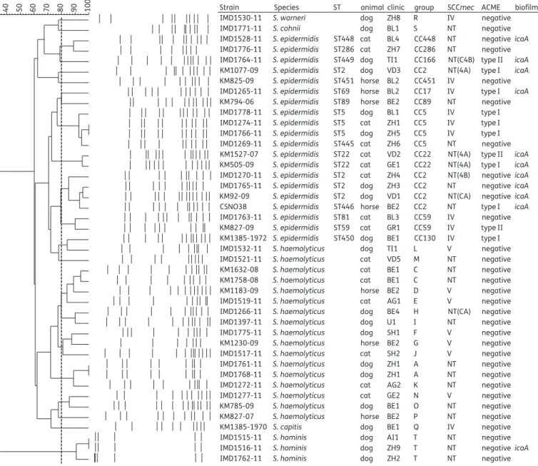
PFGE revealed a large heterogeneity between the MRCoNS of the same species. Eight different PFGE clonal lineages were identi-fied among the 17 S. haemolyticus isolates, 11 among the 20 S. epidermidis isolates and 1 among the 3 S. hominis isolates (Figure1). The S. epidermidis strains showed distinct MLST patterns and belonged to 14 different sequence types (STs), representing 11 clonal complexes (CC), namely CC2 [ST2 (n¼ 4), ST446 (n ¼1)], CC5 [ST5 (n¼ 3), ST445 (n¼ 1)], CC22 [ST22 (n ¼3)], CC59 [ST59 (n¼ 1), ST81 (n¼ 1)], CC17 [ST69 (n¼ 1), CC89 [ST89 (n¼ 1)], CC130 [ST450 (n¼ 1)], CC166 [ST449 (n¼ 1)], CC286 [ST286 (n¼1)], CC451 [ST451 (n¼ 1)] and CC448 [ST448 (n¼ 1)]. ST445, ST446, ST448, ST449, ST450 and ST451 were newly described STs [Figure1and Figure S1 (available as Supplementary data at JAC Online)]. All but one of the isolates belonging to the predominant CC2 (n¼ 5), CC5 (n¼4), CC22 (n¼ 2) and CC59 (n¼ 2) clustered into four dis-tinct PFGE branches (Figure1). However, different PFGE patterns could still be observed within these groups, indicating a larger di-versity between strains of the same CC (Figure1).
Ten of 20 S. epidermidis isolates harboured the biofilm forma-tion operon ica, including all the CC2 (ST2, ST446) (n¼5) and ST22 (n¼ 2) isolates as well as the ST69, ST448 and ST449 isolates. The icaA gene was also detected in one S. hominis isolate. ACMEs were only detected in S. epidermidis. Eight S. epidermidis isolates carried a type I ACME (arcA+ /opp3AB +) and three carried a type II ACME (arcA+ /opp3AB2). ACMEs were found in all ST5 and ST22 isolates, but were also present in one ST2 isolate and in the ST59, ST69, ST446, ST449 and ST450 isolates (Figure1). An SCCmec element could only be typed for 17 isolates. SCCmec IV was detected in one S. capitis, one S. warneri and eight S. epidermi-dis isolates. In S. epidermiepidermi-dis, SCCmec IV was associated with ACME type I in the ST5, ST69 and ST450 isolates and with ACME type 2 in the ST59 isolate. SCCmec V was detected in seven S. haemolyticus isolates (Figure1). The other 26 SCCmec elements could not be characterized as they lacked either known ccr genes or a known mecA class structure or both (Figure1).
Distribution of MRCoNS isolates in veterinary practices
Association of a specific clonal lineage with a clinic was only observed for two pairs of S. haemolyticus (IMD1761-11, IMD1768-11 and IMD1632-08, IMD1758-08) isolated from differentTable 3. C o n tinued Animal s (n ¼ 27) and C oNS Str ains Type of infection His tory of antib iotic tr ea tmen t befor e identifica tion of the Stap h ylococcus (no. of cour ses) R esis tanc e p rofile of isola ted Stap h ylococc us fr om infe ction side Antibiotics used for tr ea tmen t o f the Stap h ylococc us infe ction Inco mpa tibi lity with resis tanc e me chanism Outco me S. haemol yticus KM1230 -09 respir a tor y tr a ct (BA L) unkno wn PEN, O XA, KAN , GEN, STR, STH, TMP , TET , CIP no tr ea tment N A reco very amo x-cla v, amo xicil lin/cla vulanic a cid ; CHL , chl or amph enicol ; CIP , cipr oflo xa cin; CLI, cli ndam ycin ; E R Y, ery thr om ycin; GEN, gent amicin; KAN, kan am ycin; O XA, o xa cillin; PEN , penicillin; STR, st rept om ycin; TET , tetr a cy cline ; TIA, tiamulin; TMP , trim ethoprim; N A , not a vail able; BAL, br onchoalv eo lar la vage . mecA , methi cillin-r esis tanc e gene encod ing PBP2 a for resis tanc e to all b -la ctam antib iotics (e. g. peni cillin, amo xicillin, amo xicillin/cla vula nic a cid and cefale xin) ; aa c(6 ′)-I e – aph (2 ′)-I a , aminogl ycosi de a cetyltr an sfer ase an d pho sphotr ansfer ase tandem gene s (gent amicin/k anam ycin/n eom ycin); erm (C), ma cr olide, linc osam ide and st reptog ramin B 23S rRNA me th y-lase gene (clind am ycin); tet (K), tetr a cy clin e efflux resis tance gene (tetr a cy cline ).
Kern and Perreten
animals in two clinics. Each pair showed similar PFGE profiles (A and C) and contained a non-typeable SCCmec element (Figure 1). However, they exhibited different antibiotic resistance profiles (Table2). Otherwise, MRCoNS isolated from animals admitted to the same clinic were genetically distant. On the other hand, genet-ically related MRCoNS were isolated from different animals in differ-ent clinics (Figure 1). These isolates also displayed different antibiotic resistance profiles (Tables1and2).
Antibiotic resistance profile
All the MRCoNS isolates were resistant to b-lactam antibiotics and contained the mecA gene. None of them was resistant to
linezolid, quinupristin/dalfopristin, rifampicin or vancomycin. Nonetheless, the isolates were also resistant to gentamicin/ kanamycin owing to the bifunctional acetyltransferase/phospho-transferase gene aac(6′)-Ie– aph(2′)-Ia (n¼ 33), kanamycin [aph(3′)-III (n¼ 17)], macrolides and/or lincosamides [erm(C) (n¼ 22), erm(A) (n¼ 1), msr (n ¼12) and lnu(A) (n¼ 4], tetracyc-line [tet(K) (n¼ 22)], trimethoprim [dfr(A) (n¼ 7) and dfr(G) (n¼ 10)], streptomycin [str (n¼5) and ant(6)-Ia (n¼ 15)], strepto-thricin [sat4 (n¼ 15)], chloramphenicol [catpC221 (n¼ 5) and catpC223 (n¼ 3)], tiamulin [(vga(A) (n¼ 4)], mupirocin [mupR (n¼ 2)], fusidic acid (n¼13), sulfamethoxazole (n¼34) and fluor-oquinolones (n ¼30) (Tables 1 and 2). Resistance mechanisms Strain Species ST animal clinic group SCCmec ACME biofilm
IMD1530-11 S. warneri dog ZH8 R IV negative
IMD1771-11 S. cohnii dog BL1 S NT negative
IMD1528-11 S. epidermidis ST448 cat BL4 CC448 NT negativeicaA IMD1776-11 S. epidermidis ST286 cat ZH7 CC286 NT negative IMD1764-11 S. epidermidis ST449 dog TI1 CC166 NT(C4B) type II icaA KM1077-09 S. epidermidis ST2 dog VD3 CC2 NT(4A) type I icaA KM825-09 S. epidermidis ST451 horse BL2 CC451 IV negative IMD1265-11 S. epidermidis ST69 horse BL2 CC17 IV type I icaA KM794-06 S. epidermidis ST89 horse BE2 CC89 NT negative IMD1778-11 S. epidermidis ST5 dog BL1 CC5 IV type I IMD1274-11 S. epidermidis ST5 cat ZH1 CC5 IV type I IMD1766-11 S. epidermidis ST5 dog ZH5 CC5 IV type I IMD1269-11 S. epidermidis ST445 cat ZH6 CC5 NT negative KM1527-07 S. epidermidis ST22 cat VD2 CC22 NT(4A) type II icaA KM505-09 S. epidermidis ST22 cat GE1 CC22 NT(4A) type I icaA IMD1270-11 S. epidermidis ST2 cat ZH4 CC2 NT(4B) negativeicaA IMD1765-11 S. epidermidis ST2 dog ZH3 CC2 NT negativeicaA KM92-09 S. epidermidis ST2 dog VD1 CC2 NT(CA) negativeicaA CSNO38 S. epidermidis ST446 horse BE2 CC2 NT type I icaA IMD1763-11 S. epidermidis ST81 cat BL3 CC59 IV negative KM827-09 S. epidermidis ST59 cat GR1 CC59 IV type II KM1385-1972 S. epidermidis ST450 dog BE1 CC130 IV type I IMD1532-11 S. haemolyticus dog TI1 L V negative IMD1521-11 S. haemolyticus cat VD5 M NT negative KM1632-08 S. haemolyticus cat BE1 C NT negative KM1758-08 S. haemolyticus cat BE1 C NT negative KM1183-09 S. haemolyticus horse BE2 D V negative IMD1519-11 S. haemolyticus cat AG1 E V negative IMD1266-11 S. haemolyticus dog BE4 H NT(CA) negative IMD1397-11 S. haemolyticus dog U1 I NT negative IMD1775-11 S. haemolyticus dog SH1 F V negative KM1230-09 S. haemolyticus horse BE2 G V negative IMD1517-11 S. haemolyticus cat SH2 J V negative IMD1761-11 S. haemolyticus dog ZH1 A NT negative IMD1768-11 S. haemolyticus dog ZH1 A NT negative IMD1272-11 S. haemolyticus cat AG2 K NT negative IMD1277-11 S. haemolyticus cat GE2 N V negative KM785-09 S. haemolyticus dog BE1 O NT negative KM827-07 S. haemolyticus horse BE2 P NT negative
KM1385-1970 S. capitis dog BE1 Q IV negative
IMD1515-11 S. hominis dog AI1 T NT negative
IMD1516-11 S. hominis dog ZH9 T NT negativeicaA
IMD1762-11 S. hominis dog ZH2 T NT negative
100 90 80 70 60 50 40 30
Figure 1. Phylogenetic tree constructed from the PFGE pattern of methicillin-resistant S. epidermidis, S. haemolyticus, S. hominis, S. capitis and S. warneri. The tree was generated by UPGMA using Bionumerics 6.6 (Applied Maths, Kortrjk, Belgium) and comparison settings (Dice, optimization 1.5%, position tolerance 1.5%) as recommended by PulseNet International (www.pulsenetinternational.org). The broken line indicates the cut-off value of≥79%, determining clonality between the isolates according to Miragaia et al.47 Capital letters indicate the cantons and the numbers indicate the different clinics. AG, Argovia; AI, Appenzell Inner Rhoden; BE, Bern; BL, Basel-Land; GE, Geneva; GR, Grisons; SH, Schaffhausen; TI, Ticino; VD, Vaud; ZH, Zurich; U, unknown.
Methicillin-resistant CoNS in animal infections
1263
for fusidic acid and sulfamethoxazole were not investigated. Fluoroquinolone resistance was attributed to mutations in topo-isomerase II GyrA (n¼ 30) and topotopo-isomerase IV GrlA (n¼ 18) (Tables1and2). Mutations that cause amino acid substitutions in topoisomerases II and IV were found in ciprofloxacin-resistant S. epidermidis, S. haemolyticus and S. hominis at nucleotide pos-ition 251 [n¼ 30; Ser84Leu (n¼ 14), Ser84Phe (n¼ 13), Ser84Tyr (n¼ 3)] in gyrA and at positions 239 [n¼ 8; Ser80Tyr (n¼ 4), Ser80Phe (n¼4)] and 250 [n¼ 10; Asp84Tyr (n¼ 7), Gly84Tyr (n¼ 3)] in grlA. An amino acid substitution in GrlB (Glu473Lys) was also present in two S. epidermidis isolates (KM505-09 and KM1527-07) and two S. haemolyticus isolates (IMD1277-11 and IMD1532-11). This mutation was not considered as being re-sponsible for fluoroquinolone resistance in CoNS, as a mutation at the same location has been shown not to confer resistance to fluoroquinolones in Staphylococcus aureus.38 The resistance mechanism could not be explained for one strain with resistance to gentamicin and kanamycin, for one strain with resistance to clindamycin and for two strains with decreased susceptibility to tiamulin (MIC .4 mg/L), suggesting new mechanisms of resistance.
Clinical data of infected animals
Clinical data were obtained for 27 animals (11 dogs, 11 cats and 5 horses) admitted to 19 different clinics (Table3). Twenty-four animals had a history of antibiotic treatment, and 20 of them underwent antimicrobial treatment more than twice with up to 14 courses. The most commonly used antibiotics in dogs and cats prior to the identification of the staphylococcal species were amoxicillin/clavulanic acid, cephalosporins and fluoroquino-lones. In 20 dogs and cats treated, amoxicillin was given 15 times, fluoroquinolones 8 times, cephalosporins 6 times, and both a b-lactam and a fluoroquinolone antibiotic were given 7 times. Antibiotics such as gentamicin, chloramphenicol, tetracyc-line and clindamycin were also used in pets, but less frequently. In horses, cefquinome, fluoroquinolones and the combination penicillin/gentamicin were the most commonly administered antibiotics. The MRCoNS infections were then treated after con-sultation of an antibiogram, most frequently using fluoroquino-lones or amoxicillin/clavulanic acid followed by tetracycline, clindamycin, chloramphenicol, cefalexin, the combination sul-phonamides/trimethoprim, rifampicin and the aminoglycosides gentamicin, framycetin and neomycin. Most of the animals (n¼ 14) recovered after antibiotic treatment: six had a relapse, two recovered without any antibiotic therapy, one recovered after amputation of the infected lower extremity, one was still under treatment at the time of writing, two were not further treated and euthanized and one died of unknown cause prior to therapy. In seven cases, antibiotics were used even in the presence of resistance, leading to relapse in two cases when cefalexin and gentamicin were used for the treatment of an abscess and skin infection, respectively. The other five animals recovered after antimicrobial treatment with amoxicillin/clavula-nic acid (n¼3), clindamycin (n¼ 1) or framycetin (n¼ 1), despite the presence of mecA, erm(C) or aac(6′)-Ie– aph(2′)-Ia in the re-spective MRCoNS (Table3). For these animals, MRCoNS were likely not the primary cause of the infection (three urinary tract and two ear infections), although MRCoNS appeared alone in the culture. No significant difference was observed in the outcome
of the disease between animals treated with an antibiotic incom-patible and an antibiotic comincom-patible with the resistance profile of the MRCoNS.
Discussion
MRCoNS are associated with serious infections in animals and have become a challenge to therapy. The CoNS species identified in this study were the same as the ones causing nosocomial infections in humans, with S. epidermidis and S. haemolyticus being the most prevalent in animals and humans.7 Similar to the case with human infections,11,13 S. hominis, S. warneri, S. cohnii and S. capitis were only occasionally isolated from animal infection sites. The population analysis by PFGE showed that the majority of the isolates are genetically diverse. Three clonal lineages sharing similar PFGE profiles appeared to be pre-dominant among S. epidermidis and were found to belong to CC2, CC5 and CC22. However, isolates of CC2 and CC22 contained divergent SCCmec elements and ACMEs, while CC5 isolates almost exclusively contained SCCmec IV and ACME type I. Add-itionally, these clonally related isolates displayed different resist-ance profiles, emphasizing the ability of CoNS to acquire antibiotic resistance genes. The presence of numerous PFGE and antibiotic resistance profiles is a well-described phenomenon for S. epidermidis ST2, which is the most widely disseminated human healthcare-associated sequence type worldwide.39–42
A study using 217 S. epidermidis isolates from humans from 17 countries detected 30.9% of the isolates as ST2.42The suc-cessful spread of ST2 in the hospital environment has been sug-gested to be associated with its ability to generate novel phenotypic and genotypic variants by recombination and acqui-sition of new elements, such as the biofilm-formation ica operon, ACMEs and antibiotic resistance genes.14,15,18,41In our study, all isolates of CC2 and CC22, which is a subcluster of CC2 (Figure S1, available as Supplementary data at JAC Online), contained the biofilm formation operon ica. On the other hand, the ica operon was absent in isolates of CC5, which was also predomin-ant in infection sites of animals; they contained ACMEs instead. Of note, CC22 contained both ica and ACMEs. The presence of the biofilm formation operon ica and ACMEs almost exclusively in S. epidermidis of the predominant clonal lineages CC2, CC5 and CC22 may have contributed to the establishment of these strains in the animal environment. In addition to the predomin-ant STs, the animals were also infected with other S. epidermidis strains, which has also been reported in human infections, such as ST35, ST59, ST81, ST69, ST89 and ST286 (Figure S1, available as Supplementary data at JAC Online),40–42and with S. haemo-lyticus. The absence of MLST methods for S. haemolyticus pre-vented us from determining whether specific STs would also be predominant within this species. However, the different PFGE and antibiotic resistance profiles support the hypothesis that MRCoNS associated with infections in animals are very heteroge-neous, unlike methicillin-resistant S. pseudintermedius (MRSP), which spread as specific clones.43 Nevertheless, MRSP and MRCoNS are resistant to the same classes of drugs and contain similar antibiotic resistance genes. Similar to MRSP, more than two-thirds of the MRCoNS exhibit resistance to fluoroquinolones, macrolides, lincosamides and aminoglycosides, in addition to resistance to b-lactams, suggesting that they have been selected
Kern and Perreten
through the frequent use of antibiotics. These classes of drugs, especially the b-lactams and fluoroquinolones, were also the most commonly used drugs in veterinary practices (Table 3). These two classes of drugs have been shown to represent a sig-nificant risk factor for the selection of methicillin-resistant S. aureus44 and similar effects are to be expected for MRCoNS. Many animals were given more than one antibiotic course, with some animals receiving 5 and up to 14 courses of an anti-biotic before the MRCoNS infection was diagnosed. The series of empirical antimicrobial treatments may have contributed to the selection of the MRCoNS in the infection sites. Additionally, the primary cause of the infection may have been overlooked and not directly related to the presence of a MRCoNS. Indeed, two animals recovered without antibiotic treatment and five recov-ered despite the presence of a resistance mechanism against the antibiotic used for treatment. Nevertheless, in the majority of the cases the staphylococcal infections could be treated with an antibiotic chosen after consultation of an antibiogram. All these criteria highlight the importance of correct diagnosis and antibiograms.
Multidrug-resistant CoNS represent a new challenge for therapy in veterinary medicine. The infections are caused by gen-etically distant strains, indicating many possible non-hospital-related reservoirs, such as animals themselves, animal owners and people working with animals that have been shown to harbour and possibly exchange MRCoNS.22,45,46The presence of clones similar to those causing infections in humans highlights the importance of careful surveillance of bacterial infection dis-eases, the need to implement infection control programmes and the prudent use of antibiotics in veterinary settings.
Acknowledgements
We thank the Centre for Zoonoses and Bacterial Animal Diseases and Antimicrobial Resistance (ZOBA), IDEXX Diavet and Laboratory Laupeneck AG for providing the strains and all the participating veterinary practices that provided clinical data. We also thank Jacques Schrenzel and Patrice Franc¸ois for providing positive control strains and A. Collaud, C. Strauss, Y. Sigrist, S. Rychener, S. Kittl and A. Candi for helpful assistance.
Funding
This study was supported by internal funding.
Transparency declarations
None to declare.
Supplementary data
Table S1 and Figure S1 are available at JAC Online (http://jac.oxfordjournals. org/).
References
1 Kloos WE, Musselwhite MS. Distribution and persistence of Staphylococcus and Micrococcus species and other aerobic bacteria on human skin. Appl Microbiol 1975; 30: 381– 5.
2 Otto M. Molecular basis of Staphylococcus epidermidis infections. Semin Immunopathol 2012; 34: 201– 14.
3 McCann MT, Gilmore BF, Gorman SP. Staphylococcus epidermidis device-related infections: pathogenesis and clinical management. J Pharm Pharmacol 2008; 60: 1551–71.
4 Vengust M, Anderson ME, Rousseau J et al. Methicillin-resistant staphylococcal colonization in clinically normal dogs and horses in the community. Lett Appl Microbiol 2006; 43: 602–6.
5 Casey AL, Lambert PA, Elliott TS. Staphylococci. Int J Antimicrob Agents 2007; 29 Suppl 3: S23–32.
6 Rogers KL, Fey PD, Rupp ME. Coagulase-negative staphylococcal infections. Infect Dis Clin North Am 2009; 23: 73–98.
7 Santos SI, Mato R, de Lencastre H et al. Patterns of multidrug resistance among methicillin-resistant hospital isolates of coagulase-positive and coagulase-negative staphylococci collected in the international multicenter study RESIST in 1997 and 1998. Microb Drug Resist 2000; 6: 199–211.
8 Hanssen AM, Ericson Sollid JU. SCCmec in staphylococci: genes on the move. FEMS Immunol Med Microbiol 2006; 46: 8 – 20.
9 International Working Group on the Classification of Staphylococcal Cassette Chromosome Elements (IWG-SCC). Classification of staphylococcal cassette chromosome mec (SCCmec): guidelines for reporting novel SCCmec elements. Antimicrob Agents Chemother 2009; 53: 4961–7. 10 Chambers HF. Methicillin resistance in staphylococci: molecular and biochemical basis and clinical implications. Clin Microbiol Rev 1997; 10: 781–91.
11 Garza-Gonza´lez E, Morfı´n-Otero R, Llaca-Dı´az JM et al. Staphylococcal cassette chromosome mec (SCC mec) in methicillin-resistant coagulase-negative staphylococci. A review and the experience in a tertiary-care setting. Epidemiol Infect 2010; 138: 645– 54.
12 Cone LA, Sontz EM, Wilson JW et al. Staphylococcus capitis endocarditis due to a transvenous endocardial pacemaker infection: case report and review of Staphylococcus capitis endocarditis. Int J Infect Dis 2005; 9: 335–9.
13 Widerstro¨m M, Wistro¨m J, Sjo¨stedt A et al. Coagulase-negative staphylococci: update on the molecular epidemiology and clinical presentation, with a focus on Staphylococcus epidermidis and Staphylococcus saprophyticus. Eur J Clin Microbiol Infect Dis 2012; 31: 7 –20.
14 Barbier F, Lebeaux D, Hernandez D et al. High prevalence of the arginine catabolic mobile element in carriage isolates of methicillin-resistant Staphylococcus epidermidis. J Antimicrob Chemother 2011; 66: 29 –36. 15 Miragaia M, de Lencastre H, Perdreau-Remington F et al. Genetic diversity of arginine catabolic mobile element in Staphylococcus epidermidis. PLoS One 2009; 4: e7722.
16 Otto M. Coagulase-negative staphylococci as reservoirs of genes facilitating MRSA infection: Staphylococcal commensal species such as Staphylococcus epidermidis are being recognized as important sources of genes promoting MRSA colonization and virulence. Bioessays 2012; 35: 4 –11.
17 Piette A, Verschraegen G. Role of coagulase-negative staphylococci in human disease. Vet Microbiol 2009; 134: 45–54.
18 Schoenfelder SM, Lange C, Eckart M et al. Success through diversity— how Staphylococcus epidermidis establishes as a nosocomial pathogen. Int J Med Microbiol 2010; 300: 380–6.
19 Bagcigil FA, Moodley A, Baptiste KE et al. Occurrence, species distribution, antimicrobial resistance and clonality of methicillin- and erythromycin-resistant staphylococci in the nasal cavity of domestic animals. Vet Microbiol 2007; 121: 307–15.
Methicillin-resistant CoNS in animal infections
1265
20 Corrente M, D’Abramo M, Latronico F et al. Methicillin-resistant coagulase negative staphylococci isolated from horses. New Microbiol 2009; 32: 311–4.
21 Karakulska J, Fijalkowski K, Nawrotek P et al. Identification and methicillin resistance of coagulase-negative staphylococci isolated from nasal cavity of healthy horses. J Microbiol 2012; 50: 444– 51.
22 Moodley A, Guardabassi L. Clonal spread of methicillin-resistant coagulase-negative staphylococci among horses, personnel and environmental sites at equine facilities. Vet Microbiol 2009; 137: 397–401.
23 Schnellmann C, Gerber V, Rossano A et al. Presence of new mecA and mph(C) variants conferring antibiotic resistance in Staphylococcus spp. isolated from the skin of horses before and after clinic admission. J Clin Microbiol 2006; 44: 4444–54.
24 Malik S, Coombs GW, O’Brien FG et al. Molecular typing of methicillin-resistant staphylococci isolated from cats and dogs. J Antimicrob Chemother 2006; 58: 428– 31.
25 Garbacz K, Zarnowska S, Piechowicz L et al. Staphylococci isolated from carriage sites and infected sites of dogs as a reservoir of multidrug resistance and methicillin resistance. Curr Microbiol 2013; 66: 169– 73. 26 Hauschild T, Wo´jcik A. Species distribution and properties of staphylococci from canine dermatitis. Res Vet Sci 2007; 82: 1 –6. 27 Patel A, Lloyd DH, Lamport AI. Antimicrobial resistance of feline staphylococci in south-eastern England. Vet Dermatol 1999; 10: 257–61. 28 Malik S, Peng H, Barton MD. Antibiotic resistance in staphylococci associated with cats and dogs. J Appl Microbiol 2005; 99: 1283– 93. 29 Medleau L, Blue JL. Frequency and antimicrobial susceptibility of Staphylococcus spp isolated from feline skin lesions. J Am Vet Med Assoc 1988; 193: 1080– 1.
30 Lilenbaum W, Veras M, Blum E et al. Antimicrobial susceptibility of staphylococci isolated from otitis externa in dogs. Lett Appl Microbiol 2000; 31: 42– 5.
31 Tenover FC, Arbeit RD, Goering RV et al. Interpreting chromosomal DNA restriction patterns produced by pulsed-field gel electrophoresis: criteria for bacterial strain typing. J Clin Microbiol 1995; 33: 2233– 9. 32 Kondo Y, Ito T, Ma XX et al. Combination of multiplex PCRs for staphylococcal cassette chromosome mec type assignment: rapid identification system for mec, ccr, and major differences in junkyard regions. Antimicrob Agents Chemother 2007; 51: 264– 74.
33 Diep BA, Stone GG, Basuino L et al. The arginine catabolic mobile element and staphylococcal chromosomal cassette mec linkage: convergence of virulence and resistance in the USA300 clone of methicillin-resistant Staphylococcus aureus. J Infect Dis 2008; 197: 1523–30.
34 Gu J, Li H, Li M et al. Bacterial insertion sequence IS256 as a potential molecular marker to discriminate invasive strains from commensal strains of Staphylococcus epidermidis. J Hosp Infect 2005; 61: 342–8.
35 Thomas JC, Vargas MR, Miragaia M et al. Improved multilocus sequence typing scheme for Staphylococcus epidermidis. J Clin Microbiol 2007; 45: 616–9.
36 Clinical and Laboratory Standards Institute. Performance Standards for Antimicrobial Susceptibility Testing: Twenty-second Informational Supplement M100-S22. CLSI, Wayne, PA, USA, 2012.
37 Perreten V, Vorlet-Fawer L, Slickers P et al. Microarray-based detection of 90 antibiotic resistance genes of Gram-positive bacteria. J Clin Microbiol 2005; 43: 2291–302.
38 Tanaka M, Onodera Y, Uchida Y et al. Quinolone resistance mutations in the GrlB protein of Staphylococcus aureus. Antimicrob Agents Chemother 1998; 42: 3044–6.
39 Widerstro¨m M, McCullough CA, Coombs GW et al. A multidrug-resistant Staphylococcus epidermidis clone (ST2) is an ongoing cause of hospital-acquired infection in a Western Australian hospital. J Clin Microbiol 2012; 50: 2147–51.
40 Francois P, Hochmann A, Huyghe A et al. Rapid and high-throughput genotyping of Staphylococcus epidermidis isolates by automated multilocus variable-number of tandem repeats: a tool for real-time epidemiology. J Microbiol Methods 2008; 72: 296– 305.
41 Li M, Wang X, Gao Q et al. Molecular characterization of Staphylococcus epidermidis strains isolated from a teaching hospital in Shanghai, China. J Med Microbiol 2009; 58: 456–61.
42 Miragaia M, Thomas JC, Couto I et al. Inferring a population structure for Staphylococcus epidermidis from multilocus sequence typing data. J Bacteriol 2007; 189: 2540– 52.
43 Perreten V, Kadlec K, Schwarz S et al. Clonal spread of methicillin-resistant Staphylococcus pseudintermedius in Europe and North America: an international multicentre study. J Antimicrob Chemother 2010; 65: 1145–54.
44 Faires MC, Traverse M, Tater KC et al. Methicillin-resistant and -susceptible Staphylococcus aureus infections in dogs. Emerg Infect Dis 2010; 16: 69– 75.
45 Davis MF, Iverson SA, Baron P et al. Household transmission of meticillin-resistant Staphylococcus aureus and other staphylococci. Lancet Infect Dis 2012; 12: 703– 16.
46 Moon BY, Youn JH, Shin S et al. Genetic and phenotypic characterization of methicillin-resistant staphylococci isolated from veterinary hospitals in South Korea. J Vet Diagn Invest 2012; 24: 489–98. 47 Miragaia M, Carric¸o JA, Thomas JC et al. Comparison of molecular typing methods for characterization of Staphylococcus epidermidis: proposal for clone definition. J Clin Microbiol 2008; 46: 118– 29. 48 Yamada M, Yoshida J, Hatou S et al. Mutations in the quinolone resistance determining region in Staphylococcus epidermidis recovered from conjunctiva and their association with susceptibility to various fluoroquinolones. Br J Ophthalmol 2008; 92: 848– 51.