COMPUTED TOMOGRAPHY
Raw data-based iterative reconstruction in body CTA: evaluation
of radiation dose saving potential
Anna Winklehner&Christoph Karlo&Gilbert Puippe&Bernhard Schmidt&
Thomas Flohr&Robert Goetti&Thomas Pfammatter&Thomas Frauenfelder&
Hatem Alkadhi
Received: 25 April 2011 / Revised: 18 June 2011 / Accepted: 1 July 2011 / Published online: 6 August 2011 # European Society of Radiology 2011
Abstract
Objective To evaluate prospectively, in patients undergoing body CTA, the radiation dose saving potential of raw data-based iterative reconstruction as compared to filtered back projection (FBP).
Methods Twenty-five patients underwent thoraco-abdominal CTA with 128-slice dual-source CT, operating both tubes at 120 kV. Full-dose (FD) images were reconstructed with FBP and were compared to half-dose (HD) images with FBP and HD-images with sinogram-affirmed iterative reconstruction (SAFIRE), both reconstructed using data from only one tube-detector-system. Image quality and sharpness of the aortic contour were assessed. Vessel attenuation and noise were measured, contrast-to-noise-ratio was calculated.
Results Noise as image quality deteriorating artefact occurred in 24/25 (96%) HD-FBP but not in FD-FBP and HD-raw data-based iterative reconstruction datasets (p<0.001). Other artefacts occurred with similar prevalence among the data-sets. Sharpness of the aortic contour was higher for FD-FBP and HD-raw data-based iterative reconstruction as compared to HD-FBP (p<0.001). Aortoiliac attenuation was similar among all datasets (p>0.05). Lowest noise was found for HD-raw data-based iterative reconstruction (7.23HU), being 9.4% lower than that in FD-FBP (7.98HU, p<0.05) and
30.8% lower than in HD-FBP images (10.44HU, p<0.001). Contrast-to-noise-ratio was lower in HD-FBP (p<0.001) and higher in HD-raw data-based iterative reconstruction (p< 0.001) as compared to FD-FBP.
Conclusion Intra-individual comparisons of image quality of body CTA suggest that raw data-based iterative recon-struction allows for dose reduction >50% while maintaining image quality.
Key Points
• Raw data-based iterative reconstruction reduces image noise and improves image quality as compared to filtered back projection
• At a similar radiation dose, raw data-based iterative reconstruction improves the sharpness of vessel contours • In body CTA a dose reduction of >50% might be possible when using raw data-based iterative reconstructions, while image quality can be maintained
Keywords Computed tomography . Angiography . Iterative reconstruction . Raw data . Radiation dose . Image quality
Introduction
The development of new computed tomography (CT) protocols usually is driven by an optimization of image quality and a reduction of associated radiation dose. Among many tools for radiation dose saving, including lowering the tube voltage [1,2], automated, attenuation-based tube current modulation demonstrated highest efficiency [3]. The downside of lowering the tube current, however, is the increase in image noise and hence possible impairment of image quality [4]. This is explained by the use of filtered back projection (FBP)
A. Winklehner
:
C. Karlo:
G. Puippe:
R. Goetti:
T. Pfammatter:
T. Frauenfelder
:
H. Alkadhi (*)Institute of Diagnostic and Interventional Radiology, University Hospital Zurich,
Raemistrasse 100, Ch-8091 Zurich, Switzerland e-mail: [email protected] B. Schmidt
:
T. Flohr Siemens Healthcare, Forchheim, Germany DOI 10.1007/s00330-011-2227-ybeing the standard reconstruction algorithm for CT, which does not produce images of consistent image quality if tube current is substantially reduced. Recently introduced image domain-based iterative reconstruction algorithms represent a valuable alternative for CT image reconstruc-tion to FBP, in which image data are corrected with an assortment of models. Indeed, several studies have now shown the benefit of image domain-based iterative reconstruction for various clinical applications, including chest [5,6] and abdominal CT [7,8], with the result of an improved image quality that can be used for a reduction in radiation dose.
Sinogram affirmed iterative reconstruction (SAFIRE) is one of the most recently introduced iterative reconstruction processes. As compared to previous image domain-based techniques, SAFIRE uses a noise modelling technique supported by the raw data (sinogram data), with the aim to reduce noise and to maintain image sharpness. SAFIRE estimates the local noise content in each image pixel by analysing the raw data contributing to this pixel, and removes it from the current image data set. This is done step-by-step with up to five iterations. Each iteration leads to further noise reduction.
In this study, we evaluated prospectively, in patients undergoing body CT angiography (CTA), the radiation dose saving potential of raw data-based SAFIRE as compared to FBP.
Material and methods
Patient population
The study population consisted of 25 consecutive patients assigned to a clinically indicated CTA study in our department following aortic surgery or aortic stent graft placement (n=20), and for assessment of aortoiliac aneu-rysm (n=3) or aortic dissection (n=2). General exclusion criteria for contrast-enhanced CTA included impaired renal function (estimated glomerular filtration rate <30 mL/min), hypersensitivity to iodine-containing contrast media, and pregnancy. Information on patient demographics is summa-rized in Table1.
The study had institutional review board approval, written informed consent was waived.
CT protocol
All CT examinations were performed on a 128-section dual-source CT machine (SOMATOM Definition Flash, Siemens Healthcare, Forchheim, Germany). A bolus of 80 mL non-ionic, iodinated contrast material (iopromidum, Ultravist 300, 300 mg iodine/mL; Bayer Schering Pharma, Berlin,
Germany), followed by 40 mL saline flush was injected into an antecubital vein for contrast-enhanced thoraco-abdominal CTA (35 mL at a flow rate of 5 mL/s followed by 45 mL at a flow rate of 2.5 mL/s). The start of CT data acquisition was defined using bolus tracking with a signal attenuation threshold of 100 HU (region of interest (ROI) in the descending aorta). A cranio-caudal table feed was chosen. CT parameters were as follows: Reference tube current 210 mAs, effective tube current 164–401 mAs by using attenuation-based tube current modulation (CAREDose4D), slice acquisition 2 × 64 × 0.6 mm, gantry rotation time 500 ms, and pitch 1. Both tubes were simultaneously operated at a tube potential of 120 kV, with equal distribution of the tube current on both x-ray tubes.
CT data reconstruction
Three datasets were reconstructed from each patient as previously shown [9]:
1. Filtered back projection (FBP) was applied to full dose (FD) images obtained from data of both tubes, serving as reference;
2. Half dose (HD) datasets reconstructed using data from only one tube-detector system (and hence only 50% of tube current) with FBP (HD-FBP); and
3. HD datasets reconstructed using data from only one tube-detector system with raw data-based SAFIRE. To obtain a certain noise reduction with iterative recon-struction, the parameters and criteria used by the noise model can be chosen by the user. Five presets (strength 1–5) are available for adaptation of the noise model and for controlling image impression and noise reduction. The strength is not related to the number of iteration loops. As recommended by the manufacturer, a medium strength level of 3 was used in all patients of this study.
All datasets were reconstructed with a slice thickness of 2 mm and an increment of 1.6 mm, using a medium soft
Table 1 Patient demographics
Total number of patients 25
Female 8
Age [years] 70.7±11.7 (39–91)
Height [cm] 172.9±11.2 (141–194)
Body weight [kg] 80.9±16.1 (52–110)
Body mass index [kg/m2] 26.9±3.3 (19.9–32.1)
Antero-posterior Diameter [cm] 28.1±2.8 (23.4–35)
Lateral Diameter [cm] 33.8±2.7 (28.5–39.6)
Values are mean ± standard deviation with range in parentheses; diameters were measured at the level of the celiac trunk.
tissue convolution kernel (B30 and I30, respectively) and were displayed in a soft tissue window/center setting (W300/C40).
For further analysis all images were transferred to an external workstation (Multi-Modality Workplace; Siemens).
CT data analysis
Image quality
Image quality was assessed by two blinded and indepen-dent radiologists (each with 4 years of experience in radiology). Before starting the assessment, the readers were instructed on the criteria of image grading and together they assessed three test cases. Factors reducing the overall image quality such as noise (contrast flow, bolus-timing), metallic artefacts (beam hardening), motion, and contrast-medium related were assessed on a 4-point scale (1 = optimum; 2 = minor; 3 = moderate, not affecting diagnostic quality, 4 = severe, affecting diagnostic image quality), based on a recently proposed scoring system [9].
Sharpness of the aortic contour was rated on a 3-point scale (1 = good to excellent; 2 = moderate, slightly blurred; 3 = severely blurred contours).
Attenuation, noise, and contrast-to-noise ratio
Aortoiliac system attenuation was calculated in Hounsfield units (HU) by measuring a circular ROI in the centre of the artery at four different locations, as previously shown [10]: The ascending aorta (ROI 1) and the descending aorta (ROI 2) at the level of the pulmonary trunk, the abdominal aorta at the level of the root of the superior mesenteric artery (ROI 3), and the proximal part of the left common iliac artery (ROI 4). Care was taken to avoid plaques in the vessel wall (range of ROI size, 30–450 mm2
). In case of stenting of the common iliac artery, measurements were performed more distally in the external iliac artery. In addition, the attenuation of the right psoas muscle was measured at the level of the lower pole of the right kidney (ROIpm). Image noise was defined as the standard deviation of attenuation measured in the air ventral to the upper abdomen at the level of the celiac trunk (ROI size 200 mm2).
To compare the overall degree of contrast in the aortoiliac system, the mean attenuation for each patient was calculated by averaging the attenuation values obtained in 4 ROIs along the aortoiliac system. Furthermore, the CNR was calculated as ((mean of ROI1 to 4)-ROIpm)/Noise [10].
Radiation dose estimations
Radiation dose parameters of FD CT examinations were assessed from the patient protocol. Effective radiation dose
in mSv was estimated by multiplying the dose-length product (DLP) with the region-specific conversion coeffi-cient. Since images of more than one region (chest, abdomen, and pelvis) were acquired, the mean value for the three regions was used (chest, 0.014 mSv/mGycm; abdomen, 0.015 mSv/mGycm; pelvis, 0.015 mSv/mGycm [11]), as previously described [12]. Radiation dose of HD scans with data obtained from one tube-detector system was assumed to be half of the FD dataset. This is supported by the published CTDI-values from the manufacturer.
Statistical analysis
Continuous variables were reported as mean ± standard deviation (range) and categorical variables as frequencies or percentages. Cohen’s Kappa statistics were calculated for interreader agreements for the assessment of image quality factors. An excellent interreader agreement was defined as a kappa value of 0.81 or more, good; 0.61–0.80, moderate; 0.41–0.60, fair; 0.21–0.40, and poor; less than 0.20. For image noise and attenuation, interreader agreement was assessed by calculating the mean difference, Pearson correlation or Spearman rank order correlation test, as appropriate.
Quantitative image quality parameters (i.e., noise, attenuation, and CNR) were tested for normal distribution with the Shapiro-Wilk W test. Normally distributed param-eters were compared using the paired t-test, nonparametric data were tested with the Wilcoxon signed ranks test.
Statistical analysis was performed using SPSS (SPSS, release 19.0 for Windows; SPSS, Chicago, IL). Statistical significance was inferred at a p-value below 0.05.
Results
All CT scans were performed without complications in all patients. For each scan, three datasets, i.e., FD-FBP, HD-FBP, and HD-SAFIRE could be reconstructed, giving rise to a total of 75 datasets.
Image quality and artefacts
Interreader agreement for image quality analysis was moderate to good (kappa = 0.579–0.701).
Noise did not affect image quality in HD-SAFIRE (median 1, range 1) and FD-FBP datasets (median 1, range 1), whereas it was highly prevalent in HD-FBP (24/25 datasets, median 2, p<0.001 vs. FD and HD-SAFIRE) (Fig.1). No significant differences were found between the three image datasets for the presence or absence of other artefacts (p>0.05). Overall, four patients showed artefacts due to beam hardening (hip endoprosthesis, n=1; shoulder
endoprosthesis, n=2; high concentration of contrast medium in the brachiocephalic vein, n=1), not affecting diagnostic image quality in any image dataset. There was no impair-ment of image quality related to motion or contrast medium (Table2).
Sharpness of the aortic contour was found to be significantly better for FD-FBP (median score 1) and HD-SAFIRE (median score 1) as compared to HD-FBP (median score 2, p<0.001) (Fig.2, Table 3).
Attenuation, noise, and CNR
Interreader agreement was excellent for measurement of the mean aortoliliac attenuation (mean difference, FD-FBP, 2.98±1.57 HU; HD-FBP, 2.80±2.26 HU; HD-SAFIRE 2.50 ± 2.3 HU), for image noise measurements (mean difference, FD-FBP, 1.57±1.26 HU; HD-FBP, 2.17±1.76 HU; HD-SAFIRE, 2.20±1.27 HU), and for the CNR (mean difference, FD-FBP, 3.85±2.77; FBP, 3.50±3.21; HD-SAFIRE, 7.24±5.16).
Lowest image noise levels were found in HD-SAFIRE datasets (7.23±1.11 HU), being on average 9.4% lower than that of FD-FBP (7.98±1.51 HU, p<0.05), and being on average 30.8% lower than that of HD-FBP (10.44 ±1.65 HU, p<0.001).
Mean aortoiliac attenuation was comparable among all image series (FD-FBP: 220.45±38.63 HU, HD-FBP: 221.30± 37.75 HU, HD-SAFIRE: 220.95±37.93 HU, p>0.05).
Correspondingly, CNR values were significantly lower in HD-FBP (16.88 ± 5.32, p < 0.001), and significantly higher in HD-SAFIRE (24.71 ±8.13, p=0.001) as compared to FD-FBP (22.10 ±7.63).
Radiation dose estimations
Mean CTDIvol was 21.5±5.1 mGy, mean DLP 1479.1±
392.5 mGy cm. Estimated effective Dose was 21.7 ±
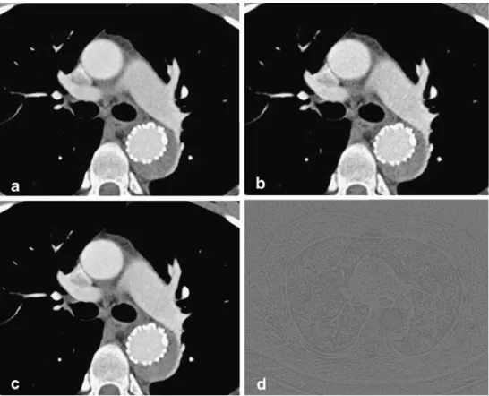
Fig. 1 Transverse images of a
male patient (BMI 29.2 kg/m2)
at the level of the left pulmonary artery displaying (a) full dose image (FD), (b) half dose image with filtered back projection (HD-FBP), and (c) half dose image with raw data-based sinogram affirmed iterative reconstruction (HD-SAFIRE). Note the improved sharpness of the vessel contours in HD-SAFIRE images, as well as the reduced image noise as displayed in the subtraction image showing the difference between HD-FBP and HD-SAFIRE (d)
Table 2 Assessment of overall image quality Image Quality Full Dose-FBP Half Dose-FBP Half Dose-SAFIRE Image noise 1 2 (1–3)*‡ 1 Motion artefacts 1 1 1 Metallic artefacts 1 (1–2) 1 (1–3) 1 (1–3) Contrast medium related 1 1 1
FBP filtered back projection; SAFIRE sinogram affirmed iterative reconstruction
Numbers are median with range in parentheses, according to 4-point scale (1 = optimum; 2 = minor; 3 = moderate, not affecting diagnostic quality, 4 = severe, affecting diagnostic image quality).
* p<0.001 compared to full dose dataset ‡ p<0.001 compared to half dose–SAFIRE
5.8 mSv for FD CT examinations and 10.9±2.9 mSv (effective Dose/2) for HD CT examinations.
Discussion
Our intra-individual comparison of image datasets obtained in a series of patients undergoing body CTA indicates that raw data-based SAFIRE has the potential of radiation dose reduction more than 50% as compared to a standard reconstruction with FBP, while maintaining diagnostic image quality.
Conventional CT image reconstruction approaches like back-projection type algorithms comprise a trade-off between sharpness and noise. Sharpness, i.e. the minimum visible detail size, can only be increased at the expense of higher image noise, or vice versa, noise can only be reduced by lowering the sharpness. This trade-off limits the minimum radiation dose required for a specific diagnostic application. SAFIRE represents an iterative optimization process which overcomes this constraint. This reconstruction type uses a noise modelling technique supported by raw data. The model utilizes the known propagation of noise in projection data into image domain [13].
Reconstructed CT data can be considered as a noise-free base (= “information”) plus additive statistical noise. The goal is to separate the information and noise with a maximum likelihood given by the model. This task can be
translated into a mathematical optimization problem and solved iteratively. The noise content is estimated and subtracted from the current dataset in each iteration loop. Afterwards, the result is compared to the initial data leading to an update image, and added to the previous dataset before the next iteration is performed. This procedure can be regarded as a validation loop. A total of five iterations are performed with SAFIRE.
In this study, we used dual-source CT, thereby enabling an intra-individual comparison of the results from the various image datasets within the same patients under same conditions as previously done by May et al. in a study assessing the value of image-domain based iterative reconstruction for saving radiation dose [9]. This approach
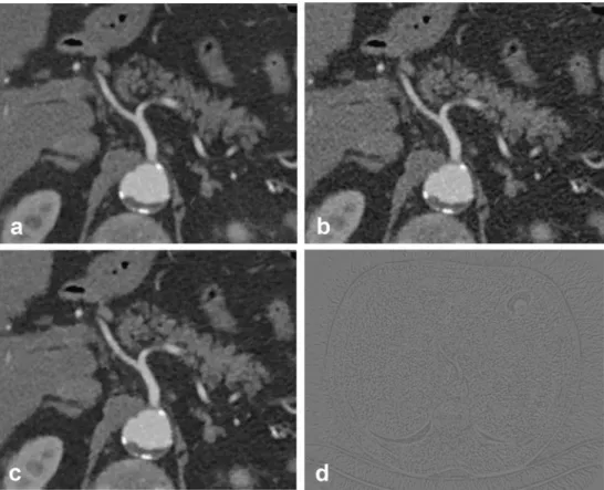
Fig. 2 Transverse images of a
male patient (BMI 28.2 kg/m2)
at the level of the celiac trunk, displaying (a) full dose image (FD), (b) half dose image with filtered back projection (HD-FBP), and (c) half dose image with raw data-based sinogram affirmed iterative reconstruction (HD-SAFIRE). Note the improved sharpness of the vessel contours in HD-SAFIRE as compared to HD-FBP images, as well as the reduced image noise as indicated in the subtraction image displaying the difference between HD-FBP and HD-SAFIRE (d)
Table 3 Sharpness of the aortic contour
Contour depiction Full
Dose-FBP Half Dose-FBP Half Dose-SAFIRE Good to excellent 16 0 18 Moderate, slightly blurred 9 17 7 Severely blurred 0 8 0 Median 1 2* 1‡
FBP filtered back projection; SAFIRE sinogram affirmed iterative reconstruction
* p<0.001 compared to full dose dataset
enables CT data acquisition with two tubes simultaneously and allows for the reconstruction of data from either a single or from both tubes. By doing so, we could eliminate the influence of different contrast enhancement or patient positioning between studies. This is reflected by the consistency of aorto-iliac vessel attenuation among the three image datasets of our patients demonstrating similar values with all reconstruction types and radiation doses.
By applying SAFIRE, image noise in half-dose datasets was significantly lower than that in full-dose datasets reconstructed with conventional FBP. This indicates that radiation dose could be reduced by more than 50% when using SAFIRE as compared to FBP, while high image quality was maintained. Moreover, sharpness of the aortic contour was significantly improved in the SAFIRE datasets as compared to the images reconstructed with conventional FBP at the same radiation dose. This difference in edge contour detection partly reflects the changes in image texture introduced by iterative reconstructions, a phenomenon which was also previously described in image domain-based methods [9].
Our study has the following limitations. First, we included only 25 patients, and further studies in larger patient populations, including also other applications such as chest or abdominal CT, are required for confirming these preliminary findings. Second, none of our patients had a BMI>35 kg/m2, so that an upper BMI limit—if present—in regard to the application of SAFIRE could not be determined. This issue could become relevant when using a strength higher than level 3, which presumably would result in a further image mottle reduction. Second, we assessed noise subjectively on a 4-point scale and objec-tively by measuring the standard deviation of attenuation, but we did not measure the noise power spectra. Finally, we did not evaluate the effect of SAFIRE on the readers’ diagnostic performance.
In conclusion, preliminary evidence suggests that raw data-based iterative reconstructions have the potential to reduce radiation dose by more than 50% in body CTA studies without deterioration in image quality.
References
1. Alkadhi H, Schindera ST (2011) State of the art low-dose CT
angiography of the body. Eur J Radiol. doi:10.1016/j.
ejrad.2010.12.099
2. Wintersperger B, Jakobs T, Herzog P et al (2005) Aorto-iliac multidetector-row CT angiography with low kV settings: im-proved vessel enhancement and simultaneous reduction of
radiation dose. Eur Radiol 15:334–341
3. McCollough CH, Bruesewitz MR, Kofler JM Jr (2006) CT dose reduction and dose management tools: overview of available
options. Radiographics 26:503–512
4. Kalra MK, Maher MM, Toth TL et al (2004) Strategies for CT
radiation dose optimization. Radiology 230:619–628
5. Pontana F, Pagniez J, Flohr T et al (2010) Chest computed tomography using iterative reconstruction vs filtered back projec-tion (Part 1): evaluaprojec-tion of image noise reducprojec-tion in 32 patients.
Eur Radiol 21:627–635
6. Pontana F, Duhamel A, Pagniez J et al (2010) Chest computed tomography using iterative reconstruction vs filtered back projec-tion (Part 2): image quality of low-dose CT examinaprojec-tions in 80 patients. Eur Radiol 21:636–643
7. Prakash P, Kalra MK, Kambadakone AK et al (2010) Reducing abdominal CT radiation dose with adaptive statistical iterative reconstruction technique. Invest Radiol 45:202–210
8. Sagara Y, Hara AK, Pavlicek W, Silva AC, Paden RG, Wu Q (2010) Abdominal CT: comparison of low-dose CT with adaptive statistical iterative reconstruction and routine-dose CT with filtered
back projection in 53 patients. AJR Am J Roentgenol 195:713–719
9. May MS, Wust W, Brand M et al (2011) Dose reduction in abdominal computed tomography: intraindividual comparison of image quality of full-dose standard and half-dose iterative reconstructions with dual-source computed tomography. Invest
Radiol 46:465–470
10. Schindera ST, Graca P, Patak MA et al (2009) Thoracoabdominal-aortoiliac multidetector-row CT angiography at 80 and 100 kVp: assessment of image quality and radiation dose. Invest Radiol
44:650–655
11. American Association of Physicists in Medicine (2008) The Measurement, Reporting, and Management of Radiation Dose in CT. American Association of Physicists in Medicine task group
23, College Park, MD.http://www.aapm.org/pubs/reports/rpt_96.
pdf. Accessed 05 Feb 2011
12. Macari M, Chandarana H, Schmidt B, Lee J, Lamparello P, Babb J (2006) Abdominal aortic aneurysm: can the arterial phase at CT evaluation after endovascular repair be eliminated to reduce
radiation dose? Radiology 241:908–914
13. Barrett HH, Swindell W (1981) Radiological imaging: the theory of image formation, detection, and processing. Academic, New York