Introduction
The leg chronic exertional compartment syndrome (CECS) is often seen in athletes. It is generally accepted that this condition is related to repetitive exertion or overuse and often interfere with the performances [1,2]. The pathophysiology is related to elevation of intra-muscular pressure, during exercise, to a point that the tissue within the affected compartment becomes tight and painful, thus preventing further activity. The pain, often, disappears within some minutes after rest and there are usually no permanent sequelae in the affected tissue. The problem is that, over time, this condition becomes more severe, and a raised intra-compartimental pressure may cause relative ischemia of the involved muscles [1,20]. Some authors [7,15] had suggested that
activity induced muscle swelling is restricted by fascial tightness; this theory is used to propose fasciotomy as the treatment of choice for this condition [3,5–7,14,17,
18, 22]. The most commonly affected compartments in the leg are the anterior followed by the deep posterior, lateral, and rarely the superficial posterior compartment [1, 2, 8]. Different techniques have been described for fasciotomy and fasciectomy of the leg, such as open one [5, 6, 8, 12], or two incisions fasciotomy [2, 17, 19], endoscopically assisted one [10,13] or two [11] incisions fasciotomy, and fasciectomy [3,4]. The use of fascioto-my or fasciectofascioto-my has been debated, with some authors performing fasciotomy with a partial fasciectomy [19] and others [3, 7] advocates fasciectomy only for recur-rent cases when a previous compartment fasciotomy had failed. In this paper we describe the open minimal E. Mouhsine
R. Garofalo B. Moretti G. Gremion A. Akiki
Two minimal incision fasciotomy for chronic
exertional compartment syndrome
of the lower leg
Received: 31 March 2004 Accepted: 2 November 2004 Published online: 18 May 2005
Ó Springer-Verlag 2005
Abstract Chronic exertional com-partment syndrome (CECS) of the leg is a pathological condition often related to overuse in subject who engage repetitive physical activities. Fascial release is the mainstay of surgical management. The purpose of this study was to evaluate the results obtained with a double inci-sion decompressive fasciotomy. Eighteen consecutive athletes with a diagnosis of anterior and/or lateral CECS of the leg were operated on with a minimal double incision fas-cial release after a mean period of 4 months after onset of symptoms. In 11 cases (61%) CECS was bilat-eral. Surgery was performed without tourniquet and active mobilization was starting immediately. Sports
activities were resumed gradually at a mean period of 25 days. The ath-letes were followed until 2 years. All resumed pre-injury level sports activity. Two patients (18%) of the 11 who underwent to bilateral fas-ciotomy referred a sensation of leg weakness for an average period of 3 months. The surgical technique presented in this paper seems to be a good mean to treat anterior and lateral leg CECS. The use of tourniquet is deconselled to obtain an accurate intraoperative haemostasis so reducing the risk of post-operative haematoma.
Keywords Leg Æ Compartment syndrome Æ Exertional Æ Fasciotomy
DOI 10.1007/s00167-004-0613-6
E. Mouhsine (&) Æ R. Garofalo B. Moretti Æ G. Gremion Æ A. Akiki Department of Orthopaedic and Traumatology,
OTR-BH 14, CHUV, 1011 Lausanne, Switzerland
E-mail: [email protected] Tel.: +41-21-3142791
double incision surgical technique to treat leg CECS, and we present the outcome obtained in a serie of 18 consecutive athletes.
Material and method
In the period between 1999 and 2002, 18 consecutive athletes with a clinical diagnosis of leg CECS underwent fasciotomy. The group of patients consisted of ten men and eight women with an average age of 25 years (19– 38 years). The type of sport activity causing symptoms are principally jogging, classic dance and foot-ball playing. Before coming to our institute the athletes were treated for a mean period of 4 months (1 to 6) with a conservative management consisting in modification of exercise program, stretching, changing of shoes, shoe inserts, or local injection of xylocaine in the area of maximal tenderness. In eleven cases the symptoms were bilateral and in the remaining seven only unilateral. In all cases the muscle complex of the anterior compart-ment was involved for a total of 29 legs and in the four patients, three of whom complained of bilateral symp-toms, the anterior and the lateral compartments were involved. Finally 36 compartements were released (Ta-ble 1). At clinical examinations, at rest, no gross physi-cal signs were noted, but in eight cases the anterior compartment of the leg was firm on palpation. Neuro-logical examination was normal in all cases.
Surgical technique
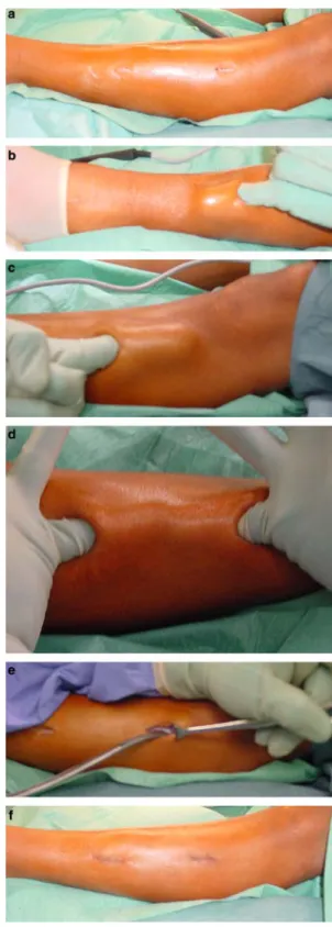
The same senior surgeon (E.M) performed all of the 36 fasciotomy. Surgery was performed always without a tourniquet. The technique consisted to perform two vertical skin incision of 2 cm length (Fig.1a) over the
anterior compartment. The distance between the 2 incisions averaged 15 cm as described by Rorabeck [17]. Once the fascia i identified, the bridge of skin and subcutaneous tissue between the two incisions is care-fully dissected down to the level of the fascia with gloved
Table 1 Patients treated for leg CECS Unilateral surgery Bilateral surgery Anterior compartment surgery only
Anterior and lateral compartment surgery
Men 3 7 7 3
Women 4 4 7 1
Total 7 11 14 4
Fig. 1 Technique and sequence of double mini-incision for fasciot-omy. a Two incisions of 2 cm length over the anterior compartment are performed. b–d After skin incision, a subcutaneous flap is developed with the two gloved fingers. e Anterior and/or lateral fasciotomy is performed under direct vision. The skin and the subcutaneous flap is easily mobilised with a retractor allowing a good vision. Sometimes, between the two incisions gloved fingers can be used as scissor to complete fascial release. f Sterile adhesive strips closed skin incisions
fingers both proximally and distally (Fig. 1b–d). Once that this subcutaneous flap is developed, it may be easily mobilised with retractors allowing a clear vision of the deep fascia.
The anterior intermuscular septum and the superficial peroneal nerve were identified through the lower incision at the point where it pierces the deep fascia, at an average of 10–12 cm proximal to the lateral malleolus. Retracting the skin anteriorly and posteriorly, respec-tively the anterior and lateral compartment fascia (seven times in our series) was divided with scissors under direct visualisation. The fascia is incised at an average of 1.0 cm distance from the intermuscular septum (Fig.1e). In the cases where the visualisation of the fascia between the two incisions was not good enough, a gloved finger was used to complete the release. Practi-cally it avoids nerve damage. The two incisions were closed using a superficial subcutaneous biosine 3.0 monofilament (Davis and Geck, Danbury, Conneticut) and sterile adhesive strips (Fig.1f). A firm bandage was then applied from midfoot to the knee. In the postop-erative period, patients remained in bed for 24 h with the operated leg elevated. Ice was applied. After that, supervised active range of motion exercises and deam-bulation were instituted. Most patients needed to use crutches for at least 10 days after the operation. Fifteen days postoperatively, the surgical incisions were con-trolled and sports activities were started gradually on a mean period of 25 days (21–32) after surgery.
The patients were reviewed at 6 weeks, 3 months, 6 months, 1 year and 2 years after surgery. At last fol-low-up a self-administered questionnaire of satisfaction was given. Clinical results were classified as none (min-imal or no relief), moderate (min(min-imal to significant re-lief), good (return to full activity with minimal or no symptoms). The results are shown in Table2.
Discussion
Surgical fascial release is the mainstay of treatment of CECS of the leg [3, 7, 12, 15, 17, 18], with report of improvement ranging from 65% to 100% [6–8,11, 15,
17, 18, 20]. Our series with double incision fasciotomy show good results in 100% of patients with an anterior or antero-lateral leg CECS, at 2 years of follow up. The pattern of recovery from the operation indicated an
early gradual improvement that remained stable with time.
The specific method for performing fascia release is variable and various authors have used both a one [5,8,
12] and two incisions [16,17,19,21] techniques; Detmer [7] have suggested three or more incisions for patients with excessively long legs. Recently, the use of endo-scope has been proposed to assist visualisation of fas-ciotomy with single [10,13] or double incisions [11]. The reported complication rate of fascia release, is between 4.5% and 13% including nerve injury, infections, post-operative haematoma, recurrence of symptoms second-ary to incomplete release, cosmetically unacceptable scarring, anaesthetic problems, muscle fascia adhesions, swelling, lymphocele or haemorrhage [6–8, 17, 23]. In our series differently to another report [6] in any case an altered skin sensation over the fasciotomy site was found. So also if,the subcutaneous tissue was under-mined by fascia using gloved finger, the risk of nerve stripping with local denervation of the skin is very low. A transitory weakness was revealed in two patients; this data was also present in another report [19]. Dif-ferently from Slimmon [19], we did not perform fasci-ectomy, but caracteristically the two patients presenting these symptoms have a bilateral CECS that involved both the anterior ant the lateral compartment.
The good outcome in term of symptoms improve-ment in our series is probably related to three factors: first, the compartment involved, second, the surgical technique used, and third, the time of preoperative symptoms duration.
In literature, the most of reports has showed a better long term outcome for antero-lateral compared with posterior compartment decompression [1,3,4,6,–8,11,
17,18,20]. In fact, the reported success for anterior and lateral compartment decompression surgery varies be-tween 65% and 100% [6–8,11,15,17,18,20], while for the posterior compartment the percentage is between 50% and 75% [1,3,4,8,17–19].
About surgical technique, a some variety is reported in literature. A study performed on cadaver specimen [10] concluded that neither techniques, endoscopic or percutaneous, was without risk of nerve injury, but a single incision endoscopically assisted fascial release of the anterior and the lateral compartments showed a lower risk of peroneal nerve injury. This nerve is most frequently at risk at the junction of the middle and distal thirds of the calf, where it pierces the fascia at an average distance of 10–12 cm proximal to the lateral malleolus and begins to head more obliquely and anteriorly. The technique of two vertical incisions as proposed by Rorabeck [17], allows in our advice to perform a complete anterior and lateral fascial release without taking major risks. Undermining the skin and the subcutaneous tissue is a very important step that allows a decompression of compartments under direct
Table 2 Clinical results
Surgical complications 0
Resumed sports activities 18
Neurological impairment 0
Reoperation 0
control, after that the anterior intermuscular septum and the superficial peroneal nerve are identified. Using two retractors during surgery is possible to perform fascial release respect the two incisions at the proximal and distal margins of the compartments with scissors. Only a little area of 1.5–2 cm, located between the two incisions can remain blind, and in this area the gloved finger of the surgeon is used as scissors, thus reducing the risk of nerve damage. Differently to the original description of Rorabeck [17], we do not use a tourni-quet during surgery in order to better control haemo-stasis. This surgical step is very important because a postoperative haematoma may lead to draining wounds, delayed recovery and potential infection. Moreover various authors [3, 4, 9, 17] have proposed an early mobilisation of the knee and the ankle and walking on the affected limb after surgery to prevent excessive scarring and adhesions. This was the protocol adapted in our department. In case of postoperative haematoma formation, this would compromise the mobilisation protocol and thus leading to poor results due to scarring, adhesions, stiffness and pain.
The duration of preoperative symptoms is another important point. In our series, no-one patient presented duration longer than 7 months. Slimmon [19] have showed that the duration of preoperative symptoms
affected significantly the proportion of satisfactory re-sults in patients with an isolated anterior compartment surgery, suggesting a time more than 12 months after the onset of symptoms associated with poor results. This argument stresses the necessity to perform early the diagnosis of CECS. So when a trainer or a sports doctor face an athlete with leg pain responding to physiother-apy or to activity modification, a CECS should be ex-cluded.
Although this series presents limitations related to the retrospective series, some points of strength are present: in fact, it was a consecutive series, in which the operation was performed by a single senior surgeon. Moreover, the outcomes at the follow up was evaluated by a indepen-dent reviewer who did not participate to any of the surgeries through a self-administered questionnaire of satisfaction.
In conclusion, minimal open double incision fasciot-omy seems to be a viable surgical method to treat anterior and lateral CECS of the leg. This technique allows a good surgical site visualisation minimising the risks of incomplete compartment release and soft tissue injury. The surgery should be performed without a tourniquet, so a good haemostasis can be carried out, to avoid postoperative haematoma, and thus lowering risk of scarring or adhesions.
References
1. Allen MJ, Barnes MR (1986) Exercise pain in the lower leg. Chronic com-partment syndrome and medial tibial syndrome. J Bone Joint Surg Br 68(5):818–823
2. Balduini FC, Shenton DW, O’Connor KH, Heppenstall RB (1993) Chronic exertional compartment syndrome: correlation of compartment pressure and muscle ischemia utilizing 31P-NMR spectroscopy. Clin Sports Med 12(1):151–165
3. Bell S (1986) Repeat compartment decompression with partial fasciectomy. J Bone Joint Surg Br 68(5):815–817 4. Black KP, Schultz TK, Cheung NL (1990) Compartment syndromes in athletes. Clin Sports Med 9(2):471–487 5. Blackmann PG (2000) A review of
chronic exertional compartment syn-drome in the lower leg. Med Sci Sports Exerc 32 (3 Suppl):S4–S10
6. Cook S, Bruce G (2002) Fasciotomy for chronic compartment syndrome in the lower limb. ANZ J Surg 72(10):720–723
7. Detmer DE, Sharpe K, Sufit RL, Girdley FM (1985) Chronic compart-ment syndrome: diagnosis, manage-ment, and outcomes. Am J Sports Med 13(3):162–170
8. Howard JL, Mohtadi NG, Wiley JP (2000) Evaluation of outcomes in pa-tients following surgical treatment of chronic exertional compartment syn-drome in the leg. Clin J Sport Med 10(3):176–184
9. Hutchinson M, Ireland ML (1994) Common compartment syndromes in athletes. Treat Rehabilit Sports Med 17(3):200–208
10. Hutchinson MR, Bederka B, Kopplin M (2003) Anatomic structures at risk during minimal-incision endoscopically assisted fascial compartment releases in the leg. 14. Am J Sports Med 31(5):764– 769
11. Leversedge F, Casey PJ, Seiler JG 3rd, Xerogeanes JW (2002) Endoscopically assisted fasciotomy: description of technique and in vitro assessment of lower-leg compartment decompression. Am J Sports Med 30(2):272–278
12. Martens MA, Backaert M, Varmaut G, Mulier JC. (1984) Chronic leg pain in athletes due to a recurrent compartment syndrome. Am J Sports Med 12(2):148– 51
13. Ota Y, Senda M, Hashizume H, Inoue H (1999) Chronic compartment syn-drome of the lower leg: a new diagnostic method using near-infrared spectros-copy and a new technique of endoscopic fasciotomy. Arthroscopy 15(4):439–43 14. Puranen J, Alavaikko A (1981) Intra-compartmental pressure increase on exertion in patients with chronic com-partment syndrome in the leg. J Bone Joint Surg Am 63(8):1304–1309 15. Qvarfordt P, Christenson JT, Eklof B,
Ohlin P, Saltin B (1983) Intramuscular pressure, muscle blood flow, and skele-tal muscle metabolism in chronic ante-rior tibial compartment syndrome. Clin Orthop 179:284–290
16. Reneman RS (1975) The anterior and the lateral compartmental syndrome of the leg due to intensive use of muscles. Clin Orthop 113:69–80
17. Rorabeck CH, Bourne RB, Fowler PJ (1983). The surgical treatment of exer-tional compartment syndrome in ath-letes. J Bone Joint Surg Am 65(9):1245– 1251
18. Schepsis AA, Martini D, Corbett M (1993) Surgical management of exer-tional compartment syndrome of the lower leg. Long-term followup. Am J Sports Med 21(6):811–817
19. Slimmon D, Bennell K, Brukner P, Crossley K, Bell SN (2002) Long-term outcome of fasciotomy with partial fasciectomy for chronic exertional compartment syndrome of the lower leg. Am J Sports Med 30(4):581–588 20. Styf J, Korner L, Suurkula M (1987).
Intramuscular pressure and muscle blood flow during exercise in chronic compartment syndrome. J Bone Joint Surg Br 69(2):301–305
21. Turnipseed W, Detmer DE, Girdley F (1989) Chronic compartment syndrome. An unusual cause for claudication. Ann Surg 210(4):557–562
22. Wallensten R, Eriksson E (1984) Intra-muscular pressures in exercise-induced lower leg pain. Int J Sports Med 5(1):31–5
23. Wiley JP, Clement DB, Doyle LD, Tauton JE (1987) A primary case per-spective of chronic compartment syn-drome of the leg. Physician Sportsmed 15(3) :111–120