K . A . S i e b e n r o c k • T. G e r i c h • R . P. J a k o b
Sequential intramedullary nailing
of open tibial shaft fractures after external fixation
Received: 9 October 1995
A b s t r a c t s W e r e v i e w e d 32 tibial shaft fractures in 31 pa- tients treated w i t h s e q u e n t i a l i n t r a m e d u l l a r y n a i l i n g after p r i m a r y e x t e r n a l fixation. T h e r e w e r e 30 o p e n fractures and 2 c l o s e d injuries with s e v e r e b l u n t t r a u m a r e q u i r i n g f a s c i o t o m y . F i f t y p e r c e n t o f the fractures w e r e c l a s s i f i e d as G u s t i l o t y p e I I I A and B injuries [13]. T h e m e a n exter- nal f i x a t i o n t r e a t m e n t a v e r a g e d 6.6 w e e k s , and s e c o n d a r y i n t r a m e d u l l a r y n a i l i n g w a s d o n e on a v e r a g e 7.4 w e e k s af- ter injury. I n 50% o f the fractures, s e c o n d a r y n a i l i n g was d o n e at the s a m e p r o c e d u r e as r e m o v a l o f the e x t e r n a l fix- ation. O v e r a l l , the i n c i d e n c e o f o s t e o m y e l i t i s and non- u n i o n was 3.1% e a c h and o f m a l u n i o n 19%. T h e t i m e to full w e i g h t - b e a r i n g a v e r a g e d 31.2 weeks. T h e results w e r e s e p a r a t e l y a n a l y z e d a c c o r d i n g to G u s t i l o t y p e s and s u b t y p e s . In the G u s t i l o t y p e III B injuries, the i n c i d e n c e o f o s t e o m y e l i t i s and n o n - u n i o n was 11%, w h i l e m a l u n i o n o c c u r r e d in 33%. T h e t i m e to full w e i g h t - b e a r i n g aver- a g e d 53 w e e k s . T h e s e results s u p p o r t the c o n c l u s i o n that this t r e a t m e n t m o d a l i t y is a v a l i d a l t e r n a t i v e to other treat- m e n t options. H o w e v e r , p r e v i o u s p i n t r a c t infections s h o u l d b e r e g a r d e d as a c o n t r a i n d i c a t i o n for s e c o n d a r y nailing.
Introduction
T h e d e b a t e on h o w to treat h i g h - e n e r g y and o p e n tibial shaft fractures has i n t e n s i f i e d w i t h the i n t r o d u c t i o n o f un- r e a m e d i n t e r l o c k i n g i n t r a m e d u l l a r y d e v i c e s [26, 32, 35]. C o n t r o v e r s y still exists about the optimal treatment m e t h o d
Beispiel: Presented at the 15th Annual Meeting on Mycorrhizae, Chicago, 1992
K. A. Siebenrock (N~) • R. P. Jakob
Department of Orthopaedic Surgery, Inselspital, University of Bern, CH-3010 Bern, Switzerland T. Gerich
Unfallchirurgische Klinik der Medizinischen Hochschule, Hannover, Germany
for s e v e r e o p e n G u s t i l o t y p e III fractures [13]. Recently, s o m e authors h a v e f a v o u r e d p r i m a r y u n r e a m e d i n t e r l o c k - ing n a i l i n g in these fracture t y p e s [26, 35]. C o m m o n p r o b - l e m s seen w i t h e x t e r n a l f i x a t i o n in the m a n a g e m e n t o f G u s t i l o t y p e III fractures i n c l u d e pintract, soft-tissue and b o n e infections, as w e l l as h i g h n o n - u n i o n and m a l u n i o n rates [9, 10, 18]. I n an a t t e m p t to l o w e r the c o m p l i c a t i o n rates a s s o c i a t e d w i t h e x t e r n a l f i x a t i o n t r e a t m e n t in these injuries, s e v e r a l i n v e s t i g a t o r s h a v e s u g g e s t e d s e q u e n t i a l i n t r a m e d u l l a r y n a i l i n g after c o m p l e t e soft-tissue h e a l i n g [4, 23, 24]. In our d e p a r t m e n t this m e t h o d o f t r e a t m e n t has i n c r e a s i n g l y b e e n a p p l i e d as a p l a n n e d p r o c e d u r e in r e c e n t years. T h i s study r e p o r t s the results o f 32 fractures treated w i t h s e q u e n t i a l n a i l i n g o f o p e n tibial shaft frac- tures after p r i m a r y t r e a t m e n t w i t h an e x t e r n a l f i x a t i o n de- v i c e o v e r a t i m e p e r i o d o f 6 years.
Materials and methods
A retrospective analysis of 31 consecutive patients with 32 tibial shaft fractures (initial injury occurred between 1986 and 1992) was done (Table 1). All patients were primarily treated at our depart- ment with an external fixation device and subsequent conversion to an intramedullary nail after soft-tissue healing. The charts and the preoperative X-rays as well as the radiographs from the last follow-up control of all 32 cases were retrospectively reviewed. Follow-up information could be obtained in all cases at an average of 22.6 months (range 6-54 months) after injury. The mean age of the patients was 33 years (16-75 years) with a male to female ra- tio of 4: 1. Seventy-one per cent of the patients had multiple in- juries. According to the AO classification [25] there were 28% type A, 20% type B and 52% type C (comminuted) fractures. Two high-energy closed fractures required fasciotomy within 24 h. In addition, two fasciotomies were done for two grade II open frac- tures within 36 h after injury. The 30 open fractures were classified according to Gustilo et al. [13]: 4 were type I, 10 as type II, 7 as type I I I A and 9 as type III B. There were no type III C injuries. All patients were treated within 24 h after injury with soft-tissue de- bridement and application of external fixation. In all but two cases a unilateral device (AO/ASIF tube external fixator) was used with two 5-mm Schanz screws both proximal and distal to the fracture site and two longitudinal bars. In the remaining two cases on AO/ASIF external point clamp fixator was used, also with two clamps each on either side of the fracture. Conversion to an in- tramedullary nail was not done as a standard procedure at our de-
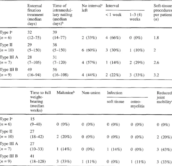
Table 1 Average treatment data for open tibial shaft frac- tures classified according to Gustilo et al. [131
a Type I group includes 2 pa- Type I a
tients with closed fractures (n = 6) b Time of intramedullary nail-
ing after injury Type II interval = time between re- (n = 10) moval of external fixation and Type III intramedullary nailing. Soft- (n = 7) tissue procedures included de-
bridement, secondary wound Type III closure, skin grafts and muscle (n = 9) flaps
Table 2 Treatment results for open tibial shaft fractures clas- sified according to Gustilo et al. [13]
Type I group includes 2 pa- Type I a tients with closed fractures (n = 6) b Malunion is defined as angu- Type II lation > 5 ° in frontal plane and
> 10 ° in sagittal plane, > 10 ° (n = 10) of rotation and > 1 cm of leg Type III discrepancy (n = 7) c Reduced joint mobility is de-
fined as loss of motion in ankle Type III joint > 10 ° and knee joint > (n = 9) 20 °
A
B
A
B
External Time of No interval c Interval fixation intramedul- left
treatment lary nailing < 1 week 1-3 (4)
(median (median weeks
days) days) b 32 (12-75) 29 (5-150) 28 (%105) 49 (16-94) Time to full weight- bearing (median weeks) Soft-tissue procedures per patient (n) 39 (14-77) 2 (33%) 4 (66%) 0 (0%) 1.8 38 (5-150) 6 (60%) 3 (30%) 1 (10%) 2 31 (7-120) 4 (57%) 1 (14%) 2 (29%) 2.6 56 (16-108) 4 (44%) 2 (22%) 3 (33%) 3.2
Malunion b Non-union Infection
soft tissue osteo- myelitis Reduced joint mobility c 15 (9-40) 0 (0%) 0 (0%) 0 (0%) 0 (0%) 0 (0%) 27 (18-42) 2 (20%) 0 (0%) 0 (0%) 0 (0%) 2 (20%) 27 (10-33) 1 (14%) 0 (0%) 1 (14%) 0 (0%) 3 (43%) 41 (18-128) 3 (33%) 1 (11%) 0 (0%) l (11%) 3 (33%)
partment, but it has been performed more frequently in recent years, according to the surgeon's preference. Reasons for change included primary planning after soft-tissue healing in 19 fractures, bone healing problems in 10 fractures, loosening of the external fixation device in 2, and loss of reduction during treatment with external fixation in 1. The majority of the cases were fixed with se- quential nails before the routine use of the new solid unreamed AO tibial nail. Thus, in 26 of 32 cases (81%) a reamed AO universal tibial nail was inserted, an unreamed solid AO tibial nail in 5 (16%) and Ender rods in 1 (3%). In 7 of 9 fractures (78%) with a grade III B open lesion, fixation with a reamed nail was per- formed. The nails were statically locked in 10 cases (31%) with subsequent dynamization in 8 of them at an average period of 6.5 months (4-9 months) after insertion. In two other cases (6%) the nail was dynamically locked. At the last follow-up 11 nails (34%) had been removed at an average period of 22 months (11-44 months) after insertion. The treatment results were analysed for all 32 fractures together and separately reviewed according to the Gustilo types and subtypes.
Results
The m e a n external fixation t r e a t m e n t time averaged 6.6 weeks ( 1 - 2 0 weeks). S e q u e n t i a l nail fixation was per- f o r m e d at an average of 7.4 weeks (1-20 weeks) after in- jury. P i n tract i n f e c t i o n was d e f i n e d as a n y persistent d r a i n a g e from a p i n site r e q u i r i n g i n t e r v e n t i o n (enlarge- m e n t o f incision, etc.) or positive bacterial cultures from
the pin-site area [5, 23]. It was seen in three cases (9.4%). L a b o r a t o r y results c o n c e r n i n g i n f e c t i o n parameters (ery- throcyte s e d i m e n t a t i o n rate, ESR, white cell counts, W B C , C-reactive protein) w i t h i n 2 weeks prior to s e q u e n - tial nail fixation were available i n 10 cases. O f these re- sults 60% was w i t h i n n o r m a l limits, 20% m i l d l y elevated ( W B C b e t w e e n 1 0 0 0 0 a n d 1 2 0 0 0 , E S R < 30), a n d i n 2 0 % there was a m o d e r a t e e l e v a t i o n of E S R (45 a n d 68, respectively). A l l of these 10 cases u n d e r w e n t n a i l i n g with u n e v e n t f u l healing. Thus, n o correlation could be d r a w n b e t w e e n e l e v a t i o n o f these parameters a n d subse- q u e n t infection. I n 16 fractures (50%) n a i l i n g was d o n e at the same procedure as external fixation removal. In 10 fractures there was an interval of 1 w e e k b e t w e e n r e m o v a l of the external fixation a n d s e c o n d a r y nailing. I n 5 cases the interval s p a n n e d 1-3 weeks, and in 1 case the interval was 4 weeks. T i m e to full w e i g h t - b e a r i n g averaged 31.2 weeks ( 9 - 1 2 6 weeks). M a l u n i o n was d e f i n e d as a n g u l a - tion in the frontal p l a n e of more than 5 deg, a n g u l a t i o n in the sagittal p l a n e or rotation of m o r e than 10 deg, as well as s h o r t e n i n g of the leg b y m o r e than 1 cm. R o t a t i o n a n d leg s h o r t e n i n g were evaluated clinically. I n cases with fractures of the contralateral leg or ipsilateral f e m u r frac- tures, leg length m e a s u r e m e n t was o b t a i n e d b y c o m p a r i n g the l e n g t h of the lower legs on radiographs taken from both sides. Routinely, X - r a y o f the u n i n v o l v e d lower leg
34
was done before secondary nail fixation to plan the cor- rect length of the nail. The long bones of the contralateral leg were fractured in five and the ipsilateral femur in three cases. In three of these eight cases a conclusive evaluation of leg length discrepancy could not be done. According to the definitions above a malunion was observed in six frac- tures (19%). Non-union occurred in one fracture (3%) due to bone infection. Soft-tissue infection was seen in one patient who went on to uneventful bone healing. Os- teomyelitis developed in the above-listed case (3%) with subsequent non-union and breakage of a nail. In this case another nail was inserted after intramedullary reaming. However, union was not achieved until removal of the second nail and plating of the shaft with additional bone grafting. Further complications included hypaesthesia in the medial calf in two cases, extensor motor weakness due to traumatic peroneal nerve palsy in two, incomplete com- partment syndrome in one, asymptomatic non-union of the fibula in one and chronic pain at the fracture site in two. Reduced joint mobility of the knee and/or ankle joint of more than 10 deg was recorded in eight cases (25%). Cases with concomitant injuries to the ligaments of the knee or ankle joint were not excluded and were counted as possibly secondary to the fracture of the lower leg. In the vast majority (seven cases) there was a reduced dorsal ex- tension of the ankle joint. In two cases this was due to posttraumatic incomplete peroneal nerve palsy. In the re- maining case there was a 20 deg loss of flexion of the knee joint (Table 2).
The results were separately analysed according to dif- ferent Gustilo fracture (sub)types and are listed in Tables 1 and 2. The two patients with closed fractures were added to the patients with type I fractures. In all nine cases with Gustilo type III B fractures, a muscle flap for soft-tissue coverage was performed. Muscle flaps in- cluded five hemisoleus flaps, a combined hemisoleus and medial gastrocnemius flap in one case, and three free latissimus dorsi flaps. Definite soft-tissue coverage in- cluding muscle flaps was achieved at primary surgery in three (33%) of the Gustilo type III B cases. In the remain- ing six fractures, coverage with a muscle flap was per- formed between the 2nd and 17th day after injury.
Bone grafting was done in 5 cases (56%) with type III B fractures at an average of 4 months (2 w e e k - l l months) after injury. In the seven cases with type I I I A fractures, wound closure was obtained by skin grafting after an av- erage of two soft-tissue debridements. In this latter group bone grafting was performed only in one case (14%) 6 weeks after injury.
Discussion
The reported infection rates for treatment of open tibial shaft fractures with external fixation range from 7% to 30% depending on the severity of the fractures [2, 3, 8, 18, 30, 32]. Malunion rates range from 10.5% to 46%, and non-union is reported in 11% to 48% [1, 2, 18, 30, 32]. Time to union in type III fractures with external fixation
ranged from 30 to 38 weeks and averaged 47.2 weeks when only type III B injuries were included [5, 10, 18]. Infection rates ranged from 29% to 35% in type III B frac- tures [8, 10]. A malunion rate of 71% was reported in this injury type treated with external fixation alone by Court- Brown et al. [10].
Infection rates for primary reamed nailing of acute tib- ial shaft fractures in the literature range from 6% to 20% [6, 15, 17, 19, 28]. As this procedure is not recommended in severe open fractures [7, 9, 14], most patients included in these studies had type I and type II open fractures. To our knowledge only Court-Brown et al. [11, 12] have pub- lished results of primary reamed nailing in type III B open tibial shaft fractures. They found infection rates of 9.5% in type III injuries. In type III B fractures they reported in- fection rates of 12.5% and 23%. In the latter patient groups time to union averaged 50.1 and 71 weeks, and mal-union was found in 15%.
In patient groups with various types of open tibial frac- tures, infection rates after primary unreamed nailing with flexible and solid devices were found to be within 3.3%- 8% [16, 21, 22, 33-35]. Malunion with flexible devices was recorded in 17% to 21% and non-union with both flexible and solid nails in 2.4%-6.3% [18, 36]. Infection rates ranged from 6% to 17% for type III fractures and were reported in 25% after the same treatment in type III B fractures [18, 32, 34-36]. In type III fractures non- union rates ranged from 5.8% to 26% [18, 35, 36], malu- nion was reported in 26% [32] with solid devices, and the average time for healing is reported to be 6.8 months. In their prelimary report about treating 19 open tibial shaft fractures (47% type III) with solid interlocking nails, Pic- cioni and Guanche [26] found no infection at all. How- ever, at the time of the report, 5 fractures had not defi- nitely healed and 3 (16%) had already undergone sec- ondary nailing for delayed bone healing. Krettek et al. [20] could not find infection in 10 patients with type III B fractures primarily treated with an unreamed solid nail. However, the study is limited by low patient numbers. Additionally, they reported muscle flap coverage in only 10% of their cases, whereas in the present study in all of the patients with type III B injuries, a muscle flap was performed. This may indicate classification problems as well.
A number of authors advocate against secondary nail- ing of open fractures after primary treatment with external fixation [5, 7, 14, 24]. Infection rates were reported to range from 25% to 44% with this treatment regimen in two studies [23, 24]. However, in both of these studies there was pin tract infection during external fixation treat- ment in five of six and in five of seven patients who con- sequently developed deep infection after sequential nail- ing. In 80% of the patients with pin tract infection and fur- ther osteomyelitis, the same bacteria were isolated before and after changing to a nail. Maurer et al. [23] demon- strated the significant increase in risk (to 71%) of devel- oping subsequent deep infection when nailing was done after evidence of pin tract infection. Consequently, they listed pin tract infection as a contraindication for sec-
Table 3 Interval length between removal of external fixation and sequential nailing and number of cases with consequent infections Reference Number of Number of Interval length Interval in cases
patients infections in study with infections
Conclusions regarding interval length and incidence of infection
Ahlers et al. [1] 13 0 2 weeks (cast) - Recommended as done
Blachut et al. [4] 39 2 0-3 weeks 1 × 5 days No correlation to interval 1 x no interval
Bone and Johnson [6] 5 0 7-10 days - Recommended as done
Maurer et al. [23] 24 7 3 days-54 weeks 9 days-t4 weeks > 3 weeks (based on study in canine model)
McGraw and Lira [24] 16 7 mean 3 weeks 1 × no interval No correlation to interval 6 x intervaP length found
Puno et al. [27] a, b 1 5--7 days Recommended as done
Siebenrock et al. [3 l] 24 1 0-4 weeks 1 x 2 weeks No correlation to interval found a Total number of patients not listed
b Detailed interval length not listed
ondary nailing. We strongly support this. The only patient who developed ostesmyelitis after sequential nailing in this study had clinical signs of former pin tract infection, al- though no pathological bacteria could be isolated from the pin tract site. In studies with a low incidence of pin tract infections, sequential nailing resulted in subsequent deep infections in only 4 % - 6 % [4, 31]. Also, those studies comprised patient groups with different types of soft-tis- sue injuries. However, none of the 12 patients with type III fractures reported by Blachut et al. [4] developed in- fection after sequential nailing. In the present study the re- sults were separately analysed for the different types of open injuries. The non-union and infection rates of 6% in type III fractures (11% in type III B) and the malunion rates of 25% (33% in type III B) indicate favorable results with the sequential treatment method, especially when compared with the treatment of type III injuries by exter- nal fixation alone [5, 8, 10]. Furthermore, two of the three malunion cases of the type III B fractures consisted only in leg shortening (1-3 cm). This is partially due to bone loss and comminuted fracture type. Primary nailing of type III injuries m a y be an alternative option. However, there are few studies that document the final outcome of severe, open, type Ill A and type III B injuries. Also, the limited stability with unreamed devices and the com- monly found comminuted fracture pattern in these injuries m a y render primary unreamed nailing a less than optimal treatment option [7, l 1, 14, 26, 34]. External fixation still has the advantage of a rigid fixation which can easily be achieved, especially in patients with multiple injuries.
When performing a sequential intramedullary nailing after external fixation, early exchange as soon as the soft tissues have healed safely seems to decrease the infection rates. Pin tract and deep infections are definitely lower when nailing was performed 2-6 weeks after the injury [4, 31] than 12-16 weeks after injury [23, 24]. R o m m e n s et al. [29] demonstrated that a shorter treatment period with external fixation reduces the risk of pin tract colonization. The soft-tissue management itself plays another decisive role in the final outcome. A variety of studies show es-
sential advantages of repeated aggressive soft-tissue man- agement aiming at early definite skin cover and prophy- lactic bone grafting in severely comminuted fractures [5, 8, 34]. Muscle flaps and definite skin cover at primary surgery were performed in 33% of the patients with type III B injuries in this study. In 56% of the patients with type III B injuries, bone grafting was performed in the further course.
There is still debate about leaving an interval between removal of external fixation and sequential nail implanta- tion. However, even in the studies with high infection rates, no significant benefit of an interval could be proven [23, 24]. A number of authors have recommended differ- ent interval lengths, as shown in Table 3. In other studies no increased risk could be found when there was no inter- val at all, and nailing was done in the same procedure as the removal of external fixation [4, 31]. In this study there was no interval for 50% of the patients. The only infection seen was in a patient in w h o m nailing was done 6 weeks after injury without leaving an interval. However, 2 weeks before this procedure there were clinical signs of pin tract infection, although cultures were negative. Reviewing the literature, we believe that this indicates a high risk of in- fection subsequent to a pin tract infection rather than the need to leave an interval. A previous pin tract infection should be strictly regarded as a contraindication for se- quential nailing [24]. Definition of pin tract infection has been proposed as any drainage from pin tract sites (a) with positive cultures from pin site or (b) which requires any kind of intervention [5, 23].
In conclusions, sequential nailing after external fixa- tion treatment is an effective treatment method for severe tibial shaft fractures including Gustilo type I I I A and B in- juries. Based on our data as well as on a literature review, there is no significantly higher risk of infection when se- quential intramedullary nailing is performed at the same procedure with external fixation removal given the strict criterion of absence of former pin tract infection. Aggres- sive soft-tissue management aiming at early wound cover- age (including muscle flaps) and bone grafting plays an-
other d e c i s i v e r o l e in the t r e a t m e n t o f s e v e r e o p e n tibial shaft fractures.
References
1.Ahlers J, Ritter G, Weigand H (1983) Die Marknagelung als Sekund~ireingriff nach vorausgegangener Anwendung des Fix- ateur externe. Unfallchirurgie 9 : 83-91
2. Bach AW, Hansen ST (1989) Plates versus external fixation in severe open tibial shaft fractures. Clin Orthop 241:89-94 3. Behrens F0 Searls K (1986) External fixation of the tibia. Basic
concepts and prospective evaluation. J Bone Joint Surg [Br] 68 : 246-254
4. Blachut A, Meek RN, O'Brien PJ (1990) External fixation and delayed intramedullary nailing of open fractures of the tibial shaft. J Bone Joint Surg [Am] 72 : 729-735
5. Blick SS, Brumback RJ, Lakatos R, Poka A, Burgess AR (1989) Early prophylactic bone grafting of high-energy tibial fractures. Clin Orthop 240:21-4i
6. Bone LB, Johnson KD (1986) Treatment of tibial fractures by reaming and intramedullary nailing. J Bone Joint Surg [Am] 68 : 877-887
7. Brumback RJ (1992) Open tibial fractures. Current orthopaedic management. Instructional Course Lectures, Vol XLI, pp 101- 117
8. Candle RJ, Stern PJ (1987) Severe open fractures of the tibia. J Bone Joint Surg [Am] 69:801-807
9. Chapman MW (1986) The role of intramedullary fixation in open fractures. Clin Orthop 212:26-34
10. Court-Brown CM, Wheelwright EF, Christie J, McQueen MM (1990) External fixation for type III open fractures. J Bone Joint Surg [Br] 72 : 801-804
11. Court-Brown CM, McQueen MM, Quba AA, Christie J (1991) Locked intramedullary nailing of open tibial fractures. J Bone Joint Surg [Br] 73 : 959-964
12. Court-Brown CM, Keating JF, McQueen MM (1992) Infection after intramedullary nailing of the tibia. J Bone Joint Surg [Br] 74 : 770-774
13.Gustilo RB, Mendoza RM, Williams DN (1984) Problems in the management of type III (severe) open fractures: a new clas- sification of type III open fractures. J Trauma 24 : 742-746 14.Gustilo RB, Merkow RL, Templeman D (1990) Current con-
cepts review - the management of open fractures. J Bone Joint Surg [Am] 72 : 299-304
15. Hamza KN, Dunkerley GE, Murray CMM (1977) Fractures of the tibia. A report on fifty patients treated by intramedullary nailing. J Bone Joint Surg [Br] 53 : 696-700
16.Harvey FG, Hodgkinson ATH, Harvey PM (1975) In- tramedullary nailing in the treatment of open fractures of the tibia and fibula. J Bone Joint Surg [Am] 57:909-915
17. Henley B (1989) Intramedullary devices for tibial fracture sta- bilization. Clin Orthop 240 : 87-96
18.Holbrook JL, Swiontkowski MF, Sanders R (1989) Treatment of open fractures of the tibial shaft: Ender nailing versus exter- nal fixation. J Bone Joint Surg [Am]71 : 1231-1238
19. Klemm KW, B 6rner M (1986) Interlocking nailing of complex fractures of the femur and tibia. Clin Orthop 212:89-100 20. Krettek C, Haas N, Schandelmaier P, Frigg R, Tscherne H
(1991) Unreamed tibial nail in tibial shaft fractures with severe soft tissue damage. Initial clinical experiences. Unfallchirurg 94 : 579-587
21.Merle d'Aubigne R, Maurer P, Zucman J, Masse Y (1974) Blind intramedullary nailing for tibial fractures. Clin Orthop
105 : 267-275
22.Lottes JO (1974) Medullary nailing of the tibia with the tri- flange nail. Clin Orthop 105 : 253-266
23. Maurer DJ, Merkow RL, Gustilo RB (1989) Infection after in- tramedullary nailing of severe open tibial fractures initially treated with external fixation. J Bone Joint Surg [Am] 71 : 835- 838
24. McGraw JM, Lim EVA (1988) Treatment of open tibial shaft fractures. J Bone Joint Surg [Am] 70 : 900-911
25. MtHler ME, Allg6wer M, Schneider R, Willenegger H (1991) Manual of internal fixation. Techniques recommended by the AO/ASIF Group. Springer, Berlin Heidelberg New York 26.Piccioni L, Guanche CA (1992) Clinical experience with un-
reamed locked nails for open tibial fractures. Orthop Rev 21 : 1213-1219
27.Puno RM, Teynor JT, Nagano J, Gustilo RB (1986) Critical analysis of results of treatment of 201 tibial shaft fractures. Clin Orthop 212 : 113-121
28.Rommens PM, Coosemans W, Broos PLO (1989) The difficult healing of segmental fractures of the tibial shaft. Arch Orthop Trauma Surg 108 : 238-242
29. Rommens P, Gielen J, Broos P, Gruwez J (1989) Intrinsic problems with the external fixation device of Hoffmann-Vidal- Adrey: a critical evaluation of 117 patients with complex tibial shaft fractures. J Trauma 29 : 630-638
30. Rosso R, Martinoli s, Leutenegger A, Ruedi T (1989) Der dy- namisierte Fixateur exteme. Anwendung und Probleme. Helv Chir Acta 56 : 241
31.Siebenrock KA, Schillig B, Jakob RP (1993) Treatment of complex tibial shaft fractures. Arguments for early secondary intramedullary nailing. Clin Orthop 290 : 269-274
32. Swanson TV, Spiegel JD, Sutherland TB, Bray TJ, Chapman MW (1990) A prospective, comparative study of the Lottes nail versus external fixation in 100 open tibial fractures. Orthop Trans 14:716-717
33. Velazco A, Whitesides TE, Fleming LL (1983) Open fractures of the tibia treated with the Lottes nail. J Bone Joint Surg [Am] 65 : 879-885
34.Whittle AP, Russell TA, Taylor C, Lavelle DG (1990) Treat- ment of open tibial shaft fractures with non-reamed interlock- ing intramedullary nails. Orthop Trans 14:717
35.Whittle AP, Russell TA, Taylor C, Lavelle DG (1992) Treat- ment of open fractures of the tibial shaft with the use of inter- locking nailing without reaming. J Bone Joint Surg [Am] 74:
1162-1171
36. Wiss DA (1986) Flexible medullary nailing of acute tibial shaft fractures. Clin Orthop 212:122-132