Purpose: To review the pathophysiology of coagulopathy in
massively transfused, adult and previously hemostatically com-petent patients in both elective surgical and trauma settings, and to recommend the most appropriate treatment strategies.
Methods: Medline was searched for articles on “massive
trans-fusion,” “transtrans-fusion,” “trauma,” “surgery,” “coagulopathy” and “hemostatic defects.” A group of experts reviewed the findings.
Principal findings: Coagulopathy will result from
hemodilu-tion, hypothermia, the use of fractionated blood products and disseminated intravascular coagulation. The clinical significance of the effects of hydroxyethyl starch solutions on hemostasis remains unclear. Maintaining a normal body temperature is a first-line, effective strategy to improve hemostasis during mas-sive transfusion. Red cells play an important role in coagulation and hematocrits higher than 30% may be required to sustain hemostasis. In elective surgery patients, a decrease in fibrinogen concentration is observed initially while thrombocytopenia is a late occurrence. In trauma patients, tissue trauma, shock, tissue anoxia and hypothermia contribute to the development of dis-seminated intravascular coagulation and microvascular bleeding. The use of platelets and/or fresh frozen plasma should depend on clinical judgment as well as the results of coagulation testing and should be used mainly to treat a clinical coagulopathy.
Conclusions: Coagulopathy associated with massive
transfu-sion remains an important clinical problem. It is an intricate, multifactorial and multicellular event. Treatment strategies include the maintenance of adequate tissue perfusion, the cor-rection of hypothermia and anemia, and the use of hemostatic blood products to correct microvascular bleeding.
Objectif-: Revoir la physiopathologie de la coagulopathie chez les adultes transfusés massivement et auparavant compétents sur le plan hémostatique, à la fois dans le contexte d’une intervention chirurgicale réglée ou à la suite d’un traumatisme. Recommander les stratégies thérapeutiques les plus appropriées.
Méthode-: Dans Medline, nous avons cherché les articles traitant de “massive transfusion,” “transfusion,” “trauma,” “surgery,”“coagulopathy” et “hemostatic defects.” Un groupe d’experts a examiné les résultats.
Constatations principales-: La coagulopathie résulte de l’hémo- dilution, l’hypothermie, l’usage de produits sanguins fractionnés et la coagulation intravasculaire disséminée. La portée clinique des effets des solutions d’hydroxyéthyl-amidon sur l’hémostase n’est toujours pas claire. Le maintien d’une température corporelle nor-male est une stratégie de première intention efficace pour amélio-rer l’hémostase pendant la transfusion massive. Les globules rouges sont importants dans la coagulation et des hématocrites supérieurs à 30 % pourraient être nécessaires à une hémostase adéquate. Chez les patients en chirurgie réglée, une baisse de la concen-tration de fibrinogène est observée précocement tandis que la thrombocytopénie est plus tardive. Chez les traumatisés, le trauma tissulaire, le choc, l’anoxie et l’hypothermie tissulaires contribuent au développement d’une coagulation intravasculaire disséminée et du saignement microvasculaire. L’utilisation de plaquettes et/ou de plasma frais congelé dépendra du jugement du clinicien ainsi que des résultats des tests de coagulation. La transfusion devra sur-tout viser le traitement d’une coagulopathie clinique (saignement microvasculaire).
Conclusion-: La coagulopathie associée à la transfusion massive demeure un important problème clinique. C’est un événement complexe, multifactoriel et multicellulaire. Le traitement comprend le maintien d’une perfusion tissulaire adéquate, la correction de l’hypothermie et de l’anémie et l’usage de produits sanguins hémo-statiques pour corriger le saignement microvasculaire.
CAN J ANESTH 2006 / 53: 6 / pp S40–S58
Massive transfusion and coagulopathy:
pathophys-iology and implications for clinical management
[Transfusion massive et coagulopathie-: physiopathologie et implications cliniques]
Jean-François Hardy MDFRCPC, Philippe de Moerloose MD, Charles Marc Samama MDPhD, Members of the Groupe d’Intérêt en Hémostase Périopératoire
From the Department of Anesthesiology, Centre Hospitalier de l’Université de Montréal,* Hôpital Notre-Dame, Montréal, Québec, Canada; the Hemostasis unit, Hôpital Universitaire de Genève,† Genève, Suisse; and the Département d’anesthésie-réanimation, Hôpital Avicenne,‡ Bobigny, France.
Address correspondence to: Dr. Jean-François Hardy, Département d’anesthésiologie, Porte AS-1115-3, Pavillon Lachapelle, CHUM Hôpital Notre Dame, 1560 rue Sherbrooke est, Montréal, Québec H2L 4M1, Canada. E-mail: [email protected] Reprints will not be available from the authors.
Accepted for publication May 28, 2003. Revision accepted January 5, 2004.
List of abbreviations
aPTT activated partial thromboplastin time BT bleeding time
DIC disseminated intravascular coagulation FFP fresh frozen plasma
HES hydroxyethyl starch MT massive transfusion MVB microvascular bleeding MWB modified whole blood PRBC packed red blood cells PT prothrombin time RBC red blood cells
U
NCONTROLLED hemorrhage, and by way of consequence, massive transfu-sion (MT) is a frequent complication of trauma and surgery. MT is commonly defined as the replacement of one blood mass in a period of 24 hr. A dynamic definition of MT, such as the transfusion of four or more red cell concentrates within one hour when ongoing need is foreseeable,1or the replacement of 50% of the total blood volume within three hours, is more relevant in the acute clinical setting. Massively transfused patients will show evidence of defective hemostasis in a high percent-age of cases. In this review, the term “coagulopathy” is used interchangeably with the more general term “defective hemostasis” and encompasses both defects of primary hemostasis (related to platelet function) and coagulopathy (related to alterations of the plasma phase of coagulation).
The incidence of hemostatic defects associated with MT will vary according to the clinical context (blunt vs penetrating trauma, presence of brain injury, elective surgery),2,3 according to the definition of
coagulopa-thy (clinical findings vs laboratory test results),3 and
according to the blood products administered to the massively bleeding patient (fresh whole blood, stored whole blood, “modified whole blood” (MWB-whole blood from which platelets and/or cryoprecipitate are salvaged before storage),4,5 packed red blood cells
(PRBC), concentrated red cells, etc.).6 For example,
prior to the era of blood fractionation, the transfusion of large volumes of stored blank blood did not result in a hemorrhagic diathesis in young and previously healthy soldiers wounded during the Vietnam war.7
More recently, it has been shown that abnormalities of the prothrombin time (PT) and of the activated partial thromboplastin time (aPTT) occur after the transfusion of 12 units of PRBC and that thrombo-cytopenia develops after the transfusion of 20 units.8
Yet, despite several attempts at defining meaningful
laboratory indicators of impending or established coagulopathy, the relationship between laboratory hemostatic abnormalities and abnormal clinical bleed-ing remains unclear.
Most studies of MT have been conducted in trauma patients and most are retrospective or uncon-trolled observational studies,9 for obvious reasons.
Given the variable and complex clinical context, the results of these studies have seldom led to defini-tive conclusions. Furthermore, factors other than the transfusion strategy, related to trauma itself, may have led to the observed hemostatic abnormalities.10
Unfortunately, conventional teaching has sometimes failed to appreciate the evolution of transfusion prac-tices and the context in which these were developed. As a result, anesthesiologists may have been led to apply transfusion strategies, e.g., those developed for trauma patients at a time when MWB was available,5,11
inappropriately to patient receiving red cell concen-trates for massive bleeding during elective surgery. The situation may become even more confusing when disseminated intravascular coagulation (DIC) is asso-ciated with trauma and/or MT.
In this article on MT and coagulopathy, we will attempt to review:
- 1. the pathophysiology of coagulopathy in mas-sively transfused, adult and previously hemostatically competent patients in both the elective surgical and trauma settings; and
- 2. the management strategies used in these set-tings.
We searched Medline for original articles published on “massive transfusion,” “transfusion,” “trauma,” “surgery,” “coagulopathy” and “hemostatic defects.” In addition, the reference lists of obtained articles were searched to identify other relevant articles. This review does not deal with the obstetrical and pediatric patient populations, nor with specific hemostatic anomalies such as those associated with cardiopulmonary bypass (CPB), liver transplantation and pre-existing coagu-lation disorders. Nevertheless, when relevant to the discussion, a few articles from those areas have been included in the review.
A preliminary version of the text was circulated to experts of the “Groupe d’intérêt en hémostase périopératoire” [Perioperative Hemostasis Interest Group] for comments and suggestions at a meeting of the GIHP held in La Fouly, Switzerland, in February 2003. A narrative format was adopted since the het-erogeneity of published studies is considerable and was not conducive to a more formal review. Because the nature of blood products available to clinicians
has changed over time, the retrieved articles were organized and reviewed by date of publication, to understand the evolution of our knowledge on MT and coagulopathy.
I. Pathophysiology of coagulopathy in MT
Volume replacement with crystalloids and colloids and temperature control are initial resuscitation measures; these are followed by the transfusion of red cells, coagulation factors and platelets; finally, DIC may become a problem that will require treatment.
A. Hemodilution
1. CRYSTALLOIDS
In elective surgery, rapid hemodilution with crystalloids has been shown to induce changes on thromboelastog-raphy suggestive of increased thrombin generation and a hypercoagulable state.12,13 The clinical significance
of this effect, specially in trauma patients who are ini-tially hypercoagulable,7 remains unclear. Nonetheless,
crystalloid-induced hypercoagulability casts a doubt on studies of the effects of colloids on coagulation that used crystalloids as a control.14
2. COLLOIDS
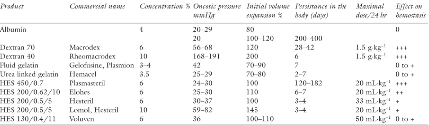
The characteristics of the different colloids and their effects on coagulation are presented in Table I.
Gelatins have the reputation of not influencing coagulation other than by their hemodiluting effect.15,16
Nonetheless, dilution of whole blood samples with two gelatin solutions resulted in a reduction of clot quality (less extensive fibrin mesh formation, reduced clot weight and mean shear modulus) compared to dilution with normal saline.17 In volunteers, 1 L of a
gelatin-based plasma substitute significantly impaired primary hemostasis and thrombin generation compared to normal saline.18 A study comparing a 3%
modi-fied fluid gelatin to 6% hydroxyethyl starch (HES) in patients undergoing primary total hip replacement concluded that the gelatin-based plasma expander had a poorer volume effect than HES and that blood losses were less important with gelatin compared to HES.15
Coagulation tests were similar in both groups except for the higher incidence of an abnormal bleeding time (BT) in the HES group. The clinical significance of these findings remains unclear. Perhaps the effects on coagulation of gelatin-based plasma substitutes are not clinically important. On the other hand, they may have been underestimated, given the difficulties of studying coagulation in the uncontrolled context of trauma, MT and ongoing bleeding.
HES solutions are effective plasma expanders in common use both in Europe and in North America.
It has long been known that HES solutions interfere with coagulation19 and that the effects vary
accord-ing to the dose and type of solution administered.20
Solutions with a high molecular weight and a high degree of substitution accumulate in the tissues and are responsible for more pronounced hemostatic effects.21
Abnormal platelet function occurs more frequently after high molecular weight HES.22 Conversely, HES
solutions with a low molecular weight and a lesser degree of substitution are eliminated more rapidly and tend to affect measures of hemostasis less.23–25
In addition to their effects on hemostasis, the infu-sion of large volumes of HES solutions will result in significant hemodilution. The resulting drop in hemoglobin and platelet concentrations may com-promise primary hemostasis. In addition, Innerhofer
et al. have shown that the administration of HES or
modified gelatin in patients undergoing knee replace-ment surgery results in reduced clot strength due to impaired fibrinogen polymerization and that reduced fibrinogen concentrations might be reached earlier than expected.26
Adverse events associated with the use of HES solutions for the resuscitation of patients requiring MT have not been reported, inasmuch as the allowed maximal daily dose is not exceeded.16 A large
retro-spective study suggested that the use of 6% HES in primary cardiac surgery with CPB may increase bleed-ing and transfusion requirements, despite the infusion of volumes smaller than the manufacturer’s recom-mended dose.27 Conversely, large volumes (up to 5 L)
have been infused without major complications,28 but
the safety of this practice remains controversial.29
The clinical significance of the effects of HES solu-tions on hemostasis remains unclear. It may never be possible to determine precisely the effect of volume replacement in massively transfused patients with ongo-ing bleedongo-ing. In these patients, hemostasis is stressed severely by numerous factors other than the type of non-blood-related iv fluid used for resuscitation and the underlying cause of coagulopathy is difficult to ascer-tain. Nonetheless, it is interesting to note that the use of colloids in trauma patients is not mentioned in the rec-ommendations by the American College of Surgeons.30
B. Hypothermia
An important measure to reduce blood loss through preservation of hemostasis is the maintenance of nor-mothermia both during and after the operation. The definition of hypothermia has varied amongst studies; however, most definitions have used temperatures below 35°C. Hypothermia slows the activity of the coagulation cascade, an enzymatic process, reduces
the synthesis of coagulation factors, increases fibrino-lysis and affects platelet function.
1. ANIMAL DATA
Several animal studies conducted under hypother-mic conditions have shown reversible platelet count decreases and platelet function defects, altered coagu-lation patterns and an enhanced fibrinolytic response. In dogs cooled to 19°C, Yoshihara et al. found a severe decrease in platelet count and collagen-induced platelet aggregability and an increase in fibrinolysis.31
No variations of the PT and aPTT were observed but these tests were performed in vitro at 37°C. These modifications, which could potentially increase bleed-ing, were not documented in the normothermic con-trol group. Other experimental studies have shown an important prolongation of the PT and aPTT that was inversely and linearly correlated to temperature.32,33
Pina-Cabral et al. also observed a decrease in platelet count in hypothermic dogs.34 The presence
of platelet clumps was detected inside the hepatic sinusoids. The authors concluded that hepatic platelet sequestration could explain the decrease in platelet count in this setting.
In swine, Oung et al. showed a prolongation of the BT (10.9 min vs 5.5 min in the control group), confirming the impairment of hemostasis induced by hypothermia (30°C).35 Prolongation of the BT
has also been observed by Valeri et al. in baboons subjected to systemic hypothermia at 32°C and skin hypothermia at 27°C.36
2. HUMAN DATA
In humans, many studies have emphasized the major role of hypothermia on the onset of bleeding during surgical procedures. Valeri et al. observed the effects of skin temperature in 33 patients undergoing CPB.37
Local hypothermia produced an increased BT and a significant reduction in thromboxane B2, an indicator of platelet activation, at the BT site. Local rewarming produced a significant increase in shed blood throm-boxane B2. Thus, hypothermia caused a reversible platelet dysfunction and rewarming improved platelet function and reduced the BT. These data have been confirmed by Michelson et al. who demonstrated the involvement of platelet glycoprotein receptor (GP Ib and GMP 140) alterations in this hemostatic defect.38
Again, rewarming completely reversed the activation defect as soon as temperature returned to 37°C.
These abnormalities of routine coagulation tests are clinically significant: mild hypothermia (35 ± 0.5°C) increased bleeding and allogeneic blood requirements in patients undergoing total hip arthroplasty.39 Thus,
surgery under normothermic conditions may help prevent bleeding complications and decrease the intra-operative use of transfusions. The additional contribu-TABLE I Characteristics of the available colloids and their effects on coagulation
Product Commercial name Concentration % Oncotic pressure Initial volume Persistance in the Maximal Effect on mmHg expansion % body (days) dose/24 hr hemostasis
Albumin 4 20–29 80 0
20 100–120 200–400
Dextran 70 Macrodex 6 56–68 120 28–42 1.5 g·kg–1 +++
Dextran 40 Rheomacrodex 10 168–191 200 6 1.5 g·kg–1 +++
Fluid gelatin Gelofusine, Plasmion 3–4 42 70–90 7 0 to +
Urea linked gelatin Hemacel 3.5 25–29 70–80 2–7 0 to +
HES 450/0.7 Plasmasteril 6 24–30 100 120–182 20 mL·kg–1 +++
HES 200/0.62/10 Elohes 6 25–30 110 6–7 20 mL·kg–1 ++
HES 200/0.5/5 Hesteril 6 30–37 100 3–4 33 mL·kg–1 +
HES 200/0.5/5 Lomol, Hesteril 10 59–82 145 3–4 20 mL·kg–1 +
HES 130/0.4/11 Voluven 6 36 100–110 50 mL·kg–1 0 to +
Effect on hemostasis: 0 = none; + = weak; ++ = moderate; +++ = important. Commercial names are European. Some data are not avail-able. In the “product” column, the first number is the molecular weight in Daltons, the second is the degree of substitution and the third is the C2/C6 ratio of hydroxyethylstarch (HES) substitution. The higher the molecular weight and the degree of substitution, the lon-ger the plasma half-life and the effects on hemostasis. Adapted from Ickx, BE and Van der Linden P. Interactions entre solutés colloides et l’hémostase. Sang Thrombose Vaisseaux 2002; 7: 408–16 [article in French] Hetastarch (480/0.7), used in the USA, has important effects on hemostasis. Pentastarch (260/0.45), available in Canada, is similar to Hesteril 10% and has weak effects on hemostasis at the maximal dose (28 mL·kg–1) recommended by the manufacturer.
tion of hypothermia to the hemorrhagic diathesis may be overlooked since coagulation testing is normally performed at 37°C.40
Hypothermia is an important contributor to coagu-lopathy in trauma patients.41 Hypothermia
(tempera-ture less than 34°C) occurred in 80% of non-survivors and 36% of survivors in the 45 trauma patients report-ed by Ferrara et al.42 Patients who were hypothermic
and acidotic developed clinically significant bleeding despite adequate blood, plasma, and platelet replace-ment. The authors concluded that avoidance or cor-rection of hypothermia may be critical in preventing or correcting coagulopathy in the patient receiving MT.
C. Blood components and alterations of hemostasis
1. RED CELLS
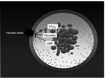
An often-ignored effect of red blood cell (RBC) transfusion is the improvement of hemostatic func-tion. The contributions of RBC to hemostasis are illustrated in Figure 1.
Erythrocytes have been shown to modulate the biochemical and functional responsiveness of acti-vated platelets, suggesting that erythrocytes contrib-ute to thrombosis and hemostasis and supporting the concept that thrombus formation is a multicellular event.43–45 Red cells contain adenosine diphosphate
that may activate platelets.46 Also, they activate platelet
cyclooxygenase,44 increase the generation of
throm-boxane A244 and may directly increase the thrombin
burst through exposure of procoagulant phospholip-ids.47 This proaggregatory property of erythrocytes is
enhanced by diabetes48 and can be decreased by an
appropriate aspirin regimen.49,50
Another mechanism by which erythrocytes modu-late hemostasis is the rheological effect of red cells on the margination of platelets.51 Under normal
circum-stances, red cell flow is maximal at the centre of a ves-sel, tending to push platelets towards the periphery of the vessel lumen, thereby optimizing their interaction with injured endothelium and promoting hemosta-sis. In rabbit arterioles, platelet numbers are highest near the vessel wall52 and platelets align themselves
with their equatorial plane parallel to the vessel wall as they move closer toward the periphery of the ves-sel.53 Experimental data have shown that platelets are
expelled toward the RBC depleted marginal layer near the tube wall by mutual interaction with erythrocytes. Under these circumstances, the near wall concentra-tion of platelets is enhanced up to about seven times the average concentration.54
The correlation between the BT and hematocrit levels has been studied experimentally. In non-throm-bocytopenic rabbits, the microvascular BT varied inversely with the hematocrit, animals with hematocrit levels above 35% having shorter BTs than animals with hematocrits lower than 35%.55 In a rabbit model
of cyclic arterial thrombosis and clot lysis, Ouaknine-Orlando et al. have shown that decreases in the hema-tocrit reduced the cyclic arterial thrombosis rate and increased the BT.46 Interestingly, normalization of the
hematocrit caused thrombosis to reappear.
Transfusion of RBC shortens the BT in anemic thrombocytopenic patients despite persistent throm-bocytopenia.56 Further, perfusion of blood from
trans-fused, previously anemic thrombocytopenic patients improved the interaction of platelets with the subendo-thelium in an experimental perfusion model. However, this interaction remained lower than in non-ane-mic non-thrombocytopenic subjects.56 Similarly, RBC
shorten the BT and control the hemorrhagic diathesis of uremic patients.57 In humans with a normal renal
function and platelet counts over 100,000·mm-3, a
modest but statistically significant inverse correlation (r = -0.47; P < 0.001) exists between the hematocrit and the BT.58 Patients with a pretransfusion hemoglobin
concentration ≤ 60 g·L–1 have a greater chance (relative
risk 4.07; 95% confidence interval 1.03 to 16.2; P = 0.04) of a posttransfusion decrease in BT than patients with a hemoglobin of more than 60 g·L–1.59 Valeri et al.
have shown that, in healthy volunteers, an acute 15% FIGURE 1 The contribution of red cells to hemostasis
Red cells contribute to the margination of platelets against the vessel wall and their availability to act at the site of a vascular lesion. They have also been shown to modulate the biochemical and functional responsiveness of activated platelets. (Courtesy of Sylvain Bélisle, MD, FRCPC, Montreal Heart Institute).
reduction in hematocrit produced a 60% increase in the BT while the BT remained normal despite a 32% reduc-tion in platelet count.60 All this being said, the clinical
significance of these findings remains unclear since the relationship between the BT and perioperative blood losses is highly controversial.60,61
The hemostatic effects of profound normovole-mic hemodilution were investigated in eight patients undergoing surgical correction of idiopathic sco-liosis by McLoughlin et al.62 Abnormal hemostasis
developed prior to compromise of global tissue oxygenation suggesting that, in healthy anesthetized subjects, normovolemic hemodilution may be limited more by preservation of normal coagulation. In this report, reinfusion of all collected blood at the end of the procedure did not normalize the PT or the aPTT. Unfortunately, the authors did not describe if the microvascular bleeding (MVB) observed during hemodilution was corrected by increasing the hema-tocrit. Modified ultrafiltration in pediatric open heart operations has been shown to increase the hematocrit and attenuate the dilutional coagulopathy associated with CPB in infants.63 Use of modified ultrafiltration
to increase the hematocrit to 36–42% reduced the rise in total body water and the need for donor blood in children undergoing open heart surgery.64
Thus, further investigations into the role of the hemoglobin concentration or hematocrit on hemosta-sis in massively transfused patients appear warranted. The data presented above tend to support the con-cept of a minimal hematocrit for optimal hemostasis. At present the optimal hematocrit or hemoglobin concentration to prevent or to initiate treatment of coagulopathy remains unknown. Experimental evi-dence suggests that relatively high hematocrits, pos-sibly as high as 35%, are required to sustain hemostasis in bleeding patients undergoing MT.
2. COAGULATION FACTORS
It is difficult to isolate precisely the role of coagulation factors in the pathophysiology of coagulopathy associ-ated with MT. Impaired hemostasis is most probably multifactorial in origin and results from the adverse hemostatic effect of multiple concurrent coagulation factor deficits combined with variable degrees of ane-mia and thrombocytopenia.6
While numerous studies have measured changes in the PT and aPTT in relationship with bleeding and MT, few have examined individual hemostatic factors. In 1979, transfusing MWB to patients who sustained major trauma, massive gastrointestinal hemorrhage or aortic aneurysm rupture, Counts et al. reported that the number of units of blood transfused explained less
than 20% of the variance in factors V and VIII and was not related to factor VII, X, XI, XII and fibrinogen levels. The authors concluded that it was not neces-sary to supplement transfusions of stored, MWB with fresh blood or fresh frozen plasma (FFP).5 In 1995,
using plasma poor red cell concentrates, Hiippala et
al. showed that a concentration of fibrinogen of 1.0
g·L–1 was reached when the blood loss was 1.42 times
the calculated blood volume and that blood losses in excess of two blood volumes caused the deficiency of prothrombin, factor V, platelets and factor VII, in this order.65 These observations were made in ASA
physi-cal status I or II patients undergoing elective major urologic or abdominal surgery and, as a result, may not apply to emergent operations or trauma. Nonetheless, they illustrate well the difference between the times when red cell solutions contained significant amounts of plasma and modern component therapy.
It is important to realize that studies conducted at a time when fresh, stored or modified MWB was in common use seldom reported low levels of coagulation factors as the primary factor responsible for impaired hemostasis.4,5,7,11,66 Since the widespread use of red cell
solutions containing minimal amounts of plasma in the late 1980’s and early 1990’s, dilution or consumption of coagulation factors has become a significant issue requiring specific treatment with, primarily, FFP.3,6,8,65
Even today, clinicians seldom administer “pure” blood products. PRBC contain a small amount of plasma (30– 60 mL), as do platelet concentrates (80 mL approxi-mately). Therefore, it may be difficult to differentiate precisely the therapeutic effect of the different blood components transfused.
3. PLATELETS
Primary hemostasis is characterized by the formation of the “platelet plug.” The mechanism is complex and involves the presence of fibrinogen and activa-tion of several glycoprotein receptors on platelets.67–71
Hemostasis is initiated by injury to the vascular wall, leading to the deposition of platelets adhering to blood vessel subendothelial matrix proteins (collagen and von Willebrand factor) via interactions with plate-let membrane glycoprotein receptors. Subsequently, the GP IIb/IIIa receptor is activated. This receptor has a high affinity for fibrinogen (and also for von Willebrand factor at high shear stress). Binding of fibrinogen to adjacent platelets results in irreversible platelet aggregation and the formation of the platelet aggregate.
Since the publication of Miller’s classic study on coagulation defects associated with massive blood transfusions,11 thrombocytopenia resulting from
hemodilution has been thought to be the most important hemostatic abnormality associated with MT. This explanation is intuitively appealing: replace-ment of lost blood with fluids that do not contain platelets (or coagulation factors) results in a dilutional coagulopathy. However, models based on the washout equation (a simple mathematical model of exchange transfusion that calculates the decay of blood com-ponents when bleeding and replacement rates are constant and equal) may not apply to bleeding trauma patients where blood volumes fluctuate, bleeding rates vary with blood pressure, and replacement often lags behind blood loss.72 Clinically, simple hemodilution
has failed to explain several observations.
In young, previously healthy soldiers with wounds excluding burns and head injuries, platelet levels fell rapidly to about 100,000·mm-3 during rapid
transfu-sion of stored whole blood and remained at that level after the first 6 L of stored whole blood. The PT, aPTT
and fibrinogen levels were less severely affected and, most important, significant operative bleeding was not encountered in conjunction with these mild dilutional coagulation changes.7 On average, the platelet count
fell below 100,000·mm-3 after transfusion of 18 units
of blood in the study by Counts et al.,5 but slightly less
than half (43%) of the variation in platelet counts could be ascribed to the functional relationship between the amount of blood transfused and the platelet count. In a study conducted to determine the efficacy of prophy-lactic platelet administration to prevent MVB associated with MT, Reed et al. observed that platelet counts were not different between patients who received prophylac-tic platelet transfusions and those who did not.4 Further,
both groups had higher platelet counts than predicted by a standard washout equation. This finding implies that platelets are being released into the circulation and counteract the effects of dilution. Sequestered platelets can be released from the spleen and the lung, in addi-tion to the premature release of platelets from the bone marrow. Elevated stress hormones and the administra-tion of catecholamines, a situaadministra-tion more likely to occur in the trauma patient, will influence release.
As in the study by Counts, Reed et al. found the relationship between platelet counts and units of blood transfused to be significant albeit highly variable (r2 =
0.24 for patients receiving platelets and r2 = 0.35 for
patients receiving FFP),4 suggesting that factors other
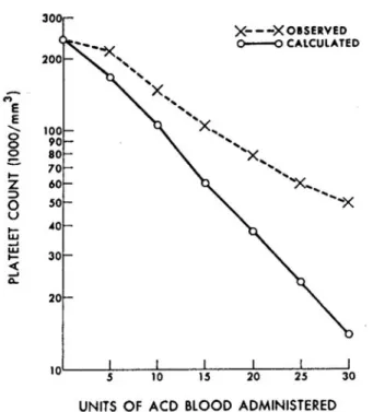
than simple dilution affect platelet count. Finally, in the study by Miller, observed platelet counts did not parallel calculated platelet counts (Figure 2).11
In elective surgery, the situation appears to be quite different, possibly because tissue trauma is more controlled, normovolemia is maintained and blood losses are replaced in a timely manner. In this context, coagulopathy is primarily related to a coagulation factor deficit3,65 and the role of fibrinogen appears to
be preponderant. The deficiency in fibrinogen con-centration develops earlier than any other hemostatic abnormality when plasma-poor red cell concentrates and colloid plasma substitutes are used for the replace-ment of major blood loss. Approximately 90% of the variation in fibrinogen concentration is explained by blood loss and the critical level of 1.0 g·L–1 is reached
when blood losses reach 142% (95% confidence inter-val 117%–169%) of the calculated blood volume.65
In this same study, thrombocytopenia (defined as a platelet count below 50,000·mm-3) was a late
occur-rence and, to a great extent, quite variable from patient to patient. Murray et al. studied coagulation changes during crystalloid and PRBC replacement of major blood loss during elective surgery.73 Four out
of 12 patients presented a coagulopathy. All patients FIGURE 2 Calculated vs observed platelet counts in a person
receiving platelet-free blood
Observed platelet counts are higher than those predicted by hemodilution alone: the observed platelet count after the admin-istration of 25 units of blood is of the order of 60,000·mm–3
while hemodilution predicted a platelet count of approximately 20,000·mm–3. Reproduced with permission from Miller RD,
Robbins TO, Tong MJ, Barton SL. Coagulation defects associated with massive blood transfusions. Ann Surg 1971; 174: 794–801.
with inadequate clinical hemostasis had a low platelet count (83,000·mm-3 or below) and transfusion of
platelet concentrates corrected the problem in the two who had fibrinogen concentrations above 1.0 g·L–1. Of note, platelet transfusion was ineffective
in the two patients with concurrent low fibrinogen concentrations (0.73 and 0.40 g·L–1). The subsequent
transfusion of two and four units of FFP (respectively) normalized hemostasis. The report by Ciavarella et
al. supports the association between MVB and a low
platelet count (below 50,000·mm-3) or a low
fibrino-gen concentration (below 0.5 g·L–1).66
Thus, it appears logical to consider that coagu-lopathy after MT can be a problem resulting from a combined deficit of platelets and fibrinogen. Focusing only on platelet levels or concentrations of specific coagulation factors may not lead to the most appropri-ate therapeutic approach.
D. DIC
1. DEFINITION
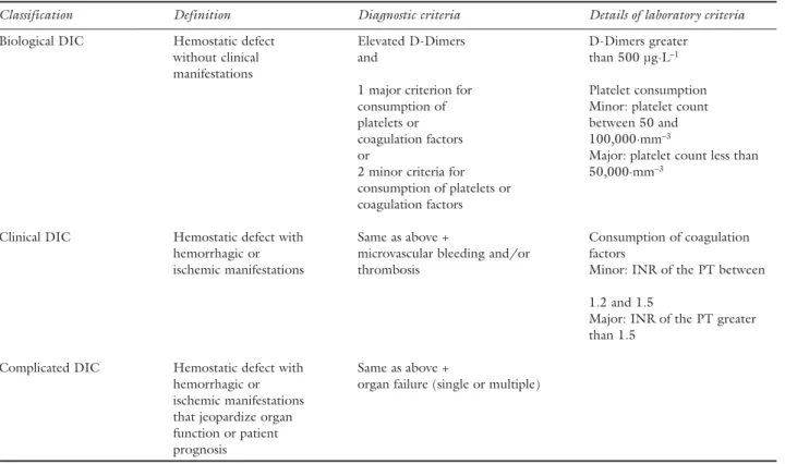
DIC is an acquired syndrome secondary to the sys-temic and excessive activation of coagulation. It may be defined by the association of hemostatic defects related to the exaggerated generation of thrombin and fibrin (with or without clinical signs) and the excessive con-sumption of platelets and coagulation factors. Criteria for the diagnosis of DIC are presented in Table II. The syndrome can be seen in numerous clinical situations and often complicates the management of MT.
2. THE TRAUMA SETTING
In earlier studies of trauma patients, DIC leading to MVB was, apparently, relatively infrequent, occurring for example in one out of 21 patients11 and three out
of 27 patients.5 In wounded, previously healthy,
sol-diers requiring MT, DIC was mild and was not associ-ated with clinical bleeding.7,11 Recent studies suggest
that there is no significant correlation between total TABLE II Diagnostic criteria for disseminated intravascular coagulation (DIC)
Classification Definition Diagnostic criteria Details of laboratory criteria
Biological DIC Hemostatic defect Elevated D-Dimers D-Dimers greater
without clinical and than 500 µg·L–1
manifestations
1 major criterion for Platelet consumption
consumption of Minor: platelet count
platelets or between 50 and
coagulation factors 100,000·mm–3
or Major: platelet count less than
2 minor criteria for 50,000·mm–3
consumption of platelets or coagulation factors
Clinical DIC Hemostatic defect with Same as above + Consumption of coagulation
hemorrhagic or microvascular bleeding and/or factors
ischemic manifestations thrombosis Minor: INR of the PT between 1.2 and 1.5
Major: INR of the PT greater than 1.5
Complicated DIC Hemostatic defect with Same as above +
hemorrhagic or organ failure (single or multiple) ischemic manifestations
that jeopardize organ function or patient prognosis
INR of the PT = international normalized ratio of the prothrombin time.The elevation of D-Dimers is not specific to DIC. Similarly, the clinical manifestations of DIC are not specific. Adapted from the Proceedings of the XXIInd consensus conference of the “Société de
Réanimation de Langue Française” held in Lille, France, in October 2002 and published in Réanimation 2002; 11: 575–666. A French summary is available at www.srlf.org/conferences/XXII-conf-Lille.html.
units of blood transfused and the severity of impaired hemostasis, suggesting that consumption of coagu-lation factors and platelets is more important than simple hemodilution.41,74,75
In trauma patients, two major mechanisms are responsible for the occurrence of DIC. The first relates to the nature and to the importance of tissue trauma. The second relates to shock and tissue anoxia. Brain injury is associated with a particularly high inci-dence of coagulopathy. In the study by Faringer et al., more than 40% of patients with penetrating or blunt trauma with associated brain injuries had abnormal coagulation tests on admission, compared with 0% in patients with blunt trauma but without brain injury.2
After blunt brain injury, a DIC syndrome secondary to the extravasation of tissue factor can rapidly (within one to four hours after injury) lead to a consumptive coagulopathy that is associated with a high frequency of death.76 Regarding the importance of tissue trauma,
in the absence of massive head injury and pre-existing disease, life-threatening coagulopathy was associated with a pH of less than 7.10, a temperature of less than 34°C, an injury severity score greater than 25, and a systolic blood pressure of less than 70 mmHg. When all risk factors were present, the incidence of coagu-lopathy was 98%.41
At least two studies have attempted to determine which populations of massively transfused patients are most at risk of sustaining DIC. Mannucci et al. studied changes in the hemostatic system in 172 patients undergoing MT for excessive bleeding dur-ing or in the early postoperative period after elective, emergent or trauma surgery. Of these, 52 (30%) suf-fered decompensated DIC and there was no signifi-cant relation between the number of whole blood or PRBC units transfused and the values of any variable of hemostasis measured.77 In a series of 64 patients
receiving more than ten units of red cell concentrates during elective or urgent surgical procedures or resus-citation of multiple trauma, Hewson et al. observed that coagulopathy is related to hemodilution initially, but, within three hours, is related to the duration of antecedent hypotension. Thus, on the basis of the available information, it is difficult to determine which patient population (elective or urgent operations, trauma) is most likely to sustain DIC.
3. THE ELECTIVE SURGICAL SETTING
In the purely elective setting, DIC complicating MT is infrequent. No patient suffered from DIC amongst the 32 young ASA physical status I or II patients undergo-ing posterior spinal stabilization and MT.3 Clinically
increased surgical bleeding was present in 17 patients
and in 14 of the 17 patients hemostasis improved after the administration of FFP (approximately 10 mL·kg–1).
Again, these results suggest that when tissue anoxia is avoided and surgical trauma is controlled, the occur-rence of DIC may remain low despite MT.
III. Management of coagulopathy in MT
A. Diagnosis
Numerous attempts have been made to monitor changes of the coagulation system in relationship to trauma, surgery and MT. Unfortunately, results have been disappointing and, at present, there is no simple, reliable and rapid diagnostic test that allows clinicians to manage massively transfused, critically ill patients. The available tests quantify specific factors in clotting or attempt to measure clotting function.
The platelet count is the only indicator of coagula-tion that can be obtained rapidly through the use of automated counters, in contrast to conventional tests of hemostasis that take a minimum of 30 min since they require centrifugation of blood samples. Even if platelets play an important role in coagulation, a decreased platelet count is not a specific indicator of coagulopathy. Rather, the significance of a decreased platelet count should be interpreted in the patient’s specific clinical context: is platelet function expected to be normal? Is the fibrinogen concentration suf-ficient? Is the hemoglobin concentration adequate? Is there any evidence of a consumptive coagulopathy? Answering these questions will assist in making the appropriate diagnosis of coagulopathy.
Usefulness of the BT to diagnose or predict coagu-lopathy in massively transfused patients has been inves-tigated in a few studies. Overall, the BT increases early in the course of surgery and transfusion,78 remains
elevated for several days postoperatively79 and does
not allow the differentiation between bleeding and non-bleeding patients.5 In their critical reappraisal of
the BT, Rodgers and Levin concluded that there is no evidence that abnormalities in the test occur suf-ficiently in advance of other indicators of bleeding to allow actions to be taken that could alter outcome favourably.80 Consequently, the BT is of no use in the
context of MT.
Reliable bedside monitors have become available to monitor the PT and aPTT81 and may add to the timely
management of the bleeding patient. Controversy remains, though, on the proper use of coagulation screening tests to guide replacement therapy. The PT and aPTT are expected to become elevated when lev-els of factor V, VIII and IX are less than 50% of values found in a control patient population.82 It should be
when fibrinogen levels are low in the presence of normal concentrations of coagulation factors (e.g., congenital afibrinogenemia).
As for decreased platelet counts and prolongations of the BT, prolongations of the PT and aPTT are very common in massively transfused patients.2,3,5,7,8,11,41,6 5,73,75,77,83,84 Minor prolongations of the PT or aPTT
ratio (ratio = measured value/control value) are poor predictors of bleeding in massively transfused patients.5
Ciavarella et al. have shown that, in 36 massively trans-fused patients, patients with a PT or aPTT ratio greater or equal to 1.8 had an 80 to 85% chance of exhibiting MVB.66 When the fibrinogen level is adequate, a PT or
aPTT ratio greater or equal to 1.8 reliably predicts that factor V and VIII levels are less than 30%,66 a
percent-age that has been cited as an indication for coagulation factor replacement.4,66 In the presence of a decreased
fibrinogen concentration (less than 0.75 g·L–1), a PT or
aPTT ratio greater than 1.5 is associated with factors V and VIII of less than 20%.73
In summary, only marked prolongations of the PT or aPTT (1.5 and 1.8 times higher than control when the fibrinogen level is low and normal respectively) are likely to be significant from a clinical perspective.
Other instruments have been developed to study coagulation at the bedside, using whole blood. While their use in the context of MT appeared promising, results have, unfortunately, been disappointing. The best known is the thromboelastograph, a device that
measures the viscoelastic properties of the clot during its formation and subsequent lysis. In general surgi-cal patients, thromboelastography analysis showed a trend toward increased coagulability with progressive blood loss and contributed to the identification of two patients who required treatment of a coagulopathy.85
Thromboelastography has a high negative predictive value (82%) for bleeding after routine cardiac surgery, allowing the differentiation between surgical bleeding and coagulopathy. Unfortunately, the positive predic-tive value of the test is low (41%).86 More studies will
be required to establish its usefulness in the manage-ment of MT.
Another instrument, the PFA-100® (Platelet Function Analyzer; Dade-Behring, Miami, FL, USA), measures an ex-vivo BT in the presence of different agonists. The device has been used extensively for the diagnosis of hereditary coagulation disorders, particu-larly von Willebrand’s disease. In patients undergoing routine cardiac operations, the PFA was not correlated with mediastinal chest drainage.87,88 A Medline search
on the use of the PFA during MT failed to retrieve any relevant references.
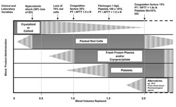
B. Treatment of coagulopathy
The treatment of coagulopathy associated with MT is not simple. Two approaches are outlined in Table III and in Figure 3. Both approaches should be viewed as complementary.
TABLE III Proposals for the administration of hemostatic blood products in the massively transfused patient Presence of clinical bleeding
Yes No
Yes Transfusion of FFP and PC Possible transfusion of FFP
according to the results of and PC if the hemorrhagic risk coagulation testing related to the surgical procedure is
high
Abnormal No Search for a cause other than a No indication to transfuse
coagulation tests* hemostatic defect
Re-evaluate the importance of red cell transfusion and consider FFP and PC if transfusion is massive Repeat coagulation tests
Unknown Transfusion guided by clinical No indication to transfuse probability of a specific hemostatic defect Obtain coagulation tests
*Platelet count less than 50,000·mm–3, fibrinogen concentration less than 1.0 g·L–1, prothrombin time (PT) and activated partial
throm-boplastin time (aPTT) more than 1.5 to 1.8 times control. FFP = fresh frozen plasma; PC = platelet concentrate. Adapted from the rec-ommendations of the “Agence française de sécurité sanitaire des produits de santé” for the transfusion of allogeneic red cells published in Transfusion clinique et biologique 2002; 9: 333–56 (article in French).
1. GOALS OFTREATMENT
The causal diagnosis of MVB remains elusive and the clinician must intervene rapidly if the patient is to survive. The situation is further complicated by the management of risks associated with transfusion, mandating that patients receive the smallest number of blood products possible. In Canada, in the year 2000, the risk of human immunodeficiency virus and hepatitis C virus transmission was minimal: one in four million and one in three million, respectively.89
However, the aggregate risk of potentially severe reactions was much higher: 43.2 per 100,000 red cell units and 125.7 per 100,000 platelet pools of five units, affecting 337 and 88 recipients, respectively.89
The most frequent potentially severe outcomes were hemolytic reactions and volume overload for red cell transfusion and major allergic reactions and bacterial contamination for platelet transfusion.89
In general, clinicians assume that all blood products are comparable and that the conclusions of studies conducted in similarly developed countries can be applied locally. We compared the characteristics of
blood products (PRBC, FFP and platelet concen-trates) available in Canada and in three European countries (Belgium, France and Switzerland; char-acteristics of allogeneic blood products available as Additional Material at www.cja-jca.org). While some differences exist, these are minor and unlikely to result in clinically significant management disparities (Douglas Fish, personal communication).
In light of the above risks, we recommend that blood products be transfused to correct a physiologic deficit likely to be detrimental to the patient. Obviously, this is, most often, not possible in the context of MT, in the presence of ongoing hemorrhage for example. Hemostatic blood components should be transfused mainly to treat a clinical coagulopathy, unless important surgical factors justify transfusion, e.g., prior to invasive procedures in patients with a known hemostatic defect, in neurosurgical procedures where bleeding may have catastrophic consequences, etc., (note that the latter recommendations are based on expert opinion only).
No prophylactic regimen involving the admin-istration of FFP and/or platelet concentrates has FIGURE 3 The management of massive transfusion in the elective surgical setting
Reproduced with permission from Erber WN. Massive blood transfusion in the elective surgical setting. Transfus Apheresis Sci 2002; 27: 83–92.
been shown to be effective in massively transfused patients.4,5,77 The prophylactic administration of
(potentially) unnecessary transfusions exposes patients to multiple donors, increases the risks to the patient and is unacceptable.90,91 Nonetheless, given the delay
required to prepare and/or obtain blood products, MT associated with evidence of an evolving labora-tory coagulopathy may warrant ordering of blood components that will, then, be readily available to treat MVB should it occur.92,93 Whenever possible, the
use of single donor plasma or platelets is preferred to decrease donor exposure.
2. APPROACHES TO THE TREATMENT OFCOAGU -LOPATHY
Figure 3, by Erber et al., summarizes well the use of blood and non-blood products involved in the resus-citation of a bleeding patient.94 Initially, crystalloids
or colloids are infused to maintain normovolemia. As the percentage of blood volumes lost and replaced increases, PRBC, FFP and platelets will be required, in that order, guided by clinical and laboratory variables. Pharmacological agents are usually administered rela-tively late in the process [protamine sulfate antagonizes the effect of excess heparin; desmopressin has been used to control hemorrhage, specially after cardiac surgery, with varying success; aprotinin has been suggested in the context of trauma and MT but its use is not support-ed by any controllsupport-ed trial; recombinant activatsupport-ed factor VII (rFVIIa) is a promising hemostatic agent under
investigation but the optimal timing of administration of rFVIIa remains to be determined]. While this scheme is appealing and applicable in the elective surgical setting where bleeding is usually progressive and coagulopathy can be monitored and anticipated, the situation can be quite different in the traumatized patient.
In the interval between injury and arrival to the hos-pital, the trauma patient has lost an undefined amount of blood that has been replaced, in part, by crystalloids or colloids.72 Tissue trauma, shock, tissue anoxia and
hypothermia contribute to the development of DIC and the results of coagulation testing are not immedi-ately available. Clinicians are called upon to intervene rapidly in a very unclear and unstable situation.
Basic recommendations include the maintenance of normothermia and correction of a low hemo-globin concentration. Maintaining a normal body temperature is a relatively simple and probably one of the most effective strategies of blood conserva-tion. Unfortunately, it can easily be overlooked in the trauma setting. When a hypothermic patient bleeds without an apparent surgical cause, restoration of temperature towards normal should be a first-line intervention. Correction of the hypothermia-induced hemostatic defect can be expected as soon as the patient is rewarmed.
A low hemoglobin concentration should be cor-rected prior to the administration of hemostatic blood products, in view of normalizing hemostasis in bleed-ing patients. When circumstances allow, allogeneic RBC should be transfused one unit at a time and the effects of transfusion on hemostasis monitored before administering supplemental units. The optimal hemo-globin concentration to sustain hemostasis in the con-text of MT remains unknown but is probably higher than that required for oxygen transport and delivery.
The use of platelets and/or FFP should depend on the results of coagulation testing and clinical judg-ment. A markedly prolonged PT and aPTT suggests a coagulation factor deficiency and is best treated with FFP. Decreased levels of fibrinogen will also require treatment with FFP. FFP should be administered in doses large enough to increase coagulation factor lev-els and maintain them above critical levlev-els. Doses rang-ing from 5 to 20 mL·kg–1 have been recommended.
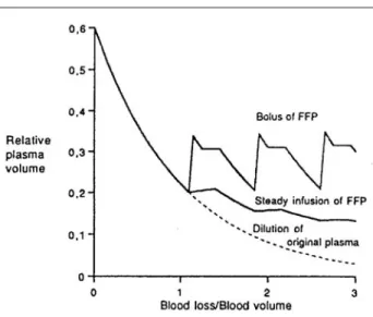
In the average adult, four units of FFP (800 to 1,000 mL) should suffice initially but additional bolus doses should be administered according to ongoing blood losses and transfusions.6 As shown in Figure 4, it is
important to administer sufficient bolus doses of FFP to maintain adequate concentrations of coagulation factors. Further, using a three compartment dynamic computer simulation, Hirshberg et al. suggest that, in FIGURE 4 Simulation of relative plasma volume according to
mode of fresh frozen plasma (FFP) supplementation
Reproduced with permission from Hiippala S. Replacement of massive blood loss. Vox Sang 1998; 74: 399–407.
trauma patients with exsanguinating hemorrhage, FFP
should be given rapidly (with the first units of PRBC replacement) in order to effectively prevent dilutional coagulopathy.72
If the fibrinogen concentration remains below 1.0 g·L–1 despite therapy with FFP, the administration of
cryoprecipitate, a concentrated source of fibrinogen, factor VIII, factor XIII and von Willebrand factor may be indicated95 despite the associated infectious
risks (ten to 20 units - containing 5 mL each - will be required). In Europe specially, cryoprecipitates are no longer available and, when fibrinogen substitution is required, virus-inactivated fibrinogen preparations are administered.
Platelets should be administered to correct a clinical coagulopathy associated with a decreased platelet count and/or platelet function. Clinicians must remember that platelet counts will decrease in a majority of massively transfused patients but that not all patients will bleed excessively as a result of thrombocytopenia. Other hemostatic anomalies may warrant a more urgent (a marked prolongation of the PT and aPTT) or concurrent (a decreased fibrinogen level) correction.
From the time PRBC have replaced whole blood, there is laboratory and clinical evidence in the lit-erature that FFP may be required prior to platelet concentrates to treat a coagulopathy.3,6,8,73 On the
other hand, two studies have shown that survival in massively transfused trauma patients is associated with the increased transfusion of platelet concentrates.41,96
One may postulate that a more aggressive control of coagulopathy was beneficial in these patients, a situa-tion somewhat analogous to the benefits derived from early goal-directed therapy in patients with severe sepsis and septic shock.97 Unfortunately, at present,
no monitor allows clinicians to identify which patient is likely to benefit from the increased transfusion of hemostatic blood components (such as platelets or FFP).
Several animal experiments, case reports and case series report the successful use of rFVIIa to treat bleed-ing that could not be controlled by the administration of hemostatic blood components.98–106 Originally, rFVIIa
was developed to treat hemophiliacs with inhibitors to exogenous factor VIII and its use has been extended to the correction of several hemostatic defects.107 Since
factor VII must interact with tissue factor to initiate the generation of thrombin, coagulation occurs at the site of injury108 and the risk of thromboembolic effects
appears minimal.109 The initial results with this novel
hemostatic agent for “rescue” therapy of MVB are impressive and warrant the conduct of randomized controlled trials to evaluate further the efficacy and safety of this expensive drug.110,111
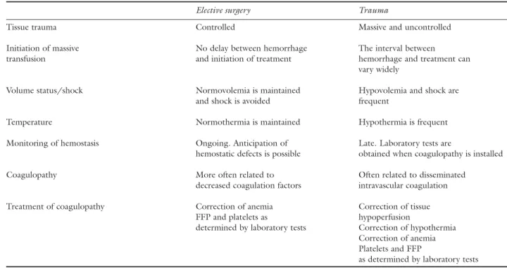
TABLE IV Massive transfusion: the main differences between elective surgery and trauma
Elective surgery Trauma
Tissue trauma Controlled Massive and uncontrolled
Initiation of massive No delay between hemorrhage The interval between
transfusion and initiation of treatment hemorrhage and treatment can
vary widely
Volume status/shock Normovolemia is maintained Hypovolemia and shock are
and shock is avoided frequent
Temperature Normothermia is maintained Hypothermia is frequent
Monitoring of hemostasis Ongoing. Anticipation of Late. Laboratory tests are
hemostatic defects is possible obtained when coagulopathy is installed
Coagulopathy More often related to Often related to disseminated
decreased coagulation factors intravascular coagulation
Treatment of coagulopathy Correction of anemia Correction of tissue
FFP and platelets as hypoperfusion
determined by laboratory tests Correction of hypothermia Correction of anemia Platelets and FFP
IV. Conclusions
Our first objective was to review the pathophysiology of coagulopathy in massively transfused, adult and pre-viously hemostatically competent patients in both the elective surgical and trauma settings. The pathophysi-ology is complex and coagulopathy associated with MT requires a multidisciplinary approach involving anesthesiologists, blood bankers, hematologists, labo-ratory specialists and surgeons. In the trauma patient, DIC secondary to tissue injury and anoxia often com-plicates the management of MT. As a result, it is often difficult to ascertain the exact cause of MVB. The “classic” view that thrombocytopenia is responsible for the majority of bleeding complications in mas-sively transfused patients should be abandoned.93 As
we have seen, decreased levels of platelets and coagu-lation factors cannot be predicted by dilution alone. Coagulopathy associated with MT and surgery is an intricate, multicellular and multifactorial event. While the interaction between platelets, fibrinogen and red cells is particularly important, the contribution of fluid replacement therapy, hypothermia and coagulation factor deficiency must not be overlooked.
Our second objective was to review the evidence for strategies used to manage coagulopathy in both the elective surgical and trauma settings. Whether for elective surgery or for the management of trauma patients, reliable bedside monitors of hemostasis are needed urgently. By assisting clinicians in making the correct diagnosis in a timely manner, monitors will contribute to the optimal use of blood products.
Times have changed, blood products have changed, and so has the management of the bleeding patient. Therapy should take into account all the factors known to affect hemostasis in these patients. Transfusion of blood components should be based on appropriate hematological testing and initiated mainly in those patients who bleed actively. Nevertheless, a standard-ized management of MVB may not always be possible in massively bleeding patients, especially in the context of trauma.
The situation appears to be different in the elec-tive surgical situation. Tissue trauma is more con-trolled, normovolemia can usually be maintained, tissue anoxia is avoided and there is more time to monitor coagulation variables and anticipate deficits. In this context, coagulopathy is more often related to decreased coagulation factors and best treated initially with FFP. The main differences between elective sur-gery and trauma are summarized in Table IV.
Finally, anesthesiologists tend to focus on hemor-rhage and MT that occur as an immediate consequence of trauma or surgery. Yet, two other important
consid-erations should be kept in mind. First, it is important to realize that many pre-existing bleeding disorders are clinically silent and only manifest at times of hemostatic stress such as trauma and major surgery.112 Thus, a
postoperative assessment may be warranted in patients with a history or clinical examination suggestive of such a bleeding disorder. Second, in the postoperative period, many of these patients will experience throm-botic complications112 related to the consumption of
coagulation factors associated with DIC. Once the ini-tial, hemorrhagic, phase of coagulopathy is controlled, efforts should be directed towards the prevention of later, potentially disastrous thrombotic complications. Only then will the management of coagulopathy associ-ated with MT be complete.
References
1 Crosson JT. Massive transfusion. Clin Lab Med 1996; 16: 873–82.
2 Faringer PD, Mullins RJ, Johnson RL, Trunkey DD. Blood component supplementation during massive transfusion of AS-1 red cells in trauma patients. J Trauma 1993; 34: 481–5; discussion 485–7. 3 Murray DJ, Pennell BJ, Weinstein SL, Olson JD.
Packed red cells in acute blood loss: dilutional coagu-lopathy as a cause of surgical bleeding. Anesth Analg 1995; 80: 336–42.
4 Reed RL, Ciavarella D, Heimbach DM, et al. Prophylactic platelet administration during massive transfusion. A prospective, randomized, double-blind clinical study. Ann Surg 1986; 203: 40–8.
5 Counts RB, Haisch C, Simon TL, Maxwell NG,
Heimbach DM, Carrico CJ. Hemostasis in massively
transfused trauma patients. Ann Surg 1979; 190: 91–9.
6 Hiippala S. Replacement of massive blood loss. Vox Sang 1998; 74(Suppl 2): 399–407.
7 Simmons RL, Collins JA, Heisterkamp CA 3rd, Mills
DE, Andren R, Phillips LL. Coagulation disorders in
combat casualties. I. Acute changes after wounding. II. Effects of massive transfusion. III. Post-resuscita-tive changes. Ann Surg 1969; 169: 455–82. 8 Leslie SD, Toy PT. Laboratory hemostatic
abnormali-ties in massively transfused patients given red blood cells and crystalloid. Am J Clin Pathol 1991; 96: 770–3.
9 Stainsby D, MacLennan S, Hamilton PJ. Management of massive blood loss: a template guideline. Br J Anaesth 2000; 85: 487–91.
10 Guay J, Ozier Y, de Moerloose P, Samana CM, Belisle
S, Hardy JF. Polytrauma and hemostatic anomalies
(French). Can J Anaesth 1998; 45: 683–91. 11 Miller RD, Robbins TO, Tong MJ, Barton SL.
Coagulation defects associated with massive blood transfusions. Ann Surg 1971; 174: 794–801. 12 Ng KF, Lam CC, Chan LC. In vivo effect of
haemo-dilution with saline on coagulation: a randomized controlled trial. Br J Anaesth 2002; 88: 475–80. 13 Ruttmann TG, James MF, Finlayson J. Effects on
coagulation of intravenous crystalloid or colloid in patients undergoing peripheral vascular surgery. Br J Anaesth 2002; 89: 226–30.
14 Ruttmann TG. Haemodilution enhances coagulation. Br J Anaesth 2002; 88: 470–2.
15 Mortelmans YJ, Vermaut G, Verbruggen AM, et al. Effects of 6% hydroxyethyl starch and 3% modified fluid gelatin on intravascular volume and coagulation during intraoperative hemodilution. Anesth Analg 1995; 81: 1235–42.
16 Samama CM. Traumatic emergencies and hemostasis (French). Cah Anesthesiol 1995; 43: 479–82. 17 Mardel SN, Saunders FM, Allen H, et al. Reduced
quality of clot formation with gelatin-based plasma substitutes. Br J Anaesth 1998; 80: 204–7.
18 de Jonge E, Levi M, Berends F, van der Ende AE, ten
Cate JW, Stoutenbeek CP. Impaired haemostasis by
intravenous administration of a gelatin-based plasma expander in human subjects. Thromb Haemost 1998; 79: 286–90.
19 Strauss RG. Review of the effects of hydroxyethyl starch on the blood coagulation system. Transfusion 1981; 21: 299–302.
20 Traylor RJ, Pearl RG. Crystalloid versus colloid ver-sus colloid: all colloids are not created equal. Anesth Analg 1996; 83: 209–12.
21 de Jonge E, Levi M. Effects of different plasma sub-stitutes on blood coagulation: a comparative review. Crit Care Med 2001; 29: 1261–7.
22 Boldt J, Muller M, Heesen M, Heyn O, Hempelmann
G. Influence of different volume therapies on platelet
function in the critically ill. Intensive Care Med 1996; 22: 1075–81.
23 Boldt J, Haisch G, Suttner S, Kumle B, Schellhaass A. Effects of a new modified, balanced hydroxyethyl starch preparation (Hextend) on measures of coagula-tion. Br J Anaesth 2002; 89: 722–8.
24 Strauss RG, Pennell BJ, Stump DC. A randomized, blinded trial comparing the hemostatic effects of pentastarch versus hetastarch. Transfusion 2002; 42: 27–36.
25 Langeron O, Doelberg M, Ang ET, Bonnet F,
Capdevila X, Coriat P. Voluven, a lower substituted
novel hydroxyethyl starch (HES 130/0.4), causes fewer effects on coagulation in major orthopedic surgery than HES 200/0.5. Anesth Analg 2001; 92: 855–62.
26 Innerhofer P, Fries D, Margreiter J, et al. The effects of perioperatively administered colloids and crystal-loids on primary platelet-mediated hemostasis and clot formation. Anesth Analg 2002; 95: 858–65. 27 Knutson JE, Deering JA, Hall FW, et al. Does
intra-operative hetastarch administration increase blood loss and transfusion requirements after cardiac sur-gery? Anesth Analg 2000; 90: 801–7.
28 Gan TJ, Bennett-Guerrero E, Phillips-Bute B, et al. Hextend, a physiologically balanced plasma expander for large volume use in major surgery: a randomized phase III clinical trial. Hextend Study Group. Anesth Analg 1999; 88: 992–8.
29 Haynes GR, Bailey MK. Excessive use of hetastarch: an iatrogenic cause of bleeding and hypocalcemia? Anesth Analg 2000; 90: 1455–6.
30 American College of Surgeons. Advanced Trauma Life Support Course Manual. Chicago, Illinois: American College of Surgeons; 1997: 103–12.
31 Yoshihara H, Yamamoto T, Mihara H. Changes in coagulation and fibrinolysis occurring in dogs during hypothermia. Thromb Res 1985; 37: 503–12. 32 Rohrer MJ, Natale AM. Effect of hypothermia on the
coagulation cascade. Crit Care Med 1992; 20: 1402– 5.
33 Reed RL 2nd, Johnston TD, Hudson JD, Fischer RP. The disparity between hypothermic coagulopathy and clotting studies. J Trauma 1992; 33: 465–70.
34 Pina-Cabral JM, Ribeiro-da-Silva A, Almeida-Dias
A. Platelet sequestration during hypothermia in dogs
treated with sulphinpyrazone and ticlopidine - revers-ibility accelerated after intra-abdominal rewarming. Thromb Haemost 1985; 54: 838–41.
35 Oung CM, Li MS, Shum-Tim D, Chiu RC, Hinchey
EJ. In vivo study of bleeding time and arterial
hemor-rhage in hypothermic versus normothermic animals. J Trauma 1993; 35: 251–4.
36 Valeri CR, Feingold H, Cassidy G, Ragno G, Khuri S,
Altschule MD. Hypothermia-induced reversible
plate-let dysfunction. Ann Surg 1987; 205: 175–81. 37 Valeri CR, Khabbaz K, Khuri SF, et al. Effect of skin
temperature on platelet function in patients undergo-ing extracorporeal bypass. J Thorac Cardiovasc Surg 1992; 104: 108–16.
38 Michelson AD, MacGregor H, Barnard MR, Kestin
AS, Rohrer MJ, Valeri CR. Reversible inhibition of
human platelet activation by hypothermia in vivo and in vitro. Thromb Haemost 1994; 71: 633–40. 39 Schmied H, Kurz A, Sessler DI, Kozek S, Reiter A.
Mild hypothermia increases blood loss and transfu-sion requirements during total hip arthroplasty. Lancet 1996; 347: 289–92.