. . . .
. . . .
Impact of cardiac magnetic resonance imaging on
human lymphocyte DNA integrity
Michael Fiechter
1,2†
, Julia Stehli
1†
, Tobias A. Fuchs
1†
, Svetlana Dougoud
1
,
Oliver Gaemperli
1
, and Philipp A. Kaufmann
1,2
*
1
Department of Radiology, Cardiac Imaging, University Hospital Zurich, Ramistrasse 100, NUK C 42, Zurich CH-8091, Switzerland; and2
Zurich Center for Integrative Human Physiology (ZIHP), University of Zurich, Zurich, Switzerland
Received 1 March 2013; revised 23 April 2013; accepted 8 May 2013; online publish-ahead-of-print 21 June 2013
Guest edited by Jeroen Bax, Professor of Cardiology, Leiden University Medical Centre, Leiden, Netherlands.
See page 2337 for the editorial comment on this article (doi:10.1093/eurheartj/eht214)
Aims
Magnetic resonance (MR) imaging is widely used for diagnostic imaging in medicine as it is considered a safe alternative to
ionizing radiation-based techniques. Recent reports on potential genotoxic effects of strong and fast switching
electro-magnetic gradients such as used in cardiac MR (CMR) have raised safety concerns. The aim of this study was to analyse
DNA double-strand breaks (DSBs) in human blood lymphocytes before and after CMR examination.
Methods
and results
In 20 prospectively enrolled patients, peripheral venous blood was drawn before and after 1.5 T CMR scanning. After
density gradient cell separation of blood samples, DNA DSBs in lymphocytes were quantified using immunofluorescence
microscopy and flow cytometric analysis. Wilcoxon signed-rank testing was used for statistical analysis.
Immunofluores-cence microscopic and flow cytometric analysis revealed a significant increase in median numbers of DNA DSBs in
lym-phocytes induced by routine 1.5 T CMR examination.
Conclusion
The present findings indicate that CMR should be used with caution and that similar restrictions may apply as for
X-ray-based and nuclear imaging techniques in order to avoid unnecessary damage of DNA integrity with potential carcinogenic
effect.
-Keywords
Cardiac MRI † DNA damage † g-H2AX † Flow cytometry † Immunofluorescence microscopy
Introduction
Magnetic resonance (MR) imaging is a widely used and
well-established non-invasive medical diagnostic imaging tool. By using a
static and a gradient magnetic field in combination with a
radiofre-quency field (RF), MR provides excellent contrast among different
tissues of the body including the brain, musculoskeletal system, and
the heart. Although long-term effects on human health from
expos-ure to strong static magnetic fields seem unlikely,
1acute effects such
as vertigo, nausea, change in blood pressure, reversible arrhythmia,
2and neurobehavioural effects have been documented from
occupa-tional exposition to 1.5 T.
3Cardiac MR (CMR) imaging requires
some of the strongest and fastest switching electromagnetic
gradi-ents available in MR exposing the patigradi-ents to the highest administered
energy levels accepted by the controlling authorities.
4Studies
focus-ing on experimental teratogenic
5–9or carcinogenic
10–12effects
of MR revealed conflicting results. Since CMR is emerging as one
of the fastest growing new fields of broad MR application,
13it is of
particular concern that a recent in vitro study with CMR sequences
has reported on CMR-induced DNA damages in white blood
cells up to 24 h after exposure to 1.5 T CMR.
4It is in this context
that the European Parliament,
14the International Commission on
Non-Ionizing Radiation Protection (ICNIRP),
15,16and the World
Health Organization (WHO)
17have urgently called for an action
†Contributed equally to this work.
*Corresponding author. Tel:+41 44 255 41 96; Fax: +41 44 255 44 14; Email:pak@usz.ch
&
The Author 2013. Published by Oxford University Press on behalf of the European Society of Cardiology.This is an Open Access article distributed under the terms of the Creative Commons Attribution Non-Commercial License (http://creativecommons.org/licenses/by-nc/3.0/), which permits non-commercial re-use, distribution, and reproduction in any medium, provided the original work is properly cited. For commercial re-use, please contact
in order to evaluate adverse biological effects of clinical MR
scanning.
The aim of the present study was to assess the impact of routine
CMR scanning on DNA double-strand breaks (DSBs) of peripheral
blood mononuclear cells (PBMCs) as a measure of the carcinogenic
potential of this examination.
Methods
Twenty consecutive patients referred for cardiac evaluation were
included. After obtaining written informed consent, 10 mL of
periph-eral blood was drawn before and after undergoing routine contrast
(gadobutrolum, Gadovist, Bayer Schering Pharma, Germany) enhanced
CMR examination
18on a 1.5 T MR scanner (Philips Achieva, Best, NL,
USA) as approved by the local ethics committee (KEK-Nr. 849).
PBMCs were obtained using density gradient separation (Histopaque
1077, Sigma-Aldrich) as previously established.
19The clinical CMR protocol used in our daily routine has been recently
reported in detail.
20In brief, a commercially available MR scanner (Philips
1.5 T, Achieva, software release 3.2.1) equipped with a maximum
gradient strength of 42 mT/m and a maximum gradient speed of
180 mT/m/ms was used. The following standard pulse sequences to
gen-erate images were used: gradient echo, steady-state free precession,
FastSE, T
2-weighted double-inversion black-blood spin-echo sequence
for oedema imaging, balanced SSFP sequence for perfusion and inversion
recovery segmented gradient echo sequence for late gadolinium
enhancement.
DSBs were detected by immunofluorescence microscopy using a
rabbit-anti-human phospho-histone g-H2AX and a
goat-anti-rabbit-AlexaFluor-488 antibody (CST Cell Signalling Technology, adapted
from May et al.
21). Cell nuclei were counterstained with 4
′,6-diamidino-2-phenylindole (DAPI, Vector Laboratories) and the g-H2AX foci per
lymphocyte were visualized on an inverse confocal microscope
(CLSM-Model SP5, Leica Microsystems) and quantified by a blinded
observer.
With flow cytometry (FACScanto, BD Bioscience), DSBs were
add-itionally quantified in T-lymphocytes
22,23previously identified by a
mouse-anti-human CD3-APC antibody (Life Technologies). Based on
forward and side light scattering, PBMCs were gated for viable single-cell
events and proper compensation controls were used in flow cytometric
analyses to correct for spectral overlap. Data from flow cytometric
quan-tification
(MFI,
geometric
mean
of
fluorescence
intensity
of
g-H2AXpositive T-lymphocytes) was evaluated using FlowJo software
(V10.0.2, Tree Star, Inc.).
Based on a variation of g-H2AX assessment at 20% as reported by
Muslimovic et al.,
22an average difference in g-H2AX findings reported
in ex vivo experiments,
4aiming at alpha ¼ 0.05 and a power (1 2
b
) of
0.8, the number of patients necessary was calculated between 10 and 15.
SPSS 20.0 (SPSS, Chicago, IL, USA) was used for all statistical analysis.
The Shapiro – Wilk test was applied to exclude normal distribution of
data sets. This was followed by testing for significant differences
between DSBs before and after CMR examination by using the Wilcoxon
signed-rank test. P-values of ,0.05 (two-tailed) were considered
statis-tically significant.
Results
Mean age of patients was 53 + 13 years and 16 (80%) were males.
Ten patients were referred for evaluation of cardiomyopathy and
10 for the assessment of myocardial ischaemia. The mean CMR
scan duration was 68 + 22 min with an average contrast media
bolus of 15 + 4 mL. The patient baseline characteristics are given
in Table
1
.
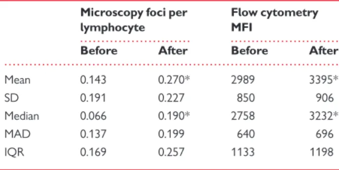
By immunofluorescence microscopy (Figure
1
), the median
number of DSBs (foci, Table
2
) per lymphocyte in baseline samples
was 0.066 (range: 0 – 0.661) and increased significantly (P , 0.05)
after CMR exposure to 0.190 (range: 0 – 1.065, Figure
2
).
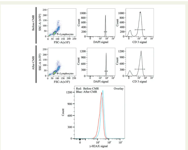
In T-lymphocytes, flow cytometry (Figure
3
) revealed a median MFI
(arbitrary units) of 2758 (range: 1907 – 5109) before and 3232 (range:
2413 – 5484) after CMR (P , 0.005, Table
2
and Figure
4
).
Discussion
We show here that clinical routine CMR scanning exerts genotoxic
effects. Although many experimental in vitro studies have suggested
DNA damage after exposure to MR imaging, we present the first
in vivo results documenting that contrast CMR scanning in daily clinical
routine is associated with increased lymphocyte DNA damage.
The different components of the magnetic field during CMR may
have contributed to the observed DNA damage. The gradient field
generated during MR scanning includes extremely low frequencies
(ELF), which have been classified by the International Agency for
Re-search on Cancer (IARC) as possible human carcinogen (group 2B)
24based on a large body of literature on the genotoxic effects of ELF
magnetic fields.
25–28The latter seem to be involved directly and
in-directly in DNA and chromosomal damage by inducing reactive
oxygen species.
29Similarly, DNA damage and chromosome
altera-tions have been discussed after exposure to RF.
Our results do not allow commenting on the persistence of the
induced DNA damage, although this is a key issue of genetic risk
as-sessment, because damage can trigger DNA instability and exert
tumourigenic effects. Due to the long time delay between DSB
induc-tion and resulting cancer development, our study cannot quantify
such long-term effects as this was beyond the scope of the present
study. This, however, is true in principle for any observation of DSB
. . . .
. . . .
Table 1
Patient baseline characteristics (n 5 20)
Age (years + SD) 53 + 13 BMI (kg/m2+ SD) 25 + 4
Male, n (%) 16 (80)
Cardiovascular risk factors, n (%)
Arterial hypertension 6 (30) Diabetes mellitus 4 (20)
Dyslipidaemia 4 (20)
Smoking 2 (10)
Positive family history 1 (5) Medications, n (%)
Aspirin 7 (35)
Beta-blocker 9 (45)
ACE/angiotensin II inhibitor 8 (40)
Statin 7 (35)
SD, standard deviation; BMI, body mass index.
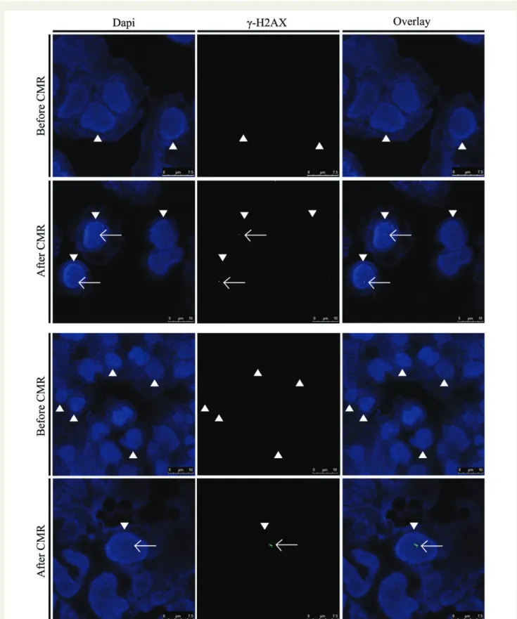
Figure 1
Visualization of double-strand breaks (DSBs) in nuclei (arrow heads) of human lymphocytes of two patients before and after cardiac
magnetic resonance scans by immunofluorescence microscopy. DSBs (foci, white arrows) are detected by g-H2AX staining (green).
induction from any diagnostic radiation exposure including ionizing
radiation, for which no direct observational proof of its adverse
impact on outcome is available due to the small scale of damage
and the long delay between exposure and event. In view of the
growing use of new generation MR scanners with increasing magnetic
field strength (higher Tesla), our results seem to support the
sugges-tions of the ICNIRP for an urgent need of monitoring workers and for
epidemiologic studies on subjects with high levels of exposure or
par-ticular conditions such as for example pregnant occupational
workers.
30Despite activation of repair mechanisms, persistence of DNA
damage has been found in human lymphocytes more than 24 h
after exposing patients and blood samples to CMR scanning.
4Co-genotoxic effects of MR in combination with the administered
gadolinium-based contrast material may further have contributed
to DNA damage due to the potentiating effect of gadolinium-based
contrast material and MR exposure.
31As in our study all patients
underwent contrast enhanced CMR, reflecting widely used clinical
practice,
32we cannot differentiate the precise contribution of the
known genotoxic effect of the gadolinium-based contrast material
from the effects of the magnetic field. However, the use of contrast
material is generally an integrated part of CMR scanning and
there-fore our results may appropriately represent the effect of a routine
CMR scan. The absolute amount of DNA damage is certainly larger
in our study compared with previous in vitro studies, as the entire
blood of each patient rather than a blood sample was exposed
during CMR. According to the assumptions used in the field of
radi-ation protection, an increased number of DNA damages confer a
lin-early increased risk of cancer. Conversely, even a low number of
DSBs may represent a carcinogenic risk according to the linear-no
threshold theory. Our results compare well to the more than
two-fold increase in DSBs induced by CMR and assessed by
immuno-fluorescence microscopy as reported by Simi et al.,
4which was
sub-stantially less pronounced than the almost six-fold increase observed
after cardiac CT by Kuefner et al.
33Although only a few data are
avail-able using FACS analyses for this low scale of signal, the excellent
agreement between microscopy and FACS over a large range of
signal including the present study strengthens the validity of our
results.
34Of note, observations in several subsets of patients seem to
suggest increased sensibilities to MRI exposition, as higher
suscepti-bility for DNA damage by MRI has been found for example in
lympho-cytes of patients with Turner’s syndrome.
35Thus, inappropriate
examinations should be avoided and CMR should be used with
caution and similar restrictions may apply as for X-ray-based and
nuclear imaging techniques where the potential harm is carefully
weighted against the obvious benefit offered by each examination
in order to avoid unnecessary damage of DNA integrity with
poten-tial carcinogenic effect.
. . . .
. . . .
. . . .
Table 2
Increase in double-strand breaks after cardiac
magnetic resonance assessed by immunofluorescence
Microscopy foci per lymphocyte
Flow cytometry MFI
Before After Before After
Mean 0.143 0.270* 2989 3395*
SD 0.191 0.227 850 906
Median 0.066 0.190* 2758 3232*
MAD 0.137 0.199 640 696
IQR 0.169 0.257 1133 1198
IF, immunofluorescence (units are foci per lymphocyte); MFI, geometric mean of T-lymphocyte fluorescence intensity (arbitrary units); g-H2AX, marker of DSBs; SD, standard deviation; MAD, median absolute deviation; IQR, interquartile range. *Indicates P , 0.05 vs. before.
Figure 2
Amount of double-strand breaks before and after cardiac magnetic resonance (CMR) scan by immunofluorescence microscopy. After
CMR scanning, there was a significant increase (*P , 0.05) in g-H2AX foci per lymphocyte by immunofluorescence microscopy. Bars indicate
median values with median absolute deviation (left panel) and individual values are interconnected with a line (right panel).
Figure 3
Flow cytometric analysis of double-strand breaks (g-H2AXpositive T-lymphocytes) before and after cardiac magnetic resonance (CMR)
scan. T-lymphocytes were readily identified by representative dot plots and histograms (lymphocytes, DAPI, and CD3). The shift of the left curve
(red, before CMR) to the right curve (blue, after CMR) in the presented overlay indicates an increase in double-strand breaks (g-H2AXpositive
T-lymphocytes). SSC-A: side scatter channel area. FSC-A: forward scatter channel area. DAPI: 4
′,6-diamidino-2-phenylindole, counterstaining
cell nuclei. CD3: mouse-anti-human CD3-APC antibody counterstaining specifically the T-lymphocytes.
Figure 4
Amount of double-strand breaks before and after cardiac magnetic resonance scan by flow cytometry of g-H2AXpositive
T-lymphocytes using geometric mean fluorescence intensity (MFI). The median MFI increased significantly after cardiac magnetic resonance scanning
(*P , 0.005, left panel). Individual values are interconnected with a line (right panel).
Acknowledgements
We thank Christiane Koenig, Jose Maria Mateos, PhD, Stefano
Ferrari, PhD, and Florian Mair, MSc, from the University of Zurich,
Zurich, Switzerland, and Arnold von Eckardstein, MD, Institute for
Clinical Chemistry, University Hospital Zurich, Zurich, Switzerland,
for their invaluable advice and excellent technical support for the
im-munofluorescence analysis.
Conflict of interest: none declared.
Funding
Grants from the Swiss National Science Foundation to P.A.K. and to M.F.
are gratefully acknowledged.
References
1. Kangarlu A, Robitaille P. Biological effects and health implications in magnetic reson-ance imaging. Concepts Magn Reson 2000;12:321 – 359.
2. Franco G, Perduri R, Murolo A. Health effects of occupational exposure to static magnetic fields used in magnetic resonance imaging: a review. Med Lav 2008;99: 16 – 28.
3. de Vocht F, van-Wendel-de-Joode B, Engels H, Kromhout H. Neurobehavioral effects among subjects exposed to high static and gradient magnetic fields from a 1.5 Tesla magnetic resonance imaging system—a case-crossover pilot study. Magn Reson Med 2003;50:670 – 674.
4. Simi S, Ballardin M, Casella M, De Marchi D, Hartwig V, Giovannetti G, Vanello N, Gabbriellini S, Landini L, Lombardi M. Is the genotoxic effect of magnetic resonance negligible? Low persistence of micronucleus frequency in lymphocytes of individuals after cardiac scan. Mutat Res 2008;645:39 – 43.
5. Carnes KI, Magin RL. Effects of in utero exposure to 4.7 T MR imaging conditions on fetal growth and testicular development in the mouse. Magn Reson Imaging 1996;14: 263 – 274.
6. High WB, Sikora J, Ugurbil K, Garwood M. Subchronic in vivo effects of a high static magnetic field (9.4 T) in rats. J Magn Reson Imaging 2000;12:122 – 139.
7. Rodegerdts EA, Gronewaller EF, Kehlbach R, Roth P, Wiskirchen J, Gebert R, Claussen CD, Duda SH. In vitro evaluation of teratogenic effects by time-varying MR gradient fields on fetal human fibroblasts. J Magn Reson Imaging 2000;12: 150 – 156.
8. Saito K, Suzuki H, Suzuki K. Teratogenic effects of static magnetic field on mouse fetuses. Reprod Toxicol 2006;22:118 – 124.
9. Schiffer IB, Schreiber WG, Graf R, Schreiber EM, Jung D, Rose DM, Hehn M, Gebhard S, Sagemuller J, Spiess HW, Oesch F, Thelen M, Hengstler JG. No influence of magnetic fields on cell cycle progression using conditions relevant for patients during MRI. Bioelectromagnetics 2003;24:241 – 250.
10. Greenland S, Sheppard AR, Kaune WT, Poole C, Kelsh MA. A pooled analysis of mag-netic fields, wire codes, and childhood leukemia. Childhood Leukemia-EMF Study Group. Epidemiology 2000;11:624 – 634.
11. Repacholi MH, Greenebaum B. Interaction of static and extremely low frequency electric and magnetic fields with living systems: health effects and research needs. Bioelectromagnetics 1999;20:133 – 160.
12. Vijayalaxmi OG. Controversial cytogenetic observations in mammalian somatic cells exposed to extremely low frequency electromagnetic radiation: a review and future research recommendations. Bioelectromagnetics 2005;26:412 – 430.
13. Greenwood JP, Maredia N, Younger JF, Brown JM, Nixon J, Everett CC, Bijsterveld P, Ridgway JP, Radjenovic A, Dickinson CJ, Ball SG, Plein S. Cardiovascular magnetic resonance and single-photon emission computed tomography for diagnosis of cor-onary heart disease (CE-MARC): a prospective trial. Lancet 2012;379:453 – 460. 14. Mattsson MO, Auvinen A, Bridges J, Norppa H, Schu¨tz J. European Parliament and the
Council. Research needs and methodology to address the remaining knowledge gaps on the potential health effects of EMF. Scientific Committee on Emerging and Newly Identi-fied Health Risks (SCENIHR). http://ec.europa.eu/health/ph_risk/committees/ 04_scenihr/docs/scenihr_o_024.pdf(23 February 2013).
15. Vecchia P, Hietanen M, Ahlbom A, Anderson LE, Breitbart E, de Gruijl FR, Lin JC, Matthes R, Peralta APT, So¨derberg P, Stuck BE, Swerdlow AJ, Taki M, Saunders R, Veyret B. International Commission on Non-Ionizing Radiation Protection
(ICNIRP). Guidelines on limits of exposure to static magnetic fields. Health Phys 2009;96:504 – 514.
16. Vecchia P, Hietanen M, Ahlbom A, Anderson LE, Breitbart E, de Gruijl FR, Lin JC, Matthes R, Peralta APT, So¨derberg P, Stuck BE, Swerdlow AJ, Taki M, Saunders R, Veyret B. International Commission on Non-Ionizing Radiation Protection (ICNIRP). Amendment to the ICNIRP ‘Statement on medical magnetic resonance (MR) procedures: protection of patients’. Health Phys 2009;97:259 – 261. 17. Belyaev I. Static Fields Environmental Health Criteria No. 232. Geneva, Switzerland:
World Health Organization, WHO. http://www.who.int/pehemf/publications/ EHC_232_Static_Fields_full_document.pdf(23 February 2013).
18. Myerson S, Francis J, Neubauer S. Cardiovascular Magnetic Resonance. 2nd ed. Oxford: Oxford University Press; 2010.
19. Winchester R, Ross G. Methods for enumerating lymphocyte populations. In: Rose NR, Friedman H, (eds). Manual of Clinical Immunology. 1st ed. Washington, DC: American Society for Microbiology; 1976. p64 – 76.
20. Fiechter M, Fuchs TA, Gebhard C, Stehli J, Klaeser B, Stahli BE, Manka R, Manes C, Tanner FC, Gaemperli O, Kaufmann PA. Age-related normal structural and function-al ventricular vfunction-alues in cardiac function assessed by magnetic resonance. BMC Med Imaging 2013;13:6.
21. May MS, Brand M, Wuest W, Anders K, Kuwert T, Prante O, Schmidt D, Maschauer S, Semelka RC, Uder M, Kuefner MA. Induction and repair of DNA double-strand breaks in blood lymphocytes of patients undergoing (18)F-FDG PET/CT examina-tions. Eur J Nucl Med Mol Imaging 2012;39:1712 – 1719.
22. Muslimovic A, Ismail IH, Gao Y, Hammarsten O. An optimized method for measure-ment of gamma-H2AX in blood mononuclear and cultured cells. Nat Protoc 2008;3: 1187 – 1193.
23. Andrievski A, Wilkins RC. The response of gamma-H2AX in human lymphocytes and lymphocytes subsets measured in whole blood cultures. Int J Radiat Biol 2009; 85:369 – 376.
24. World Health Organization (WHO), International Agency for Research on Cancer (IARC), Working Group on the Evaluation of Carcinogenic Risks to Humans. Non-ionizing radiation, Part 1: static and extremely low-frequency (ELF) electric and magnetic fields. IARC Monogr Eval Carcinog Risks Hum 2002;80:1 – 395. 25. Jian W, Wei Z, Zhiqiang C, Zheng F. X-ray-induced apoptosis of BEL-7402 cell line
enhanced by extremely low frequency electromagnetic field in vitro. Bioelectromag-netics 2009;30:163 – 165.
26. Juutilainen J. Do electromagnetic fields enhance the effects of environmental carci-nogens? Radiat Prot Dosimetry 2008;132:228 – 231.
27. Nordenson I, Mild KH, Andersson G, Sandstrom M. Chromosomal aberrations in human amniotic cells after intermittent exposure to fifty hertz magnetic fields. Bioe-lectromagnetics 1994;15:293 – 301.
28. Winker R, Ivancsits S, Pilger A, Adlkofer F, Rudiger HW. Chromosomal damage in human diploid fibroblasts by intermittent exposure to extremely low-frequency electromagnetic fields. Mutat Res 2005;585:43 – 49.
29. Phillips JL, Singh NP, Lai H. Electromagnetic fields and DNA damage. Pathophysiology 2009;16:79 – 88.
30. Bassen H, Bernhardt JH, Brix G, Lejeune JJ, Owen RD, de Seze R, Saunders R, Ueno S, Veyret B, Zaremba L. Medical magnetic resonance (MR) procedures: protection of patients. Health Phys 2004;87:197 – 216.
31. Yildiz S, Cece H, Kaya I, Celik H, Taskin A, Aksoy N, Kocyigit A, Eren MA. Impact of contrast enhanced MRI on lymphocyte DNA damage and serum visfatin level. Clin Biochem 2011;44:975 – 979.
32. Bruder O, Schneider S, Nothnagel D, Dill T, Hombach V, Schulz-Menger J, Nagel E, Lombardi M, van Rossum AC, Wagner A, Schwitter J, Senges J, Sabin GV, Sechtem U, Mahrholdt H. EuroCMR (European Cardiovascular Magnetic Resonance) registry: results of the German pilot phase. J Am Coll Cardiol 2009;54:1457 – 1466. 33. Kuefner MA, Hinkmann FM, Alibek S, Azoulay S, Anders K, Kalender WA,
Achenbach S, Grudzenski S, Lobrich M, Uder M. Reduction of X-ray induced DNA double-strand breaks in blood lymphocytes during coronary CT angiography using high-pitch spiral data acquisition with prospective ECG-triggering. Invest Radiol 2010;45:182 – 187.
34. MacPhail SH, Banath JP, Yu TY, Chu EH, Lambur H, Olive PL. Expression of phos-phorylated histone H2AX in cultured cell lines following exposure to X-rays. Int J Radiat Biol 2003;79:351 – 358.
35. Scarfi M, Prisco M, Lioi M, Zeni O, Della Noce M, Di Pietro R, Franceschi C, Iafusco D, Motta M, Bersani F. Cytogenetic effects induced by extremely low frequency pulsed magnetic fields in lymphocytes from Turner’s syndrome subjects. Bioelectrochem Bioenerg 1997;43:221 – 226.