University of Neuchâtel, UniNE
Institute of Biology Laboratory of Plant Physiology
The role of alpha-tocopherol in the protection of
tomato plants against abiotic stress
Livia Spicher
Supervisor
Prof. Felix Kessler
Institute of Biology
University of Neuchâtel, UniNE
Reviewers
Dr. Gaétan Glauser
Neuchâtel Platform of Analytical Chemistry, NPAC University of Neuchâtel
Dr. Laurent Mène-Saffrané
Department of BiologyUniversity of Fribourg
Defended on February 07, 2017 A dissertation submitted for the degree of Doctor of Philosophy in Biological Sciences
Imprimatur pour thèse de doctorat www.unine.ch/sciences
Faculté des Sciences
Secrétariat-décanat de Faculté Rue Emile-Argand 11 2000 Neuchâtel – Suisse Tél : + 41 (0)32 718 21 00 E-mail : [email protected]
IMPRIMATUR POUR THESE DE DOCTORAT
La Faculté des sciences de l'Université de Neuchâtel autorise l'impression de la présente thèse soutenue par
Madame Livia SPICHER
Titre:“The role of alpha-tocopherol
in the protection of tomato plants
against abiotic stress”
sur le rapport des membres du jury composé comme suit:
• Prof. Felix Kessler, directeur de thèse, Université de Neuchâtel, Suisse • Dr Gaétan Glauser, Université de Neuchâtel, Suisse
• Dr Laurent Mène-Saffrané, Université de Fribourg, Suisse
Abstract
The ability of energy conversion by the photosynthetic machinery under stress and its capacity to adjust to an ever-changing environment is crucial for plant survival. The photosynthetic light reactions occur at the photosystems in the thylakoids of chloroplasts. The photosystems are composed of proteins in a specific lipid environment. It includes not only membrane lipids but also lipophilic pigments (chlorophylls, carotenoids) and prenylquinones (plastoquinone, phylloquinone, tocopherol). Apart from their respective roles in light harvesting and electron transport, carotenoids and prenylquinones have important antioxidant properties and protect plant cells against reactive oxygen species. The focus of this work is to understand how plants resist and adapt to environmental stress in particular high light, high temperature and the combination of the two. Lipid metabolism takes places in plastid subcompartments, at the level of envelopes, at thylakoid microdomains called plastoglobules. Plastoglobules are involved in various essential biosynthetic metabolic pathways and accumulation of prenylquinone molecules. In this thesis, we use tomato as the model system to address the role of (prenyl) lipids synthesis and remodelling to protect photosynthetic function under stress. After an introduction on the implication of photosynthetic machinery in lipid metabolism, in Chapter 2 we summarized recent advances in plastoglobule research and their findings on biosynthesis and metabolism of Vitamins E and K1. Then in Chapter 3, we investigate the question
of how the photosynthetic machinery is protected against heat stress. Amongst many hundreds of compounds that change under heat stress, we identified α-tocopherol and plastoquinone as the most significantly increased antioxidants. This finding suggests a new role for these two prenylquinones in protecting the photosynthetic apparatus against temperature stress. In Chapter 4, through a joint effort, we provided valuable information on the metabolic fluxes and biosynthesis of Vitamin E in tomato. Finally, in Chapter 5, we intended to identify molecules that contribute to the protection against combined high temperature and high light stress. To perturb α-tocopherol levels we used the tomato vte5 knock down-line. The data indicate that VTE5 protects against combined high light and high temperature stress and does so by supporting α-tocopherol production. Overall, this
thesis contributes to a better understanding of the role of prenylquinone compounds, in the resistance of tomato plants against high light and high temperature stresses.
Keywords: lipidomics, prenylquinones, photosynthesis, temperature stress, high light
stress, tocopherol, Vitamin E, plastoquinone, plastochromanol, carotenoids, Fv/Fm, plas-toglobules, phytol, phytol kinase, Solanum lycopersicum, tomato.
Résumé
La capacité de conversion de l’énergie par les chloroplastes en condition de stress et son aptitude à s’adapter à un environnement en constante évolution est cruciale pour la survie des plantes. Les réactions photosynthétiques se produisent au niveau des photosystèmes dans les thylakoïdes des chloroplastes. Les photosystèmes sont composés de protéines dans un environnement lipidique spécifique. Ce dernier comprend non seulement des lipides membranaires mais aussi des pigments lipophiles (chlorophylles, caroténoïdes) et des prénylquinones (plastoquinone, phylloquinone, tocophérol). À part leurs rôles de col-lecteur de lumière et leurs implications dans le transport d’électrons, les caroténoïdes et les prénylquinones (respectivement) ont d’importantes propriétés antioxydantes et protègent les cellules végétales contre les espèces réactives à l’oxygène. Le but de ce travail est de comprendre comment les plantes résistent et s’adaptent aux stress environnementaux, en particulier aux fortes intensités lumineuses, à la hausse de température et à la combinaison des deux. Le métabolisme des lipides prend place dans les sous-compartiments des plas-tides, au niveau des enveloppes, des membranes des thylacoïdes et de ses microdomaines, appelés plastoglobules. Les plastoglobules sont impliqués dans diverses voies métaboliques biosynthétiques essentielles et dans l’accumulation des molécules de prénylquinone. Dans cette thèse, nous avons utilisé la tomate comme système modèle afin d’étudier le rôle de la synthèse des (prényl) lipides ainsi que leur remodelage dans la protection de la fonction photosynthétique en condition de stress. Après une introduction sur l’implication des chloroplastes dans le métabolisme des lipides, nous avons résumé, dans le chapitre 2, les récents progrès de la recherche sur les plastoglobules et leurs implications sur la biosyn-thèse et le métabolisme de la vitamine E et de la vitamine K1. Ensuite, dans le chapitre
3, nous avons étudié comment le chloroplaste est protégé contre le stress dû à la hausse de la température. Parmi les centaines de composés qui changent sous stress thermique, nous avons identifié l’α-tocophérol et la plastoquinone comme étant les antioxydants les plus significativement en hausse. Cette découverte suggère un nouveau rôle pour ces deux prénylquinones dans la protection de l’appareil photosynthétique contre le stress thermique. Dans le chapitre 4, nous avons fourni des informations précieuses sur les flux métaboliques et de biosynthèse impliqués dans l’accumulation de la vitamine E chez la tomate. Enfin, au chapitre 5, nous avons cherché à identifier les molécules qui contribuent à la protection contre le stress dû à la hausse des températures combiné au stress de haute
intensité lumineuse. Pour perturber les niveaux d’α-tocophérols, nous avons utilisé un mutant chez la tomate ayant perdu la fonction du VTE5 (vte5). Les données indiquent que le VTE5 protège la plante contre la hausse des températures combinée au stress de forte intensité lumineuse en soutenant la production d’α-tocophérol. D’une manière générale, cette thèse contribue à une meilleure compréhension du rôle des prénylquinones impliqués dans la résistance chez la tomate au stress de forte intensité lumineuse combiné à la hausse de température.
Mots-clés: lipidomique, prénylquinones, photosynthèse, stress de température, lumière
de forte intensité, tocophérol, Vitamin E, plastoquinone, plastochromanol, caroténoïdes, Fv/Fm, plastoglobules, phytol, phytol kinase, Solanum lycopersicum, tomate.
Contents
1 General introduction 1
1.1 Introduction . . . 1
1.1.1 Plastids adapt to changes in environmental conditions . . . 1
1.1.2 Chloroplasts - a primary site of diverse metabolic pathways . . . . 3
1.1.3 Regulatory roles of plastids in isoprenoid compounds biosynthesis . 4 1.1.4 Prenylquinone biosynthesis also derives from the MEP pathway . . 6
1.1.5 Tocochromanols essential plastid antioxidant metabolites . . . 6
1.1.6 Plastoglobules are more than lipid droplets . . . 9
1.2 General thesis outline . . . 10
2 Unexpected roles of plastoglobules (plastid lipid droplets) in vitamin K1 and E metabolism 23 2.1 Introduction . . . 24
2.2 Plastoglobules change with plant developmental stages and function as microdomains for (prenyl-) lipid metabolism . . . 25
2.3 Vitamin K1 in the thylakoids and plastoglobules . . . 28
2.4 Tocopherol cyclase (VTE1) located at plastoglobules fulfills diverse roles in the tocopherol metabolism . . . 29
2.5 VTE1 and NDC1 enzymes are directly implicated in the redox cycle of α-tocopherol . . . 30
2.6 ABC1-like kinases affect prenyl lipid composition of the chloroplast . . . . 31
2.7 Conclusion . . . 32
3 Lipid antioxidant and galactolipid remodeling under temperature stress in tomato plants 37 3.1 Introduction . . . 38
3.2 Material and Methods . . . 39
3.2.1 Plant material and stress treatments . . . 39
3.2.2 Determination of photosynthetic parameters . . . 40
3.2.3 Chlorophyll quantification . . . 40
3.2.4 Lipid profiling . . . 40
3.3 Results . . . 42
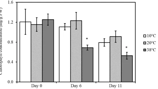
3.3.1 Photosynthetic efficiency is reduced after cold temperature treatment 42 3.3.2 Heat stress reduces chlorophyll content in tomato leaves . . . 42
3.3.3 Untargeted lipidomics identify changes in lipid composition in tomato
leaves after high temperature treatment . . . 43
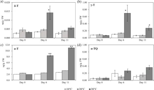
3.3.4 High temperature increases prenylquinones in tomato leaves . . . . 44
3.3.5 Temperature modulates saturation of galactolipids mostly present in thylakoid membranes . . . 50
3.3.6 Discussion . . . 52
3.3.7 Acknowledgments . . . 55
3.4 Supplementary data . . . 55
4 Down-regulation of tomato PHYTOL KINASE strongly impairs tocopherol biosynthesis and affects prenyllipid metabolism in an organ-specific manner 63 4.1 Introduction . . . 64
4.2 Materials and Methods . . . 67
4.2.1 Plant material, growth conditions and sampling . . . 67
4.2.2 Phylogenetic analysis . . . 67
4.2.3 Generation of SlVTE5-RNA interference (RNAi) transgenic lines . . 67
4.2.4 Identification of the folk-1 tomato mutant by TILLING . . . 68
4.2.5 qPCR analysis . . . 68
4.2.6 Leaf gas exchange and fluorescence measurements . . . 68
4.2.7 Tocopherol, free phytol and fatty acid phytyl ester quantification . 69 4.2.8 Prenylquinone and carotenoid profile . . . 69
4.2.9 Chlorophyll and chlorophyll catabolites . . . 70
4.2.10 Quantification of soluble sugars and starch . . . 70
4.2.11 Trolox equivalent antioxidant capacity (TEAC) assay . . . 71
4.2.12 Transmission electron microscopy . . . 71
4.2.13 Data analyses . . . 71
4.3 Results . . . 72
4.3.1 Tomato tocopherol contents are highly dependent on SlVTE5 but not on SlFOLK . . . 72
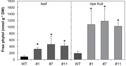
4.3.2 Down-regulation of SlVTE5 boosted phytyl ester synthesis in leaves 75 4.3.3 Chlorophyll content is not affected in SlVTE5-RNAi lines . . . 76
4.3.4 SlVTE5 knockdown alters prenyllipid metabolism in fruits . . . 76
4.3.5 VTE5 deficiency affects the expression of tocopherol metabolism-related genes . . . 81
4.3.6 Measurements of carbohydrate metabolism, photosynthesis and yield parameters suggest carbon export impairment in SlVTE5-knockdown plants . . . 82
4.4 Discussion . . . 83
4.5 Funding . . . 88
5 Essential role for phytol kinase and tocopherol/vitamin E in tolerance to
combined light and temperature stress in tomato 107
5.1 Introduction . . . 108
5.2 Materials and Methods . . . 109
5.2.1 Plant material . . . 109
5.2.2 Stress treatments . . . 109
5.2.3 Transmission electron microscopy . . . 109
5.2.4 Determination of photosynthetic parameters . . . 110
5.2.5 Untargeted lipid profiling . . . 110
5.2.6 Targeted lipid profiling: prenylquinone, carotenoid and glycolipids profile . . . 110
5.2.7 Free phytol and fatty acid phytyl ester quantification . . . 111
5.2.8 qPCR analysis . . . 111
5.2.9 Isolation of thylakoid membranes and Hill reaction . . . 112
5.3 Results . . . 112
5.3.1 Untargeted lipidomics demonstrated changes in lipid composition in tomato leaves after combined high light and HT . . . 112
5.3.2 vte5 develops a chlorotic phenotype under combined high-light and high-temperature stress . . . 113
5.3.3 Combination of high light and high temperature triggers photoinhi-bition in vte5 plants . . . 113
5.3.4 Plastoglobules accumulate under HT+HL in vte5 . . . 115
5.3.5 Stress treatments change prenylquinone and carotenoid metabolism 115 5.3.6 Fatty acid phytyl esters accumulate massively in the vte5 mutant . . 118
5.3.7 Free phytol levels increased under combined high light and temper-ature stress . . . 123
5.3.8 Phytol toxicity assessed by electron transport activity . . . 123
5.3.9 Gene expression profiles . . . 124
5.3.10 Discussion . . . 125 5.3.11 Acknowledgements . . . 127 5.4 Supplementary data . . . 127 6 General conclusion 139 6.1 Future directions . . . 143 Contents xi
1
General introduction
1.1 Introduction
Plants have to adapt continually to environmental changes. With the changes in climate, including increased temperatures in combination with high light intensity, plants incur losses and shifts of their original habitat (Pretty et al., 2010; Streb et al., 2003; Walther et al., 2002). The inability of plants to adapt to environmental changes leads to significant physiological perturbations that affect photosynthesis (Mishra and Singhal, 1992). As a consequence climate changes increase pressure on agriculture, especially crop plant productivity.
Plant growth and development depends on photosynthesis. Photosynthesis is an essential process that enables life on Earth (Jajoo, 2014) and the major bioenergetic activity that takes place at the thylakoid membranes in the chloroplasts (Eberhard et al., 2008). The photosynthetic light reactions generate ATP, NADPH, and molecular oxygen. Using the ATP and NADPH, the “dark” reactions assimilate carbon dioxide into organic compounds, initially in the form of starch (Waters and Langdale, 2009). Starch degradation products will then either be exported to the cytosol or directly used in the chloroplast stroma as a primary carbon source for plant biomass production.
Light is essential for plant growth and productivity. However excessive light can cause severe stress in plants by damaging the photosynthetic reaction centers (Lichtenthaler, 1999; Takahashi and Badger, 2011). In combination with increased temperature, excessive light leads to dramatic changes in the structure of photosynthetic machinery, consequently altering the photosynthetic capacity of plants, and disrupting cellular homeostasis (Bita and Gerats, 2013; Wahid et al., 2007).
1.1.1 Plastids adapt to changes in environmental conditions
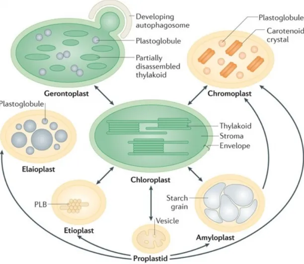
Plastids are essential organelles of endosymbiotic origin. Plastid occur in higher plants in various sizes, shapes, and functions (Thomson and Whatley, 1980). Depending on environmental stimuli and developmental signals, plastids develop into distinct, tissue-specific types, including chloroplasts, chromoplasts, amyloplasts, elaioplasts, gerontoplasts (Figure 1.1) (Jarvis and López-Juez, 2013; Lopez-Juez and Pyke, 2005; Wise, 2006).
Fig. 1.1: Plastid diversity and interconversions. Plastids differentiate in various functional
or-ganelles depending on tissue, developmental stage, hormonal and environmental cues. Proplastids are totipotent plastids present in the meristematic tissue. Etioplasts are chloro-plast precursors that develop in the absence of light and accumulate the chlorophyll precursor protochlorophyllide in the prolamellar body (PLB, paracrystalline membra-nous structure). Upon illumination etioplasts, rapidly differentiate into chloroplasts containing the vast thylakoid membrane system. Leucoplasts are non-pigmented, storage plastids. This group comprises amyloplasts and elaioplasts. Amyloplasts store starch and are present in storage tissues such as cotyledons, endosperm, and tubers. Elaio-plasts store lipids and exist, for example, in the functional layer of nutritive cells within the anther during pollen development or epidermal cells of some monocotyledonous families. Chromoplasts accumulate colored carotenoids and are associated with repro-ductive tissues in flowers and fruits, having roles in attracting pollinators and seed disseminators. Gerontoplasts differentiate from chloroplasts during senescence and have highly regulated catabolic activities including the disassembly of the photosynthetic machinery, autophagic recycling and reallocation of resources to seed and perennial tissues (Jarvis and López-Juez, 2013; Neuhaus and Emes, 2000; Rottet et al., 2015; Waters and Langdale, 2009)
Moreover, in its photosynthetic role, the chloroplast adapts to varying environmental conditions, remodeling its membrane system to increase or decrease light harvesting surface (Figure 1.2) (Lichtenthaler and Burkart, 1999).
Fig. 1.2: Chloroplast ultrastructural adaptation in response to high-light and low-light.
Sun-type chloroplasts exhibit a reduction of the grana stacks and abundance of light-harvesting complex-LHCII, large starch grains, and enlargement of plastoglobules when compared to chloroplasts in shade-grown plants (Lichtenthaler, 2010). Adapted from Lichtenthaler and Burkart (1999)
1.1.2 Chloroplasts - a primary site of diverse metabolic
pathways
Besides supplying energy through photosynthesis, chloroplasts, fulfill other important metabolic roles (Lopez-Juez and Pyke, 2005; Neuhaus and Emes, 2000; Rolland et al., 2012). Chloroplasts synthesize an immense variety and quantity of lipophilic compounds that serve different purposes within the cell. The implication of these chloroplast lipids range from structural roles, such as membrane biogenesis (Andersson et al., 2001; Benning, 2008, 2009; Kobayashi et al., 2007, 2013) and remodeling (Kelly and Dörmann, 2004; Kirchhoff, 2014; Shimojima and Ohta, 2011) to protection against oxidative stress under high light (Nowicka et al., 2016; Szyma´nska and Kruk, 2010) and to lipid-derived signaling in response to biotic stress, e.g. wounding, herbivory and pathogens (Howe and Schilmiller, 2002; Upchurch, 2008; Wasternack, 2007).
Lipid metabolism takes places in plastid subcompartments, at the level of envelopes, thylakoid membranes and at thylakoid microdomains called plastoglobules. The processes involved in chloroplast lipid metabolism are tightly regulated depending on developmental stage and environmental factors (Zhang et al., 2010).
The chloroplast structural core is constituted by a complex membrane system, the thy-lakoids, that are formed mainly by galactolipids, approximated 50%
diacylglycerol, 26% digalactosyldiacylglycerol, sulfoquinovosyldiacylglycerol and phos-phatidylglycerol comprise most of the remaining lipids (Boudière et al., 2014; Dörmann, 2013; Kobayashi, 2016). Chloroplast photosynthetic membranes also contain embedded proteins and lipophilic compounds including chlorophylls, carotenoids (β-carotene, lutein, neoxanthin and the three xanthophyll cycle carotenoids zeaxanthin, violaxanthin and antheraxanthin) and prenylquinones (plastoquinone, phylloquinone, tocopherol and plas-tochromanol) (Dekker and Boekema, 2005; Lichtenthaler, 2007; Lichtenthaler and Calvin, 1964; Lichtenthaler and Park, 1963; Matringe et al., 2008). While carotenoids play struc-tural and light harvesting roles and have a protective function against excessive light Frank and Cogdell, 1996; Gruszecki and Strzałka, 2005; Niyogi et al., 2000, prenylquinones have essential roles as antioxidants and in electron transport (Havaux et al., 2005). Together, the carotenoids and prenylquinones unfold protective functions under environmental stress and shield plant cells against ROS (reactive oxygen species) (Havaux and Kloppstech, 2001; Horvath et al., 2006).
1.1.3 Regulatory roles of plastids in isoprenoid compounds
biosynthesis
The prenylquinones, as well as the carotenoids, belong to the plastid isoprenoid family. Due to their diverse roles in a wide range of biological processes, they are essential for plant growth and development. Also known as terpenoids, isoprenoid compounds are the most abundant class of metabolites and more than 50 000 different molecules have been identified and reported to date (Banerjee and Sharkey, 2014; Thulasiram et al., 2007). In higher plants, two independent biosynthetic pathways supply the universal C5 precursor, isopentenyl diphosphate (IPP) and its isomer, dimethylallyl diphosphate (DMAPP) (Eisenreich et al., 1998), the cytosolic mevalonate (MVA) and the plastidial methylerythritol-4-phosphate (MEP) pathways (Lange and Croteau, 1999; Lange et al., 1998; Lichtenthaler, 1999, 2010; Paseshnichenko, 1998; Rodríguez-Concepción and Boronat, 2015; Vranová et al., 2013; Wanke et al., 2001). Whilst the MVA pathway supplies precursors for the synthesis of sesquiterpenes, triterpenes and sterols (e.g. brassinosteroid hormones) (Mendoza-Poudereux et al., 2015; Rodríguez-Concepción, 2006), the MEP pathway is involved in the production of monoterpenes, plastoquinones, carotenoids, the phytol tail of chlorophylls, and phytohormones (e.g. cytokinins, gibberellins, abscisic acid) (Boronat, 2010; Rodríguez-Concepción, 2006; Rodríguez-Concepción and Boronat, 2002). The separation of the two pathways in different compartments and its advantages are not yet fully understood (Hemmerlin et al., 2012). The plastid pathway is probably conserved from the endosymbiotic event, whereas the cytosolic one is eukaryotic. Moreover, many isoprenoid compounds from the MEP pathway are required in the plastid for their roles in photosynthesis (Figure 1.3). Thus the physical separation is likely to facilitate the optimal supply of precursors for each pathway (Rodríguez-Concepción and Boronat,
2015). In this chapter, I will focus on plastidial derived pathway, with particular emphasis into prenyllipids and tocopherol metabolism.
Fig. 1.3: Plastid MEP pathway in plant cells. Solid-line arrows indicate a single enzymatic
step, dashed-line arrows indicate more than one enzymatic step, and circle-ended ar-rows indicate cross-membrane transport. White arar-rows indicate enzymatic reactions in plastids. Short-chain prenyl diphosphates such as isopentenyl diphosphate (IPP), dimethylallyl diphosphate (DMAPP), geranyl diphosphate (GPP), and farnesyl diphos-phate (FPP) translocate across the plastid membrane (Bick and Lange, 2003). Geranyl-geranyl diphosphate (GGPP) can be used for protein prenylation in the cytosol of tobacco cells (Gerber et al., 2009). Specific transporters for prenyl diphosphates have not yet been identified. Other abbreviations: 1-Deoxy-D-xylulose 5-phosphate synthase (DXS); 1-Deoxy-D-xylulose 5-phosphate reductoisomerase (DXR); 2-C-methyl-D-erythritol 4-phosphate cytidylyltransferase (MCT); 4-(Cytidine 5’-diphospho)-2-C-methyl-D-erythritol kinase (CMK); 2-C-methyl-D-erythritol 2,4-cyclodiphosphate synthase (MDS); 4-Hydroxy-3-methylbut-2-enyl-diphosphate synthase (HDS); 4-Hydroxy-3-methylbut-2-enyl diphos-phate reductase (HDR); Isopentenyl diphosdiphos-phate-isomerase (IPPI); Geranyl diphosdiphos-phate synthase (GPPS); Geranylgeranyl diphosphate synthase (GGPPS). Adapted from Vranová et al. (2013).
The MEP pathway (also known as 1-deoxy-D-xylulose 5-phosphate (DXP) pathway or pyruvate/glyceraldehyde-3-phosphate pathway) consists of seven enzymatic steps (Rodríguez-Concepción and Boronat, 2002; Rohdich et al., 2001; Rohmer, 1999; Rohmer et al., 1996; Sharkey et al., 2007). DXP is biosynthesized from pyruvate and D-glyceraldehyde 3-phosphate (GAP) that are catalyzed by the enzyme 1-deoxy-D-xylulose-5-phosphate syn-thase (DXS) (Rodríguez-Concepción and Boronat, 2002; Rohmer et al., 1993). Next, the enzyme 1-deoxy-D-xylulose-5-phosphate reductoisomerase (DXR) converts DXP into MEP. Then, through three consecutive enzymatic steps involving cytidylation (CTP-dependent), phosphorylation (ATP-dependent), and cyclization, MEP is converted to the cyclic inter-mediate methylerythritol 2,4-cyclodiphosphate (MEcDP). In the following step, catalyzed
by HMBDP synthase (HDS) MEcDP is converted into hydroxymethylbutenyl diphosphate (HMBDP). Finally, HMDBP is reduced to IDP and DMADP by HMBDP reductase (HDR). IDP and DMADP are also isomerized by isopentenyl diphosphate isomerase (IDI) (Banerjee and Sharkey, 2014)).
1.1.4 Prenylquinone biosynthesis also derives from the MEP
pathway
Prenylquinones, like all isoprenoids, are assembled from the condensation of monomers of isopentenyl diphosphate (IPP) and its double bond isomer dimethylallyl diphosphate (DMAPP). A series of subsequent condensation reactions yields geranylgeranyl diphosphate (GGDP) from IPP. GGDP is an essential intermediate for isoprenoid-derived metabolism shared in the biosynthesis of carotenoids, tocochromanols, a group that includes to-copherols, tocotrienols and plastochromanol (PC-8), and other isoprenoid metabolic pathways isoprenoids (DellaPenna and Pogson, 2006; Kruk et al., 2014). Prenylquinone biosynthesis also depends on aromatic precursors from the shikimate pathway: p-hydroxy-phenylpyruvate for plastoquinone, plastochromanol, and tocopherols, or chorismate, for phylloquinone, ubiquinone, and menaquinones (Figure 1.4).
1.1.5 Tocochromanols essential plastid antioxidant metabolites
Tocochromanols make up a small family of amphipathic molecules that is composed of four tocopherols and four tocotrienols. This group of molecules does not only have potent lipid-soluble antioxidant activity, but its members are also essential as nutrients for human health (Kamal-Eldin and Appelqvist, 1996; Munné-Bosch and Alegre, 2002; Schneider, 2005; Wolf, 2005). Discovered as a reproductive factor in 1922 (Evans and Bishop, 1922), the tocopherol biosynthetic pathway in plants and algae was decoded at the beginning of the 1970ies from precursor and products studies using radiolabeled intermediates (Grusak and DellaPenna, 1999; Whistance and Threlfall, 1970). Tocopherol biosynthetic reactions were unraveled in the following decade (Soll et al., 1980; Soll and Schultz, 1980; Soll et al., 1983, 1985), whereas the genes involved were only recently identified (Cheng et al., 2003; Collakova and Dellapenna, 2001; Porfirova et al., 2002; Rohmer, 2003; Savidge et al., 2002; Shintani and Dellapenna, 1991; van Eenennaam et al., 2003). Almost all of the enzymes responsible for tocopherol biosynthesis were localized to the inner envelope membrane (Bouvier et al., 2005; DellaPenna and Pogson, 2006) with the exception of VTE1 that is present predominantly in plastoglobules (Austin II et al., 2006; Lundquist et al., 2012; Vidi et al., 2006; Ytterberg et al., 2006).
Tocochromanols occur naturally as α, β, γ, and δ-tocopherol and -tocotrienols that differ in the number and position of methyl groups at the aromatic ring (Grusak and DellaPenna,
Fig. 1.4: Overview of plastidic isoprenoid, tocopherol, and carotenoid biosynthesis in plants.
The MEP pathway provides IPP for the synthesis of GGDP. Orange arrows relate to abun-dant carotenoids. Minor products of the carotenoid pathway are not shown. Green arrows regard tocopherol synthesis starting with homogentisate, a product of the shiki-mate pathway. The tocotrienol product of the condensation of GGDP with homogentisate produced by the same pathway is not shown, adapted from DellaPenna and Pogson (2006).
1999) (Figure 1.5). α-tocopherol is the most active form as vitamin E due to its retention by a hepatic α-tocopherol binding protein (α-TTP) (Hosomi et al., 1997; Terasawa et al., 2000).
Fig. 1.5: Tocopherol and tocotrienol structures. Differences are indicated in red. The table
shows the number/position of methyl substituents in α, β, γ, and δ-tocopherol and tocotrienols. *Vitamin E activity of each tocopherol and tocotrienol with α-tocopherol being 100% (DellaPenna, 2005).
Tocopherols and tocotrienols are differentiated by their isoprenyl side chains, derived from phytyl-PP or GGDP, respectively. The polar head group (homogentisate) of tocochromanols is derived from the Shikimate pathway of aromatic amino-acid metabolism (DellaPenna and Pogson, 2006; Kamal-Eldin and Appelqvist, 1996; Munné-Bosch and Alegre, 2002). The condensation of the prenyl moiety phytyl diphosphate and homogentisate by ho-mogentisate phytyl transferase, (HPT, VTE2) initiates tocopherol synthesis. The 2-methyl-6-phytylbenzoquinol (MPBQ) derived from the previous step can be methylated on the C-3 position on the aromatic ring by the dimethyl-phytylquinol methyl transferase (VTE3) to give 2,3-dimethyl-6-phytyl-1,4-benzoquinone (DMPBQ). The cyclization of either MPBQ or DMPBQ by the tocopherol cyclase (VTE1) results in δ- and γ-tocopherol, respectively. The further methylation at the C-5 position of these two molecules, mediated by the tocopherol γ-methyl transferase (VTE4), leads to the formation of β- and α-tocopherol (DellaPenna and Pogson, 2006; Zbierzak et al., 2010). Phytyl diphosphate, besides de novo synthesis, may originate from an alternative salvage pathway dependent on the phytol kinase (VTE5) and phytyl-phosphate kinase (VTE6), which by phosphorylation recycle the phytol moiety released from the chlorophyll tetrapyrrole ring into tocopherol biosynthesis (Almeida et al., 2016; Ischebeck et al., 2006; Valentin et al., 2006; vom Dorp et al., 2015).
Tocochromanols react with polyunsaturated acyl groups and protect membrane lipids from oxidative damage by scavenging lipid peroxyl radicals and quenching highly reactive singlet oxygen (1O
2) as well as other reactive oxygen species (DellaPenna and Pogson, 2006;
Mène-Saffrané and DellaPenna, 2010; Wolf, 2005). Their chromanol ring is responsible for their spectral and antioxidant properties (Kruk et al., 2014). In the first mechanism, the tocochromanol ring hydroxyl will donate a hydrogen atom to a highly reactive PUFA peroxyl radical, which will convert into its much less reactive hydroperoxide form, preventing the propagation of PUFAs peroxidation in the cell membranes (Serbinova et al., 1991; Traber and Atkinson, 2007). In the second mechanism,1O
2will be either quenched physically or
chemically (Krieger-Liszkay and Trebst, 2006; Triantaphylidès and Havaux, 2009). The physical quench of 1O
2 relies on charge transfer and thermal dissipation mechanisms
to return the oxygen to its ground state (3O
2) (Dellapenna and Mène-Saffrané, 2011).
The1O
2 chemical quenching occurs by the opening of the chromanol ring, resulting in
the tocopherol oxidation product, α-tocopherolquinone. The latter can still fulfil roles in electron transfer, and it can be reconverted and recycled into α-tocopherol, by the re-introduction of the chromanol ring by the activity of tocopherol cyclase (VTE1) (Gruszka et al., 2008; Kobayashi and DellaPenna, 2008; Kruk et al., 2005; Munné-Bosch, 2005; Triantaphylidès and Havaux, 2009).
Even though relative tocopherol composition can vary among species (Grusak and Del-laPenna, 1999; Horvath et al., 2006). Tocopherols are found in most plant organs, while tocotrienols are mostly present in monocotyledon seeds. The levels of α-tocopherol in most seeds are low and limited by the expression of VTE4. Arabidopsis and soybean plants overexpressing VTE4 have strikingly increased α-tocopherol levels (Shintani and Dellapenna, 1991; van Eenennaam et al., 2003). Besides its photoprotective roles (Falk and Munné-Bosch, 2010), tocopherol participates in seed longevity and germination (Chen et al., 2016; Mène-Saffrané and DellaPenna, 2010; Sattler et al., 2004). The loss of function mutant vte2 exhibited reduced seed longevity, and during germination, seedlings showed severe growth defects, presumably caused by increased levels of lipid hydroperox-ides and hydroxyl fatty acids. VTE5 was initially characterized in Arabidopsis. The vte5 mutant had only 20% of the tocopherol normally found in wild-type seeds, and even less so in leaves. Surprisingly, however, no effect on seed germination has been reported (Valentin et al., 2006).
1.1.6 Plastoglobules are more than lipid droplets
Chloroplast lipid droplets, also known as plastoglobules are microdomains of the thylakoid membranes (Austin II et al., 2006). They were first believed to be just a storage site for lipid metabolites (Bailey and Whyborn, 1963; Greenwood et al., 1963). Plastoglobules are physically connected to thylakoids through the outer membrane leaflet (Austin II et al., 2006) and their lipid monolayer boundary hosts a small number of proteins (Kessler et al.,
1999), that are mostly involved in lipid metabolism (Eugeni Piller et al., 2012; Grennan, 2008). Since the analysis of plastoglobule proteomes of Arabidopsis chloroplasts and red pepper chromoplasts, an active metabolic role of plastoglobules in the biosynthesis of neutral lipids, has been established (Lundquist et al., 2012; Vidi et al., 2006; Ytterberg et al., 2006).
It has been demonstrated that plastoglobules are involved in various essential biosyn-thetic metabolic pathways. In the context of this thesis, metabolism and/or accumulation of prenyl quinone molecules, e.g. tocopherol, plastoquinone and plastochromanol in plastoglobules (Vidi et al., 2006; Zbierzak et al., 2010). Beyond lipid metabolism and storage, plastoglobule protein composition changes in response to stress or developmental transitions. During chloroplast senescence, which entails catabolic processes such as the dismantling of the thylakoid membrane, plastoglobules play roles in metabolic detoxifica-tion. The degradation of chlorophyll (liberating phytol) and monogalactosyldiacylglycerol (MGDG) was correlated with the accumulation of triacylglycerol (TAG) and fatty acid phytyl ester (FAPE). Both TAG and FAPE are products of phytyl ester synthases 1 (PES1) and 2 (PES2) that localize to plastoglobule (Besagni and Kessler, 2013; Gaude et al., 2007; Ischebeck et al., 2006; Lippold et al., 2012; Rottet et al., 2015; Tevini and Steinmüller, 1985). Likewise, in reproductive tissues and fruit maturation, plastoglobules have roles in disassembly of thylakoids, and concomitant accumulation of carotenoids in plastoglobules and plastoglobule-derived carotenoid fibrils (Deruère et al., 1994; Steinmüller and Tevini, 1985).
Not only plastoglobules fascinate by their dynamic ultrastructural changes, in constant adaptation to plant development stage, stresses or changes in the environment; but also their direct involvement in essential lipid metabolism highlights their importance in plant development and survival. This present thesis explores and confirms the role of prenylquinone lipids, which metabolism takes place in the plastoglobules, in the resistance of tomato plants against high light and high temperature stresses.
1.2 General thesis outline
The focus of this work is to understand how plants resist and adapt to environmental stress in particular high light, high temperature and the combination of the two. I took strong interest in lipid synthesis and re-modeling as the two are key factors in photosynthetic function under environmental stress conditions I addressed the role of (prenyl) lipids in these processes taking a broad metabolomics approach to lipophilic compounds. I chose tomato as the model system. As a crop plant this prominent member of the Solanaceae family is not only of importance in agriculture, but it has also become an important laboratory model system for fleshy fruit plants. In addition to its nutritional value, tomato
is also a suitable model to study prenylquinone metabolism. The use of a crop systems may allow a more rapid translation of the findings into agriculture. Our knowledge of the role of prenylquinones in stress resistance was expanded by the use of new lipidomic-based methods, which allowed the simultaneous and rapid profiling of prenylquinones and carotenoids in plant extract based-methods.
In Chapter 2, I present a review published in Current Opinions in Plant Biology entitled “Unexpected roles of plastoglobules (plastid lipid droplets) in vitamin K1and E metabolism.”
In this review, I summarized recent advances on plastoglobule research and their findings on biosynthesis and metabolism of Vitamins E and K1.
Climate change and rising temperatures are amongst the most serious issues on this planet. In Chapter 3, I present a manuscript that was published in Frontiers in Plant Science entitled “Lipid antioxidant and galactolipid remodeling under temperature stress in tomato plants.” To investigate the question of how the photosynthetic machinery is protected against heat stress, I employed a non-targeted lipidomics approach. Amongst many hundreds of compounds that change under heat stress, I identified α-tocopherol and plastoquinone as the most significantly increased antioxidants. This suggest a new role for these two prenyl quinones in protecting the photosynthetic apparatus against temperature stress.
In Chapter 4, I present an article published in the Journal of Experimental Botany entitled “Down-regulation of tomato PHYTOL KINASE strongly impairs tocopherol biosynthesis and affects prenyllipid metabolism in an organ-specific manner.” This article was the result of a collaboration with the University of São Paulo. My contribution to this study, together with Neuchâtel Platform of Analytical Chemistry (NPAC), was the non-targeted lipidomic analysis of prenyllipids and galactolipids in leaves and fruits of a tomato vte5 knock down-line (SIVTE5). I designed the extraction method for mature-green and ripe fruit analysis. The results obtained through this joint effort provided valuable information on the metabolic fluxes implicated in Vitamin E accumulation as well as biosynthetically related pathways which impact tomato physiology.
In Chapter 5, I intended to identify molecules that contribute to the protection against combined high temperature and high light stress. The ability of energy conversion by the photosynthetic machinery under stress and its capacity to adapt to an ever-changing environment is crucial for plant survival. Here, we use the non-targeted lipidomics approach in the tomato model system to identify lipophilic molecules that may contribute to protection against this combined stress. Among several hundred compounds, the two most strongly upregulated compounds were α-tocopherol and plastoquinone/-ol. To perturb α-tocopherol levels we used the tomato vte5 knock down-line (SIVTE5). The data indicate that VTE5 protects against combined high light and high temperature stress and do so by supporting α-tocopherol production. The manuscript entitled “VTE5 phytol
kinase is essential for resistance to combined light and temperature stress in tomato” will be submitted shortly.
The use of plants perturbed in biosynthetic pathways has deepened our understanding of the importance of prenyllipid metabolism in plants (Mène-Saffrané and DellaPenna, 2010). Currently, climate change scenarios include rising temperatures in conjunction with high light (HL) intensity that may undermine plant survival and affect agricultural yields (Streb et al., 2003; Walther et al., 2002). This thesis provides valuable insights into how prenyllipids, notably α-tocopherol, contributes to plant resistance to high light and high temperature stress.
References
Almeida, J. et al. (2016). “Down-regulation of tomato PHYTOL KINASE strongly impairs tocopherol biosynthesis and affects prenyllipid metabolism in an organ-specific manner”. In: Journal of Experimental Botany 67.3, pp. 919–934.ISSN: 0022-0957.DOI: 10.1093/ jxb/erv504.
Andersson, M. X. et al. (2001). “Chloroplast biogenesis. Regulation of lipid transport to the thylakoid in chloroplasts isolated from expanding and fully expanded leaves of pea.” In: Plant physiology 127.1, pp. 184–93.ISSN: 0032-0889.DOI: 10.1104/pp.127.1.184.
Austin II, J. R. et al. (2006). “Plastoglobules are lipoprotein subcompartments of the chloro-plast that are permanently coupled to thylakoid membranes and contain biosynthetic enzymes”. In: Plant Cell 18.7, pp. 1693–1703.DOI: 10.1105/tpc.105.039859.
Bailey, J. L. and A. Whyborn (1963). “The osmiophilic globules of chloroplasts II. Globules of the spinach-beet chloroplast”. In: Biochimica et Biophysica Acta 78.1, pp. 163–174.
ISSN: 00063002.DOI: 10.1016/0006-3002(63)91621-4.
Banerjee, A. and T. D. Sharkey (2014). “Methylerythritol 4-phosphate (MEP) pathway metabolic regulation”. In: Natural Product Reports 31.8, p. 1043.ISSN: 0265-0568.DOI: 10.1039/C3NP70124G.
Benning, C. (2008). “A role for lipid trafficking in chloroplast biogenesis.” In: Progress in Lipid Research 47.5, pp. 381–389.ISSN: 01637827.DOI: 10.1016/j.plipres.2008.04. 001.
Benning, C. (2009). “Mechanisms of lipid transport involved in organelle biogenesis in plant cells.” In: Annual review of cell and developmental biology 25, pp. 71–91. ISSN: 1081-0706.DOI: 10.1146/annurev.cellbio.042308.113414.
Besagni, C. and F. Kessler (2013). “A mechanism implicating plastoglobules in thylakoid disassembly during senescence and nitrogen starvation.” In: Planta 237.2, pp. 463–470.
ISSN: 1432-2048.DOI: 10.1007/s00425-012-1813-9.
Bick, J. A. and B. M. Lange (2003). “Metabolic cross talk between cytosolic and plastidial pathways of isoprenoid biosynthesis: unidirectional transport of intermediates across the chloroplast envelope membrane.” In: Archives of biochemistry and biophysics 415.2, pp. 146–154.ISSN: 0003-9861. DOI: http://dx.doi.org/10.1016/S0003-9861(03) 00233-9.
Bita, C. E. and T. Gerats (2013). “Plant tolerance to high temperature in a changing environment: scientific fundamentals and production of heat stress-tolerant crops”. In: Frontiers in Plant Science 4.273. ISSN: 1664-462X.DOI: 10.3389/fpls.2013.00273. Boronat, A. (2010). “The methylerythritol 4-phosphate pathway: regulatory role in plastid
isoprenoid biosynthesis”. In: The chloroplast: Basics and applications. Ed. by C. Rebeiz et al. Springer Netherlands. Chap. 8, pp. 119–126. ISBN: 978-90-481-8531-3. DOI: 10.1007/978-90-481-8531-3_8.
Boudière, L. et al. (2014). “Glycerolipids in photosynthesis: Composition, synthesis and trafficking”. In: Biochimica et Biophysica Acta - Bioenergetics 1837.4, pp. 470–480.ISSN:
00052728.DOI: 10.1016/j.bbabio.2013.09.007.
Bouvier, F. et al. (2005). “Oxidative tailoring of carotenoids: a prospect towards novel functions in plants.” In: Trends in plant science 10.4, pp. 187–194.ISSN: 1360-1385.DOI:
10.1016/j.tplants.2005.02.007.
Chen, D. et al. (2016). “Specific roles of tocopherols and tocotrienols in seed longevity and germination tolerance to abiotic stress in transgenic rice.” In: Plant science : an international journal of experimental plant biology 244, pp. 31–39.ISSN: 1873-2259.DOI: 10.1016/j.plantsci.2015.12.005.
Cheng, Z. et al. (2003). “Highly divergent methyltransferases catalyze a conserved reac-tion in tocopherol and plastoquinone synthesis in cyanobacteria and photosynthetic eukaryotes.” In: The Plant cell 15.10, pp. 2343–2356.ISSN: 1040-4651.DOI: 10.1105/ tpc.013656.
Collakova, E. and D. Dellapenna (2001). “Isolation and functional analysis of homogenti-sate phytyltransferase from Synechocystis sp . PCC 6803”. In: Plant physiology 127.3, pp. 1113–1124.DOI: 10.1104/pp.010421.
Dekker, J. P. and E. J. Boekema (2005). “Supramolecular organization of thylakoid mem-brane proteins in green plants.” In: Biochimica et biophysica acta 1706.1-2, pp. 12–39.
ISSN: 0006-3002.DOI: 10.1016/j.bbabio.2004.09.009.
DellaPenna, D. (2005). “A decade of progress in understanding vitamin E synthesis in plants.” In: Journal of plant physiology 162.7, pp. 729–737. ISSN: 0176-1617. DOI: 10.1016/j.jplph.2005.04.004.
Dellapenna, D. and L. Mène-Saffrané (2011). “Vitamin E”. In: Advances in botanical research. Ed. by J.-C. Kader and M. Delseny. London: Academic Press, pp. 179–227.
ISBN: 9780123858511.
DellaPenna, D. and B. J. Pogson (2006). “Vitamin synthesis in plants: tocopherols and carotenoids.” In: Annual review of plant biology 57, pp. 711–738.ISSN: 1543-5008.DOI:
10.1146/annurev.arplant.56.032604.144301.
Deruère, J. et al. (1994). “Fibril assembly and carotenoid overaccumulation in chromo-plasts: A model for supramolecular lipoprotein structures”. In: The Plant cell 6.1, pp. 119– 133.ISSN: 10404651.DOI: 10.1105/tpc.6.1.119.
Dörmann, P. (2013). “Galactolipids in plant membranes”. In: eLS. Chichester, UK: John Wi-ley & Sons, Ltd, pp. 1–6.ISBN: 0470016175.DOI: 10.1002/9780470015902.a0020100. pub2.
Eberhard, S. et al. (2008). “The dynamics of photosynthesis”. In: Annual Review of Genetics 42.1, pp. 463–515. ISSN: 0066-4197. DOI: 10 . 1146 / annurev . genet . 42 . 110807 . 091452.
Eisenreich, W. et al. (1998). “The deoxyxylulose phosphate pathway of terpenoid biosyn-thesis in plants and microorganisms.” In: Chemistry & biology 5.9, R221–R233.ISSN: 10745521.DOI: 10.1016/S1074-5521(98)90002-3.
Eugeni Piller, L. et al. (2012). “Plastid lipid droplets at the crossroads of prenylquinone metabolism”. In: Journal of Experimental Botany 63.4, pp. 1609–1618.ISSN: 00220957. DOI: 10.1093/jxb/ers016.
Evans, H. M. and K. S. Bishop (1922). “On the existence of a hitherto unrecognized dietary factor essential for reproduction”. In: Science 56.1458, pp. 650–651. ISSN: 0036-8075. DOI: 10.1126/science.56.1458.650.
Falk, J. and S. Munné-Bosch (2010). “Tocochromanol functions in plants: antioxidation and beyond.” In: Journal of experimental botany 61.6, pp. 1549–1566. ISSN: 1460-2431.
DOI: 10.1093/jxb/erq030.
Frank, H. A. and R. J. Cogdell (1996). “Carotenoids in photosynthesis.” In: Photochemistry and photobiology 63.3, pp. 257–264.ISSN: 0033-4545.DOI: 10.1351/pac198557050723. Gaude, N. et al. (2007). “Nitrogen deficiency in Arabidopsis affects galactolipid composition
and gene expression and results in accumulation of fatty acid phytyl esters”. In: Plant Journal 49.4, pp. 729–739. ISSN: 09607412.DOI: 10.1111/j.1365-313X.2006.02992. x.
Gerber, E. et al. (2009). “The plastidial 2-C-methyl-D-erythritol 4-phosphate pathway provides the isoprenyl moiety for protein geranylgeranylation in tobacco BY-2 cells.” In: The Plant cell 21.1, pp. 285–300.ISSN: 1040-4651.DOI: 10.1105/tpc.108.063248.
Greenwood, A. et al. (1963). “The osmiophilic globules of chloroplasts: I. Osmiophilic globules as a normal component of chloroplasts and their isolation and composition in Vicia faba L”. In: Biochimica et Biophysica Acta 78.1, pp. 148–162.ISSN: 00063002.DOI: 10.1016/0006-3002(63)91620-2.
Grennan, A. K. (2008). “Plastoglobule proteome”. In: Plant physiology 147.2, pp. 443–445.
ISSN: 0032-0889.DOI: 10.1104/pp.104.900261.
Grusak, M. A. and D. DellaPenna (1999). “Improving the nutrient composition of plants to enhance human nutrition and health”. In: Annual review of plant physiology and plant molecular biology 50.1, pp. 133–161.ISSN: 1040-2519.DOI: 10.1146/annurev.arplant. 50.1.133.
Gruszecki, W. I. and K. Strzałka (2005). “Carotenoids as modulators of lipid membrane physical properties”. In: Biochimica et Biophysica Acta (BBA) - Molecular Basis of Disease 1740.2, pp. 108–115.ISSN: 09254439.DOI: 10.1016/j.bbadis.2004.11.015.
Gruszka, J. et al. (2008). “Tocochromanols, plastoquinol, and other biological prenyllipids as singlet oxygen quenchers-determination of singlet oxygen quenching rate constants and oxidation products.” In: Free radical biology & medicine 45.6, pp. 920–928. ISSN: 0891-5849.DOI: 10.1016/j.freeradbiomed.2008.06.025.
Havaux, M. and K. Kloppstech (2001). “The protective functions of carotenoid and flavonoid pigments against excess visible radiation at chilling temperature investigated in Arabidopsis npq and tt mutants”. In: Planta 213.6, pp. 953–966.ISSN: 0032-0935. DOI: 10.1007/s004250100572.
Havaux, M. et al. (2005). “Vitamin E protects against photoinhibition and photooxidative stress in Arabidopsis thaliana”. In: The Plant cell 17.12, pp. 3451–3469.ISSN: 1040-4651. DOI: 10.1105/tpc.105.037036.radicals.
Hemmerlin, A. et al. (2012). “A raison d’être for two distinct pathways in the early steps of plant isoprenoid biosynthesis?” In: Progress in Lipid Research 51.2, pp. 95–148.ISSN:
01637827.DOI: 10.1016/j.plipres.2011.12.001.
Horvath, G. et al. (2006). “Differential distribution of tocopherols and tocotrienols in photosynthetic and non-photosynthetic tissues.” In: Phytochemistry 67.12, pp. 1185– 1195.ISSN: 0031-9422.DOI: 10.1016/j.phytochem.2006.04.004.
Hosomi, A. et al. (1997). “Affinity for alpha-tocopherol transfer protein as a determinant of the biological activities of vitamin E analogs.” In: FEBS letters 409.1, pp. 105–108.
ISSN: 0014-5793.DOI: 10.1016/S0014-5793(97)00499-7.
Howe, G. A. and A. L. Schilmiller (2002). “Oxylipin metabolism in response to stress”. In: Current Opinion in Plant Biology 5.3, pp. 230–236.ISSN: 13695266.DOI: 10.1016/S1369-5266(02)00250-9.
Ischebeck, T. et al. (2006). “A salvage pathway for phytol metabolism in Arabidopsis”. In: Journal of Biological Chemistry 281.5, pp. 2470–2477.ISSN: 00219258.DOI: 10.1074/ jbc.M509222200.
Jajoo, A. (2014). “Stress and Photosynthesis”. In: Journal of Photochemistry and Photobiol-ogy B: BiolPhotobiol-ogy 137, pp. 1–3. ISSN: 10111344. DOI: 10.1016/j.jphotobiol.2014.05.
006.
Jarvis, P. and E. López-Juez (2013). “Biogenesis and homeostasis of chloroplasts and other plastids.” In: Nature reviews. Molecular cell biology 14.12, pp. 787–802.ISSN: 1471-0080. DOI: 10.1038/nrm3702.
Kamal-Eldin, A. and L.-Å. Appelqvist (1996). “The chemistry and antioxidant properties of tocopherols and tocotrienols”. In: Lipids 31.7, pp. 671–701.ISSN: 0024-4201.DOI:
10.1007/BF02522884.
Kelly, A. A. and P. Dörmann (2004). “Green light for galactolipid trafficking”. In: Current Opinion in Plant Biology 7.3, pp. 262–269.ISSN: 13695266.DOI: 10.1016/j.pbi.2004. 03.009.
Kessler, F. et al. (1999). “Identification of proteins associated with plastoglobules isolated from pea (Pisum sativum L.) chloroplasts”. In: Planta 208.1, pp. 107–113.ISSN: 0032-0935.DOI: 10.1007/s004250050540.
Kirchhoff, H. (2014). “Structural changes of the thylakoid membrane network induced by high light stress in plant chloroplasts”. In: Philosophical Transactions of the Royal Society B: Biological Sciences 369.1640, p. 20130225.ISSN: 0962-8436.DOI: 10.1098/rstb.
2013.0225.
Kobayashi, K. (2016). “Role of membrane glycerolipids in photosynthesis, thylakoid bio-genesis and chloroplast development”. In: Journal of Plant Research 129.4, pp. 565–580.
ISSN: 0918-9440.DOI: 10.1007/s10265-016-0827-y.
Kobayashi, K. et al. (2007). “Galactolipid synthesis in chloroplast inner envelope is essential for proper thylakoid biogenesis, photosynthesis, and embryogenesis”. In: Proceedings of the National Academy of Sciences of the United States of America 104.43, pp. 17216– 17221.ISSN: 0027-8424.DOI: 10.1073/pnas.0704680104.
Kobayashi, K. et al. (2013). “Role of galactolipid biosynthesis in coordinated development of photosynthetic complexes and thylakoid membranes during chloroplast biogenesis in Arabidopsis”. In: Plant Journal 73.2, pp. 250–261.ISSN: 09607412.DOI: 10.1111/tpj. 12028.
Kobayashi, N. and D. DellaPenna (2008). “Tocopherol metabolism, oxidation and recycling under high light stress in Arabidopsis”. In: The Plant Journal 55.4, pp. 607–618.ISSN: 09607412. DOI: 10.1111/j.1365-313X.2008.03539.x.
Krieger-Liszkay, A. and A. Trebst (2006). “Tocopherol is the scavenger of singlet oxygen produced by the triplet states of chlorophyll in the PSII reaction centre.” In: Journal of experimental botany 57.8, pp. 1677–1684. ISSN: 0022-0957.DOI: 10.1093/jxb/erl002.
Kruk, J. et al. (2005). “Tocopherol as singlet oxygen scavenger in photosystem II”. In: Journal of Plant Physiology 162.7, pp. 749–757.ISSN: 01761617.DOI: 10.1016/j.jplph.
2005.04.020.
Kruk, J. et al. (2014). “Plastochromanol-8: fifty years of research.” In: Phytochemistry 108, pp. 9–16. ISSN: 1873-3700.DOI: 10.1016/j.phytochem.2014.09.011.
Lange, B. M. and R. Croteau (1999). “Isopentenyl diphosphate biosynthesis via a mevalonate-independent pathway: Isopentenyl monophosphate kinase catalyzes the terminal enzy-matic step”. In: Proceedings of the National Academy of Sciences 96.24, pp. 13714–13719.
ISSN: 0027-8424.DOI: 10.1073/pnas.96.24.13714.
Lange, B. M. et al. (1998). “A family of transketolases that directs isoprenoid biosynthesis via a mevalonate-independent pathway.” In: Proceedings of the National Academy of Sciences of the United States of America 95.5, pp. 2100–2104.ISSN: 0027-8424.
Lichtenthaler, H. K. (1999). “The 1-deoxy-D-xylulose-5-phosphate pathway of isoprenoid biosynthesis in plants”. In: Annual Review of Plant Physiology and Plant Molecular Biology 50.1, pp. 47–65. ISSN: 1040-2519.DOI: 10.1146/annurev.arplant.50.1.47.
Lichtenthaler, H. K. (2007). “Biosynthesis, accumulation and emission of carotenoids, α-tocopherol, plastoquinone, and isoprene in leaves under high photosynthetic irradiance”. In: Photosynthesis research 92.2, pp. 163–179.ISSN: 0166-8595.DOI:
10.1007/s11120-007-9204-y.
Lichtenthaler, H. K. (2010). “The non-mevalonate DOXP/MEP (deoxyxylulose 5-phosphate/ methylerythritol 4-phosphate) pathway of chloroplast isoprenoid and pigment biosyn-thesis”. In: The Chloroplast - Basics and Applications. Ed. by C. A. Rebeiz et al. Vol. 31. Dordrecht, The Netherlands: Springer Netherlands. Chap. 7, pp. 95–118.ISBN:
978-90-481-8530-6 (HB).DOI: 10.1007/978-90-481-8531-3_7.
Lichtenthaler, H. K. and S. Burkart (1999). “Photosynthesis and high light stress”. In: Bulgarian Journal of Plant Physiology 25.3-4, pp. 3–16.
Lichtenthaler, H. K. and M. Calvin (1964). “Quinone and pigment composition of chloro-plasts and quantasome aggregates from Spinacia Oleracea”. In: Biochimica et Biophysica Acta (BBA) - Specialized Section on Biophysical Subjects 79.1, pp. 30–40. ISSN: 09266577.
DOI: 10.1016/0926-6577(64)90035-X.
Lichtenthaler, H. K. and R. B. Park (1963). “Chemical composition of chloroplast lamellæ from spinach”. In: Nature 198.4885, pp. 1070–1072.ISSN: 0028-0836.DOI: 10.1038/
1981070a0.
Lippold, F. et al. (2012). “Fatty acid phytyl ester synthesis in chloroplasts of Arabidopsis”. In: The Plant Cell 24.5, pp. 2001–2014.ISSN: 1040-4651.DOI: 10.1105/tpc.112.095588.
Lopez-Juez, E. and K. A. Pyke (2005). “Plastids unleashed: their development and their integration in plant development.” In: The international journal of developmental biology 49.5-6, pp. 557–577.ISSN: 0214-6282. DOI: 10.1387/ijdb.051997el.
Lundquist, P. K. et al. (2012). “The functional network of the Arabidopsis plastoglobule proteome based on quantitative proteomics and genome-wide coexpression analysis”. In: Plant physiology 158.3, pp. 1172–1192.ISSN: 0032-0889.DOI: 10.1104/pp.111.193144. Matringe, M. et al. (2008). “Tocotrienols, the unsaturated forms of vitamin E, can function
as antioxidants and lipid protectors in tobacco leaves.” In: Plant physiology 147.2, pp. 764–778.ISSN: 0032-0889.DOI: 10.1104/pp.108.117614.
Mendoza-Poudereux, I. et al. (2015). “Metabolic cross-talk between pathways of terpenoid backbone biosynthesis in spike lavender”. In: Plant Physiology and Biochemistry 95, pp. 113–120.ISSN: 09819428.DOI: 10.1016/j.plaphy.2015.07.029.
Mène-Saffrané, L. and D. DellaPenna (2010). “Biosynthesis, regulation and functions of tocochromanols in plants.” In: Plant physiology and biochemistry : PPB / Société française de physiologie végétale 48.5, pp. 301–309.ISSN: 1873-2690. DOI: 10.1016/j.plaphy.
2009.11.004.
Mishra, R. K. and G. S. Singhal (1992). “Function of photosynthetic apparatus of intact wheat leaves under high light and heat stress and its relationship with peroxidation of thylakoid lipids.” In: Plant physiology 98.1, pp. 1–6.ISSN: 0032-0889.DOI: 10.1104/pp. 98.1.1.
Munné-Bosch, S. (2005). “The role of α-tocopherol in plant stress tolerance”. In: Journal of Plant Physiology 162.7, pp. 743–748.ISSN: 01761617.DOI: 10.1016/j.jplph.2005. 04.022.
Munné-Bosch, S. and L. Alegre (2002). “The function of tocopherols and tocotrienols in plants”. In: Critical Reviews in Plant Sciences 21.1, pp. 31–57.ISSN: 0735-2689. DOI:
10.1080/0735-260291044179.
Neuhaus, H. E. and M. J. Emes (2000). “Nonphotosynthetic metabolism in plastids”. In: Annual Review of Plant Physiology and Plant Molecular Biology 51.1, pp. 111–140.ISSN:
1040-2519.DOI: 10.1146/annurev.arplant.51.1.111.
Niyogi, K. K. et al. (2000). “A pigment-binding protein essential for regulation of photo-synthetic light harvesting”. In: Nature 403.6768, pp. 391–395. ISSN: 00280836. DOI:
10.1038/35000131.
Nowicka, B. et al. (2016). “Prenyllipid antioxidants participate in response to acute stress induced by heavy metals in green microalga Chlamydomonas reinhardtii”. In: Environmental and Experimental Botany 123, pp. 98–107. ISSN: 00988472. DOI: 10. 1016/j.envexpbot.2015.11.008.
Paseshnichenko, V. A. (1998). “A new alternative non-mevalonate pathway for isoprenoid biosynthesis in eubacteria and plants.” In: Biochemistry. Biokhimiia 63.2, pp. 139–148.
ISSN: 0006-2979.
Porfirova, S. et al. (2002). “Isolation of an Arabidopsis mutant lacking vitamin E and identification of a cyclase essential for all tocopherol biosynthesis.” In: Proceedings of the National Academy of Sciences of the United States of America 99.19, pp. 12495–12500.
ISSN: 0027-8424.DOI: 10.1073/pnas.182330899.
Pretty, J. et al. (2010). “The top 100 questions of importance to the future of global agriculture”. In: International Journal of Agricultural Sustainability 8.4, pp. 219–236.
ISSN: 14735903.DOI: 10.3763/ijas.2010.0534.
Rodríguez-Concepción, M. (2006). “Early steps in isoprenoid biosynthesis: multilevel regulation of the supply of common precursors in plant cells”. In: Phytochemistry Reviews 5.1, pp. 1–15.ISSN: 1568-7767. DOI: 10.1007/s11101-005-3130-4.
Rodríguez-Concepción, M. and A. Boronat (2002). “Elucidation of the methylerythritol phosphate pathway for isoprenoid biosynthesis in bacteria and plastids. A metabolic milestone achieved through genomics.” In: Plant physiology 130.3, pp. 1079–1089.ISSN: 0032-0889.DOI: 10.1104/pp.007138.
Rodríguez-Concepción, M. and A. Boronat (2015). “Breaking new ground in the regulation of the early steps of plant isoprenoid biosynthesis”. In: Current Opinion in Plant Biology 25, pp. 17–22.ISSN: 13695266.DOI: 10.1016/j.pbi.2015.04.001.
Rohdich, F. et al. (2001). “The non-mevalonate pathway of isoprenoids: genes, enzymes and intermediates”. In: Current Opinion in Chemical Biology 5.5, pp. 535–540. ISSN: 13675931.DOI: 10.1016/S1367-5931(00)00240-4.
Rohmer, M. (1999). “The discovery of a mevalonate-independent pathway for isoprenoid biosynthesis in bacteria, algae and higher plants”. In: Natural Product Reports 16.5, pp. 565–574. ISSN: 02650568.DOI: 10.1039/a709175c.
Rohmer, M. (2003). “Mevalonate-independent methylerythritol phosphate pathway for isoprenoid biosynthesis. Elucidation and distribution”. In: Pure and Applied Chemistry 75.2-3, pp. 375–388.ISSN: 1365-3075.DOI: 10.1351/pac200375020375.
Rohmer, M. et al. (1993). “Isoprenoid biosynthesis in bacteria: a novel pathway for the early steps leading to isopentenyl diphosphate.” In: The Biochemical journal 295.Pt 2, pp. 517–524. ISSN: 0264-6021.
Rohmer, M. et al. (1996). “Glyceraldehyde 3-phosphate and pyruvate as precursors of isoprenic units in an alternative non-mevalonate pathway for terpenoid biosynthesis”. In: Journal of the American Chemical Society 118.11, pp. 2564–2566.ISSN: 0002-7863. DOI: 10.1021/ja9538344.
Rolland, N. et al. (2012). “The biosynthetic capacities of the plastids and integration between cytoplasmic and chloroplast processes”. In: Annual Review of Genetics 46.1, pp. 233–264. ISSN: 0066-4197.DOI: 10.1146/annurev-genet-110410-132544. Rottet, S. et al. (2015). “The role of plastoglobules in thylakoid lipid remodeling during
plant development”. In: Biochimica et Biophysica Acta (BBA) - Bioenergetics 1847.9, pp. 889–899. ISSN: 00052728.DOI: 10.1016/j.bbabio.2015.02.002.
Sattler, S. E. et al. (2004). “Vitamin E is essential for seed longevity and for preventing lipid peroxidation during germination.” In: The Plant cell 16.6, pp. 1419–1432.ISSN:
1040-4651.DOI: 10.1105/tpc.021360.
Savidge, B. et al. (2002). “Isolation and characterization of homogentisate phytyltrans-ferase genes from Synechocystis sp. PCC 6803 and Arabidopsis.” In: Plant physiology 129.1, pp. 321–332. ISSN: 0032-0889.DOI: 10.1104/pp.010747.
Schneider, C. (2005). “Chemistry and biology of vitamin E”. In: Molecular Nutrition & Food Research 49.1, pp. 7–30.ISSN: 1613-4125.DOI: 10.1002/mnfr.200400049.
Serbinova, E. et al. (1991). “Free radical recycling and intramembrane mobility in the antioxidant properties of alpha-tocopherol and alpha-tocotrienol.” In: Free radical biology & medicine 10.5, pp. 263–275.ISSN: 0891-5849.
Sharkey, T. D. et al. (2007). “Isoprene emission from plants: Why and how”. In: Annals of Botany 101.1, pp. 5–18.ISSN: 0305-7364.DOI: 10.1093/aob/mcm240.
Shimojima, M. and H. Ohta (2011). “Critical regulation of galactolipid synthesis controls membrane differentiation and remodeling in distinct plant organs and following envi-ronmental changes”. In: Progress in Lipid Research 50.3, pp. 258–266.ISSN: 01637827.
DOI: 10.1016/j.plipres.2011.03.001.
Shintani, D. and D. Dellapenna (1991). “Elevating the vitamin E content of plants through metabolic engineering”. In: Science 282.5396, pp. 2098–2100.DOI: 10.1126/science. 282.5396.2098.
Soll, J. et al. (1980). “Tocopherol and plastoquinone synthesis in spinach chloroplasts subfractions.” In: Archives of biochemistry and biophysics 204.2, pp. 544–550. ISSN: 00039861.DOI: http://dx.doi.org/10.1016/0003-9861(80)90066-1.
Soll, J. and G. Schultz (1980). “2-Methyl-6-phytylquinol and 2,3-dimethyl-5-phytylquinol as precursors of tocopherol synthesis in spinach chloroplasts”. In: Phytochemistry 19.2, pp. 215–218.ISSN: 00319422.DOI: 10.1016/S0031-9422(00)81963-9.
Soll, J. et al. (1983). “Hydrogenation of geranylgeraniol: two pathways exist in spinach chloroplasts.” In: Plant physiology 71.4, pp. 849–854.ISSN: 0032-0889.DOI: 10.1104/
pp.71.4.849.
Soll, J. et al. (1985). “Localization and synthesis of prenylquinones in isolated outer and inner envelope membranes from spinach chloroplasts.” In: Archives of biochemistry and biophysics 238.1, pp. 290–299.ISSN: 00039861.DOI: 10.1016/0003-9861(85)90167-5. Steinmüller, D. and M. Tevini (1985). “Composition and function of plastoglobuli. I. Isolation and purification from chloroplasts and chromoplasts”. In: Planta 163.2, pp. 201– 207.ISSN: 0032-0935.DOI: 10.1007/BF00393507.
Streb, P. et al. (2003). “High temperature effects on light sensitivity in the two high mountain plant species Soldanella alpina (L.) and Rannunculus glacialis (L.)” In: Plant Biology 5.4, pp. 432–440.ISSN: 1435-8603. DOI: 10.1055/s-2003-42713.
Szyma´nska, R. and J. Kruk (2010). “Plastoquinol is the main prenyllipid synthesized during acclimation to high light conditions in arabidopsis and is converted to plastochromanol by tocopherol cyclase”. In: Plant and Cell Physiology 51.4, pp. 537–545.ISSN: 00320781. DOI: 10.1093/pcp/pcq017.
Takahashi, S. and M. R. Badger (2011). “Photoprotection in plants: a new light on photo-system II damage.” In: Trends in plant science 16.1, pp. 53–60. ISSN: 1878-4372. DOI:
10.1016/j.tplants.2010.10.001.
Terasawa, Y. et al. (2000). “Increased atherosclerosis in hyperlipidemic mice deficient in alpha-tocopherol transfer protein and vitamin E”. In: Proceedings of the National Academy of Sciences 97.25, pp. 13830–13834.ISSN: 0027-8424. DOI: 10.1073/pnas.240462697. Tevini, M. and D. Steinmüller (1985). “Composition and function of plastoglobuli. II. Lipid
composition of leaves and plastoglobuli during beech leaf senescence”. In: Planta 163.1, pp. 91–96. ISSN: 0032-0935.DOI: 10.1007/BF00395902.
Thomson, W. W. and J. M. Whatley (1980). “Development of Nongreen Plastids”. In: Annual Review of Plant Physiology 31.1, pp. 375–394.ISSN: 0066-4294.DOI: 10.1146/ annurev.pp.31.060180.002111.
Thulasiram, H. V. et al. (2007). “Chimeras of two isoprenoid synthases catalyze all four coupling reactions in isoprenoid biosynthesis”. In: Science 316.5821, pp. 73–76. ISSN: 0036-8075.DOI: 10.1126/science.1137786.
Traber, M. G. and J. Atkinson (2007). “Vitamin E, antioxidant and nothing more”. In: Free Radical Biology and Medicine 43.1, pp. 4–15. ISSN: 08915849. DOI: 10.1016/j.
freeradbiomed.2007.03.024.
Triantaphylidès, C. and M. Havaux (2009). “Singlet oxygen in plants: production, detoxifi-cation and signaling.” In: Trends in plant science 14.4, pp. 219–228. ISSN: 1360-1385. DOI: 10.1016/j.tplants.2009.01.008.
Upchurch, R. G. (2008). “Fatty acid unsaturation, mobilization, and regulation in the response of plants to stress”. In: Biotechnology Letters 30.6, pp. 967–977.ISSN: 01415492. DOI: 10.1007/s10529-008-9639-z.
Valentin, H. E. et al. (2006). “The Arabidopsis vitamin E pathway gene5-1 mutant reveals a critical role for phytol kinase in seed tocopherol biosynthesis.” In: The Plant cell 18.1, pp. 212–224. ISSN: 1040-4651.DOI: 10.1105/tpc.105.037077.
van Eenennaam, A. L. et al. (2003). “Engineering vitamin E content : from Arabidopsis mutant to soy oil we report the identification and biotechnological utility of a plant gene encoding the tocopherol (vitamin E) biosynthetic enzyme”. In: 15.December, pp. 3007– 3019.DOI: 10.1105/tpc.015875.2.
Vidi, P. A. et al. (2006). “Tocopherol cyclase (VTE1) localization and vitamin E accumula-tion in chloroplast plastoglobule lipoprotein particles”. In: Journal of biological chemistry 281.16, pp. 11225–11234.ISSN: 00219258.DOI: 10.1074/jbc.M511939200.
vom Dorp, K. et al. (2015). “Remobilization of phytol from chlorophyll degradation is essential for tocopherol synthesis and growth of Arabidopsis.” In: The Plant cell 27.10, pp. 2846–2859.ISSN: 1532-298X.DOI: 10.1105/tpc.15.00395.
Vranová, E. et al. (2013). “Network Analysis of the MVA and MEP Pathways for Isoprenoid Synthesis”. In: Annual Review of Plant Biology 64.1, pp. 665–700.ISSN: 1543-5008.DOI: 10.1146/annurev-arplant-050312-120116.
Wahid, A. et al. (2007). “Heat tolerance in plants: an overview”. In: Environmental and experimental botany 61.3, pp. 199–223.ISSN: 00988472.DOI: 10.1016/j.envexpbot.
2007.05.011.
Walther, G.-R. et al. (2002). “Ecological responses to recent climate change”. In: Nature 416.6879, pp. 389–395.ISSN: 0028-0836. DOI: 10.1038/416389a.
Wanke, M. et al. (2001). “Isoprenoid biosynthesis via 1-deoxy-D-xylulose 5-phosphate/2-C-methyl-D-erythritol 4-phosphate (DOXP/MEP) pathway.” In: Acta biochimica Polonica 48.3, pp. 663–672.ISSN: 0001-527X.
Wasternack, C. (2007). “Jasmonates: an update on biosynthesis, signal transduction and action in plant stress response, growth and development”. In: Annals of Botany 100.4, pp. 681–697.ISSN: 03057364.DOI: 10.1093/aob/mcm079.
Waters, M. T. and J. a. Langdale (2009). “The making of a chloroplast”. In: The EMBO Journal 28.19, pp. 2861–2873.ISSN: 0261-4189.DOI: 10.1038/emboj.2009.264. Whistance, G. R. and D. R. Threlfall (1970). “Biosynthesis of phytoquinones. Homogentisic
acid: a precursor of plastoquinones, tocopherols and alpha-tocopherolquinone in higher plants, green algae and blue-green algae.” In: The Biochemical journal 117.3, pp. 593– 600.ISSN: 0264-6021.
Wise, R. R. (2006). The structure and function of plastids, pp. 4–26.ISBN:
978-1-4020-4060-3.DOI: 10.1007/978-1-4020-4061-0.
Wolf, G. (2005). “The discovery of the antioxidant function of vitamin E: the contribution of Henry A. Mattill.” In: The Journal of nutrition 135.3, pp. 363–366.ISSN: 0022-3166. Ytterberg, A. J. et al. (2006). “Protein profiling of plastoglobules in chloroplasts and chromoplasts. A surprising site for differential accumulation of metabolic enzymes”. In: Plant physiology 140.3, pp. 984–997.ISSN: 0032-0889. DOI: 10.1104/pp.105.076083. Zbierzak, A. M. et al. (2010). “Intersection of the tocopherol and plastoquinol metabolic pathways at the plastoglobule.” In: The Biochemical journal 425.2, pp. 389–399.ISSN: 1470-8728.DOI: 10.1042/BJ20090704.
Zhang, R. et al. (2010). “Moderate heat stress of Arabidopsis thaliana leaves causes chloroplast swelling and plastoglobule formation.” In: Photosynthesis research 105.2, pp. 123–134.ISSN: 1573-5079.DOI: 10.1007/s11120-010-9572-6.
2
Unexpected roles of plastoglobules
(plastid lipid droplets) in vitamin K
1
and E metabolism
Livia Spicher, Felix Kessler
Current Opinion in Plant Biology, 2015, 25: 123-129
Abstract
Tocopherol (Vitamin E) and phylloquinone (Vitamin K1) are lipid-soluble antioxidants
that can only be synthesized by photosynthetic organisms. These compounds function primarily at the thylakoid membrane but are also present in chloroplast lipid droplets, also known as plastoglobules (PG). Depending on environmental conditions and stage of plant development, changes in the content, number and size of PG occur. PG are directly connected to the thylakoid membrane via the outer lipid leaflet. Apart from storage, PG are active in metabolism and likely trafficking of diverse lipid species. This review presents recent advances on how plastoglobules are implicated in the biosynthesis and metabolism of Vitamin E and K.