morphogenesis during Drosophila dorsal closure
The MIT Faculty has made this article openly available.
Please share
how this access benefits you. Your story matters.
Citation
Ducuing, A., C. Keeley, B. Mollereau, and S. Vincent. “A
DPP-Mediated Feed-Forward Loop Canalizes Morphogenesis During
Drosophila Dorsal Closure.” The Journal of Cell Biology 208, no. 2
(January 19, 2015): 239–248.
As Published
http://dx.doi.org/10.1083/jcb.201410042
Publisher
Rockefeller University Press
Version
Final published version
Citable link
http://hdl.handle.net/1721.1/94532
Terms of Use
Creative Commons Attribution
JCB:
Article
The Rockefeller University Press $30.00 J. Cell Biol. Vol. 208 No. 2 239–248
www.jcb.org/cgi/doi/10.1083/jcb.201410042 JCB 239
Correspondence to Stéphane Vincent: [email protected] Abbreviations used in this paper: ANOVA, analysis of variance; Brk, Brinker; DC, dorsal closure; DPP, Decapentaplegic; FFL, feed-forward loop; Jar, Jaguar; LE, leading edge; tkv, thick veins; UAS, upstream activation sequence.
Introduction
Mechanisms that achieve robustness evolved to cope with
environmental stress or genomic instability. This buffering pro
cess, known as canalization (Waddington, 1959), stores ge
notypic diversity and minimizes phenotypic plasticity (Paaby
and Rockman, 2014). When canalization is overwhelmed, cryp
tic genetic variations are unleashed for natural selection to act
upon (Rutherford and Lindquist, 1998; Rohner et al., 2013).
A wellknown biological network that conveys robustness is the
feedforward loop (FFL), in which molecule A controls the ex
pression of a branch component B, and A and B together act on
a common target (Milo et al., 2002; Mangan and Alon, 2003).
FFLs control patterning both in the Drosophila melanogaster
embryo (Xu et al., 2005), the wing imaginal disc (Zecca and
Struhl, 2007), and in the developing eye (Tsuda et al., 2002). In
addition, miRNAs have been shown to form FFLs that regulate
canalization (Posadas and Carthew, 2014).
Dorsal closure (DC) in the Drosophila embryo provides
an elegant system to study robustness: hundreds of leading edge
(LE) cells differentiate and act in concert to seal the dorsal
opening in a process reminiscent of wound healing (Martin
and Parkhurst, 2004; Belacortu and Paricio, 2011). LE cells are
polarized, display strong adherent junctions, accumulate a dense
microtubule network, and produce a transcellular actomyosin
cable and filopodia (Jacinto et al., 2000, 2002; Kaltschmidt
et al., 2002; Jankovics and Brunner, 2006; Fernández et al., 2007;
Millard and Martin, 2008; Solon et al., 2009). The closure dy
namics are highly reproducible at a given temperature, indicat
ing that DC is a robust and quantifiable process (Kiehart et al.,
2000; Hutson et al., 2003).
Two major developmental pathways control DC: the stress
response pathway JNK acts upstream and induces the bone mor
phogenetic protein homologue Decapentaplegic (DPP; Glise and
Noselli, 1997; Hou et al., 1997; Kockel et al., 1997; RiesgoEscovar
and Hafen, 1997). These two signaling pathways are crucial for
DC since embryos mutant for either JNK or DPP pathway com
ponents fail to close dorsally and exhibit a dorsal open phenotype
(Affolter et al., 1994; Glise et al., 1995). However, how JNK and
DPP contribute to DC and how the signals are integrated in a robust
manner remain unclear (RiesgoEscovar and Hafen, 1997; Martin
and Parkhurst, 2004; RíosBarrera and RiesgoEscovar, 2013).
Here we report that DPP and JNK are wired in a coherent
FFL that controls LE cell identity and differentiation. At the
D
evelopment is robust because nature has selected
various mechanisms to buffer the deleterious effects
of environmental and genetic variations to deliver
phenotypic stability. Robustness relies on smart network
motifs such as feed-forward loops (FFLs) that ensure the
reliable interpretation of developmental signals. In this
paper, we show that Decapentaplegic (DPP) and JNK
form a coherent FFL that controls the specification and
dif-ferentiation of leading edge cells during Drosophila
me-lanogaster dorsal closure (DC). We provide molecular
evidence that through repression by Brinker (Brk), the DPP
branch of the FFL filters unwanted JNK activity.
High-throughput live imaging revealed that this DPP/Brk branch
is dispensable for DC under normal conditions but is
re-quired when embryos are subjected to thermal stress. Our
results indicate that the wiring of DPP signaling buffers
against environmental challenges and canalizes cell
iden-tity. We propose that the main function of DPP pathway
during Drosophila DC is to ensure robust morphogenesis,
a distinct function from its well-established ability to spread
spatial information.
A DPP-mediated feed-forward loop canalizes
morphogenesis during Drosophila dorsal closure
Antoine Ducuing,
1Charlotte Keeley,
2Bertrand Mollereau,
1and Stéphane Vincent
11Laboratory of Molecular Biology of the Cell, UMR5239, Ecole Normale Supérieure de Lyon, Centre National de la Recherche Scientifique, 69007 Lyon, France 2Department of Biological Engineering, Massachusetts Institute of Technology, Cambridge, MA 02139
© 2015 Ducuing et al. This article is distributed under the terms of an Attribution– Noncommercial–Share Alike–No Mirror Sites license for the first six months after the pub-lication date (see http://www.rupress.org/terms). After six months it is available under a Creative Commons License (Attribution–Noncommercial–Share Alike 3.0 Unported license, as described at http://creativecommons.org/licenses/by-nc-sa/3.0/).
THE
JOURNAL
OF
CELL
BIOLOGY
on February 11, 2015
jcb.rupress.org
Downloaded from
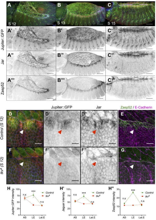
Published January 19, 2015 http://jcb.rupress.org/content/suppl/2015/01/15/jcb.201410042.DC1.html Supplemental Material can be found at:Figure 1. DPP signaling is required for Jupiter, Jar, and Zasp52 LE expression during DC. (A–C) Embryos at stage (S) 12 (A), 13 (B), and 15 (C) display-ing Jupiter::GFP (green; gray in A, B, and C), Jar (red; gray in A, B, and C), and Zasp52 (blue; gray in A, B, and C). Bars, 50 µm. (D–G) Control (D and E) and tkv8 (F and G) stage 12 embryos marked for Jupiter::GFP (green in D and F; gray in D and F), Jar (red in D and F; gray in D and F) and E-Cadherin (blue in D and F), or Zasp52 (green in E and G) and E-Cadherin (magenta in E and G). Bars, 10 µm. (H–H) Plot profile of Jupiter::GFP (n = 8), Jar (n = 8), and Zasp52 (n = 10) intensity in control and tkv8 embryos. AS, amnioserosa; LE, leading edge; Lat.E, lateral epidermis. (Two-way ANOVA and Bonferroni post-hoc test: ***, P < 0.001.) Accumulation of Jupiter::GFP, Jar, and Zasp52 at the LE is lost in tkv embryos (arrowheads). Error bars
are means ± SEM.
on February 11, 2015
jcb.rupress.org
241 A JnK-DPP FFl ensures dorsal closure robustness • Ducuing et al.
mechanistic level, we provide evidence that derepression by the
transcription factor Brk is sufficient to mediate DPP input. We
show that the DPP/Brk indirect branch of the FFL does not pat
tern the LE but can filter unwanted JNK signaling so that the
developmental JNK input remains preserved. Interestingly, al
though the DPP/Brk indirect branch of the FFL is dispensable
for DC at 25°C, it is critical at 32°C. We propose that DPP func
tion during DC is to ensure the robust interpretation of the posi
tional information provided by JNK. By being wired into the
FFL, DPP signaling acts as a filter rather than a positional signal
and fosters the canalization of morphogenesis.
Results
DPP is required for Jupiter, Jaguar (Jar), and Zasp52 accumulation at the LE
We first analyzed three markers that display a strong accumulation
at the LE during DC: the myosin VI homologue Jar (Kellerman
and Miller, 1992), the microtubule binding molecule Jupiter
(Morin et al., 2001; Karpova et al., 2006), and Zasp52, which
promotes integrinmediated adhesion (Morin et al., 2001; Jani
and Schöck, 2007). To determine whether DPP signaling is re
quired for their accumulation, we analyzed these three markers
in embryos mutant for the DPP receptor thick veins (tkv) at
stage 12, during which morphological defects are not yet de
tected. We observed that the LE accumulation of all three mark
ers is lost in tkv mutant embryos compared with controls (Fig. 1,
D–G; see Fig. 1, H–H for quantifications). Therefore, LE ac
cumulation of all three targets requires DPP activity.
We next wondered how DPP mediates its effect on the
markers. Indeed, DPP is known to induce two classes of targets
that are both repressed by brinker (brk). Upon DPP action, Brk
is transcriptionally repressed (Ja
źwińska et al., 1999), leading
to the induction of the first set of targets. The expression of the
second set, however, requires the concomitant activation by the
SMAD family of transcriptional activators (Affolter and Basler,
2007). Interestingly, loss of Brk is sufficient to rescue DC in
the absence of pathway activation, suggesting that the DPP tar
gets required for DC are expressed upon Brk derepression only
(Marty et al., 2000). We hence tested whether removing Brk ac
tivity in the absence of DPP activation rescues Jar, Jupiter, and
Zasp52 expression at the LE. To do so, we generated embryos
double mutant for brk and tkv, to simultaneously disable DPP
activation and prevent repression by Brk (
Fig. S1 A
). In these
embryos, Jar, Jupiter, and Zasp52 expression is restored to wild
type (Fig. 2, A–F). In addition, brk overexpression represses
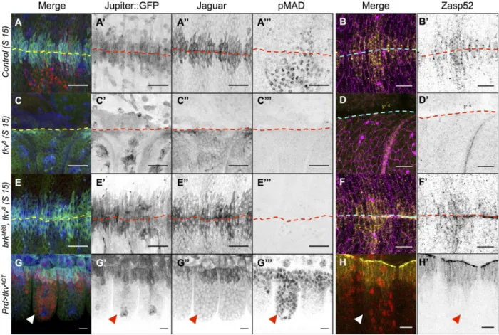
Figure 2. DPP is required to derepress Jupiter, Jar, and Zasp52 but cannot induce them ectopically. (A–F) Control (A and B), tkv8 (C and D), and brkM68,
tkv8 (E and F) stage (S) 15 embryos marked for Jupiter::GFP (blue in A, C, and E; gray in A, C, and E) Jar (green in A, C, and E; gray in A, C, and E), phospho-Mad (pMad; red in in A, C, and E; gray in A, C, and E), Zasp52 (yellow in B, D, and F; gray in B, D, and F), and E-Cadherin (magenta). The dashed lines delineate the midline. Accumulation of Jupiter::GFP, Jar, and Zasp52 at the LE is lost in tkv8 mutant embryos and restored in brkM68, tkv8 embryos. (J and K) Prd-Gal4, UAS-tkvACT embryos marked for Jupiter::GFP (blue in G; gray in G), Jar (green in G; gray in G), phospho-Mad (red in G; gray in G), or Zasp52::GFP (yellow in H; gray in H) and phospho-Mad (red). Ectopic activation of the DPP pathway does not lead to Jupiter, Jar, or Zasp52 accumulation (arrowheads). Bars, 10 µm.
on February 11, 2015
jcb.rupress.org
Downloaded from
brk
embryos (Fig. 2, E–F; and Fig. S1, C–H). In addition,
the phosphoMad pattern is broader than the Jupiter, Jar, and
Zasp52 pattern, suggesting that, instead of delineating the
boundaries of the expression of these targets, DPP may fulfill
a function different from its wellestablished patterning ac
tivity (Fig. 2, G and H; Dorfman and Shilo, 2001). We fur
ther confirmed that ectopic activation of the DPP pathway in
paired
stripes fails to induce these targets outside the LE, indi
cating that DPP does not define the boundary of the expression
patterns of the three markers during DC (Fig. 2, G–H). What
then, is the factor that limits their expression pattern, and what
is the biological significance of DPP control of Jar, Jupiter,
and Zasp52?
the three markers (Fig. S1, B–B). We conclude that repression
of brk alone is sufficient for the accumulation of Jar, Jupiter, and
Zasp52 at the LE.
DPP does not delineate Jupiter, Jar, and Zasp52 expression pattern
DPP is the best example of a secreted morphogen, a factor that
patterns gene expression in a concentrationdependent manner
(Nellen et al., 1996). In the wing imaginal disc, Brk activ
ity dictates the boundaries of the DPP targets Salm and Omb,
whose expression patterns expand in brk
clones (Ja
źwińska
et al., 1999). In contrast, at the LE, the expression patterns
of Jar, Jupiter, and Zasp52 remain unchanged in tkv brk or
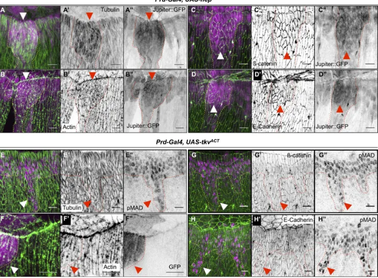
Figure 3. JNK and DPP form a coherent FFL that regu-lates cell differentiation. (A) Experimental design. The wild-type (WT) cell (black rectangle) secretes DPP (red dots) that induces its pathway in all cells (red nuclei). The absence of target (green) in the Prd>BskDN cell abutting the wild-type cell indicates the presence of a JNK/DPP FFL. (B–C) Prd-Gal4, UAS-bskDN, Dpp-lacZ embryos marked for Jupiter::GFP (green in B; gray in B) or Zasp52::GFP (green in C; gray in C), phospho-Mad (red in B and C; gray in B and C), and lacZ (blue in B and C; gray in B and C). The brackets indicate the
BskDN domain, where DPP-lacZ (blue) is off. Anti–phos-pho-Mad (red) indicates that all cells receive DPP. Ju-piter (B) and Zasp52 (C) in green are excluded from the BskDN territory, even though DPP signaling is active (arrowheads), indicating that JNK acts also in parallel of DPP. (D–D) Prd-Gal4, UAS-bskDN, Dpp-lacZ embryos marked for Jupiter::GFP (green in D; gray in D) Jar (red in D; gray in D) and lacZ (blue in D; gray in D). (E) Prd-Gal4, UAS-bskDN, Dpp-lacZ embryos marked for Zasp52::GFP and lacZ. All the markers are lost in the entire BskDN territory (brackets in B–D or dotted lines
in E). (F) Prd-Gal4, UAS-hepACT, Dpp-lacZ, Jupiter::GFP embryos marked for Jupiter::GFP (green in F; gray in F), Jar (red in F; gray in F), and lacZ (blue in F; gray in F). (G–H) Prd-Gal4, UAS-hepACT, Dpp-lacZ embryos marked for lacZ (magenta in G and H; gray in G) and Zasp52 (green in G; gray in G) or Zasp52::GFP (green in H). Ectopic JNK activity (dotted lines) induces Jar, Jupiter, and Zasp52 accumulation (arrowheads). Bars, 10 µm.
on February 11, 2015
jcb.rupress.org
243 A JnK-DPP FFl ensures dorsal closure robustness • Ducuing et al.
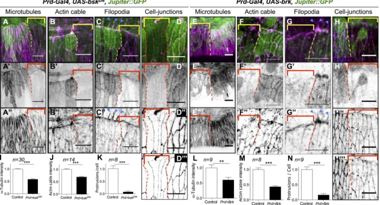
by using bsk
DN(Fig. 4, A–D) or DPP input by overexpress
ing brk (Fig. 4, E–H) and analyzed microtubule polarization,
actomyosin cable, filopodia formation, and junctional integrity.
Impairing either JNK or DPP signal affects the hallmarks of
LE cell differentiation: First, microtubules fail to polarize and
to accumulate (Fig. 4, A and E). Second, filopodia and the
actomyosin cable are absent (Fig. 4, B, C, F, and G). Last,
both ECadherin and catenin expression are reduced, indicat
ing weaker adhesion (Fig. 4, D, D, H, and H; see Fig. 4,
I–N for quantifications). We conclude that both branches of
the FFL are absolutely required for LE cell differentiation and
morphogenesis.
A prediction of this model is that ectopic JNK, but
not ectopic DPP, should redirect lateral cells to the LE cell
identity and path of differentiation. We tested this prediction
by inducing either JNK activity or DPP signaling in stripes
(Fig. 5, A–D and E–H, respectively). As expected for an
FFL, ectopic JNK induces ectopic accumulation of micro
tubules (Fig. 5, A–A) and actin (Fig. 5, B–B) as well as
ECadherin and catenin (Fig. 5, C–D). Conversely, ecto
pic activation of the DPP pathway has no effect on microtu
bules, actin, ECadherin, or catenin accumulation (Fig. 5,
E–H). Altogether, these data indicate that we identified a
novel FFL that plays a pivotal role in LE cells specification
and differentiation.
JNK and DPP are wired into a coherent FFL that controls LE cell differentiation
JNK acts upstream of DPP and determines LE identity (Glise
and Noselli, 1997; Hou et al., 1997; Kockel et al., 1997; Riesgo
Escovar and Hafen, 1997). To test whether JNK activates the tar
gets in parallel to DPP, we expressed a dominantnegative form
of the JNK homologue basket (bsk) in paired stripes so that cells
in the paired domain are deficient for JNK signaling but still re
ceive DPP from their wildtype neighbors by diffusion (Fig. 3 A).
We reasoned that if the expression of the markers does not re
quire JNK activity in parallel to DPP, the markers should remain
expressed in the cells in which JNK is affected as long as they
receive DPP. We found that DPP produced by the neighboring
cells efficiently induces Mad phosphorylation in the paired do
main, yet the targets are not expressed (Fig. 3, B–E). Therefore,
JNK acts both upstream and in parallel to DPP to control Jar,
Jupiter, and Zasp52. To confirm that JNK directs the pattern of Jar,
Jupiter, and Zasp52, we induced ectopic JNK signaling in paired
stripes and used DPPlacZ as a reporter of JNK activity. All the
cells in which DPPlacZ is induced also express Jar, Jupiter, and
Zasp52 (Fig. 3, F–H). These observations indicate that JNK and
DPP form a coherent FFL, in which JNK induces DPP, and both
signals are absolutely required for target gene expression.
We next asked whether the FFL controls LE cell differen
tiation. We selectively inactivated in paired stripes, either JNK
Figure 4. Cytoskeletal components crucial for DC are also regulated by the JNK/DPP FFL. (A–H) Prd-Gal4, UAS-bskDN, Jupiter::GFP embryos (A–D) and
Prd-Gal4, UAS-brk, Jupiter::GFP (E–H) marked for Jupiter::GFP (green in all panels; gray in A–H), -tubulin (magenta in A and E; gray in A and E) or
actin (magenta in B, C, F, and G; gray in B, C, F, and G), or -catenin (red in D and H; gray in D and H) and E-Cadherin (blue in D and H; gray in D and H). In all panels, the BskDN or the Brk overexpression territory is marked by the absence of Jupiter::GFP (brackets), and the border between the
wild-type and the BskDN or Brk overexpression territory is delineated by the dotted lines. (I–N) Quantification of microtubule intensity, actin cable intensity,
and filopodia numbers. Error bars: ±SEM (for all panels, Mann–Whitney’s U test: **, P < 0.01; ***, P < 0.001). bskDN or brk overexpression affects mi-crotubules, -catenin, and DE-Cadherin accumulation as well as actin cable formation at the LE and filopodia (arrowheads in C and G). Bars, 10 µm.
on February 11, 2015
jcb.rupress.org
Downloaded from
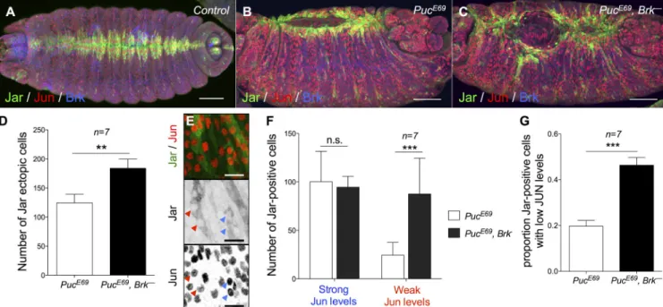
activation throughout the lateral epidermis, suggesting the pres
ence of nonuniform, ectopic JNK signal that varies in strength
(MartínBlanco et al., 1998). To test whether the FFL can fil
ter the ectopic JNK signal in puc embryos, we generated puc
brk
double mutants and found that the ectopic Jar expression
and the morphological defects are magnified compared with
puc
single mutants, suggesting that more cells respond errone
ously to the action of the unwanted JNK signal when the FFL
is disabled (Fig. 6, A–D). A critical aspect of the FFL is that the
filtering ability depends on the delay between the activation of
the direct and the indirect branch: any signal shorter than the
delay is filtered out. We reasoned that the uneven JNK activity
pattern reflects signal duration and could provide us with a nice
system to test whether transient and robust JNK inputs are dis
criminated by the FFL: weak Jun staining corresponds to short
accumulation of Jun and reveals transient signaling; strong Jun
staining corresponds to an accumulation of Jun synthesis over
The JNK/DPP FFL can filter unwantedJNK signaling
FFLs can act as filters of short bursts of signaling (Milo et al.,
2002; Mangan and Alon, 2003), which are random noises that
make biological processes error prone if unchecked. In this par
adigm, signaling robustness is achieved in that the synchrony
between the two branches of the FFL is absolutely required
for a response to occur. If the direct signal switches off before
the indirect signal fires, no response can be elicited. We rea
soned that in the JNK/DPP FFL, brkmediated repression is
the sentinel that prevents unwanted JNK activity from speci
fying ectopic LE identity. To test this hypothesis, we needed
to first produce a source of ectopic JNK signal that is non
uniform and subsequently verify whether the FFL can indeed
filter out such unwanted JNK activity to canalize LE identity.
A previous study and our observations indicate that puc mu
tant embryos display a saltandpepper pattern of ectopic JNK
Figure 5. Ectopic JNK but not ectopic DPP activity leads to accumulation of cytoskeletal components crucial for DC. (A–D) Prd-Gal4, UAS-hepACT, Jupiter::
GFP embryos marked for Jupiter::GFP (magenta in A–D; gray in A–D) and -tubulin (green in A; gray in A) or actin (green in B; gray in B), -catenin
(green in C; gray in C), or DE-Cadherin (green in D; gray in D). In all panels, the ectopic JNK activity is marked by the ectopic accumulation of Jupiter:: GFP (arrowheads) and is delineated by dotted lines. Ectopic JNK signaling leads to accumulation of microtubules, -catenin, DE-Cadherin, and actin. (E–E) Prd-Gal4, UAS-tkvACT embryo stained for phospho-Mad (magenta in E; gray in E) and -tubulin (green in E; gray in E). (F–F) Prd-Gal4, UAS-tkvACT,
UAS-GFP embryos marked for GFP (magenta in F; gray in F) and actin (green in F; gray in F). (G–H) Prd-Gal4, UAS-tkvACT embryos stained for phospho-Mad (pphospho-Mad; magenta in G and H; gray in G and H) and -catenin (green in G; gray in G) or E-Cadherin (green in H; gray in H). In all panels, the ectopic DPP activity is marked by either ectopic phospho-Mad nuclei or the presence of GFP (arrowheads) and is delineated by dotted lines. Ectopic DPP signaling activity does not lead to any accumulation of microtubules, -catenin, E-Cadherin, or actin. Bars, 10 µm.
on February 11, 2015
jcb.rupress.org
245 A JnK-DPP FFl ensures dorsal closure robustness • Ducuing et al.
Discussion
We present a novel mechanism that weaves two classic signal
ing pathways into an FFL to canalize morphogenesis. This FFL
is coherent as both JNK and DPP act positively and belong to
the “and” type, as either signal alone does not trigger a response.
Both experimental and computational evidence indicate that the
general function of the indirect branch of a coherent FFL is to
filter the input received by the direct branch (Mangan and Alon,
2003). Here, we find that during DC, patterning information is
given by JNK, and the DPP/Brk branch filters this spatial infor
mation. In the presence of ectopic JNK generated by puckered
loss of function, Brk filters out unwanted JNK signaling in two
thirds of the cells displaying weak, but not strong, JNK activa
tion. This is a prediction of the FFL model in which the network
filters out only short bursts of signal and not longer, more robust
signaling events. Interestingly, under normal laboratory condi
tions, at 25°C, Brk activity is not required for DC to proceed
normally; LE markers are patterned correctly, and the dynamics
of DC are nearly wildtype. Conversely, when embryos are sub
jected to thermal stress, at 32°C, Brk becomes critical to prevent
the presence of ectopic LE cells in the lateral epidermis and to en
sure proper closure dynamics. These observations provide strong
evidence to support that DPP function during DC is to provide
robustness to the system: under difficult conditions, phenotypic
variation remains minimal, and cell identity remains canalized.
miRNAs are major players in the canalization of cell
decisions in the face of environmental challenges (Posadas
and Carthew, 2014): mir7 stabilizes gene expression and al
lows the correct determination of sensory organs in flies sub
jected to temperature fluctuations (Li et al., 2009). miRNAs are
time and indicates robust signaling. We therefore compared Jar
induction in cells displaying robust and weak Jun staining: al
though Brk activity does not modify Jar induction by robust
ectopic JNK signaling, a cell that receives weak JNK signaling
is 2.5 times more likely to wrongfully express Jar in a brk
mutant (Fig. 6, E–G). We conclude that the FFL buffers weak
ectopic JNK signaling to prevent the ectopic differentiation of
lateral cells into LE cells.
The JNK/DPP FFL canalizes DC
Having confirmed that the FFL filters unwanted JNK noise,
we sought to test whether the indirect branch of the FFL cana
lizes morphogenesis in the presence of environmental pertur
bations. We compared how wildtype or FFLdeficient (brk
)
embryos cope with thermal stress, a classical assay for robust
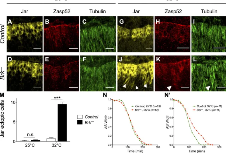
ness in Drosophila (Perry et al., 2010). At 25°C, brk mutants
show wildtype Jar and Zasp52 expression and microtubule
accumulation (Fig. 7, A–F). In contrast, brk mutants raised at
32°C display cells that ectopically express Jar and Zasp52 and
accumulate microtubules, indicating that they differentiate into
LE cells erroneously (Fig. 7, G–M; and
Fig. S2, A–M
). There
fore, brk canalizes LE specification by counteracting the delete
rious effects of environmental stress. Next, we quantified DC
dynamics in brk mutants at 32°C. Although closure speed is
undistinguishable between wildtype and brk embryos at 25°C,
a 1h delay is recorded in brk at 32°C compared with wild type
(Fig. 7, N and N;
Fig. S3
; and
Videos 1 and 2
). Hence, brk
activity renders embryonic morphogenesis more resilient to en
vironmental challenge. Altogether, our data indicate that during
DC, the DPPmediated FFL canalizes LE identity to foster DC
robustness (Fig. 8).
Figure 6. The JNK/DPP FFL filters weak ectopic JNK activity. (A–C) Control (A), PucE69 (B), and PucE69, brkM68 (C) stage 15 embryos stained for Jar, Jun, and Brk. Bars, 50 µm. (D) Quantification of Jar ectopic cells in the lateral epidermis. (n = 7; Mann–Whitney’s U test: **, P < 0.01.) Error bars: ±SEM. (E) Close-up of the lateral epidermis of a PucE69 embryo showing weak (red arrowheads) or strong (blue arrowheads) Jun expression. Bars, 10 µm. (F and G) Quantification of Jar expression in cells expressing low or high Jun levels in PucE69 versus PucE69, brkM68 embryos. (F: two-way ANOVA and Bonferroni post-hoc test: ***, P < 0.001; G: Mann–Whitney’s U test: ***, P < 0.001.) Error bars: ±SEM. Brk represses Jar in about two thirds of the cells displaying weak Jun expression.
on February 11, 2015
jcb.rupress.org
Downloaded from
prediction is that DPPmediated FFL filters JNK inputs that are
on a long time scale: DPP would not only filter out JNK noise
but could also filter out authentic JNK signaling that is impor
tant for nonpatterning functions. JNK is the main messenger
of stress, and mechanisms must exist to distinguish stress
related and developmentrelated JNK inputs within a given cell.
This would explain why brk mutants close normally in favor
able conditions. Environmental perturbations such as tempera
ture excess are bound to have pleiotropic effects on biological
systems. The FFL appears as the generic remedy to enforce
robustness at several levels. Factors acting at specific kinetics
form the indirect branches of FFLs adapted to specific needs:
miRNAs cancel noise, and DPP ensures the proper interpreta
tion of JNK signaling.
DPP is one of the main architects of fly development and
as such fulfills many functions during embryogenesis: DPP
specifies dorsal tissues, including the amnioserosa early and the
dorsal epidermis at midembryogenesis (Ferguson and Anderson,
1992; Xu et al., 2005) and also directs dorsal tracheal migration
(Vincent et al., 1997). At stage 5, DPP induces zerknüllt, and
both DPP and Zerknüllt control the amnioserosaspecific gene
Race
, thus forming a coherent FFL (Xu et al., 2005). In addition,
posttranscriptional regulators that produce moderate but rapid
effects on gene expression. This rapid action appears to have
favored their recruitment into network motifs dedicated to tune
gene expression in a prompt manner: a transcription factor controls
the miRNA and both together control a common target, form
ing an FFL. The major difference between miRNA and DPP
mediated FFL is the time scale: compared with the swiftacting
miRNAs, DPP needs to be translated, secreted, reach a threshold
to activate its pathway, to finally repress brk transcription. The
Figure 7. The JNK/DPP FFL canalizes LE specification and fosters DC robustness. (A–L) Control (top) and brkM68 (bottom) embryos at 25°C (left) or 32°C (right) marked for Jar (yellow), Zasp52 (red), and Tubulin (green). Ectopic Jar, Zasp52, and microtubule accumulations are detected only in brkM68 embryos at 32°C (arrowheads). Bars, 10 µm. (M) Quantification of Jar ectopic cells in control and brkM68 embryos at 25°C or 32°C. Only brkM68 embryos at 32°C exhibit Jar ectopic cells. n ≥ 7. Two-way ANOVA and Bonferroni post-hoc test: ***, P < 0.001. (N and N) Width of the dorsal opening measured over time of control and brkM68 embryos imaged at 25°C or 32°C. Only brkM68 embryos at 32°C exhibit slower closure dynamics.
Figure 8. Model of JNK and DPP wiring during DC. JNK and DPP form a coherent FFL that ensures a canalized and robust DC.
on February 11, 2015
jcb.rupress.org
247 A JnK-DPP FFl ensures dorsal closure robustness • Ducuing et al.
Phalloidin staining
Embryos were dechorionated with bleach and fixed in a 1:1 mix of 4% PFA–heptane. After PFA removal, embryos were stuck on double-sided tape, immerged in 0.1% Triton X-100 and PBS with Rhodamine Phalloidin (1:500; Sigma-Aldrich), and hand devitellinized with a needle. Devitellinized em-bryos were quickly rinsed twice with 0.1% Triton X-100 and PBS and mounted in Vectashield.
Image processing
Images were acquired on the acousto-optical beam splitter confocal laser-scanning microscope (SP5; Leica) with the following objectives: HC Plan Fluotar 20×, 0.5 multi-immersion (numerical aperture: 0.7), HCX Plan Apo-chromat 40× 1.25–0.75 oil (numerical aperture: 1.25), and HCX Plan Apochromat 63× 1.4–0.6 oil (numerical aperture: 1.4) using the acquisi-tion software LAS AF (Leica) at the PLATIM imaging facility and analyzed with ImageJ (National Institutes of Health). Unless otherwise indicated, all images are projections of confocal sections.
Live imaging
Unless otherwise indicated, all crosses were performed at 25°C. Stage 10 or 11 embryos were staged and aligned in Halocarbon oil 27 (Sigma-Aldrich) and then imaged at 25°C or 32°C with a spinning disk (Leica), with a 20× dry objective (numerical aperture: 0.4) and a camera (iXon3; Andor Technology) using the acquisition software MetaMorph (Molecular Devices). brkM68/FM7 females were crossed with Jupiter::GFP males. In addition, wild-type females were crossed with Jupiter::GFP males as con-trols. Brk mutant embryos were identified by the absence of spontaneous movements at stage 17 and confirmed by the absence of hatching. For every sample, the length and width over time were normalized with the maximal length or maximal width, respectively.
Quantification and statistical analyses
We used the Prism software (GraphPad Software) to generate graphs. For Figs. 1, 4, 6, and 7 M, bar graphs represent means ± SEM. For Figs. 7 (N and N) and S4, graphs represent the mean. Mann–Whitney’s U test was used to determine significant differences for Figs. 4 and 6 (D and G). For Figs. 1 (H–H), 6 F, and 7 M, we used a two-way analysis of vari-ance (ANOVA) followed by a Bonferroni post-hoc test. **, P < 0.01; ***, P < 0.001.
Online supplemental material
Fig. S1 describes the experimental strategy used to determine whether the three targets belong to the derepressed only or to the derepressed and in-duced class of DPP targets as well as the effects of the overexpression and the loss of function on the targets’ expression. Fig. S2 reports the effects of temperature on brk mutants. Fig. S3 displays the analysis of the dynam-ics of DCs in brk mutants at 25°C and 32°C. Video 1 is a live recording of the closure of embryos representative of the controls and brk mutants we analyzed at 25°C. Video 2 is a live recording of the closure of em-bryos representative of the controls and brk mutants we analyzed at 32°C. Online supplemental material is available at http://www.jcb.org/cgi/ content/full/jcb.201410042/DC1.
We thank the DROSO-TOOLS and PLATIM facilities of the UMS3444 and Bloomington and the Developmental Studies Hybridoma Bank for reagents. We thank Dali MA for the critical reading of this manuscript and Markus Affolter, Uri Alon, and Arezki Boudaoud for discussions.
This work was supported by a Chair from the Centre National de la Recherche Scientifique to S. Vincent.
The authors declare no competing financial interests.
Submitted: 10 October 2014 Accepted: 9 December 2014
References
Affolter, M., and K. Basler. 2007. The Decapentaplegic morphogen gradient: from pattern formation to growth regulation. Nat. Rev. Genet. 8:663–674. http://dx.doi.org/10.1038/nrg2166
Affolter, M., D. Nellen, U. Nussbaumer, and K. Basler. 1994. Multiple re quirements for the receptor serine/threonine kinase thick veins reveal novel functions of TGF homologs during Drosophila embryogenesis.
Development. 120:3105–3117.
Beira, J.V., A. Springhorn, S. Gunther, L. Hufnagel, G. Pyrowolakis, and J.P. Vincent. 2014. The Dpp/TGFdependent corepressor schnurri protects
DPP also controls the spatial distribution of targets such as Ush
aped
, in both the dorsal epidermis and the amnioserosa (Lada
et al., 2012). This regulation is important for the interaction
between these two tissues that is critical for DC. Recently, a
study reported how DPP can protect from JNKinduced apopto
sis in the dorsal epidermis (Beira et al., 2014). They show that
the DPP pathway repressor Schnurri directly represses the pro
apoptotic gene reaper. Therefore, JNK fails to induce reaper
expression or apoptosis in the pannier domain. This indicates
that JNK and DPP signaling pathways are reiteratively inte
grated during Drosophila embryogenesis. To get a full picture
of this network, we will also need to integrate the two negative
feedback loops mediated by Puc and scarface that dampen JNK
activity (MartínBlanco et al., 1998; Rousset et al., 2010). A
likely possibility is that these feedback loops improve fidelity in
signaling. Altogether, the dorsal epidermis provides an elegant
model system to understand how different inputs are integrated
to modulate cell decisions during development. Although some
of these functions are paramount to cell specification, we show
that some, such as the JNK/DPP FFL, can also counteract delete
rious environmental stimuli and canalize development, a func
tion distinct from DPP wellestablished, non–cellautonomous
patterning activity.
Materials and methods
Fly strains and genetics
We used the following lines: Canton-S (wild type), tkv8 (amorphic allele; Bloomington Stock Center [BL] 34509), BrkM68 (loss-of-function allele, see Jaźwińska et al., 1999), gift from M. Affolter (University of Basel, Basel, Switzerland), PucE69 (loss-of-function allele, see Martín-Blanco et al., 1998),
Prd-Gal4 (BL 1947), upstream activation sequence (UAS)–tkvACT (BL 36537), gift from M. Grammont (Université de Lyon, Lyon, France), UAS-bskDN (BL 6409), UAS-hepACT (BL 9306), UAS-brk (brk coding sequence under the control of a promoter containing UAS sequence), gift from J. de Celis (Centro de Biología Molecular “Severo Ochoa,” Madrid, Spain), UAS-GFPNLS (BL 4776), Jupiter::GFP (GFP knock-in; BL 6836), Zasp52::GFP (GFP knock-in; BL 6838), and DPP-lacZNUCLEAR (lacZ-NLS coding sequence cloned after the BS 3.0 promoter of DPP, see Blackman et al., 1991). Unless otherwise indicated, all crosses were performed at 25°C.
Immunofluorescence and quantification
We used standard techniques of immunohistofluorescence as described in Ducuing et al. (2013). Embryos were dechorionated with bleach, fixed in a 1:1 mix of 4% PFA–heptane. Embryos were subsequently devitellinized by replacing the 4% PFA with methanol. Samples were incubated with pri-mary antibodies, with fluorescent-coupled secondary antibodies and mounted in Vectashield.
We used the following primary antibodies: rabbit anti-lacZ (1:1,000; Cappel), mouse anti-lacZ (1:250; G4644; Sigma-Aldrich), guinea pig anti-Brk (1:500; gift from G. Morata, Centro de Biología Molecular “Severo Ochoa,” Madrid, Spain), mouse anti-Jar 3C7 (1:100; Kellerman and Miller, 1992), rabbit anti-pMad (1:1,500; gift from P. ten Dijke, Leids Universi-tair Medisch, Leiden, Netherlands), rat anti–DE-Cadherin (1:333; Devel-opmental Studies Hybridoma Bank [DSHB]), mouse anti-Armadillo (1:250; DSHB), mouse anti–-tubulin (1:1,000; T6199; Sigma-Aldrich), rabbit anti-Jun (1:10; Santa Cruz Biotechnology, Inc.), and rabbit anti-Zasp52 (1:400; gift from F. Schöck, McGill university, Montreal, Quebec ). For Brk, pMad, Jar, and Zasp52, antigen was a full-length protein. Second-ary antibodies are from Invitrogen and were used at 1:500. We used the following secondary antibodies: Alexa Fluor donkey anti–mouse 488, Alexa Fluor goat anti–mouse 633, Alexa Fluor goat anti–rat 546, Alexa Fluor donkey anti–rabbit 488, Alexa Fluor goat anti–rabbit 546, and Alexa Fluor goat anti–guinea pig 488. For 32°C experiments, embryos where first grown at 25°C and then shifted for 4 h at 32°C and immedi-ately fixed after.
on February 11, 2015
jcb.rupress.org
Downloaded from
during development. Cell. 137:273–282. http://dx.doi.org/10.1016/j.cell .2009.01.058
Mangan, S., and U. Alon. 2003. Structure and function of the feedforward loop network motif. Proc. Natl. Acad. Sci. USA. 100:11980–11985. http://dx .doi.org/10.1073/pnas.2133841100
Martin, P., and S.M. Parkhurst. 2004. Parallels between tissue repair and em bryo morphogenesis. Development. 131:3021–3034. http://dx.doi.org/ 10.1242/dev.01253
MartínBlanco, E., A. Gampel, J. Ring, K. Virdee, N. Kirov, A.M. Tolkovsky, and A. MartinezArias. 1998. puckered encodes a phosphatase that mediates a feedback loop regulating JNK activity during dorsal closure in Drosophila.
Genes Dev. 12:557–570. http://dx.doi.org/10.1101/gad.12.4.557 Marty, T., B. Müller, K. Basler, and M. Affolter. 2000. Schnurri mediates Dpp
dependent repression of brinker transcription. Nat. Cell Biol. 2:745–749. http://dx.doi.org/10.1038/35036383
Millard, T.H., and P. Martin. 2008. Dynamic analysis of filopodial interactions during the zippering phase of Drosophila dorsal closure. Development. 135:621–626. http://dx.doi.org/10.1242/dev.014001
Milo, R., S. ShenOrr, S. Itzkovitz, N. Kashtan, D. Chklovskii, and U. Alon. 2002. Network motifs: simple building blocks of complex networks.
Science. 298:824–827. http://dx.doi.org/10.1126/science.298.5594.824 Morin, X., R. Daneman, M. Zavortink, and W. Chia. 2001. A protein trap strat
egy to detect GFPtagged proteins expressed from their endogenous loci in Drosophila. Proc. Natl. Acad. Sci. USA. 98:15050–15055. http:// dx.doi.org/10.1073/pnas.261408198
Nellen, D., R. Burke, G. Struhl, and K. Basler. 1996. Direct and longrange action of a DPP morphogen gradient. Cell. 85:357–368. http://dx.doi .org/10.1016/S00928674(00)811149
Paaby, A.B., and M.V. Rockman. 2014. Cryptic genetic variation: evolution’s hidden substrate. Nat. Rev. Genet. 15:247–258. http://dx.doi.org/10.1038 /nrg3688
Perry, M.W., A.N. Boettiger, J.P. Bothma, and M. Levine. 2010. Shadow en hancers foster robustness of Drosophila gastrulation. Curr. Biol. 20: 1562–1567. http://dx.doi.org/10.1016/j.cub.2010.07.043
Posadas, D.M., and R.W. Carthew. 2014. MicroRNAs and their roles in devel opmental canalization. Curr. Opin. Genet. Dev. 27:1–6. http://dx.doi.org/ 10.1016/j.gde.2014.03.005
RiesgoEscovar, J.R., and E. Hafen. 1997. Drosophila Jun kinase regulates expression of decapentaplegic via the ETSdomain protein Aop and the AP1 transcription factor DJun during dorsal closure. Genes Dev. 11:1717–1727. http://dx.doi.org/10.1101/gad.11.13.1717
RíosBarrera, L.D., and J.R. RiesgoEscovar. 2013. Regulating cell morphogen esis: the Drosophila Jun Nterminal kinase pathway. Genesis. 51:147– 162. http://dx.doi.org/10.1002/dvg.22354
Rohner, N., D.F. Jarosz, J.E. Kowalko, M. Yoshizawa, W.R. Jeffery, R.L. Borowsky, S. Lindquist, and C.J. Tabin. 2013. Cryptic variation in mor phological evolution: HSP90 as a capacitor for loss of eyes in cavefish.
Science. 342:1372–1375. http://dx.doi.org/10.1126/science.1240276 Rousset, R., S. BonoLauriol, M. Gettings, M. Suzanne, P. Spéder, and S. Noselli.
2010. The Drosophila serine protease homologue Scarface regulates JNK signalling in a negativefeedback loop during epithelial morphogenesis.
Development. 137:2177–2186. http://dx.doi.org/10.1242/dev.050781 Rutherford, S.L., and S. Lindquist. 1998. Hsp90 as a capacitor for morphological
evolution. Nature. 396:336–342. http://dx.doi.org/10.1038/24550 Solon, J., A. KayaCopur, J. Colombelli, and D. Brunner. 2009. Pulsed forces
timed by a ratchetlike mechanism drive directed tissue movement dur ing dorsal closure. Cell. 137:1331–1342. http://dx.doi.org/10.1016/j.cell .2009.03.050
Tsuda, L., R. Nagaraj, S.L. Zipursky, and U. Banerjee. 2002. An EGFR/Ebi/ Sno pathway promotes delta expression by inactivating Su(H)/SMRTER repression during inductive notch signaling. Cell. 110:625–637. http:// dx.doi.org/10.1016/S00928674(02)008759
Vincent, S., E. Ruberte, N.C. Grieder, C.K. Chen, T. Haerry, R. Schuh, and M. Affolter. 1997. DPP controls tracheal cell migration along the dorsoven tral body axis of the Drosophila embryo. Development. 124:2741–2750. Waddington, C.H. 1959. Canalization of development and genetic assimila
tion of acquired characters. Nature. 183:1654–1655. http://dx.doi.org/ 10.1038/1831654a0
Xu, M., N. Kirov, and C. Rushlow. 2005. Peak levels of BMP in the Drosophila embryo control target genes by a feedforward mechanism. Development. 132:1637–1647. http://dx.doi.org/10.1242/dev.01722
Zecca, M., and G. Struhl. 2007. Recruitment of cells into the Drosophila wing primordium by a feedforward circuit of vestigial autoregulation. Devel
opment. 134:3001–3010. http://dx.doi.org/10.1242/dev.006411 epithelial cells from JNKinduced apoptosis in Drosophila embryos. Dev.
Cell. 31:240–247. http://dx.doi.org/10.1016/j.devcel.2014.08.015 Belacortu, Y., and N. Paricio. 2011. Drosophila as a model of wound healing
and tissue regeneration in vertebrates. Dev. Dyn. 240:2379–2404. http:// dx.doi.org/10.1002/dvdy.22753
Blackman, R.K., M. Sanicola, L.A. Raftery, T. Gillevet, and W.M. Gelbart. 1991. An extensive 3 cisregulatory region directs the imaginal disk expres sion of decapentaplegic, a member of the TGF family in Drosophila.
Development. 111:657–666.
Dorfman, R., and B.Z. Shilo. 2001. Biphasic activation of the BMP pathway patterns the Drosophila embryonic dorsal region. Development. 128: 965–972.
Ducuing, A., B. Mollereau, J.D. Axelrod, and S. Vincent. 2013. Absolute require ment of cholesterol binding for Hedgehog gradient formation in Drosophila.
Biol. Open. 2:596–604. http://dx.doi.org/10.1242/bio.20134952 Ferguson, E.L., and K.V. Anderson. 1992. Decapentaplegic acts as a morpho
gen to organize dorsalventral pattern in the Drosophila embryo. Cell. 71:451–461. http://dx.doi.org/10.1016/00928674(92)90514D Fernández, B.G., A.M. Arias, and A. Jacinto. 2007. Dpp signalling orches
trates dorsal closure by regulating cell shape changes both in the am nioserosa and in the epidermis. Mech. Dev. 124:884–897. http://dx.doi .org/10.1016/j.mod.2007.09.002
Glise, B., and S. Noselli. 1997. Coupling of Jun aminoterminal kinase and Decapentaplegic signaling pathways in Drosophila morphogenesis.
Genes Dev. 11:1738–1747. http://dx.doi.org/10.1101/gad.11.13.1738 Glise, B., H. Bourbon, and S. Noselli. 1995. hemipterous encodes a novel
Drosophila MAP kinase kinase, required for epithelial cell sheet movement.
Cell. 83:451–461. http://dx.doi.org/10.1016/00928674(95)90123X Hou, X.S., E.S. Goldstein, and N. Perrimon. 1997. Drosophila Jun relays the Jun
aminoterminal kinase signal transduction pathway to the Decapentaplegic signal transduction pathway in regulating epithelial cell sheet movement.
Genes Dev. 11:1728–1737. http://dx.doi.org/10.1101/gad.11.13.1728 Hutson, M.S., Y. Tokutake, M.S. Chang, J.W. Bloor, S. Venakides, D.P. Kiehart,
and G.S. Edwards. 2003. Forces for morphogenesis investigated with laser microsurgery and quantitative modeling. Science. 300:145–149. http://dx.doi.org/10.1126/science.1079552
Jacinto, A., W. Wood, T. Balayo, M. Turmaine, A. MartinezArias, and P. Martin. 2000. Dynamic actinbased epithelial adhesion and cell match ing during Drosophila dorsal closure. Curr. Biol. 10:1420–1426. http:// dx.doi.org/10.1016/S09609822(00)00796X
Jacinto, A., W. Wood, S. Woolner, C. Hiley, L. Turner, C. Wilson, A. Martinez Arias, and P. Martin. 2002. Dynamic analysis of actin cable function dur ing Drosophila dorsal closure. Curr. Biol. 12:1245–1250. http://dx.doi .org/10.1016/S09609822(02)009557
Jani, K., and F. Schöck. 2007. Zasp is required for the assembly of functional integrin adhesion sites. J. Cell Biol. 179:1583–1597. http://dx.doi.org/ 10.1083/jcb.200707045
Jankovics, F., and D. Brunner. 2006. Transiently reorganized microtubules are essential for zippering during dorsal closure in Drosophila melanogaster.
Dev. Cell. 11:375–385. http://dx.doi.org/10.1016/j.devcel.2006.07.014 Jaźwińska, A., N. Kirov, E. Wieschaus, S. Roth, and C. Rushlow. 1999. The
Drosophila gene brinker reveals a novel mechanism of Dpp target gene regulation. Cell. 96:563–573. http://dx.doi.org/10.1016/S00928674 (00)806601
Kaltschmidt, J.A., N. Lawrence, V. Morel, T. Balayo, B.G. Fernández, A. Pelissier, A. Jacinto, and A. Martinez Arias. 2002. Planar polarity and actin dynamics in the epidermis of Drosophila. Nat. Cell Biol. 4:937– 944. http://dx.doi.org/10.1038/ncb882
Karpova, N., Y. Bobinnec, S. Fouix, P. Huitorel, and A. Debec. 2006. Jupiter, a new Drosophila protein associated with microtubules. Cell Motil.
Cytoskeleton. 63:301–312. http://dx.doi.org/10.1002/cm.20124 Kellerman, K.A., and K.G. Miller. 1992. An unconventional myosin heavy chain
gene from Drosophila melanogaster. J. Cell Biol. 119:823–834. http:// dx.doi.org/10.1083/jcb.119.4.823
Kiehart, D.P., C.G. Galbraith, K.A. Edwards, W.L. Rickoll, and R.A. Montague. 2000. Multiple forces contribute to cell sheet morphogenesis for dor sal closure in Drosophila. J. Cell Biol. 149:471–490. http://dx.doi.org/ 10.1083/jcb.149.2.471
Kockel, L., J. Zeitlinger, L.M. Staszewski, M. Mlodzik, and D. Bohmann. 1997. Jun in Drosophila development: redundant and nonredundant functions and regulation by two MAPK signal transduction pathways. Genes Dev. 11:1748–1758. http://dx.doi.org/10.1101/gad.11.13.1748
Lada, K., N. Gorfinkiel, and A. Martinez Arias. 2012. Interactions between the amnioserosa and the epidermis revealed by the function of the ushaped gene. Biol. Open. 1:353–361. http://dx.doi.org/10.1242/bio.2012497 Li, X., J.J. Cassidy, C.A. Reinke, S. Fischboeck, and R.W. Carthew. 2009.
A microRNA imparts robustness against environmental fluctuation