E2F6 in Axial Skeletal Development and Gliosis
by
Laurie Beth Friesenhahn
B.S., Texas A&M University (2002)
Submitted to the Department of Biology
in partial fulfillment of the requirements for the degree of
Doctor of Philosophy at the
Massachusetts Institute of Technology July 2008 MASSACHUSETTS INSTITUTE MASSACHUSETTS INSTITUT7I-OF TECH•0LOGY
SEP 0 9
2008
LIBRARIES
ARCHIVES
© 2008 Massachusetts Institute of Technology. All Rights ReservedSignature of Author_
(/
Laurie B. Friesenhahn Department of BiologyjI
A
Certified by Jacqueline A. Lees Thesis Supervisor Accepted by Stephen P. Bell Graduate Committee ChairmanABSTRACT
E2F transcription factors were originally identified as regulators of cell cycle and cellular proliferation. In vivo mouse models have uncovered novel roles for these proteins in different developmental processes. This dissertation examines the biological role of E2F6 in mammalian development. E2F6 functions as a repressor of transcription in concert with the polycomb group (PcG) proteins and chromatin modifiers. PcG proteins regulate processes required for proper embryonic development and differentiation.
E2F6 interacts with core components of Polycomb Repressive Complex 1 (PRC1) and participates in PcG-mediated repression of Hox genes. Hox genes are required for
correct patterning of the mammalian skeleton. Loss of E2f6 results in posterior axial
skeletal transformations. Mice deficient for both E2f6 and Bmil, a component of PRC1, exhibit increased penetrance of axial skeletal transformations. Thus, E2F6 and Bmil cooperate in the regulation of Hox genes and axial skeletal development. Bmi 1 also represses transcription of the Ink4a-Arf locus, and it is consequently required to maintain the proliferative and self-renewal properties of hematopoietic and neural stem cells.
However, E2F6 does not participate in the repression of the Ink4a-Arflocus. These findings underscore the significance of the E2F6-Bmil interaction in vivo and suggest that the Hox and Ink4a-Arfloci are regulated by somewhat different mechanisms.
In addition to axial skeletal transformations, E2f6' mice exhibit a suppressed gliotic response after neural injury. Gliosis occurs in response to neurodegeneration, ischemia, and neuronal cell death. This process provides neuronal protection by
restricting inflammation and regulating the concentration of molecules in the extracellular environment. However, gliosis has potentially detrimental effects such as the inhibition of axonal regeneration or the release of cytotoxic agents that trigger degeneration of neighboring neurons. The molecular mechanisms required to initiate and sustain a gliotic response are poorly understood. Gliosis is the focus of therapy for neurodegenerative diseases and ischemia, and complete understanding of the mechanisms underlying this process will lead to more effective therapies for neurodegenerative disease and ischemia.
ACKNOWLEDGEMENTS
I would like to thank my advisor, Jackie Lees, for her helpful advice and direction throughout the years. I would also like to thank the members of the Lees lab who have created a collaborative and inspiring atmosphere that has truly made my experience at MIT memorable. Additionally, I would like to express my gratitude to past and present members of my thesis committee, Phil Sharp, David Housmann, Mike Yaffee, Phil Hinds, and Laurie Boyer for the time and thought they have contributed to my graduate work.
I am especially indebted to my parents, Lawrence and Kathy. Without their confidence, love, and support, I would never have been as successful as I am. Finally I would like to thank my boyfriend for the companionship and love we have shared for the past four years. I am also grateful for his help in editing my thesis.
Table of Contents
Chapter 1: Introduction 6
I. The E2F Family of Transcription Factors 7
II. E2Fs in Development and Disease 19
III. Regulation of Hox Genes by Polycomb Group Proteins 25
IV. Polycomb Group Proteins in Development and Disease 31
V. Reactive Gliosis 43
Bibliography 47
Chapter 2: E2f6 and Bmi 1 cooperate in axial skeletal development 67
Abstract 68 Introduction 69 Results 72 Discussion 87 Experimental Procedures 90 Bibliography 94
Chapter 3: E2f6 loss suppresses gliosis after neuronal injury 99
Abstract 100 Introduction 101 Results 104 Discussion 110 Experimental Procedures 111 Bibliography 113 Chapter 4: Discussion 116
1. E2F6 is not involved in the regulation of Ink4a-Arf 118
2. E2F6 and Bmil co-regulate Hox genes and synergize in axial skeletal 119 development
3. Regulation of Hox genes and chromatin by PcG proteins 120
4. E2F6 in the regulation of gliosis 122
5. Possible molecular mechanisms of E2F6's role in gliosis 125
6. Concluding Remarks 127
Bibliography 128
Appendix A: E2F3a and E2F3b make overlapping but different contributions
to total E2F3 activity 134
Abstract 135
Introduction 136
Results 138
Discussion 154
Bibliography 166
Supplemental Figure 1 169
Supplemental Figure 2 170
Supplemental Figure 3 171
Appendix B: A role for RYBP in chromatin condensation 172
Introduction 173
Results 174
Discussion 181
Chapter 1
I. The E2F Family of Transcription Factors
The E2F transcription factors are classically described as key regulators of the cell cycle and cellular proliferation. The cellular targets of E2F include cell-cycle regulators; components of the DNA replication machinery; proteins involved in chromatin
modification, assembly, condensation, segregation; and nucleotide biosynthesis (some classic E2F target genes are listed in Table 1). It is now clear that E2F also plays
important roles in development and disease. Many studies have linked the amplification of E2Fs to various cancers (Oeggerli et al., 2006; Orlic et al., 2006; Hurst et al., 2007). Further research has reported a requirement for different E2Fs in developmental
processes such as heart development (Cloud et al., 2002), axial skeletal development (Courel et al., 2008), and embryonic development (Li et al., 2008).
1. Discovery and cloning of E2F
E2F was first identified in research based upon the observation that the
adenovirus E1A (early region LA) protein stimulates the transcription of several viral and cellular promoters (Nevins, 1981). When Kovesdi et al. infected cells with E1A, they discovered that the induction of transcription by E1A was dependent upon a cellular factor, E2F (E2 factor) (Kovesdi et al., 1986). The induction was determined to be independent of protein synthesis (Reichel et al., 1988). Subsequent research found that ElA induces transcription by dissociating cellular E2F complexes. This dissociation releases free E2F and activates E2F's transcriptional activity (Bagchi et al., 1990). Yee et
Cell Cycle Cdc2 Cdc25 E2fl Cdk2 P107 pRB CycE CycA B-myb C-myc CycD Replication PolA Orcl 1 Mcm Cdc6 PCNA TK1 TOP2A DHFR
Table 1: Classic E2F Target Genes
and DNA mutagenesis studies revealed two distinct E2F binding sites that were required for transcription of the E2 protein (Yee et al., 1987). The discovery of an E2F binding site allowed purification of E2F from crude extracts by chromatography and DNA purification schemes. This purification yielded a 54kD protein that could bind to the E2F consensus site and stimulate transcription in an in vitro reporter assay (Yee et al., 1989).
8 Cell Division Cycle 2 Cell Division Cycle25
E2 promoter binding factor 1 Cyclin dependent kinase 2 Retinoblastoma-associated protein homolog 107 Retinoblastoma-associated protein Cyclin E Cyclin A Cyclin D DNA polymerase a
Origin recognition complex 1 Minichromosome maintenance
Cell division cycle 6
Proliferation cell nuclear antigen Thymidine Kinase
Topoisomerase 2 a Dihydrofolate reductase
References
(Tommasi and Pfeifer, 1995)
(Stevaux and Dyson, 2002) (DeGregori et al., 1995a; Li
et al., 2008)
(Ren et al., 2002; Stevaux and Dyson, 2002)
(Zhu et al., 1995) (Ren et al., 2002)
(Botz et al., 1996) (Ohtani
et al., 1995; Geng et al.,
1996)
(Schulze et al., 1995) (Zwicker et al., 1997) (DeGregori et al., 1995a) (DeGregori et al., 1995a) (DeGregori et al., 1995a) (Ohtani et al., 1996) (Ren et al., 2002) (Lavia and Jansen-Durr, 1999)
(DeGregori et al., 1995a) (DeGregori et al., 1995a) (Ren et al., 2002)
(Means et al., 1992; Wade
et al., 1992; Lavia and
In addition to E2Fs known role in the activation of viral genes, many scientists hypothesized that E2F may have a similar cellular function. Indeed, many genes involved in proliferation contain an E2F binding site, and E2F binding activity increases upon serum stimulation of cells (Mudryj et al., 1990). This observation was the first step to realizing E2F' s larger role in the control of proliferation and the cell cycle.
An important breakthrough in the elucidation of the mechanism of E2F's transcriptional transactivation was the discovery that E2F is a cellular target of the retinoblastoma protein, pRB (Chellappan et al., 1991; Chittenden et al., 1991; Kaelin et
al., 1991). pRB was known to control cell cycle and proliferation, but the mechanisms
were unknown (Chittenden et al., 1991; Kaelin et al., 1991). pRB and two related proteins, p107 and p 130, are a family of transcription factors known as the pocket proteins. The Rb-E2F interaction was the basis for cloning E2F. Helin and colleagues screened a X expression library for cDNAs whose protein products had the ability to bind the pocket region of pRB. They characterized a cDNA and its protein product that
interacts with pRB, binds to E2F recognition sequences, and transactivates the E2 promoter. Additionally, E1A disrupted the binding of this cDNA's protein and pRB (Helin et al., 1992). This cDNA was ultimately determined to be the coding sequence of E2F. The cloning of E2F was a milestone because it led to the discovery of an entire family of E2F transcription factors that have the ability to either activate or repress transcription. The E2F transcription factors work in concert with the pocket proteins to control the entry and progression through the cell cycle.
2. The E2F/RB pathway
The pocket proteins are named based on their pocket-like structure. This pocket is made of two sub-domains separated by a spacer (Ewen et al., 1991). Although the pocket proteins have a similar structure, it is clear that they have different affinities for different E2Fs. The activating E2Fs (E2F1, E2F2, and E2F3) associate almost exclusively with pRB, and the repressive E2Fs (E2F4 and E2F5) associate mainly with p107 and p130. pRB is often referred to as the restriction point switch of the cell cycle. Indeed, the phosphorylation of pRB is a major determinant of whether the cell commits to a cell cycle or re-enters a resting state (Go) (Bartek et al., 1996). A cell's ability to surpass the restriction point when conditions for cell division are unfavorable is the basis for many cancers. For this reason, it is important to know the factors and signaling pathways that regulate this critical point in the cell cycle.
A resting cell in Go has low levels of pRB, low levels of the activating E2Fs, high levels of cyclin dependent kinase inhibitors (CDKIs), and high levels of p130 (Weinberg,
1995; Dyson, 1998). The predominant complexes are p130-E2F4 and p130/p107-E2F5 (Dyson, 1998). This concerted gene expression pattern results in suppression of S-phase and proliferation genes by two mechanisms. First, repressive pocket protein-E2F
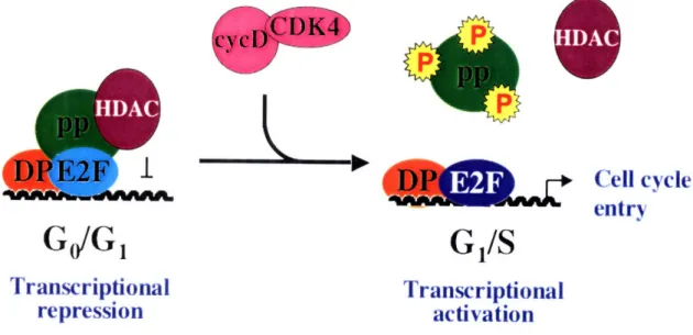
complexes allow for active repression by recruitment of chromatin modifiers and histone deacetylases that actively repress transcription. Second, pRB complexed with activating E2Fs renders them functionally inactive and leads to a loss of transcriptional activation of these genes (Figure 1) (Dyson, 1998; Trimarchi and Lees, 2002). The high levels of CDKIs also suppress cell cycle progression by inhibiting the activity of any cyclin/cdk complex that may be present (Dyson, 1998; Trimarchi and Lees, 2002).
WanE2FI
SCell
cycle
entry
G
/S
Transcriptional
repression
Transcriptional
activation
Figure 1: Transcriptional repression and activation by E2F.
E2F target genes are regulated in GdG, by a repressive complex consisting of repressive E2Fs/DP, histone deacetylases (HDAC), and pocket proteins (pp). Upon mitogenic stimulation, cycD/CDK complexes phosphorylate the pocket proteins. This
phosphorlylation leads to the disruption of pocket protein-E2F complexes, causing the activating E2Fs to bind the promoters of cell cycle regulated target genes and activate their transcription.
I
,
HDAC
Upon mitogenic stimulation, expression of cyclin Dl is induced (Matsushime et
al., 1991) and expression of CDKIs are reduced (Polyak et al., 1994). This gene
expression pattern shifts the CDKI-to-cyclin ratio and relieves inactivation of the cyclins by the CDKIs (Weinberg, 1995). Cyclin D forms an active complex with cdk4 or cdk6 and then phosphorylates pRB. Phosphorylation of pRB causes the dissociation of pRB complexes, and the activating E2Fs are now free to stimulate transcription of E2F-target genes (Figure 1) (Weinberg, 1995; Dyson, 1998; Mittnacht, 1998). This first wave
of pRB phosphorylation and E2F transcriptional activity leads to increased transcription of Cyclin E, an E2F-target gene. Cyclin E and cdk2 form an active complex that hyperphosphorylates pRB, p107, and p130 (Weinberg, 1995; Dyson, 1998; Mittnacht, 1998). This results in disassembly of E2F-pocket protein complexes, a process that is essential for entry into phase. Free, activating E2Fs now initiate transcription of S-phase genes (Weinberg, 1995; Dyson, 1998; Mittnacht, 1998; Trimarchi and Lees, 2002). Conversely, E2F4 and E2F5 lack a nuclear localization signal, and dissociation from the pocket proteins results in their translocation from the nucleus and loss of transcriptional repression of S-phase genes (Muller et al., 1997; Chestukhin et al., 2002). Activation of gene expression, control of cyclin-cdk activity, and regulation of pRB phosphorylation are critical for a cell's entry and committal to cell division.
3. Regulation, structural identity, and transcriptional activity of different E2Fs
The E2F transcription factors are essential for coordinated control of cell division and proliferation. For this reason, it is not surprising that deregulation of E2F is the hallmark of many cancers (Sherr, 1996). Understanding the regulation of E2F and the
mechanisms by which E2F confers control on the cell cycle is essential to studies in cancer and other diseases.
To date, nine E2F and two DP (DRTF1 1olypeptide) proteins have been identified. DP is the heterodimerization partner of E2F transcription factors, and it is required for the DNA binding activity of E2Fs 1-6 (Bandara et al., 1993; Huber et al.,
1993). The E2F-DP complex creates a basic helix-loop-helix transcription factor that recognizes the consensus DNA sequence 5'-TTTCGCGC-3' (Zheng et al., 1999; Trimarchi and Lees, 2002). E2Fs 1-8 can be divided into subclasses based on their structure, expression pattern, ability to bind pocket proteins, and transcriptional activity (Figure 2). The E2Fs 1-6 share two structural domains: a DNA binding domain and a dimerization domain (Trimarchi and Lees, 2002). The dimerization domain is necessary for heterodimerization with a DP (DRTF1 polypeptide) subunit (Zheng et al., 1999). This interaction is required for high affinity binding to E2F consensus sites (Slansky and Farnham, 1996). E2F7 and E2F8, the newest members of the E2F family, have two conserved DNA binding domains but lack a dimerization domain. Instead of
heterodimerizing with DP, E2F7 and E2F8 can heterodimerize or homodimerize with themselves or with each other (Li et al., 2008). Exogenous expression of E2F7 or E2F8 can repress E2F target genes and block cell proliferation (de Bruin et al., 2003; Di
Stefano et al., 2003; Maiti et al., 2005).
Similar to E2F7 and E2F8, E2Fs 4-6 are also active repressors of transcription of E2F-target genes (Muller et al., 1997; Verona et al., 1997; Trimarchi et al., 1998;
Giangrande et al., 2004). E2F4, E2F5, and E2F6 have all been shown to interact with histone deacetylases, histone methylases, and other chromatin remodeling enzymes
('Cvelin A Binding Iomain I I ITransactivation I I
E IZ
I
I
L
"'Y
DNA Binding
Marked Box
Dimeriz ation
Pocket Protein
Binding
Figure 2: The E2F family of transcription factors.
The E2F transcription factors all contain a homologous DNA-binding region. The activating E2Fs, E2Fs 1-3, each contain a nuclear localization signal (NLS), a marked box, a dimerization region, and a pocket protein-binding region. The repressive E2Fs, E2Fs 4-5, lack a NLS but have a nuclear export signal (NES). E2Fs 6-8 lack sequences required for pocket protein binding and repress transcription through mechanisms independent of the pocket proteins.
E2FI
E2F2
E2 F3a
I
EL
]
E2F3b
E2F4
E2F5
E2F6
E2F7
E2F8
Kz
1
]
z
El
LI
LI
I
NES
I
NLS
1
-I
I
associated with repressed chromatin (lavarone and Massague, 1999; Ogawa et al., 2002; Attwooll et al., 2005). However, it is clear that E2F4 and E2F5 repress transcription in a mechanism that is different from E2F6. E2F4 and E2F5 contain a pocket protein binding domain and repress transcription through a pocket protein-E2F complex. Interaction of E2F4-DP and E2F5-DP is also induces the nuclear localization of these complexes (Trimarchi and Lees, 2002). E2F6 lacks a pocket protein binding domain and represses transcription through its association with the polycomb group family of transcription factors (discussed in more detail in following sections) (Trimarchi and Lees, 2002). E2F4 and E2F5 are present throughout the cell cycle (Slansky and Farnham, 1996), but they accumulate in quiescent cells (Ikeda et al., 1996). In contrast, E2F6 levels increase as cells enter the cell cycle and peak at mid Gl (Dahme et al., 2002). While the mechanisms of repression and expression patterns of these two sub-groups of E2Fs differ, their
specific targets are overlapping, and it has been shown that E2F4 or E2F6 can compensate for the loss of the other (Giangrande et al., 2004).
The last group of E2F transcription factors includes E2Fs 1-3. As with E2F4 and E2F5, these E2Fs also have a pocket protein binding domain, a dimerization domain, and a DNA binding domain (Helin, 1998; DeGregori, 2002; Trimarchi and Lees, 2002) (Dyson, 1998). However, E2Fs 1-3 are potent activators of transcription and their expression patterns are transcriptionally regulated throughout the cell cycle (Trimarchi and Lees, 2002). The transcription of these E2Fs is induced in late G , and the gene products accumulate at this time (Slansky and Farnham, 1996). E2Fs 4-5 require the binding of a pocket protein to actively repress E2F-target genes. This repression is achieved through interactions with pocket proteins and histone deacetylases. However,
E2Fs 1-3 must be dissociated from a pocket protein before they become transcriptionally active (Sun et al., 2007). Overexpression of the activating E2Fs can induce quiescent cells to re-enter the cell cycle and can override growth-arrest signals such as TGF(3 or cyclin dependent kinase inhibitors such as p16, p21, or p27 (DeGregori et al., 1995a;
DeGregori et al., 1995b) (Schwarz et al., 1995; Mann and Jones, 1996). It is clear that E2Fs 1-3 are potent activators of the cell cycle, and overexpression of an activating E2F can induce transformation of primary cells (Johnson et al., 1994; Singh et al., 1994; Xu et
al., 1995). Similarly, loss of E2F1, E2F2, and E2F3 leads to a decrease in E2F-target
gene expression and a block in cell proliferation (Wu et al., 2001).
Together, the E2Fs and pocket proteins are critical for a cell's entry and committal to cell division when conditions are favorable. The manner in which a cell responds to unfavorable signals and aborts the cell division program is equally important. When a cell undergoes DNA damage or oncogenic stress, a different program ensures that a damaged or potentially cancerous cell does not undergo division. At the backbone of this control are pl9ARF and p53. These proteins are the basis of the ARF-p53 tumor surveillance network.
4. The Arf-p53 tumor surveillance network
pl 9ARF is one of the proteins encoded in the INK4A-ARF locus. The other protein,
p161NK4
a, is a CDKI and inhibits cyclin/cdk complexes and subsequently the G,/S transition (Sherr and Roberts, 1999). pl19ARF is a tumor suppressor that responds to
abnormal proliferative or oncogenic signals. Its activation leads to cell cycle arrest or apoptosis (Figure 3) (de Stanchina et al., 1998; Palmero et al., 1998; Radfar et al., 1998). pl9
repressor of p53 (Zhang et al., 1998). Upon activation of p53 by p19ARF, p53 increases the levels of p21 (a CDKI) or activates Bax proteins. This leads to growth arrest or apoptosis (Figure 3) (Lundberg and Weinberg, 1999).
The deregulation of INK4a-ARF or the E2F-RB pathway occurs in a significant fraction of cancers. This suggests that circumvention of this pathway leads to cellular transformation. Research of the complex mechanisms that control whether or not a cell divides is critical to understanding the process of tumorigenesis.
Ras c-Mye
oncogenic
stress
E2FARF
DNA damage
Mdm2
-L
p
5 3Cell cycle
arrest
Apoptosis
Figure 3: The Arf-p53 tumor surveillance pathway.
pl 9ARF is activated by abnormal proliferative or oncogenic signals and is repressed in
normal proliferating cells by a polycomb group protein, Bmi 1. Activation of this pathway results in cell cycle arrest or apoptosis.
Bmil
F•... C• 1.----_,___
II. E2Fs in Development and Disease
The E2F transcription factors are key regulators of the cell cycle. However, in
vivo studies of the E2Fs in mouse models have uncovered roles for these proteins beyond
cell cycle control and proliferation. E2Fsl-8 have been deleted in mice, and analysis of these mice has revealed overlapping and distinct roles for E2Fs in development,
differentiation, and disease.
1. The activating E2Fs: E2F1, E2F2, and E2F3
The activating E2Fs are critical for cellular proliferation. Deletion of E2fsl-3 results in severely impaired proliferation and cell cycle kinetics in cultured cells (Wu et
al., 2001). Disruption of activating E2Fs in mouse models reveals specific and
overlapping functions for these E2Fs. E2fl mutant mice are fully viable but exhibit testicular atrophy, exocrine abnormalities, and a spectrum of tissue-specific tumors (Yamasaki et al., 1996). Interestingly, overexpression of E2fl in mice leads to tumor development as well, suggesting that E2F1 can act as a tumor suppressor and an
oncogene in vivo (Pierce et al., 1999). In contrast to E2fl mutant mice, E2f3 mutant mice exhibit embryonic lethality in a pure genetic background and are born at one quarter the expected frequency in a mixed genetic background. Although E2f3 mutant mice do not develop tumors, they exhibit severe growth retardation and die prematurely from congestive heart failure (Cloud et al., 2002). Analysis of E2fl;E2f3 compound mutant mice revealed overlapping roles for these proteins. These mice had increased embryonic lethality, growth retardation, and increased severity in testicular atrophy and congestive heart failure. However, the compound mutant mice had no alteration in tumor incidence, timing, or spectrum (Cloud et al., 2002). Recently, E2f3a and E2f3b-specific mutant mice
have been developed. These mice are viable, and they have no histological abnormalities. This suggests that E2f3a and E2f3b have overlapping roles in vivo. E2fl ;E2f3b mutant mice are viable, and they have no tissue defects. E2fl;E2f3a mutant mice die perinatally and exhibit a defect in cartilage development. (Danielian, Friesenhahn et. al, in press; Appendix A). These studies reveal distinct roles for E2F1 and E2F3 in vivo.
E2J2 mutant mice, like E2fl, are born at the expected frequency. However, they
die prematurely from systemic autoimmune disease caused by enhanced T lymphocyte proliferation (Murga et al., 2001) and hematopoietic defects (Li et al., 2003). E2fl;E2f2 mutant mice are viable, but diabetes causes a premature death. These mice exhibit a reduction of size and cellularity of lymphoid organs, the testes, and salivary gland (Iglesias et al., 2004). In this case, the resulting diabetes is caused by the combination of the defect in hematopoiesis (caused primarily from E2J2 deletion) and exocrine
degradation (caused primarily by the E2fl deletion) (Li et al., 2003). E2F and E2F2 have distinct roles in vivo, but combinatorial loss of these two genes results in a novel disease phenotype.
2. The repressing E2Fs: E2F4 and E2F5
E2F4 and E2F5 play similar roles in the repression of cell cycle genes. However,
in vivo studies revealed that these proteins have distinct roles in different developmental
programs. Mutation of E2f4 in mice uncovered a novel role for E2F4 in the control of erythopoiesis. E2f4-' mice exhibited severe anemia, low red blood cell numbers, macrocytosis, and Howell-Jolly bodies (Humbert et al., 2000). While the mature red blood cells were abnormal, there was no defect detected in the immature erythrocytes,
mice have abnormal craniofacial development, abnormal development of the gut
epithelium, neonatal lethality, and growth retardation starting as early as E13.5 (Humbert
et al., 2000; Rempel et al., 2000). Neither the erythroid defect nor the resulting anemia is
the cause of the neonatal lethality. Instead, a defect in the development of cilia in the nasal epithelium makes these mice unable to clear infectious agents, which results in increased susceptibility to bacterial infections (Humbert et al., 2000; Danielian et al., 2007). Similar to E2f4 mutant mice, E2f5 mutant mice have normal cell cycle kinetics. These mice appear to develop normally prior to weaning age. After weaning, E2f5 mutant mice developed ataxia, ruffled coats, and dehydration. Most of these mice died at around six weeks of age due to hydrocephalus (excessive cerebrospinal fluid production) and intracerebral hemorrhage. Lindeman et al. subsequently determined that E2F5 is important for the regulation of secretion of cerebrospinal fluid (Lindeman et al., 1998). While E2F4 or E2F5 can compensate for each other in vitro during the regulation of cell cycle genes (Lindeman et al., 1998; Humbert et al., 2000), in vivo studies uncovered novel roles for these proteins in different developmental pathways.
3. E2F6
Although E2F6 is a repressor of transcription, it lacks a pocket protein binding domain and represses transcription through association with polycomb group proteins (Trimarchi and Lees, 2002). E2F6 has been shown to interact biochemically with the polycomb group proteins: EZH2, Bmil, Mel-18, RYBP, Ringla, and Ringlb (Trimarchi
et al., 2001; Ogawa et al., 2002; Attwooll et al., 2005). The polycomb group proteins
(discussed more in-depth in the next section) are repressors of homeobox (Hox) genes, which control the anterior-posterior patterning of the developing embryo. Mice deficient
for E2F6 exhibit posterior transformations of the axial skeletons. These include: (1) a T13-L1 transformation as evidenced by the absence or incomplete development of ribs
normally present on the thirteenth thoracic vertebra; and (2) a L6-S 1 conversion in which
the iliac bones associate with the sixth lumbar vertebra instead of the first sacral vertebra. These mice survive to birth and live a normal life span. In vitro studies did not reveal any cell cycle or proliferation defects (Storre et al., 2002; Courel et al., 2008). Further
analysis of E2f6 mutant mice revealed a defect in spermacyte development, but the defect was not severe enough to affect fertility (Storre et al., 2002). Recently, unpublished data
suggests that E2F6 plays a role in the gliosis response of the brain after neuronal damage (Chapter 3). E2f6-mutant mice exhibit a suppression of gliosis when they have brain damage induced by Bmil-loss or injection of a neurotoxin (Chapter 3), but it is not clear how E2f6 loss suppresses gliosis.
Beyond Hox gene regulation, little is known about which genes E2F6 controls. In an attempt to understand more about the biological role of E2F6, some laboratories have conducted microarrays or chromatin immunoprecipitation followed by CpG island
microarrays (Chip-Chip) to study which genes E2F6 regulates on a genome-wide scale. Oberley et al. used Chip-Chip on human tumor cells and found that E2F6 regulates genes involved in tumor suppression and maintenance of chromatin structure (Oberley et al., 2003). Two other laboratories conducted cDNA microarray experiments with wildtype and E2f6 mutant mouse embryonic fibroblasts (MEFs). These studies revealed that E2F6 is important for the repression of some meiotic (Smclf3 and STAG3) (Storre et al., 2005) and testes-specific (ac-tubulin 3 and 7) genes (Pohlers et al., 2005). Although E2F6 is widely expressed in most tissues, little is known about genes that E2F6 regulates in vivo.
While derepression of the testes-specific genes could explain the defect in spermacyte development, the mechanism by which E2F6 regulates gliosis is still unknown.
4. E2F7 and E2F8
E2F7 and E2F8 are the most recent E2F family members to have been identified. Homologous recombination techniques have been utilized to conditionally mutate each of these genes in mice. E2f7 or E2f8 mutant mice are viable, and they display no
abnormalities. The E2J7;E2j8 compound mutant mice die as early as E11.5. These embryos exhibit vascular defects, multifocal hemorrhages, and widespread apoptosis. RNA analysis from these embryos revealed a substantial increase in the level of E2fl mRNA. In wildtype cells, E2F7 and E2F8 occupy the promoter of E2fl and repress transcription. The elevated level of E2F1 is due to a loss of direct repression by E2F7 and E2F8. The massive apoptosis seen in the compound mutant animals is due to E2Fl
overexpression, p53 accumulation, and activation of the apoptotic response of the cell. Microarray analysis from MEFs made from these embryos indicated a deregulation of genes involved in stress responses to hypoxia, nutrient deprivation, and apoptosis (Li et
al., 2008). It is unclear whether deregulation of these genes is due to a direct loss of E2F7
and E2F8 transcriptional repression or whether it is due to an indirect consequence of the apoptotic program that is activated in these cells. It will be interesting to learn what tissue-specific abnormalities arise in these mice when E2J7 and E2f8 are acutely deleted in different tissues.
5. Specificity of E2Fs in Development
It is clear the E2Fs have distinct and overlapping roles in development. While this is not surprising, it still raises the question of where the specificity arises among different
functional groups of E2Fs. There is some evidence to suggest that specificity arises from the spatial and temporal regulation of individual E2F family members. For example, E2Fs 4 and 5 exhibit disparate embryonic expression patterns. E2F4 is expressed early in proliferating cells and the epithelium, while E2F5 is mainly expressed in terminally differentiating or differentiated cells (Lindeman et al., 1998; Humbert et al., 2000; Rempel et al., 2000). In fact, most E2Fs exhibit tissue and cell-type specific expression patterns during development (Dagnino et al., 1997). This implies that, while E2Fs in the same functional group may be able to compensate for one another, diverse expression patterns may prevent them from doing so in certain tissues. Another theory is that different combinatorial interactions achieve the specificity of function seen in
developmental processes. For example, in a yeast two-hybrid screen for E2F3-specific interacting partners, TFE3 was found to bind specifically to E2F3 but not other activating E2Fs. The E2F3-TFE3 complex synergistically controls transcription of the p68 gene (Giangrande et al., 2003). It is likely that E2F specificity is achieved by a combination of these two mechanisms.
III. Regulation of Hox Genes by Polycomb Group Proteins.
Polycomb group proteins were first characterized in metazoan development as regulators of segment identity. A central question in this field was how embryonic cells first acquired and subsequently maintained unique positional identities. Through genetic analysis and mutational studies in Drosophila, the answer came with the discovery of Polycomb group (PcG) genes and their transcriptional targets, the homeotic genes (Hom). These genes were determined to be important regulators of cellular identity in
Drosophila. The PcG genes and Hom genes (called Hox in vertebrate systems) are highly
conserved in mammals, and these genes play a similar functional role. It is clear that PcG genes have roles beyond their transcriptional regulation of the Hox genes. Mutations of PcG proteins in mammalian systems lead to deregulation of Hox genes and patterning defects as well as other developmental abnormalities.
1. The Regulation of Horn genes in Drosophila
The Drosophila embryo is composed of segmented units, or a repeated pattern of elements along the anterior-posterior axis. This metameric organization is established within the first two hours following fertilization. This process requires coordinated expression of two groups of genes: the segmentation genes and the homeotic genes
(Hom) (Akam, 1987). The segmentation genes are the first detectable zygotically active
genes and are divided into three classes based on their mutated phenotypes. The first group consists of gap genes whose mutations cause multiple adjacent segments to be missing from the embryo (Nusslein-Volhard and Wieschaus, 1980; Qian et al., 1993;
mutations cause alternate segment-size units to be missing (Nusslein-Volhard and
Wieschaus, 1980; Qian et al., 1993). The third type of gene is the segment polarity genes whose mutations lead to a deletion in part of every segment and a replacement of the deleted part with a mirror image of the remaining structure (Nusslein-Volhard and Wieschaus, 1980; Hooper and Scott, 1989; Perrimon and Smouse, 1989). Correct spatial and temporal expression of the Hom genes is required for segment identity in the
developing Drosophila embryo. The segmentation genes serve to initiate a
transcriptionally active (by the pair-rule genes) or repressed (by the gap genes) state of the Hom genes (Scott and Carroll, 1987). Early in embryogenesis (about four hours), expression of the segmentation genes disappear (Akam, 1987), and other regulatory mechanisms are required to maintain the transcriptional program created by these genes.
The polycomb group proteins and trithorax group proteins maintain the
transcriptional state of the Hom genes initiated by the segmentation genes. PcG proteins maintain repression of the Hom genes while trithorax group proteins maintain their activation. Mutations of PcG proteins lead to a derepression of Hom genes and segments that differentiate into structures characteristic of posterior segments (Struhl and Akam, 1985; Simon et al., 1992; Chiang et al., 1995). Ectopic expression or mutation of Hom genes in Drosophila changes the identity of the segment and leads to the formation of structures characteristic of a different segment (Kaufman et al., 1990; Pattatucci and Kaufman, 1991; Pattatucci et al., 1991). Polycomb and trithorax group proteins play a vital role in transcriptional regulation of the homeotic genes in Drosophila during embryonic development.
2. Regulation of Hox genes in mammals.
The Hox genes play a similar role in mammalian development. Hox genes are required for patterning of the body plan and development of the axial skeleton. Similar to
Drosophila, the mammalian axial skeleton develops from metameric units called somites.
During gastrulation, a subpopulation of the mesoderm that resides around the neural tube (paraxial mesoderm) is formed. Somites form from blocks of cells that separate from the paraxial mesoderm in a process called "initial segmentation." The dorsal portion of the somite eventually becomes the dermomyotome. The ventral portion becomes the
sclerotome, which contains stem cells that eventually give rise to the axial skeleton. In a process called "resegmentation," the sclerotome cells segregate into rostral and caudal compartments. The rostral and caudal halves then fuse with their neighbor to form a vertebra (reviewed in (Yamaguchi, 1997; Saga and Takeda, 2001).
Hox genes regulate the patterning of the mammalian axial skeleton. While
Drosophila have only two clusters of Hox genes (Antennapedia complex - ANT-C and
bithorax complex - BX-C), mammals have thirty-nine Hox genes arranged in four
clusters (HoxA, HoxB, HoxC, and HoxD) and thirteen paralogous groups (Hoxl-13). The
expression of Hox genes is described as "temporal and spatial colinearity." This term refers to how the timing of expression during embryonic development correlates with the spatial location of the Hox genes within each cluster. The 3' genes (HoxAl, HoxB1 etc...) are expressed the earliest and are detected at seven days post-conception (dpc) (Dolle et
al., 1989; Izpisua-Belmonte et al., 1991). Mutations in 3' Hox genes exhibit phenotypes
in the anterior region of the axial skeleton, while mutations in 5' Hox genes exhibit phenotypes in the posterior region (Condie and Capecchi, 1993; Davis and Capecchi,
1994). Due to the redundancy within paralogous Hox genes, mutation of any single Hox gene do not affect viability and result in relatively minor phenotypes. Mutations of the
Hox3 through Hoxll genes cause defects in the axial skeleton (Chisaka and Capecchi,
1991; Le Mouellic et al., 1992; Condie and Capecchi, 1993; Jeannotte et al., 1993; Davis and Capecchi, 1994; Rancourt et al., 1995; Suemori et al., 1995; Chen and Capecchi, 1997; van den Akker et al., 1999; van den Akker et al., 2001; McIntyre et al., 2007). Paralogous mutations have been constructed for many of the Hox gene groups. These mutations lead to synergistic and severe axial skeletal phenotypes. For example,
mutations of the Hox9 paralogous group have anterior transformations of the thoracic vertebrae, such that there are thirteen or fourteen ribbed vertebra attached to the sternum instead of the normal seven (McIntyre et al., 2007). Mutation of an entire cluster has a less severe phenotype than paralogous mutations. The exception to this is HoxB, whose mutation results in a severe sternal phenotype (Medina-Martinez et al., 2000). This is probably due to the fact that HoxB contains Hox5-9, all of which are important for the patterning of the sternum. The other Hox clusters have only two to four members of
Hox5-9. It is clear from these studies that coordinated expression of the Hox genes is vital
to proper development of the mammalian embryo. Thus, it is important to understand the mechanisms underlying the regulation of these genes.
Hox gene regulation in mammals is more complex than in Drosophila. Gap or
pair-rule type regulation has not been identified in mammals. Instead, Hox regulation is initiated by a variety of transcription factors, signaling molecules, and polycomb group proteins. During development, FGF (Ciruna and Rossant, 2001; Dubrulle and Pourquie, 2004), WNT (Aulehla et al., 2003; Forlani et al., 2003) and retinoic acid (Boncinelli et
al., 1991; Krumlauf, 1994) gradients are early regulators of Hox genes. These molecules
act by signaling to Cdx genes, which then directly stimulate the transcription of Hox genes in a dosage dependent manner (Subramanian et al., 1995; Pownall et al., 1996; Charite et al., 1998; Isaacs et al., 1998; Houle et al., 2003; Gaunt et al., 2004). Mutations of Cdx genes, hypomorphs of Fgfrl, and hypomorphs of Wnt3 lead to a deregulation of
Hox genes and axial skeletal transformations (Partanen et al., 1998; Ikeya and Takada,
2001; van den Akker et al., 2002). The mechanisms described above serve to initiate transcriptional activation of the Hox genes. Transcriptional repression of these genes early in embryonic development requires the actions of multiprotein polycomb repressive complexes.
3. Polycomb Repressive Complexes
Two core Polycomb repressive complexes (PRCs) have been purified from
Drosophila and Hela cells and were found to have many of the same homologous
proteins. PRC2, which is required for the initiation of Hox gene repression in mammals, contains the proteins EZH2, Suzl2, and EED (Cao et al., 2002; Kuzmichev et al., 2002; Muller et al., 2002). This complex initiates a heritable, repressive state of Hox genes through modifications of the chromatin structure. EZH2, a core component of the PRC2 complex methylates histone H3 at lysine 27 (Cao et al., 2002; Czermin et al., 2002; Kuzmichev et al., 2002; Muller et al., 2002). In some cases, his methyl mark serves to recruit PRC1. However, it has also been shown that PRC1 is recruited in the absense of PRC2 (Schoeftner et al., 2006). The PRC1 core complex contains the proteins Bmil, Ringla, Ringlb, HPH, HPH2, HPC2, HPC3, and Scmhl (Saurin et al., 2001; Levine et
protein, directly binds to methylated histone H3 at lysine 27 through a highly conserved chromodomain (Cao et al., 2002; Muller et al., 2002). PRC2 initiates Hox gene
repression, and PRC1 maintains the repression throughout development. Mice deficient for a component of the PRC2 complex die early in development by 7 dpc (Faust et al., 1995; O'Carroll et al., 2001; Pasini et al., 2004). In contrast, most PRC1 mice exhibit no reduction in viability and display axial skeletal transformations (van der Lugt et al., 1994; Takihara et al., 1997; del Mar Lorente et al., 2000; Tokimasa et al., 2001). The exception to this is Ringlb (Ring2) deficient mice. These mice arrest in gastrulation, and they are
predicted to have a proliferation defect. An upregulation of the Ink4a-Arf locus is
observed, and mice deficient for both Ringlb and Ink4a-Arf exhibit a partial rescue of the early embryonic lethality (Voncken et al., 2003).
IV. Polycomb Group Proteins in Development and Disease.
Polycomb group proteins are required for correct development and patterning of the mammalian embryo. Mutation or misexpression of these genes causes a wide
spectrum of defects. These include early embryonic lethality, axial skeletal
transformations, and a variety of cancers. PcG mutants also exhibit impairment in
hematopoiesis, cell cycle control, senescence, X-inactivation, stem cell maintenance, and differentiation. Described below are some of the key players of polycomb group
complexes and polycomb group proteins that have been found to biochemically interact with E2F6.
1. PRC2: EZH2, EED, Suzl2
The PRC2 complex plays a vital role in the initiation of a developmental program that is required for patterning of the mammalian embryo. The components of the PRC2 complex are required for early embryonic development, and mice deficient for these genes die by seven days post-conception (Faust et al., 1995; O'Carroll et al., 2001; Pasini
et al., 2004). EZH2 is the component of the PRC2 complex that methylates histone H3 at
lysine 27. Loss of EZH2 results in a loss of this methylation mark in early zygotes
(Erhardt et al., 2003) and severely compromises the proliferation of embryonic stem (ES) cells. Attempts to derive ES cells from blastocysts generate non-ES-like cells that
become apoptotic or necrotic (O'Carroll et al., 2001). Alternatively, overexpression of EZH2 increases the proliferative capacity of primary B cells (Visser et al., 2001) and bypasses cellular senescence in MEFs (Kamminga et al., 2006). EZH2 is strongly expressed in highly proliferative, undifferentiated cells (Visser et al., 2001). As cells
differentiate, expression of EZH2 is developmentally downregulated, and overexpression of EZH2 in undifferentiated cells prevents them from differentiating (Caretti et al., 2004). One hypothesis is that EZH2 plays a role in maintaining a cell's "stemness" during development. In support of this hypothesis, overexpression of EZH2 in hematopoietic stem cells prevents stem cell exhaustion when bone marrow is serially transplanted (Kamminga et al., 2006). EZH2 is also found to be overexpressed in a variety of
cancerous cells including lymphoma (Visser et al., 2001), bladder cancer (Arisan et al., 2005), breast carcinomas, prostate cancer (Varambally et al., 2002; Kleer et al., 2003),
and bronchial squamous cell carcinomas (Breuer et al., 2004). The precise mechanism of EZH2's involvement in cancer is unknown. An attractive model is that overexpression of EZH2 may be trapping the cell in a highly proliferative, stem cell-like state. The result is
a block in appropriate differentiation and an induction of abnormal proliferation. EED and Suzl2 also play a role in differentiation and early embryonic
development. Mice with a hypomorph for Eed exhibit posterior transformations along the AP axis (Schumacher et al., 1996) and a deregulation of Hox genes (Wang et al., 2002),
consistent with a role for this protein in Hox gene regulation. Eed homozygous mutant mice display a defect in embryonic ectoderm growth, an absence in axial structures, and early embryonic lethality (Niswander et al., 1988; Faust et al., 1995). Unlike EZH2 mutant ES cells, ES cells from Eed or Suz12 mutant blastocysts are viable, but they lack
methylation of histone H3 at lysine 27 (Montgomery et al., 2005; Boyer et al., 2006; Pasini et al., 2007). Suzl2 plays an essential role in promoting differentiation. ES cells
and embroid bodies deficient for this gene fail to differentiate properly (Pasini et al., 2004; Pasini et al., 2007). Additionally, a Suzl2 point mutation that causes aberrant
mRNA splicing results in an increase in the number of multipotent hematopoietic
progenitors and enhances hematopoietic stem cell activity (Majewski et al., 2008). These results present a requirement for Suzl2 and the PRC2 complex in differentiation of different populations of progenitor cells.
2. Bmil
Bmil was first identified as an oncogene that cooperates with the Ept-myc transgene in B-cell lymphoma (Haupt et al., 1991; van Lohuizen et al., 1991). Bmil is homologous to the Drosophila gene posterior sex combs, (Adler et al., 1991; Brunk et
al., 1991) and it has been found to be a part of the core PRC1 complex involved in
maintenance of gene repression (Lewis, 1978; Paro, 1990; Zink et al., 1991; Saurin et al., 2001; Levine et al., 2002). Bmil is highly expressed progenitor cell populations. Adult tissues have a low level of Bmil RNA with the exception of the thymus, heart, brain, and testes (van Lohuizen et al., 1991). Mice deficient for this protein exhibit a variety of axial skeletal transformations, which is consistent with a role for Bmil in Hox gene repression. The transformations include (van der Lugt et al., 1994; Courel et al., 2008):
(1) E - an extra piece of bone rostral to the first cervical vertebra, C1
(2) C1-C2 conversion in which the second cervical vertebra is transformed to the first cervical vertebra and has an axis-like appearance
(3) C7-T1 conversion evidenced by the presence of ribs on the seventh cervical vertebra, which then fuse with ribs at the first thoracic vertebra or connect directly to the sternum
(5) T13-L1 conversion in which there is an absence or incomplete development of ribs normally present on the thirteenth thoracic vertebra and
(6) L6-S 1 conversion demonstrated by an association of iliac bones with the sixth lumbar vertebra instead of the first sacral vertebra.
In addition to axial skeletal transformations, Bmil-mutant mice display a variety of other
defects, indicating that Bmi 1 has roles beyond the maintenance of repression of the Hox genes.
Bmil-mutant mice are born at expected frequency but are smaller in size and
selectively cannibalized shortly after birth. Approximately 50% survive to adulthood (van der Lugt et al., 1994; Courel et al., 2008). In addition to axial skeletal transformations, these mice also display defects in hematopoiesis, the central nervous system, and the peripheral nervous system (van der Lugt et al., 1994; Jacobs and van Lohuizen, 2002). In
the hematopoietic system, there is a loss of mature T and B cells, hypocellularity of the bone marrow, decreased spleen size, and an involuted thymus (van der Lugt et al., 1994;
Lessard and Sauvageau, 2003). The neurological defects in the Bmil mutant mice include an ataxic gait; seizures; hypocellularity of the molecular and granular layers of the
cerebellum; and astrogliosis in the cortex and cerebellum of the brain (van der Lugt et al.,
1994; Molofsky et al., 2003; Leung et al., 2004; Zencak et al., 2005). The neurological
and hematopoietic defects can be partially attributed to a deficiency in the proliferation and self-renewal capacity of the stem cells in these compartments (Lessard and
Sauvageau, 2003; Park et al., 2003; Molofsky et al., 2005).
The proliferation defects observed in vivo are consistent with a role for the PcG proteins in the control of the cell cycle. Indeed, mouse embryonic fibroblasts (MEFs)
deficient for Bmil have impaired proliferation properties and undergo premature senescence (Core et al., 1997; Jacobs et al., 1999b; Courel et al., 2008). Bmil is a
repressor of the Ink4a-Arf, and the cell cycle defects in the Bmil/' MEFs result from a
derepression of this locus (Jacobs et al., 1999b). Mice mutant for Bmil and Ink4a-Arf have a partial rescue of the neural and hematopoietic defects. BmilP;Ink4a-Arf' mice exhibit a cerebellum that is comparable in size to wildtype mice, and the number of thymocytes and splenocytes is now 50-70% of wildtype levels. The proliferative and
senescent defects of MEFs deficient for Bmil are fully rescued in Bmil'l;Ink4a-Arf' MEFs (Jacobs et al., 1999b). These studies clearly show that Bmil plays a vital role in the INK4A-ARF tumor surveillance pathway. Therefore, it is not surprising that Bmil is deregulated in a variety of cancers. These cancers include: high grade B-cell
Non-Hodgkin lymphomas (Bea et al., 2001), breast carcinomas (Dimri et al., 2002), non-small cell lung cancers (Vonlanthen et al., 2001), medulloblastomas (Leung et al., 2004), and human colorectal cancers (Kim et al., 2004). Recent studies implicate Bmil in the proliferation of bronchiolalveolar stem cells and a requirement for Bmil in lung
tumorigenesis (Dovey et. al, in press). It is clear that Bmil and INK4a-ARF are
important for the maintenance and proliferation of different populations of stem cells, and deregulation of these genes can cause inappropriate proliferation and cancer.
3. Mel-18
Mel-18 is a PcG protein that is 70% identical to Bmil (Tagawa et al., 1990; Goebl, 1991). Like the Bmil mutant mice, Mel-18 mutant mice are growth retarded and
defective in hematopoiesis. These mice also die in a similar time frame as the Bmil
include a defect in B-cell proliferation and maturation, impaired expansion of the most immature T progenitor cells, severe thymic atrophy, and an impairment in the self-renewal and proliferation of the hematopoietic stem cells (Akasaka et al., 1996; Akasaka
et al., 1997; Miyazaki et al., 2005). Mel-18/' mice also exhibit posterior axial skeletal
transformations similar to those found in Bmil deficient mice. These include E, C 1-C2, C7-T1, T13-L1, and L6-S1. These mice also have a C2-C3 transformation marked by the lack of the odontoid process from the C2 vertebra and an S4-Cal transformation in which the fourth sacral vertebra looks like the first caudal vertebra (Akasaka et al., 1996). It is apparent that Bmi 1 and Mel-18 have overlapping functions in vivo. However, there are no indications that Mel-18 is a regulator of the Ink4a-Arf locus or that it plays a role in the proliferation of neural or lung stem cells.
4. Ringl/Ringla and Ring2/Ringlb
Ring 1 a and Ring lb were first characterized on the basis of their interaction with M33 in a yeast two-hybrid screen (Schoorlemmer et al., 1997). The two genes are both found to be in the PRC 1 core complex, and they interact with many other PcG proteins (Satijn et al., 1997; Schoorlemmer et al., 1997; Hemenway et al., 1998; Satijn and Otte, 1999; Levine et al., 2002). Ringla deficient mice are viable, but they display axial skeletal transformations (del Mar Lorente et al., 2000). Interestingly, the skeletal transformations in Ringla mutant mice are anterior, while all other mutant mice for PRC 1 components have posterior transformations (van der Lugt et al., 1994; Akasaka et
al., 1996; Takihara et al., 1997). The exception to this is M33 mutant mice, in which most
C2-Cl (Core et al., 1997; Katoh-Fukui et al., 1998). The anterior axial skeletal transformations of Ringla mutant mice include (del Mar Lorente et al., 2000):
(1) an abnormal Cl and C2
(2) a C2-C1 transformation characterized by a broadening of the neural arch (3) a T3-T2 transformation in which the prominent spinous process normally found on T2 is now present on T3
(4) a T8-T7 transformation marked by the presence of eight vertebrosternal ribs instead of seven and
(5) a L1-T13 transformation evidenced by rudimentary ribs on L1.
Mice overexpressing Ringla display many of the same transformations, including the C2-C1, the T8-T7, and the L1-T13. These mice also have a T10-T9 transformation (dorsal cartilage normally found on T10 is now present on T9) (del Mar Lorente et al., 2000). Expression of Ringla in the embryo is limited to the central nervous system at E8.5. At E13.5 it is found in the central and peripheral nervous system, and at E15.5 it is also found in the thymus and epithelial cell types. In the adult mouse, Ring l a is expressed in
differentiated tissues (Schoorlemmer et al., 1997). The skeletal transformations of Ringla are consistent with its role as a member of PRC 1.
In contrast to Ringla mutant mice, Ringlb mutant embryos arrest early in embryogenesis during gastrulation (Voncken et al., 2003). Ringlb expression is found early in the blastocyst and embryonic stem cells (Voncken et al., 2003). A Ringlb hypomorph mutant mouse is viable, but it displays posterior axial skeletal
(1) a supraoccipital bone-Cl transformation in which there are ectopic bones on the Cl vertebra
(2) a C1-C2 transformation
(3) a C2-C3 transformation in which C2 lacks the odontoid process (4) a C7-T1 transformation
(5) a T1-T2 transformation in which the prominent spinous process of T2 is now present on T1
(6) a T7-T8 transformation (7) a T13-L1 transformation (8) a L6-S 1 transformation and (9) a S4-Cal transformation.
These skeletal transformations were accompanied by a deregulation of Hox genes (Suzuki et al., 2002). The diversity in the phenotypes and viability of the Ringla and
Ringlb mutant mice suggests that these two highly related proteins have different functions in vivo.
Little was known about the mechanism of Ringla and Ringlb until de Napoles et
al. found that these proteins have catalytic E3 ubiquitin ligase activity towards histone
H2A at lysine 119. Ring lb maintains global H2A ubiquitination in ES cells, and both Ringla and Ringlb can ubiquitinate histone H2A on the inactive-X chromosome (de Napoles et al., 2004; Cao et al., 2005). The big question was whether the ubiquitin chromatin mark contributes to repression or whether it has another function. To answer this question, Cao et al. found evidence that this chromatin mark participated in
27 methylation (Cao et al., 2005). Recent evidence suggests the ubiquitin mark of histone H3 interferes with the processivity of the RNA polymerase. This provides a direct
mechanism by which histone H3 ubiquitination represses transcription by interfering with the RNA polymerase (Stock et al., 2007).
5. RYBP
RYBP (Ring l and YY 1 Binding Protein) was cloned as an interactor with Ring la. RYBP also interacts with many other PcG proteins and E2Fs 2, 3, and 6 (Garcia et al.,
1999; Schlisio et al., 2002). Recent published and unpublished results provide insight into the mechanism in which RYBP may contribute to polycomb silencing and X-inactivation. RYBP contains an Np14 zinc finger (NZF) and binds to ubiquitinated
histone H2A in vivo (Arrigoni et al., 2006). Additionally, RBYP co-localizes with Ringla at the inactive-X chromosome (Arrigoni et al., 2006). Experiments from Professor Jackie Lees's laboratory identified Suv4-20 in a yeast two-hybrid assay as a possible interactor of RYBP. This protein, similar to EZH, contains a SET domain and can tri-methylate histone H4 at lysine 20 (Schotta et al., 2004). This methyl mark, which is downstream of the methylation of histone H3 lysine 27, is found at pericentric heterochromatin and the inactive-X chromosome (Plath et al., 2003; Silva et al., 2003; Schotta et al., 2004). One possible mechanism is a sequential recruitment of histone modifications that contribute to a highly condensed state of chromatin. RYBP may serve as a bridging protein between the complex that ubiquitinates histone H2A and the complex that methylates histone H4 at lysine 20. The interaction between RYBP and Suv4-20, however, has not been confirmed, and this mechanism is highly speculative.
Rybp mutant embryos die early during embryonic development at E5.5, indicating
that RYBP is important during postimplantation. Rybp"' and chimeric mice have many defects of the central nervous system and in ocular development. These include
exencephaly due to defective neural tube closure, chaotic forebrain outgrowth, retinal coloboma, ventral rotation of the lens, and an abnormal separation of the lens vesicle
from the surface of the ectoderm. There is no indication that Rybp÷' mice display any
axial skeletal transformations or deregulation of Hox genes. Clearly, Rybp is essential for proper embryonic and central nervous system development, and it will be important to study the exact mechanism and the significance of RYBP's ubiquitin binding activity.
6. Synergy between PcG proteins
Compound mutants of PRC1 components have been generated in mice and flies. Despite the fact that these proteins participate in the same complex, the result is often an exacerbation of the skeletal and developmental defects (Bel et al., 1998; Akasaka et al., 2001; Kwon et al., 2003; Courel et al., 2008). This result emphasizes the partial
functional redundancy of some PRC1 components. The synergy between PRC1 components is best studied in Bmil mutant mice. These mice exhibit posterior axial skeletal transformations as well as severe developmental defects. The posterior axial skeletal transformations are due to deregulation of Hox genes, and the developmental defects are due to a deregulation of the Ink4a-Arf locus and impaired stem cell proliferation and maintenance.
Compound mutants of Bmil, a core component of PRC 1, and other PRC1 proteins show a clear synergy in Hox gene regulation. Mice doubly deficient for Bmil and Mel] 8 or M33 exhibit exacerbated defects in axial skeletal development. Significantly, the
severity of these defects increases upon a decrease in the PcG gene dosage. These compound mutant mice also show an increased anterior shift in the boundaries of some
Hox genes in the embryos (Bel et al., 1998; Akasaka et al., 2001). Mice doubly deficient
for Bmil and E2f6 have also been generated. There is an increased penetrance of some of the axial skeletal transformations, and this is accompanied by a further derepression of
Hox genes. E2F6 was also found to occupy a subset of Hox gene promoters that Bmi 1
occupies (Courel et al., 2008). Bmil loss in combination with mutation of E2f6, Mel-18, or M33 causes further deregulation of Hox genes and exacerbated axial skeletal defects. These exemplified genetic interactions underscore the documented biochemical
interactions for these proteins and provide a biologically relevant role for this interaction. E2F6, M33, and Mel-18 synergize with Bmil in axial skeletal development, but there is no conclusive evidence to suggest that these proteins cooperate with Bmil in the regulation of Ink4a-Arf. Mice doubly deficient for Bmil and Me118 or M33 die during embryogenesis (Bel et al., 1998; Akasaka et al., 2001). The Bmil;M33 compound mutant mice exhibit increased apoptosis in the embryo, suggesting that there may be enhanced deregulation of the Ink4a-Arf locus. These mice did have a ten-fold increase in Arflevels compared to wildtype littermates, but the levels in Bmil mutant embryos were not reported in this analysis. It is inconclusive whether the levels of Arf are further increased in the compound mutant embryos when compared to the Bmil mutant embryos or whether the level of derepression seen was solely due to loss of Bmil. No analysis was done of cells from these mice, and it is unknown whether loss of Mel-18 in Bmil-mutant mice enhances the proliferation defect of primary cells (Akasaka et al., 2001). In the case of the Bmil;M33 compound mutant mice, no analysis was done of the Ink4a-Arflocus,
apoptosis, or proliferation, and there was no suggestion as to why these mice died significantly earlier than the Bmil or M33 single mutants (Bel et al., 1998).
In contrast to M33 and Me1J8, loss of E2f6 in Bmil mutant mice does not alter the lifespan of these mice. In this case, an interaction between E2F6 and Bmi 1 in the
regulation of Ink4a-Arf was well studied. Compound mutants, as well as intermediate genotypes, were analyzed for exacerbated hematopoietic and neural phenotypes. There was no significant difference between the Bmil-mutant and Bmil;E2f6 compound mutant mice. MEFs from these mice did not have enhanced proliferative defects or enhanced deregulation of Bmi 1 or E2F6 target genes. The exception to this is a subset of the Hox
genes, in which the penetrance was increased in Bmil ';E2f6'- mice compared to Bmil/'
mice. Additionally, Bmil, but not E2F6, was found to occupy the promoter of Arfin wildtype embryonic stem cells. These data clearly indicate that E2F6 does not play a role in the regulation of the Ink4a-Arf locus (Courel et al., 2008).
These studies of the genetic interactions between PRC1 components consistently show that loss of one or two PRC1 components results in a deregulation of some Hox genes. This result underscores the complexity of Hox gene regulation and suggests that there may be PRCs of different compositions that regulate different Hox genes. Many of these proteins are involved in modification of histones and chromatin compaction. It is likely that these chromatin marks play an important role in the regulation of Hox and other genes. It will be important to do a more detailed analysis of these chromatin marks and the complexes that initiate and maintain them.
V. Reactive Gliosis
The E2F proteins are classically known for their role in regulating the cell cycle, however, these proteins play other roles in development and disease. In this thesis, I have described a novel requirement for E2F6 in reactive gliosis. Reactive gliosis forms in response to brain injury and central nervous system pathologies including Parkinson's disease, Alzheimer's, stroke, amyotrophic lateral sclerosis, and pathological pain (Abraham, 2001; Teismann et al., 2003; Barbeito et al., 2004; Swanson et al., 2004; Teismann and Schulz, 2004; Wieseler-Frank et al., 2004).
Reactive gliosis refers to cellular changes that astrocytes undergo following brain trauma. Astrocytes are the most abundant cell population in the brain, and they have many supportive functions for neurons. Activation of astrocytes is characterized by an
increase in the size of the cell body and processes and an increase in the levels of an intermediate filament protein called GFAP (glial fibrillary acidic protein). The most important questions are what role gliosis plays in brain trauma and neurodegeneration and whether this process is protective or harmful to the recovery process. There is evidence to suggest that gliosis is both neuroprotective and harmful. Astrocytes have
neuroprotective properties in the absence of trauma, and these properties are especially important upon neuronal injury. After acute neuronal damage to the central nervous system, there is a release of glutamate from neurons (Swanson et al., 2004). Glutamate and potassium uptake by astrocytes prevents toxic elevations of these molecules in the brain and extracellular space (Aschner and Kimelberg, 1991; Anderson and Swanson, 2000). Neurodegeneration is also associated with a high level of oxidative stress (Sano et
al., 1997; Marcus et al., 1998). Astrocytes contain the highest concentration of