HAL Id: inserm-02962465
https://www.hal.inserm.fr/inserm-02962465
Submitted on 9 Oct 2020
HAL is a multi-disciplinary open access
archive for the deposit and dissemination of
sci-entific research documents, whether they are
pub-lished or not. The documents may come from
teaching and research institutions in France or
abroad, or from public or private research centers.
L’archive ouverte pluridisciplinaire HAL, est
destinée au dépôt et à la diffusion de documents
scientifiques de niveau recherche, publiés ou non,
émanant des établissements d’enseignement et de
recherche français ou étrangers, des laboratoires
publics ou privés.
remodeling of metabolically imbalanced arterial
macrophages
Ludovic Boytard, Tarik Hadi, Michele Silvestro, Hengdong Qu, Andrew
Kumpfbeck, Rayan Sleiman, Kissinger Hyppolite Fils, Dornazsadat
Alebrahim, Francesco Boccalatte, Matthias Kugler, et al.
To cite this version:
Ludovic Boytard, Tarik Hadi, Michele Silvestro, Hengdong Qu, Andrew Kumpfbeck, et al..
Lung-derived HMGB1 is detrimental for vascular remodeling of metabolically imbalanced
arte-rial macrophages.
Nature Communications, Nature Publishing Group, 2020, 11 (1), pp.4311.
�10.1038/s41467-020-18088-2�. �inserm-02962465�
Lung-derived HMGB1 is detrimental for vascular
remodeling of metabolically imbalanced arterial
macrophages
Ludovic Boytard
1,9
, Tarik Hadi
1,9
, Michele Silvestro
1
, Hengdong Qu
1
, Andrew Kumpfbeck
1
, Rayan Sleiman
1
,
Kissinger Hyppolite Fils
1
, Dornazsadat Alebrahim
1
, Francesco Boccalatte
2
, Matthias Kugler
3
,
Annanina Corsica
1
, Bruce E. Gelb
4
, Glenn Jacobowitz
1
, George Miller
3,5
, Chiara Bellini
6
,
Jessica Oakes
6
, Jean-Sébastien Silvestre
7
, Lior Zangi
8
& Bhama Ramkhelawon
1,3
✉
Pulmonary disease increases the risk of developing abdominal aortic aneurysms (AAA).
However, the mechanism underlying the pathological dialogue between the lungs and aorta is
unde
fined. Here, we find that inflicting acute lung injury (ALI) to mice doubles their incidence
of AAA and accelerates macrophage-driven proteolytic damage of the aortic wall. ALI-induced
HMGB1 leaks and is captured by arterial macrophages thereby altering their mitochondrial
metabolism through RIPK3. RIPK3 promotes mitochondrial
fission leading to elevated
oxida-tive stress via DRP1. This triggers MMP12 to lyse arterial matrix, thereby stimulating AAA.
Administration of recombinant HMGB1 to WT, but not Ripk3
−/−mice, recapitulates
ALI-induced proteolytic collapse of arterial architecture. Deletion of RIPK3 in myeloid cells,
DRP1 or MMP12 suppression in ALI-inflicted mice repress arterial stress and brake MMP12
release by transmural macrophages thereby maintaining a strengthened arterial framework
refractory to AAA. Our results establish an inter-organ circuitry that alerts arterial
macro-phages to regulate vascular remodeling.
https://doi.org/10.1038/s41467-020-18088-2
OPEN
1Division of Vascular Surgery, Department of Surgery, New York University Langone Health, New York, NY, USA.2Department of Pathology, New York University Langone Health, New York, NY, USA.3Department of Cell Biology, New York University Langone Health, New York, NY, USA.4Transplant Institute, Department of Surgery, New York University Langone Health, New York, NY, USA.5S. Arthur Localio Laboratory, Department of Surgery, New York University Langone Health, New York, NY, USA.6Department of Bioengineering, Northeastern University, Boston, MA, USA.7Paris Cardiovascular Research Center, Inserm, UMRS 970 Paris, France.8Cardiovascular Research Center, Icahn School of Medicine at Mount Sinai, New York, NY, USA.9These authors
contributed equally: Ludovic Boytard, Tarik Hadi. ✉email:bhama.ramkhelawon@nyumc.org
123456789
M
acrophages residing in the arterial wall are key cellular
integrators of vascular homeostasis
1, yet they can
orchestrate pathological signaling events that promote
vascular injury. Activated transmural macrophages express a
plethora of pattern recognition receptors (PRRs), including
toll-like receptors (TLR)
2, capable of recognizing damage associated
molecular pattern molecules (DAMPs) that accumulate within
the arterial territory, thereby instigating molecular networks
detrimental to the local vasculature
3–5. Notably, TLR4 deficiency
has been described to prevent the destruction of the extracellular
matrix (ECM) in murine models of abdominal aortic aneurysms
(AAA)
5–7. Mounting evidence demonstrates that macrophages
contribute to the destruction of the ECM in AAA
8–10. AAA are
often asymptomatic but account for unacceptable 90% mortality
rates upon rupture of the weakened aorta
11. The silent nature of
ECM destruction that manifests in AAA suggests that risk factors
can subject the arterial territory to proteolytic damage, induced
by matrix degrading enzymes, including matrix
metalloprotei-nases (MMP), thereby facilitating AAA growth. While the
instructive signals generated by the arterial tissue that orchestrate
proteolytic macrophage behavior is well described, whether they
can resonate to signaling waves generated in extra-arterial
terri-tories is poorly understood.
Large scale epidemiologic studies have instructed us that
inflammatory obstructive disease affecting the lungs including
chronic obstructive pulmonary disease (COPD) and asthma
contribute to AAA development
12–14. Interestingly, both COPD
and asthma have been shown to associate with AAA independent
of tobacco consumption
13,15, suggesting that additional signals
directly stemming from injured lungs could accelerate proteolytic
damage in AAA. As such, inflicting mice with asthma modeled by
ovalbumin sensitization, increased the incidence of AAA
16. While
this study begun to address the pathological crosstalk between
diseased lungs and abdominal aorta, whether macrophages within
the aorta could capture DAMPs released from diseased lung is
not defined. COPD has been described as one of the most
inflammatory pulmonary conditions
17and the initial phases of
the disease are characterized by neutrophil influx, a condition that
overlaps with acute lung infection
17. To circumvent the direct
effects of cigarette smoke on the aorta and evaluate the role of
inflammatory lung damage on AAA, we subjected mice to acute
lung injury mimicked by intranasal lipopolysaccharide (LPS)
instillation
18. Under such conditions where the alveolar
gas-exchange surfaces are destroyed, we hypothesized that
macro-phages residing in the artery could serve as cellular sensors of
exogenous DAMPs that escape from the diseased lungs thereby
remotely stimulating arterial injury.
High mobility group box 1 (HMGB1) is a DAMP that has
been consistently shown to be increased locally and disseminate
in the circulation in COPD
19–25. Typically located in the nucleus
and possessing DNA-binding capacities, HMGB1 can be
released from damaged cells to exert extracellular functions
26.
HMGB1 plays a central role in activating inflammatory
responses directly through PRRs-including TLR2 and TLR4 and
advanced glycation end-product receptor (AGER)
27. Here, we
demonstrate that the discharge of HMGB1 from injured lungs
controls mitochondrial metabolism of macrophages seeded
within the abdominal aorta by activating receptor-interacting
serine/threonine-protein kinase3 (RIPK3). Stimulation of
mac-rophages with extracts of injured lung induces the upregulation
of RIPK3 and mitochondrial
fission, which is reversed when
HMGB1 is depleted from the extracts. RIPK3-dependent
increase of mitochondrial
fission is regulated by the
phosphor-ylation of dynamin-related protein-1 (DRP1) thereby generating
mitochondrial reactive oxygen species, which induce matrix
metalloproteinase 12 (MMP12) expression in macrophages. The
absence of RIPK3 in macrophages, DRP1 inhibition or MMP12
deficiency in mice subjected to ALI reduce mitochondrial
oxi-dative stress and MMP12 expression, thereby protecting the
abdominal artery wall from proteolytic damage and refraining
AAA development.
Results
ALI increases mice susceptibility to AAA. In a retrospective
study of patients that underwent aneurysm repair procedures,
we established an association between COPD and AAA,
con-sistent with published studies
28. Data collected from 632
patients presenting with AAA revealed that ~25% of the cohort
studied suffered from COPD at the time of AAA diagnosis
(Fig.
1
a). However, the mechanisms underlying the relationship
between inflammatory lung damage and AAA are undefined.
We directly addressed this knowledge gap by assessing the
incidence of AAA in mice modeled through the subcutaneous
infusion of angiotensin II (Ang II)
29, and nasal instillation of
LPS, as a prototypical model of acute lung injury (ALI) that
recapitulates the inflammatory phases that manifest in COPD.
Severe lung damage characterized by visible infiltration of
leukocytes into the interstitial space and into the septal walls
of alveolar sacs manifested in mice subjected to LPS nasal
instillation (Supplementary Fig. 1a). This was associated with
increased pulmonary inflammation in lungs and
broncho-alveolar lavage (BAL) (Supplementary Fig. 1b, c).
Importantly, mice exposed to Ang II and ALI had doubled
incidence and increased severity of AAA compared to Ang II
challenge alone (Fig.
1
b–d). Abdominal aortic diameter increased
after 21 days of Ang II, as measured in images captured by
Doppler ultrasound analysis (Fig.
1
e–g). Microscopic analysis of
the aortic vessel wall of ALI-mice demonstrated increased
inflammation and more severe structural injuries compared to
Ang II alone. While the concentric framework of elastin
fibers
was damaged in Ang II treated mice, ALI induced exaggerated
fragmentation and thinning of the elastin layers (Fig.
1
h) that was
associated with increased CD68-positive macrophage infiltration
(Fig.
1
i). Taken together, our results indicate that experimental
lung injury increased the incidence of AAA and induced marked
damage in the abdominal aorta.
HMGB1 is increased in ALI and accumulates in abdominal
aorta. To define the molecular mechanisms underlying the
increased risk of AAA induced by ALI, we hypothesized that
lung-derived HMGB1 could instruct detrimental proteolytic signaling
cascades leading to ECM destruction in the aorta. HMGB1 levels
were elevated in ALI-lung tissues when compared to PBS (Fig.
2
a).
Immunofluorescence (IF) staining demonstrated a distinct pattern
of extracellular HMGB1 in both mouse (Supplementary Fig. 1d)
and human diseased-lung tissues (Supplementary Fig. 1e).
Confocal microscopy revealed that cytoplasmic HMGB1 was
exclusively located in alveolar type 2 epithelial cells (CD326
+) of
ALI lungs, while its expression was restricted to the nuclei in
control mice (Fig.
2
b). A similar pattern of expression was
obtained in lung sections of mice exposed to chronic cigarette
smoke (Fig.
2
c). These data suggested that lung epithelial
cell-s inflicted with LPS or cigarette cell-smoke provoked the exit of
HMGB1 from the nucleus consistent with its potential to act like a
DAMP. Enzyme-linked immunosorbent assay (ELISA) revealed a
significant increase in circulating HMGB1 concentration in mice
challenged to ALI (Fig.
2
d). This suggested that ALI-derived
HMGB1 could be systemically distributed through the circulation
and captured within the abdominal aortic tissue. IF staining of
aortic tissues from ALI mice demonstrated a marked presence
immunoblotting confirmed the accumulation of HMGB1 in mice
with AAA compared to control (Fig.
2
f). These
findings were
recapitulated in diseased human aortic specimens that stained
intensely for HMGB1 compared to non-diseased sections (Fig.
2
g).
Among the three described receptors of HMGB1, Tlr2 and
Ager mRNA expression were unchanged in diseased aorta. In
contrast, quantitative RT-PCR (qRT-PCR) revealed increased
Tlr4 mRNA in AAA aortic sections (Fig.
2
h) and IF staining
validated the increase of TLR4 protein in AAA (Fig.
2
i). These
data suggested that lung-derived HMGB1 could activate
TLR4 signaling cascades in AAA.
Lung-derived HMGB1 induces RIPK3 expression in macrophages.
To delve into the underlying mechanisms of lung-derived HMGB1
in AAA, we profiled the transcriptomic landscape of murine AAA by
RNA sequencing and performed pathway analysis to investigate
HMGB1-TLR4 downstream networks. Analysis of gene expression
between control and AAA aorta generated a contrasted library
of transcripts as evidenced by the volcano plot (Supplementary
Fig. 2a). Interestingly, pathway analysis utilizing the DAVID
Bioin-formatics Resource (version 6.8) displayed an overrepresentation of
pro-inflammatory pathways in AAA. Notably, TLR molecular
net-works that primarily mediate HMGB1 responses, were enriched in
AAA, as well as the downstream proinflammatory TNF and NF-κB
pathways (Fig.
3
a). Noteworthy, RIPK3, an enzyme that was
ori-ginally described to orchestrate necroptosis, was markedly increased
in this pathway (Fig.
3
b). TIR domain-containing adapter molecules
1 and 2 (Ticam1 and Ticam2), direct actors in TLR signaling
30,31,
were also increased in mice with AAA. Quantitative RT-PCR, ELISA
assay and IF staining confirmed the increased expression of RIPK3 in
AAA aortas relative to control (Fig.
3
c, d and Supplementary
Fig. 2b). Human AAA samples exhibited increased expression of
RIPK3 mRNA compared to control aortic tissues (Fig.
3
e), which was
confirmed by IF staining (Supplementary Fig. 2c).
To generate a vista of RIPK3 abundance amongst
inflamma-tory subsets niched within the vascular wall, we performed
single-cell RNA sequencing of AAA aorta. t-distributed stochastic
neighbor embedding (t-SNE) analysis indicated that distinct
cellular clusters were harbored in AAA (Fig.
3
f). Using canonical
markers of cells that populate the aneurysmal tissue, we identified
vascular smooth muscle cells (VSMC, Acta2
+) and endothelial
cells (Pecam1
+), as well as the immune cell types representative of
aortic inflammation: macrophages (Cd68
+, Adgre1
+), neutrophils
a
c
d
g
h
Ang II Ang II + ALI 0 20 40 60 80 100 Aneurysm incidence (%)*
Non-COPD COPD 157 25% 475 75%*
0 I II III 0 20 40 60 80 Aneurysm severity (%) 0 5 10 15Mean fluorescence intensity/area (AU)
**
CD68 IV Stagef
b
e
i
Ang II Ang II + ALI Ang II Ang II + ALI Ang II Ang II + ALI Ang II Ang II + ALI Ang II Ang II + ALI Ang II Ang II + ALIAng II Ang II + ALI
Ang II Ang II + ALI
0 7 14 21 28 1.0 1.5 2.0 2.5 Days Diameter (mm) Maximum diameter (mm)
*
0 1.6 1.8 2.0 2.2 2.4 2.6Fig. 1 Acute lung injury (ALI) increases AAA development. (a) Retrospective analysis of clinical cohort representing the prevalence of COPD in patients diagnosed with AAA. n= 632. Aneurysm incidence (b) and severity (c) of ALI mice treated with Ang II and compared to Ang II alone. n = 5 per group. (d) Photomicrographs of aortas, (e) representative color Doppler ultrasound images of aorticflow and M-mode pictures of mice as indicated. Dotted lines delineate the aortic wall. Arrows show diameter. Chronological quantifications of maximal aortic diameter (f) and at day 28 (g). n = 5 per group. *P < 0.05. (h) Representative images of Verhoeff-Van Gieson staining of elastin in murine aortic section as indicated. Arrows point to elastin breaks (scale bar= 50 µm). (i) Fluorescence staining and quantification of macrophages (CD68) in aortic sections as indicated. AU, Arbitrary units (scale bar = 200 µm). n = 3 per group. **P= 0.0049. Data is presented as mean, error bars represent s.e.m. P values were calculated using two-tailed unpaired Student’s t-tests.
(Ly6g
+), T- and B-lymphocytes (Cd3e
+and Cd20
+respectively).
Screening for Ripk3 amongst the immune subsets
demon-strated that it accumulated primarily in macrophages (Fig.
3
g).
Histochemical analysis confirmed that RIPK3 was present in
CD68 transmural macrophages both in murine and human
diseased sections (Fig.
3
h, i) as well as in vascular sections of mice
exposed to cigarette smoke (Supplementary Fig. 2d). Stimulation
of bone marrow derived macrophages (BMDM) with
recombi-nant HMGB1 enhanced RIPK3 expression which was reduced in
the presence of TLR4 inhibitor (TLR4i) (Supplementary Fig. 2e).
BMDM were stimulated with conditioned media harvested from
alveolar CD326
+isolated from lungs by
fluorescence-activated
cell sorting (Supplementary Fig. 2f, g) imbued with cigarette
smoke concentrate
32. ELISA assay indicated that HMGB1 was
significantly increased in the conditioned media of primary
CD326
+cells exposed to cigarette smoke extract (Supplementary
Fig. 2h). Ripk3 mRNA was augmented in BMDM stimulated with
tobacco smoke conditioned media. However, this was reversed
when HMGB1 was depleted from lung extracts prior to BMDM
stimulation (Supplementary Fig. 2i). These results indicate
overlap of RIPK3-mediated mechanisms between ALI and
cigarette smoke exposure in the lungs.
a
HMGB1b
HMGB1 (AU)c
β-actin * Control ALI Control ALI HMGB1 Merge CD326 ALI PBS CD326 Control Cigarette smoke HMGB1 Merge L L 45 kDa 29 kDa 0 2 4 6 8 10e
Ang II Ang II + ALI
Ang II Ang II + ALI 0 6 10 *** 0 2 4 6 HMGB1 (ng mL –1 )
*
Mean fluorescence intensity / area (AU)
Control ALI Control ALI HMGB1 ALI Control 2 4 8 12 14 L A A A A
d
L L Lf
g
AAA HMGB1 L A L A Control HMGB1 (AU) 0 1 2 3 ** Control Ang II Control Ang II HMGB1 β-Actin 45 kDa 29 kDa HMGB1h
Control Ang II 0 1 2 3 4 Tlr2 Tlr4 Ager*
mRNA (fold change)
i
TLR4 0 5 10 15 20Mean fluorescence intensity / area (AU)
*
Control Ang II
Control Ang II
TLR4
Fig. 2 HMGB1 is increased in ALI and accumulates in aorta. (a) Western Blot (WB) analysis of HMGB1 from lungs of control or ALI mice (n= 3 per group) and quantification of bands normalized to β-actin. AU, arbitrary units. *P = 0.0493. (b, c) Immunofluorescence (IF) staining of HMGB1 in lungs of mice with conditions as indicated (scale bar= 20 µm). (d) Enzyme-linked immunosorbent assay (ELISA) of HMGB1 in serum of control or ALI mice (n = 8 per group). *P= 0.0176. (e) IF staining of HMGB1 in aortas of mice as indicated and quantification of staining (scale bar = 200 µm). Dotted lines outline the adventitial region. L, lumen; A, adventitia. n= 3 per group. AU, arbitrary units. ***P = 0.0003. (f) WB of HMGB1 in aortas of mice treated as shown and quantification of bands. n= 3 per group. AU, arbitrary units. **P = 0.0061. g Representative H&E and IF images of HMGB1 in healthy and AAA aortic human tissue (scale bar H&E= 500 µm, IF = 10 µm). L, lumen; A, adventitia. Inset indicate areas of IF images. (h) Quantitative-RT-PCR (qRT-PCR) analysis of Tlr4, Tlr2 and Ager mRNA in aorta of mice treated with PBS (Control) or Ang II. n= 3 per group for Tlr4, n = 4 per group for Tlr2, n = 4 (Control) and 3 (Ang II) for Ager. *P = 0.0171. (i) IF staining and quantification of TLR4 in aortic sections as indicated (scale bar = 10 µm). AU, arbitrary units. n = 3 per group *P = 0.036. Data is presented as mean, error bars represent s.e.m. P values were calculated using two-tailed unpaired t-tests (a, d, f–i) or one-way ANOVA (e).
a
g
b
c
e
i
Osteoclast differentiationLysosome B cell receptor signaling pathway Cytokine-cytokine receptor interaction Natural killer cell mediated cytotoxicity Phagosome Chemokine signaling pathway Leukocyte transendothelial migrationToll-like receptor signaling pathway TNF signaling pathway NF-kappa B signaling pathway Fc epsilon RI signaling pathway Glycosaminoglycan degradation Jak-STAT signaling pathwayPlatelet activation Hematopoietic cell lineage Apoptosis log10(p value –1 )
f
d
h
CD68 RIPK3 Mergej
k
l
CTL extract ALI extract
ALI extract
+ α-HMGB1 ALI extract+ TLR4i
RIPK3 PBS Ang II 0 1 2 3 4 5 * Ripk3 mRN A (fold chang e ) 0.0 0.5 1.0 1.5 2.0 Ripk3
mRNA (fold change)
**
CTL ALI ALI + α-HMGB1 ALI + IgG 0.0 0.5 1.0 1.5 2.0 ALI + TLR4i ALI*
Ripk3mRNA (fold change)
*
PBS Ang II Cd80 Ccl3 Ccl4 Irf5 Pik3cg Spp1 Tlr1 Tlr2 Tlr7 Ticam1 Casp8 Ccl2 Cx3cl1 Icam1 Il1b Mmp3 Pik3r5 Ripk3 Vcam1 Bcl2a1b Lyn Syk Ticam2 0 0.5 1 1.5 2Log2 fold change
RIPK3 (ng/mL) PBS Ang II 0 50 100 150 200 * t-SNE2 t-SNE1 Macrophages (Adgre1+, Cd68+) Endothelial cells (Pecam1+) VSMC (Acta2+) B cells (Cd20+ ) T cells (Cd3e+) Neutrophils (Ly6g+) Other
*
*
α-HMGB1 – – +Mean fluorescence intensity / cell (AU)
*
TLR4i – – – – + ALI – + + + 0 5 10 15 Control AAA 0 2 4 6 8 RIPK3mRNA (fold change)
** 0 20 40 60 Ripk3 + c e lls (%) Macrophages B cells T cells Neutrophils CD68 RIPK3 Merge 101102103104105106107108
Fig. 3 HMGB1 activates RIPK3 in arterial macrophages. Pro-inflammatory signaling pathways (red) in aorta of mice with AAA (a). Heatmap representation of selected TLR activated genes in aorta of mice treated with PBS (control) and Ang II (AAA) (b). n= 4 per group. qRT-PCR of Ripk3 mRNA (c) and protein in aorta of mice as indicated (d). n= 3 for PBS group, n = 8 for Ang II group. c *P = 0.0494, d *P = 0.0121. e qRT-PCR of RIPK3 mRNA levels in control and aneurysmal human aorta. n= 4 (Control) and 3 (Ang II). **P = 0.0015. (f) t-distributed stochastic neighbor embedding (t-SNE) plot of single-cell RNA-sequencing of AAA murine aortas (n= 3, pooled). Clusters are color coded as shown. (g) Ripk3 expression amongst immune populations. Representativefluorescence images of CD68 and RIPK3 in the AAA murine aortas (h) and in human aneurysmal aortic tissue (i). Scale bars = 20 µm. Arrows show colocalization in the merge panels (yellow). Ripk3 mRNA in BMDM stimulated with PBS (CTL) or ALI lung extracts (10µg ml−1) (j) or with extracts pre-incubated with control IgG (IgG) or anti-HMGB1 (α-HMGB1) depleting antibodies, or in the presence of TLR4 inhibitor (TLR4i) (k). n = 3 per group.j **P= 0.001, k *P < 0.05. l Representative fluorescent microscopy images of RIPK3 staining in BMDM stimulated as indicated and quantification (scale bar= 20 µm). n = 3 per group. *P < 0.05. Data is presented as mean, error bars represent s.e.m. P values were calculated using KEGG pathway and Bejamini–Hochberg procedure (a), one-tailed (c, d) or two-tailed (e, j) unpaired t-tests or one-way ANOVA (k, l).
To directly demonstrate that lung-derived HMGB1 regulates
RIPK3 expression in macrophages, we stimulated BMDM with
similar concentrations of protein extracts from injured or control
lungs. Ripk3 level was significantly increased in BMDM treated
with diseased lung extracts compared to control (Fig.
3
j). This
was specific to Ripk3 since no difference was observed in the
expression of canonical members of the necroptotic complex
including
the
receptor-interacting
serine/threonine-protein
kinase 1 (Ripk1) and mixed lineage kinase domain-like
pseudokinase (Mlkl) (Supplementary Fig. 2j, k) consistent with
unchanged level of MLKL mRNA between healthy and AAA
human tissues (Supplementary Fig. 2l). Interestingly, the
deple-tion of HMGB1 in ALI extracts using anti-HMGB1 antibody
abrogated Ripk3 expression in macrophages, as opposed to
treatment with isotype IgG control antibody (Fig.
3
k).
Pretreat-ment of BMDM with TLR4 inhibitor (TLR4i) significantly
reduced HMGB1-dependent Rikp3 expression. These results were
validated by IF staining of RIPK3 in BMDM (Fig.
3
l). These data
clearly identified a central role of lung-derived HMGB1’s capacity
to trigger the expression of RIPK3 in transmural macrophages.
RIPK3 deletion prevents ALI-induced AAA development. The
bone marrow of wild-type (WT) or RIPK3 deficient (Ripk3
−/−)
mice was used to generate chimeric animals with Ripk3
−/−macrophages
(Ripk3
−/−→WT) or Ripk3
+/+macrophages
(Ripk3
+/+→WT). Interestingly, despite a high incidence of AAA
in Ripk3
+/+→WT mice subjected to ALI, Ripk3
−/−→WT
chimeras did not develop AAA under similar experimental
con-ditions (Fig.
4
a). Aortas isolated from Ripk3
+/+→WT mice
dis-played characteristic aneurysmal features including intramural
thrombus, while tissues harvested from Ripk3
−/−→WT mice
were generally free from visible signs of pathology in the presence
of ALI (Fig.
4
b) consistent with reduced severity of the disease
(Fig.
4
c). Doppler ultrasound captured turbulent blood
flow
dynamics in AAA of Ripk3
+/+→WT mice compared to laminar
flow patterns observed in aortas of Ripk3
−/−→WT animals
(Fig.
4
d). Quantification of vessel diameter illustrated that while
the size of the aorta of Ripk3
+/+→WT mice with ALI gradually
increased compared to mice with no lung disease, the dimension
of aorta of Ripk3
−/−→WT mice with or without ALI remained
comparable to baseline levels for 28 days (Fig.
4
e). A similar trend
in maximum aortic size was observed in our experimental groups
(Fig.
4
f). Focal breaks and reduced thickness of elastin
fibers were
evident in aortic sections of Ripk3
+/+→WT mice with ALI but
not in sections analyzed from Ripk3
−/−macrophages (Fig.
4
g).
The presence of RIPK3 was associated with accumulation of
inflammation characterized by influx of CD68 in aortic sections
(Fig.
4
h). These data suggested that the abundance of RIPK3 in
macrophages increased the susceptibility of mice to develop AAA.
HMGB1exposure recapitulates ALI-induced AAA via RIPK3.
We administered recombinant HMGB1 into WT and Ripk3
−/−mice (Supplementary Fig. 3a) to investigate whether exogenous
HMGB1 could recapitulate the effects of ALI on the aorta.
Treatment of WT mice with HMGB1 increased the incidence of
AAA compared to mice treated with vehicle. Ripk3
−/−mice
inoculated with HMGB1 did not develop AAA (Supplementary
Fig. 3b, c) suggesting that HMGB1 echoed arterial proteolytic
damage via RIPK3. This was in line with increased maximum
aortic diameter of WT mice administered with HMGB1
com-pared to control WT, Ripk3
−/−or Ripk3
−/−mice treated with
recombinant HMGB1 (Supplementary Fig. 3d). Importantly,
ALI-induced elastin
fiber degradation was mirrored by HMGB1
administration in WT mice which showed elevated elastin
frag-mentation in the aortic wall compared to Ripk3
−/−or Ripk3
−/−mice exposed to recombinant HMGB1 (Supplementary Fig. 3e).
Altogether, these results corroborate that administration of
HMGB1 recapitulates the proteolytic effects of ALI in the
vas-culature via RIPK3.
RIPK3 induces mitochondrial
fission in aortic macrophages.
We further delved into the molecular mechanisms underlying the
deleterious role of RIPK3 in AAA. Because we observed that
treatment of WT or Ripk3
−/−macrophages with HMGB1 did not
induce necroptotic death as opposed to RIPK3-dependent TNFα
stimulation (Supplementary Fig. 2m) and that the expression
of necroptotic effector Mlkl was not altered in macrophages
sti-mulated with diseased lung extracts (Supplementary Fig. 2k), we
hypothesized that RIPK3 could regulate mitochondrial machinery
based on recent evidence in the literature
33. Oxygen consumption
rate (OCR) was elevated in Ripk3
−/−BMDM compared to WT.
In the presence of HMGB1, OCR of WT BMDM was decreased
but no difference in Ripk3
−/−macrophages was observed (Fig.
5
a,
b). Notably, the effect of HMGB1 on WT macrophages was
reversed with RIPK3 inhibitor, GSKʹ872 (Fig.
5
c, d). WT BMDM
treated with recombinant HMGB1 emitted reduced
fluorescence
of a mitochondrial specific detection probe. In contrast, the
mitochondrial signal of Ripk3
−/−BMDM was higher compared
to WT macrophages, suggesting that mitochondrial number or
size was increased in the absence of RIPK3 (Fig.
5
e). BMDM
treated with ALI lung extracts revealed similar loss in
mito-chondrial
fluorescence as WT BMDM stimulated with HMGB1.
However, this was reversed when HMGB1 was depleted from the
extracts prior to treatment of WT macrophages (Fig.
5
f). These
data suggested that HMGB1 could modulate mitochondrial
function via RIPK3 by either
fine tuning mitochondrial volume or
number, two determinants that impact net mitochondrial load in
cells
25. Images of HMGB1-stimulated Ripk3
−/−BMDM acquired
by transmission electron microscopy (TEM) displayed distinct
elongated mitochondrial shapes in contrast to those of WT cells
which were of smaller morphology (Fig.
5
g). The total number of
mitochondria per cell was increased only in WT BMDM
stimu-lated with HMGB1 but not in Ripk3
−/−macrophages (Fig.
5
h).
This invoked that activation of RIPK3 could shred the
mito-chondria. Confocal microscopy of Ripk3
−/−BMDM confirmed
that the cells were enriched with elongated mitochondrial
network consistent with elevated mitochondrial surface area
compared to WT cells (Fig.
5
i). Indeed, treatment of BMDM with
HMGB1 increased the activation of mitochondrial
fission
pro-tein, dynamin-related protein 1 (DRP1), as revealed by its
increased phosphorylation level (pDRP1), while this was
reduced in the absence of RIPK3 (Fig.
5
j). A clear colocalization
of pDRP1 with mitochondria was detected in
HMGB1-stimulated WT BMDM. However, this pattern was completely
reversed when RIPK3 was inhibited with
GSK’872(Supplemen-tary Fig. 4a). Accordingly, mitochondrial to nuclear DNA ratio
(mt:nDNA) was increased when WT macrophages were treated
with HMGB1 but not in the presence of GSK’872 or
mito-chondrial
fission inhibitor, Mdivi-1 (Supplementary Fig. 4b).
These data indicated that the activation of Ripk3 by HMGB1
could control mitochondrial morphology via DRP1 in
macro-phages. In vivo, CD68-positive macrophages analyzed from
aortic section of Ripk3
+/+→WT mice with ALI displayed
punctate, bright
fluorescent staining of pDRP1 in contrast to
macrophages residing in aorta of Ripk3
−/−→WT mice with ALI
(Fig.
5
k). Likewise, macrophages expressed elevated levels of
pDRP1 in human aneurysmal tissues compared to non-diseased
aortic sections (Supplementary Fig. 4c). These data indicated
that RIPK3 played a decisive role in orchestrating the
mito-chondrial biology in arterial macrophages.
Targeting mitochondrial dysfunction prevents ALI-induced
AAA. To investigate whether modulation of mitochondrial
mor-phology impacts ALI-induced AAA in vivo, we exposed mice to
ALI and mitochondrial
fission inhibitor, Mdivi-1 (Fig.
5
l). Mdivi-1
treatment decreased pDRP1 levels in aorta of mice exposed to lung
injury (Supplementary Fig. 7b) and blunted ALI-induced AAA
development (Fig.
5
m). This was accompanied by diminished
maximum aortic diameter (Fig.
5
n), as depicted by representative
images of Doppler ultrasound captured on day 28 (Fig.
5
o).
Degradation of elastin
fibers of the aortic wall induced by ALI was
refrained in the presence of Mdivi-1 (Fig.
5
p). The protective effect
of mitochondrial
fission inhibition was recapitulated in a second
AAA model by using apolipoproteinE deficient (ApoE
−/−) mice
treated with Mdivi-1 (Supplementary Fig. 5a–g). Thus, our results
demonstrate that braking exaggerated mitochondrial fragmentation
during ALI, protects mice from developing AAA.
Aneurysm incidence (%) Aneurysm severity (%) Days Aortic diameter (mm)
a
b
e
f
Ripk3 +/+ WT + ALI Ripk3 -/- WT + ALI 0 I II Stage Ripk3+/+ WT + ALI Ripk3-/- WT + ALIc
d
** 0 20 40 60 80 100 Ripk3 +/+ WT Ripk3 -/- WT Ripk3+/+ WT Ripk3-/- WT 0 7 14 21 28 1.0 1.2 1.4 1.6 1.8 2.0 2.2 0 Ripk3+/+ WT + ALI Ripk3-/- WT + ALI Ripk3+/+ WT Ripk3-/- WTg
h
Ripk3+/+ WT + ALI Ripk3-/- WT + ALI Ripk3+/+ WT Ripk3-/- WT Ripk3+/+ WT + ALI Ripk3+/+ WT Ripk3-/- WT + ALI Ripk3-/- WT Maximum diameter (mm) 0 1.2 1.4 1.6 1.8 2.0 2.2 * ** Ripk3 +/+ WT + ALI Ripk3 -/- WT + ALI Ripk3 +/+ WT Ripk3 -/- WT 0 20 40 60 80 100 III IV CD68Ripk3+/+ WT + ALI Ripk3-/- WT + ALI
Ripk3+/+ WT
Ripk3-/- WT
Ripk3+/+ WT
Ripk3+/+ WT + ALI
Ripk3-/- WT Ripk3-/- WT + ALI
Elastin degradation score
* 0 1 2 3 4 * Ripk3 +/+ WT + ALI Ripk3 -/- WT + ALI Ripk3 +/+ WT Ripk3 -/- WT CD68 fluorescence intensity
**
***
0 10 20 30 40 Ripk3 +/+ WT + ALI Ripk3 -/- WT + ALI Ripk3 +/+ WT Ripk3 -/- WTFig. 4 The absence of RIPK3 in macrophages prevents AAA development induced by ALI. Aneurysm incidence (a), representative photomicrographs of aorta (b), and aneurysm severity (c) in Ripk3+/+→WT and Ripk3−/−→WT bone marrow transplant chimeras treated with Ang II in the presence of ALI or not. n= 4 (Ripk3+/+→WT, Ripk3−/−→WT, and Ripk3−/−→WT + ALI) and 5 (Ripk3+/+→WT + ALI). Representative color Doppler ultrasound images of abdominal aorta and aortic diameter measurement (arrows) on M-mode screenshots (d), time course of aortic diameter (e), and maximum aortic diameter (f) of mice as indicated. n= 4 (Ripk3+/+→WT, Ripk3−/−→WT, and Ripk3−/−→WT + ALI) and 5 (Ripk3+/+→WT + ALI). *P = 0.0453, **P = 0.002. Verhoeff-Van Gieson staining and elastin degradation score (g). Arrows point to elastin breaks. n= 4 per group. *P < 0.05. (h) Representative microscopy images of CD68 and quantification of aortic sections of mice as indicated (scale bar = 20 µm). n = 3 (Ripk3+/+→WT) and 4 (Ripk3+/+→WT + ALI, Ripk3−/−→WT, and Ripk3−/−→WT + ALI). *P < 0.05, **P < 0.01, ***P < 0.001. Data is presented as mean, error bars represent s.e.m. P values were calculated using two-tailed unpaired t-tests (e) or one-way ANOVA (f–h).
d
a
OCR (pmol min
–1)
OCR (pmol min
–1)
OCR (pmol min
–1)
OCR (pmol min
–1) Oligomycin FCCP Rotenone /antimycin A
0 20 40 60 80 100 0 100 200 300 400 500 Time (minutes) WT CTL WT HMGB1 Ripk3-/- CTL Ripk3-/- HMGB1
c
Time (minutes) Oligomycin FCCP Rotenone / antimycin A 0 20 40 60 80 0 100 200 300 400 500 HMGB1 +GSK’872 CTL HMGB1 0 100 200 300 400 *** **b
* 0 50 100 150 200 250 *** WT Ripk3 -/-CTL HMGB1 ***l
m
n
Day 0 Day 28 Ultrasound imaging Ang II Sacrifice LPS (20 μg/mice, i.n.)Mdivi-1 (12.5 μg/mice, i.p.) or vehicle (DMSO, 200µl, i.p.)
Aneurysm incidence (%) Vehicle Mdivi-1 0 20 40 60 80 100 0 1.0 1.5 2.0 2.5 3.0 Max diameter (mm) *** Vehicle Mdivi-1 Vehicle Mdivi-1
o
p
Vehicle Mdivi-1k
CD68 pDRP1 MergeRipk3+/+ WT + ALI Ripk3-/- WT + ALI
CD68 pDRP1 CD68 pDRP1 Merge CD68 pDRP1
i
WT + HMGB1 Ripk3-/-+ HMGB1 Mitotracker Mitochondrial size (fold change vs CTL) * WT Ripk3 -/-0.0 0.2 0.4 0.6 0.8 1.0 WT CTL WT HMGB1 Ripk3 -/-CTL Ripk3 -/-HMGB1 β-Actin pDRP1j
pDRP1 / actin (AU) * * WT Ripk3 -/-CTL HMGB1 45 kDa 80 kDa 0 2 4 6 8e
g
CTL HMGB1h
Ripk3 -/-WTf
Mitotracker (FITC) % of max CTL + α-HMGB1 CTL ALI ALI + α-HMGB1 0.0 0.5 1.0 1.5 * * α-HMGB1 – – + + ALI – + – +MFI (fold change)
CTL HMGB1
0 10 20 30
Mitochondria number / cel
l ** ** WT Ripk3 -/-Mitotracker (FITC) % of max * * *** 103 104 105 103 104 105 WT Ripk3 -/-CTL HMGB1 Ripk3 -/-WT WT + HMGB1 Ripk3-/- + HMGB1 0.0 0.6 0.8 1.0
MFI (fold change)
1.2
Fig. 5 HMGB1-induced RIPK3 promotes mitochondrialfission in macrophages. Oxygen consumption rate (OCR) and respiration quantifications of WT and Ripk3−/−BMDM treated with HMGB1 (a, b). n= 3 (WT + HMGB1, Ripk3−/−and Ripk3−/−+HMGB1) and 7 (WT). *P < 0.05, ***P < 0.001. OCR profiles and quantification of WT and Ripk3−/−BMDM treated with HMGB1 in the presence of GSK’872 (10 µM, 1 h) (c, d). n = 5 per group. **P < 0.01, ***P < 0.001. Representative histograms of Mitotracker and quantification of the mean fluorescence intensity (MFI) of WT or Ripk3−/−BMDM treated with HMGB1 (e). n= 3 (Ripk3−/−and Ripk3−/−+HMGB1), 5 (WT + HMGB1) and 6 (WT). *P < 0.05, ***P < 0.001. Representative histograms of Mitotracker and quantification of MFI of WT BMDM treated with lung extracts of PBS-treated mice (CTL) or ALI lung extracts (ALI) pre-incubated with anti-HMGB1 (α-HMGB1) or with control IgG (f). n = 3 per group. *P < 0.05. (g) Representative electron microscopy images of mitochondria in WT or Ripk3−/−BMDM treated with HMGB1 or PBS (CTL). Scale bar= 500 nm. Quantification of mitochondria number per cell (h) in WT or Ripk3−/−as indicated. n= 11 (WT), 10 (WT+ HMGB1), 12 (Ripk3−/−) and (17 Ripk3−/−+HMGB1). **P < 0.01. (i) IF staining of Mitotracker and quantification of mean mitochondrial area in WT or Ripk3−/−BMDM treated with HMGB1 (10 ng ml−1). Scale bar= 10 µm. n = 4 per group. *P = 0.0358. (j) Immunoblot of pDRP1 and quantification. n= 3 per group. *P < 0.05. (k) Representative IF of CD68 (green) and pDRP1 (red) staining in aortas of mice as indicated (scale bar = 20 µm). Inset show magnified images (scale bar = 10 µm) (i) Schematic representation of Mdivi-1 treatment in mice. i.n., intranasal; i.p., intraperitoneal. Aneurysm incidence (m), quantification of maximal aortic diameter (n), representative color Doppler ultrasound images of aorta and M-mode (arrow) (o) and Verhoeff-Van Gieson staining (p) of aortic sections from vehicle or Mdivi-1 treatment. Arrows indicates elastin breaks. n= 4 (Mdivi-1) and 6 (Vehicle). ***P = 0.0002. Data is presented as mean, error bars represent s.e.m. P values were calculated using two-tailed unpaired t-tests (i, n) or one-way ANOVA (b–h, j).
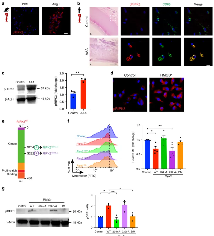
Serine 204 of RIPK3 modulates mitochondrial
fission.
Activa-tion of RIPK3 is characterized by its phosphorylaActiva-tion on distinct
serine residues
34,35. Phosphorylated form of RIPK3 (pRIPK3) was
increased in Ang II treated murine aortas compared to
saline-treatment (Fig.
6
a). Notably, pRIPK3 expression was detected in
CD68 macrophages seeded in human aneurysmal sections, but not
in healthy aortic tissues (Fig.
6
b). This was confirmed by increased
abundance of pRIPK3 (Fig.
6
c). BMDM treated with HMGB1
displayed pronounced pRIPK3 levels (Fig.
6
d). To gain further
insights into which phosphorylation sites of RIPK3 could regulate
mitochondrial function, we designed synthetic modified RNA
(modRNA) to specifically target key serine residues of RIPK3.
ModRNA strategy incorporates modified nucleotides to synthetic
mRNA coding for proteins, provides long-lasting
immuno-toler-ance and resistimmuno-toler-ance against degradation by endogenous cellular
ribonucleases
36,37. We generated modRNA to stably overexpress
wild type RIPK3 (RIPK3
WT) or constructs where either serine 204
or 232 were substituted with alanine residues (RIPK3
S204>A,
RIPK3
S232>A) as well as modRNA including both mutations
(RIPK3
DM). RIPK3 kinase domains regulated by phosphorylation
sites located on serine residue 204 (S204) and 232 (S232)
are depicted in Fig.
6
e. BMDM loaded with
fluorescent
mito-chondrial dye showed that overexpression of WT isoforms reduced
mitochondrial mass similar to RIPK3
S232>Awhile macrophages
transfected with RIPK3
S204>Amutation or RIPK3
DM, exhibited
increased
fluorescence (Fig.
6
f). In accordance, pDRP1 was
induced in BMDM transfected with RIPK3
WTconstructs
com-pared to cells treated with transfection vehicle alone.
Over-expression of RIPK3
S204>Abut not RIPK3
S232>Areduced pDRP1,
similar to RIPK3
DM(Fig.
6
g). These results suggested that the
phosphorylation site positioned on serine 204 of RIPK3 played a
critical role in regulating mitochondrial
fission.
Mitochondrial
fission regulates MMP12 in aortic macrophages.
To understand the functional significance of differential
mito-chondrial metabolism in arterial macrophages promoted by
RIPK3, we measured mitochondrial reactive oxygen species
(ROS) levels, as the mitochondria is an essential hub for
manu-facturing ROS
38. Stimulation of WT, but not Ripk3
−/−BMDM
with HMGB1 elevated mitochondrial ROS as tracked by
Mito-SOX intensity (Fig.
7
a, Supplementary Fig. 6a). HMGB1-induced
mitochondrial oxidative stress was blunted in the presence of
Mdivi-1 or by mitochondrial superoxide scavenger, MitoTEMPO
(Fig.
7
b). This was paralleled by increased expression of glutamate
dehydrogenase 1 (Glud1), a key mitochondrial matrix enzyme
responsible for catalyzing oxidative reactions, in WT BMDM
stimulated with HMGB1. Interestingly, Glud1 expression was
reversed in untreated and HMGB1 stimulated Ripk3
−/−macro-phages (Fig.
7
c). Oxidative stress determined by dihydroethidium
(DHE), indicative of ROS accumulation, was increased in aorta of
WT but not Ripk3
−/−mice treated with recombinant HMGB1or
Mdivi-1 (Fig.
7
d, Supplementary Fig. 7a).
MMP activated by ROS and secreted by activated macrophages
can facilitate the degradation of the extracellular matrix and
promote AAA
8. On the basis of our data reporting a marked
decrease in elastin fragmentation in the absence of RIPK3 in
macrophages and its ability to modulate ROS biogenesis, we
postulated that RIPK3 could
fine-tune the expression of MMP via
mitochondrial oxidative stress. Mmp12 was one of the most
upregulated genes in macrophages abundantly expressing Ripk3
as shown in the heatmap representation profiled from single-cell
RNA sequencing (Fig.
7
e). Mmp12 mRNA was increased in AAA
but reduced in aortic tissue of Ripk3
−/−mice (Fig.
7
f). Multiplex
assay of MMP indicated that while the expression of vascular
MMP12 was increased in aneurysmal WT aortas, its expression
was repressed in Ripk3
−/−aortic tissues (Fig.
7
g). Although
MMP2, MMP3 and MMP8 were significantly increased in WT
aorta with AAA, their expressions were not altered in the absence
of RIPK3 (Fig.
7
g). These results indicated that RIPK3 could
specifically temper the expression of macrophage-specific elastase
MMP12. MMP12 expression colocalized with CD68 macrophages
in the artery of WT but not Ripk3
−/−mice treated with
recombinant HMGB1 (Fig.
7
h). CD68 macrophages expressed
MMP12 in WT mice subjected to LPS nasal instillation but this
pattern was reversed in aortic samples of mice treated with
Mdivi-1 (Fig.
7
i). Notably, abundant MMP12 was detected in
macrophages and in whole human AAA tissue (Supplementary
Fig. 8a, b), as well as in aortic sections of mice exposed to cigarette
smoke (Supplementary Fig. 8c). Mmp12 mRNA increased in
BMDM stimulated with conditioned media of lung epithelial cells
imbued to cigarette smoke extract, however, its expression was
mitigated when HMGB1 was depleted from the conditioned
media or when macrophages were pretreated with RIPK3
inhibitor, GSK’872 (Supplementary Fig. 8d). Mmp12 mRNA
increased in WT BMDM stimulated with recombinant HMGB1
but was reduced in the presence of Mdivi-1 or MitoTEMPO.
A diminished signal for Mmp12 was detected in all the
aforementioned conditions in Ripk3
−/−BMDM (Fig.
7j
). These
results were confirmed by immunoblotting against MMP12
protein (Supplementary Fig. 8e).
Transfection of macrophages with increasing concentrations of
RIPK3
WTmodRNA dose-dependently elevated pRIPK3
expres-sion. This was paralleled by a similar pattern of increased
expression of MMP12 (Supplementary Fig. 8f). Notably, MMP12
expression was significantly reduced in macrophages
overexpres-sing RIPK3 with the mutation in serine residue 204 but not when
serine 232 phosphorylation site was altered compared to RIPK3
WT.
Accordingly, RIPK3
DMsignificantly decreased MMP12 levels in
macrophages (Supplementary Fig. 8g). These results reinforced a
critical role for the phosphorylation of serine 204 of RIPK3 in
regulating MMP12 in macrophages.
Deletion of MMP12 prevents ALI-induced AAA development.
Our data, reported above, strongly supported a deleterious role
for MMP12 in orchestrating aneurysmal remodeling in the
con-text of ALI. To directly demonstrate the role of MMP12, we
challenged MMP12 deficient mice (Mmp12
−/−) to ALI and
analyzed AAA (Fig.
7
k). While the incidence of AAA increased in
WT mice, induction of lung injury in Mmp12
−/−mice exhibited
reduced AAA occurrence (Fig.
7
k, bottom panel). Doppler
ultrasound imaging (Fig.
7
l) showed a significant reduction in
aortic diameter in Mmp12
−/−mice compared to WT mice
(Fig.
7
m, n). Elastin
fiber fragmentation was refrained and fibers
maintained a solid architecture in aortic sections analyzed from
Mmp12
−/−mice, consistent with non-diseased aortic integrity
and caliber (Fig.
7
o).
Discussion
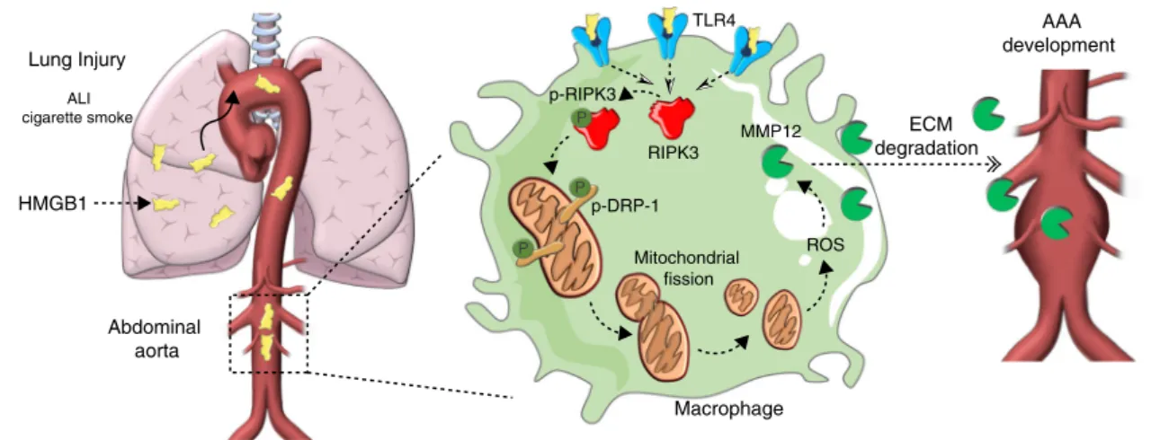
Our data provides mechanistic evidence of undescribed
spatio-temporal role of arterial macrophages in vascular remodeling. We
demonstrate that HMGB1 derived from injured lung can execute
complex mitochondrial stress responses in arterial macrophages
via RIPK3 (Fig.
8
). Our
findings indicate that RIPK3 can reroute
its necroptotic role to function as an instrumental signal capable
of regulating the expression of proteolytic enzymes in
macro-phages. To our knowledge, this is an original elucidation that
details the potential mechanisms underlying the epidemiological
mystery of why individuals suffering from chronic lung diseases
such as COPD have an increased risk of developing AAA.
g
Control Ripk3 pDRP1 WT 204>A 232>A DM β-Actin 45 kDa 80 kDa*
*
**
0 1 2 3 pDRP1 (AU) Control Ripk3 WT 204>A 232>A DMe
N-T C-T Kinase Proline-rich Binding 0 486 S204 S232 RIPK3WT RIPK3 P S204>A RIPK3 P S232>Af
0.0 0.5 1.0 1.5Relative MFI (fold change)
**
*
Control Ripk3 WT 204>A 232>A DM Mitotracker (FITC) % of max Ripk3S204>A Control Ripk3WT Ripk3S232>A Ripk3DM 103 104 105a
PBSb
pRIPK3 Ang II Control AAA pRIPK3 CD68 Merge L L Control HMGB1d
pRIPK3c
AAA pRIPK3 β-Actin 45 kDa 57 kDa 0.0 0.5 1.0 1.5 2.0 2.5pRIPK3 (fold change)
**
Control
AAA Control
Fig. 6 Phosphorylation of RIPK3 in serine 204 regulates mitochondrialfission via DRP1. (a) IF staining of phosphorylated RIPK3 (pRIPK3) in murine aortic sections of animals treated as indicated (scale bar= 20 µm). (b) Hematoxylin/Eosin (H&E) and IF staining of pRIPK3 (red) and CD68 (green) in healthy or aneurysmal human tissue (scale bar H&E= 500 µm, IF = 20 µm). Merge is shown in yellow and nucleus is stained with DAPI in blue. Dotted lines define the magnified region. L lumen. (c) Immunoblot of pRIPK3 and quantification of healthy or aneurysmal human tissue. n = 3 per group. **P= 0.0037. (d) IF staining of pRIPK3 in BMDM stimulated with HMGB1 (10 ng ml−1). Nucleus is stained with DAPI in blue (scale bar= 10 µm). (e) Schematic representation of RIPK3 phosphorylation sites. Targeted mutations designed in synthetic modified RNA (modRNA) are illustrated. WT, wild type; S, serine; A, Alanine; N-T, N-terminal; C-T, C-terminal; S204>A, modification of serine 204; S232>A modification of serine 232; DM, double serine mutations. (f) Representative histograms offlow cytometry analysis of Mitotracker and quantification of MFI in BMDM transfected with Ripk3 modRNA as indicated. n= 4 (DM) and 5 (Control, WT, 204>A and 232>A). *P < 0.05, **P < 0.01. Error bars represent s.e.m. (g) Immunoblot analysis and quantification of pDRP1 in BMDM transfected with Ripk3 modRNA as indicated. n = 3. *P < 0.05, **P < 0.01. Data is presented as mean, error bars represent s.e.m. P values were calculated using two-tailed unpaired t-tests (c) or one-way ANOVA (f, g).
Our results indicate that HMGB1 is a potent activator of RIPK3
in transmural macrophages. Utilizing single-cell RNA sequencing,
we identified that RIPK3 was harbored in macrophages with
intensified TLR4 signaling. We found that stimulation of
macro-phages with ALI extracts induced the expression of RIPK3, and that
depletion of HMGB1 from the lysates abrogated the increase of
RIPK3. These
findings clearly illustrate that danger signals, such as
HMGB1, that accumulate in injured tobacco-exposed lungs, can
leak into the circulation and alter the physiology of the abdominal
aortic wall. It is possible that the trafficking of HMGB1, and other
lung-derived DAMPs may be responsible for heightened
extra-pulmonary inflammation that manifest in COPD patients
pre-senting with ventricular dysfunction, myocardial infarction, diabetes
or hypertension
39,40. Further studies are necessary to shed light on
a
b
f
MMP12 (ng/mL ) WT Ripk3-/-e
h
j
c
g
Glud1 (fold change)***
*
***
WT Ripk3 -/-Control HMGB1 Cd14 Pgam1 Fabp4 Il1b Il1rn Lat2 Fabp5 Lpl Prdx6 Mmp12 Acp5 Epsti1 Mmp9 Ccl4 Ctsk Ccl21a Cd5l Fscn1 Ccl17 Ccl5 Ripk3-Ripk3+ 10 –10 5 0 –5 Mmp12mRNA (fold change)
***
**
WT Ripk3 -/-0 1 2 3 4 5 PBS Ang II 0 1 2 3MitoSOX MFI (fold change)
*** ***
***
HMGB1 Mdivi-1 MitoTEMPO 0 1 2 3*
*
MitoSOX MFI (fold change
) WT Ripk3 -/-Control HMGB1
d
Day 0 Day 28 Ultrasound imagingAng II pump Sacrifice
LPS (20 μg/mice, i.n.) WT Mmp12 -/-0 20 40 60 80 100 Aneurysm incidence (%) WT Mmp12
-/-*
Elastin WT Mmp12-/-k
l
m
– + + + – – + – – – – + 0 7 14 21 28 1.0 1.5 2.0 Aortic Diameter (mm) WT Mmp12 -/-0 1.0 1.5 2.0 2.5*
WT Mmp12 -/-Maximal Diameter (mm)n
o
WT Mmp12 -/-MMP2 MMP3 MMP8 pro-MMP9 MMP12 0.1 1 10 100 WT Ripk3 -/-WT Ang II + HMGB1 Ripk3 -/-Ang II + HMGB1 CD68 MMP12 Merge Ang II + ALI Ang II + ALI+ Mdivi-1
i
CD68 MMP12 Merge DHE Elastin AngII AngII + HMGB1 WT WT Ripk3-/- Ripk3-/- 0 1 2 3 4 Mmp12RNA (fold change)
**
*
**
WT Ripk3 -/-HMGB1 Mdivi-1 MitoTEMPO – + + + – – + – – – – + – + + + – – + – – – – + 0.0 0.5 1.0 1.5 2.0 2.5 0 5 10 15 20 25*
the specific mechanisms underlying these associations. We reasoned
that because macrophages are important reservoirs of HMGB1
receptors, including TLR4, they could intercept lung-derived
HMGB1 circulating in the blood and nourish pathological
signal-ing cascades in the aortic wall. Our data are in agreement with
studies showing the deleterious roles of HMGB1 and TLR4 in
AAA
5,41. Importantly, our results strongly suggest a causative role
for the HMGB1/RIPK3 axis via which cigarette consumption and
ALI could predispose to AAA development and potentially
accel-erate disease progression.
In our study, we demonstrate that recurrent LPS nasal
instil-lation models inflammation and promotes lung damage, thereby
provoking pathologic features of COPD at an accelerated scale.
Interestingly, analysis of lung sections of mice exposed to chronic
cigarette smoke revealed leakage of HMGB1 into the cytoplasm of
lung epithelial cells similar to LPS nasal instillation. Likewise,
RIPK3 and MMP12 were expressed by arterial macrophages
analyzed from aortic sections obtained from mice that consumed
tobacco. Thus, this indicates that RIPK3 might act a critical
rheostat that senses pathogenic elements disseminated in cigarette
smoke throughout the body.
Previous studies aimed at characterizing the role of transmural
macrophages in AAA have demonstrated that they are important
cellular contributors to the pool of MMPs and can drive the
sustained ECM degradation and therefore increase the incidence
of rupture
8,42. Our data are in accordance with these studies and
suggest that ALI-derived HMGB1 can selectively trigger the
expression of MMP12, which promotes AAA development and
rupture
43. Our results refine our understanding of the roles
of macrophages in the pathogenesis of AAA. We show that
macrophages seeded in AAA can intercept systemic and
extra-vascular signals, thereby fueling extra-vascular damage. The pool of
trans-activated macrophages producing MMP12 could therefore
mediate the crosstalk between lung damage and associated risk of
AAA rupture. Interestingly, MMP12 has also been shown to play
a critical role in COPD
44. MMP12 upregulation in COPD has
been shown to be in response to an increase of ambient ROS
45.
It would be of interest to investigate whether alveolar
macro-phages could regulate MMP12 via RIPK3 in COPD.
In the past decade, considerable research efforts have
eluci-dated the mechanisms underlying necroptosis
46–48, instituting
RIPK3 as the cornerstone of the necrosome leading to activation
of downstream MLKL responsible for executing necroptotic cell
death. However, mounting evidence suggests that RIPK3 can
exhibit divergent functions independent of necroptosis
49.
Nota-bly, RIPK3 was described to be a potent activator of the
inflam-masome and NF-κB pathway thereby fueling inflammation
50.
This is of particular importance in a complex multi-factorial
disease such as AAA, in which inflammation and cell death are
intertwined. Our data show that the absence of RIPK3 in
mac-rophages refrains the susceptibility of mice to develop AAA. Our
findings are in line with data reported by Wang et al., showing a
deleterious role for RIPK3 in AAA in a different model of elastase
infusion
51,52. However, more importantly, our studies diverge
mechanistically: we demonstrate that RIPK3 activation rewires
pathological macrophage mitochondrial metabolism to produce
Fig. 7 MMP12 drives AAA development triggered by lung damage. MitoSOX quantification of WT and Ripk3−/−BMDM treated with HMGB1 (a) and WT BMDM treated with HMGB1 in the presence or not of Mdivi-1 or MitoTEMPO (b). n= 3. *P < 0.05, ***P < 0.001. (c) Relative mRNA expression of Glud1 in WT or Ripk3−/−BMDM. n= 3 per group. *P < 0.05, ***P < 0.001. (d) Representative IF images of dihydroethidium (DHE) staining (red) of aortic sections of mice treated as indicated. Elastin autofluorescence (green, scale bar = 20 µm). (e) Heatmap representation of differentially expressed genes in macrophages expressing Ripk3 (Ripk3+) or not (Ripk3−). (f) qPCR analysis of Mmp12 mRNA in WT or Ripk3−/−aortas. n= 3 per group. **P < 0.01, ***P < 0.001. (g) Spider graph representation (Log10scale) of multiplex analysis of MMPs in aortic extracts from WT or Ripk3−/−mice. n= 3 (Ripk3−/−) and 4 (WT). *P= 0.0454.
Representative image of IF staining of MMP12, and its colocalization with CD68 macrophages in aortic section of mice treated as indicated (h, i). Scale bar= 20µm. (j) Mmp12 mRNA in WT or Ripk3−/−BMDM treated with HMGB1 (10 ng ml−1) in the presence or not of Mdivi-1 (10µM) or MitoTEMPO (10 µM). n = 3 per group. *P < 0.05, **P < 0.01. (k) Schematic representation of experimental protocol (top) and incidence of AAA in WT and Mmp12−/−mice (below). i.n. intranasal. n= 4–5 per group. Representative color Doppler ultrasound images of aorta and M-mode screenshots (arrow) (i). Chronological quantifications of aortic diameter (m), maximal aortic diameter (n), and representative Verhoeff-Van Gieson staining in aortic sections of WT and Mmp12−/−mice in the presence of ALI (o). n= 3 per group. Arrows indicate elastin breaks. Data is presented as mean, error bars represent s.e.m. (scale bar = 50 µm). *P = 0.0364. P va/clues were calculated by one-tailed (g) or two-tailed (m, n) unpaired t-tests, or one-way ANOVA (a–f, j).
HMGB1 Abdominal aorta Lung Injury ALI cigarette smoke Macrophage ROS MMP12 Mitochondrial fission ECM degradation AAA development RIPK3 p-RIPK3 p-DRP-1 P P P TLR4
Fig. 8 Schematic representation of our mainfindings. HMGB1 derived from injured lungs is captured by TLR4 transmural macrophages in the abdominal aorta. Activation of TLR4 by HMGB1 upregulates RIPK3 and activates its phosphorylation. RIPK3 activates phospho-DRP1 which triggers mitochondrial fission and increases mitochondrial reactive oxygen species (ROS) production. ROS stimulates the expression of MMP12 by macrophages responsible for elastinfiber degradation in the aorta. Inhibition of phosphorylation of RIPK3 on Serine 204 refrains DRP1 activation and subsequent MMP12 expression.