Biochemical and Functional Characterization of Human
RNA Binding Proteins
by
Peter Freese
A.B., Harvard University (2012)
Submitted to the Graduate Program in Computational and Systems Biology
in partial fulfillment of the requirements for the degree of
Doctor of Philosophy
at the
MASSACHUSETTS INSTITUTE OF TECHNOLOGY
February 2018
c
○ Massachusetts Institute of Technology 2018. All rights reserved.
Author . . . .
Computational and Systems Biology
December 22, 2017
Certified by . . . .
Christopher B. Burge
Professor of Biology and Biological Engineering
Thesis Supervisor
Accepted by . . . .
Christopher B. Burge
Director, Computational and Systems Biology Graduate Program
Biochemical and Functional Characterization of Human RNA
Binding Proteins
by
Peter Freese
Submitted to the Program in Computational and Systems Biology on December 22, 2017, in partial fulfillment of the
requirements for the degree of Doctor of Philosophy
Abstract
RNA not only shuttles information between DNA and proteins but also carries out many other essential cellular functions. Nearly all steps of an RNA’s life cycle are controlled by approximately one thousand RNA binding proteins (RBPs) that direct RNA splicing, cleav-age and polyadenylation, localization, translation, and degradation. Despite the central role of RBPs in RNA processing and gene expression, they have been less well studied than DNA binding proteins, in part due to the historical dearth of technologies to probe RBP binding and activity in a high-throughput, comprehensive manner. In this thesis, I describe the affinity landscapes of the largest set of human RBPs to date elucidated through a high-throughput version of RNA Bind-N-Seq (RBNS), an unbiased in vitro assay that determines the primary sequence, secondary structure, and contextual preferences of an RBP. The 78 RBPs bound an unexpectedly low diversity of RNA motifs, implying convergence of binding specificity toward a small set of RNA motifs characterized by low compositional complexity. Offsetting the low diversity of sequence motifs, extensive preferences for contextual features beyond short linear motifs were observed, including bipartite motifs, flanking nucleotide content, and preference for or against RNA structure. These features likely refine which motif occurrences are selected in cells, enabling RBPs that bind the same linear motif to act on distinct subsets of transcripts. Additionally, RBNS data is integrated with complemen-tary in vivo binding sites from enhanced crosslinking and immunoprecipitation (eCLIP) and functional (RNAi/RNA-seq) data produced through collaborative efforts with the ENCODE consortium. These data enable creation of “RNA maps” of RBP activity in pre-mRNA splic-ing and gene expression levels, either with (eCLIP) or without (RBNS) crosslinksplic-ing-based assays. The mapping and characterization of RNA elements recognized by over 200 human RBPs is also presented in two human cell lines, K562 and HepG2 cells. Together, these novel data augment the catalog of functional elements encoded in the human genome to include those that act at the RNA level and provide a basis for how RBPs select their RNA targets, a fundamental requirement in more fully understanding RNA processing mechanisms and outcomes.
Thesis Supervisor: Christopher B. Burge
Acknowledgments
I would like to thank my advisor, Prof. Chris Burge, for providing an incredibly rich envi-ronment in which to conduct research and for teaching me to be a rigorous computational biologist grounded in fundamental biological questions. Having never performed research on RNA or touched high-throughput sequencing data sets, the lab has been a wonderful place to learn new skills, formulate and test hypotheses, and receive feedback that has fueled my intellectual growth. A particular thanks goes out to Burge lab postdoc Daniel Dominguez and fellow CSB graduate student Maria Alexis, who have been phenomenal RBNS collabora-tors over the past years and central to this work as well as my overall scientific development; without either, I’m sure the RBNS project would have been nowhere near as successful or impactful as it has turned out to be. Additionally, I would like to thank all other past and present Burge lab members for their feedback and insights over the past five years, as well as their friendship and great company at our lab social events and beer hours, creating a multitude of memorable experiences outside of the lab. In particular, I’d like to thank Athma for being an always pleasant and engaging baymate, Alex and Nicole for being early RBNS collaborators who whetted my appetite for studying protein-RNA interactions, Jennifer and Yevgenia for our lighthearted coffee breaks, Matt for always being encouraging and ask-ing great biological questions, Bridget for plannask-ing great graduate student social events and group exercise class outings, Canadian Peter for his always outspoken and thought-provoking discussions, Marvin, Ana, Emma, and Kayla for their smiling faces and fresh perspectives, Genny for being a friendly face who always made me feel welcome in the Burge lab, Cassie, Myles, Amanda and Tsultrim for being amazing technicians who helped my understanding of the technical complexities of the RBNS assay, and Jason for being generous in taking me under his wing during my rotation despite being on paternity leave.
My Thesis Advisory Committee members, Phil Sharp and Manolis Kellis, have been very generous in providing their guidance and expertise at my committee meetings as well as facilitating my professional development. The connections I have made with and through them have been and will continue to be incredibly valuable. I would also like to thank Melissa Moore for agreeing to serve as my external committee member and taking time out
of her incredibly busy schedule to support my scientific endeavors.
I would like to thank my ENCODE RBP collaborators, who have been an instrumental source of feedback on my work and incredibly generous in sharing their time, data, and expertise in our group efforts. Through our work, I have learned a tremendous amount not only about RBP-RNA interactions and regulation but also the complexities and promise of studying biological problems through massive high-throughput integrative functional ge-nomic assays. In particular, Brent Graveley’s overall leadership of the RBP project has been instrumental in its success, Xintao Wei has always been a patient and responsive collaborator who made the logistics of working on a high-throughput consortium project very smooth, and Gene Yeo and his lab members Eric Van Nostrand and Gabe Pratt provided excellent scientific guidance and insights into RNA-RBP biology through their improved experimental and computational CLIP methods.
My undergraduate research advisor Irene Chen and postdoc Kirill Korolev initially sparked my interest in conducting scientific research and showed me the challenges and rewards see-ing a research project through from start to finish. I would likely not have pursued graduate studies without these formative academic role models and their support of my ongoing sci-entific development.
I would like to acknowledge the amazing friendship of my CSB 2012 classmates Vincent, Colette, Mandy, Mariana, Nezar, and Rotem, with whom I’ve shared over 5 years of incredible outings throughout MIT, Boston, and the Northeast. We’ve celebrated countless birthdays, taken summer trips to explore and relax, and supported each other through the ups and downs of a graduate career in a way that I couldn’t have imagined when we started this program together. It has been a pleasure to befriend the many other CSBs in years above and below me as we gather at retreats and other CSB events to share our research, successes, and struggles. Additionally, CSB administrator Jacquie Carota has done a phenomenal job of making the graduate program run smoothly and has always been a warm, smiling face to run into and chat with on the 2nd floor of Building 68.
Finally, I would like to thank my family members, who have been incredibly patient and generous in their support of my PhD endeavors and broader educational upbringing that has allowed me to be where I am today. My mom, dad, and sister Kelly have been endless
supporters of my scientific passions and research, and always provided me with a down-to-earth perspective of the incredible fortunes I have been lucky enough to receive through graduate education at MIT. Going home to Minnesota to visit them and my extended family as well as our phenomenal vacations have helped me contextualize my research and career within a broader perspective. This work and my intellectual growth as a graduate student would not have been possible without their unflagging support.
Contents
1 Introduction 15
1.1 RNA binding proteins . . . 16
1.1.1 RNA binding domains (RBDs) and sequence-specific recognition of RNA 16 1.1.2 RBP-mediated regulation of pre-mRNA splicing, RNA stability, and RNA translation . . . 23
1.2 Approaches for the study of RNA binding proteins . . . 31
1.2.1 In vitro RNA-RBP profiling techniques . . . 32
1.2.2 In vivo RNA-RBP profiling techniques . . . 34
1.2.3 Genetic studies of RNA profiling after RBP perturbation . . . 40
1.2.4 Structural studies of RBPs and RBP-RNA interactions . . . 40
1.3 RNA secondary structure in RBP-RNA interactions and regulation . . . 42
1.4 Overview of the thesis . . . 47
2 Sequence, Structure, and Context Preferences of Human RNA Binding Proteins 49 2.1 Abstract . . . 51
2.2 Introduction . . . 52
2.3 Results . . . 57
2.3.1 High-throughput RNA Bind-n-Seq Assay . . . 57
2.3.2 Binding specificities of a diverse set of human RNA binding proteins. 57 2.3.3 Overlapping specificities of RNA binding proteins . . . 58
2.3.4 RBPs preferentially bind low-complexity motifs . . . 62
2.3.6 Protein-bound sequences are associated with in vivo regulation of mRNA
levels . . . 67
2.3.7 RBPs with similar motifs often bind distinct transcript locations . . . 67
2.3.8 Most RBPs analyzed prefer less structured RNAs . . . 69
2.3.9 RNA structural elements influence binding of some RBPs . . . 72
2.3.10 Many RBPs favor pairs of short, spaced motifs . . . 74
2.3.11 RNA sequence context commonly influences RBP binding . . . 79
2.3.12 Towards a more complete characterization of RBP specificities . . . . 81
2.4 Discussion . . . 86
2.4.1 Towards an RNA processing parts list of RNA elements and RBPs. . 86
2.4.2 RBPs recognize a small subset of the available sequence space . . . . 86
2.4.3 RBP binding specificities harbor hidden complexity . . . 87
2.5 Supplementary Figures . . . 89
2.6 Methods . . . 99
2.6.1 Cloning of RNA binding protein domains . . . 99
2.6.2 Bacterial expression and protein purification . . . 99
2.6.3 Production of random RNAs by in vitro transcription . . . 100
2.6.4 RNA Bind-n-Seq Assay. . . 100
2.6.5 RNA Bind-n-Seq data processing and motif logo generation. . . 101
2.6.6 Clustering of RBNS motifs . . . 103
2.6.7 Comparison with RNAcompete . . . 104
2.6.8 Overlap of RBNS 6mers with splicing and stability regulatory elements104 2.6.9 Analysis of eCLIP for motif discovery, regulation and overlapping targets105 2.6.10 Analysis of RNA-seq datasets for regulation and RBNS Expression & Splicing Maps . . . 106
2.6.11 Generation of random sets of ranked 6mer lists with edit distances to top 6mer matching RBNS . . . 108
2.6.12 RBNS RBP groups without paralogs or RBPs with any RBD pair sharing 40% identity . . . 108
2.6.14 Motif entropy analysis . . . 109
2.6.15 RNA secondary structure analysis . . . 111
2.6.16 Determination of bipartite motifs . . . 113
2.6.17 Assessment of flanking nucleotide compositional preferences . . . 114
2.6.18 Filter binding assay . . . 115
2.6.19 Calculation of feature-specific 𝑅 values and relative entropy of context features . . . 115
2.6.20 Tissue specificity of RBP gene expression . . . 116
3 A Large-Scale Binding and Functional Map of Human RNA Binding Pro-teins 119 3.1 Abstract . . . 121
3.2 Introduction . . . 122
3.3 Results . . . 124
3.3.1 Overview of data and processing. . . 124
3.3.2 In vivo binding is largely determined by in vitro binding specificity . 131 3.3.3 Functional Characterization of the RBP Map . . . 136
3.3.4 RBP association with splicing regulation . . . 138
3.3.5 RBP Association with Chromatin . . . 142
3.3.6 RBP regulatory features in subcellular space . . . 146
3.3.7 Preservation of RBP regulation across cell types . . . 150
3.4 Discussion . . . 156
3.5 Supplementary Figures . . . 158
3.6 Methods . . . 178
3.6.1 RNA binding protein annotations and domains . . . 178
3.6.2 eCLIP - experimental methods . . . 178
3.6.3 eCLIP - data processing and peak identification . . . 179
3.6.4 Knockdown followed by RNA-seq (KD/RNAseq) - experimental methods180 3.6.5 KD/RNA-seq - data processing . . . 181
3.6.7 RBNS - data processing . . . 182 3.6.8 Immuno-Fluorescence, Microscopy Imaging and Data Processing . . . 183 3.6.9 ChIP-seq - experimental methods . . . 185 3.6.10 ChIP-seq - data processing . . . 185 3.6.11 Integrated Analysis . . . 186
4 Conclusion 197
4.1 Summary . . . 197 4.2 Future Directions . . . 199
4.2.1 Impact of post-transcriptional RNA and post-translational protein mod-ifications on RBP-RNA binding . . . 199 4.2.2 Role of alternative protein isoforms and low-complexity domains in
RNA binding specificity and higher-order protein assemblies . . . 200 4.2.3 Integrative analysis of RBP binding data sets to relate genetic variation
List of Figures
1-1 Common RNA Binding Domain (RBD) types . . . 18 1-2 cis-acting splicing regulatory elements and trans-acting splicing factors . . . 27 1-3 The enhanced CLIP (eCLIP) assay . . . 38 1-4 Examples of in silico-folded RNA oligos. . . 43
2-1 Overview of the high-throughput RNA Bind-n-Seq assay and computational analysis pipeline. . . 55 2-2 RBPs bind a small subset of the sequence space, characterized by low-entropy
motifs . . . 60 2-3 RBNS-derived motifs are associated with regulation of mRNA splicing and
stability in vivo . . . 64 2-4 RNA secondary structural preferences of RBPs. . . 70 2-5 Many RBPs bind bipartite motifs or prefer flanking nucleotide compositions 76 2-6 RBPs that bind similar motifs often diverge in sequence context preferences 82 2-S1 RBNS assay and comparison to RNAcompete . . . 89 2-S2 Overlapping specificities of RBPs . . . 90 2-S3 RBNS-derived splicing and stability RNA maps and RBP binding in the
tran-scriptome . . . 91 2-S4 In vitro and in vivo structural preferences of RBPs and distribution of
enrich-ments across reads . . . 93 2-S5 Bipartite core spacing, flanking nucleotide composition, and degenerate
pat-tern binding preferences . . . 95 2-S6 Sequence context effects on RBP binding . . . 97
3-1 Overview of experiments and data types . . . 125
3-2 Integrative analysis of RBP binding and function . . . 129
3-3 Sequence-specific binding in vivo is determined predominantly by intrinsic RNA affinity of RBPs . . . 133
3-4 Association between RBP binding and RNA expression upon knockdown . . 137
3-5 Integration of eCLIP and RNA-seq identifies splicing regulatory patterns . . 139
3-6 Chromatin-association of RBPs and overlap with RNA binding. . . 143
3-7 RBP subcellular localization, binding, and regulation . . . 148
3-8 Preservation of RBP binding and regulation across cell types . . . 152
3-S1 Integrative analysis of RBP data types in cryptic exon suppression . . . 158
3-S2 Saturation of RBP binding and regulation in the transcriptome . . . 159
3-S3 Comparison of in vitro RBNS-derived motifs with in vivo eCLIP-derived motifs161 3-S4 Splicing regulatory activity of RBNS+ and RBNS- eCLIP peaks . . . 163
3-S5 Association between RBP binding and RNA expression upon knockdown . . 165
3-S6 Generation of splicing maps for RBFOX2 . . . 167
3-S7 Splicing regulatory patterns of SR, HNRNP, and spliceosomal proteins . . . 169
3-S8 RNA maps for alternative 5’ and 3’ splice sites . . . 170
3-S9 eCLIP binding patterns in subcellular space . . . 172
3-S10Preservation of binding across cell types . . . 174
3-S11Expression of RBPs across tissues and cell types . . . 176
4-1 Integrative analyses of RBP data can identify genetic variants that may impact RBP regulation . . . 203
Chapter 1
Introduction
The central dogma of molecular biology, first stated nearly 60 years ago (Crick [1958]), detailed that genetic information does not transfer from protein to DNA but instead typically from DNA → RNA → protein. With proteins effecting most functions in the cell and DNA being the central molecule of heredity passed from generation to generation, RNA was originally viewed as a somewhat less important intermediary between information and action. However, work over the past decades has revealed that RNA is a highly dynamic and regulated molecule, subject to a wide range of RNA processing mechanisms in eukaryotes (Mitchell and Parker [2014]). For messenger RNA (mRNA) molecules that encode protein sequences, these processes include 5’ capping and 3’ polyadenylation of the newly transcribed mRNA; constitutive and alternative pre-mRNA splicing; RNA editing; export from the nucleus into the cytoplasm; subcellular localization within different parts of the cytoplasm; regulation of translational efficiency; RNA surveillance and quality control; and regulation of mRNA stability and eventual degradation of the RNA. RNA binding proteins (RBPs) play critical roles in these post-transcriptional pathways, with each of the 1,000+ RBPs in humans having unique RNA binding activity and protein-protein interaction partners to produce a diverse assortment of highly regulated RNA molecules from the relatively modest ∼20,000 genes encoded in the genome.
1.1
RNA binding proteins
In eukaryotic cells, each mRNA is bound by a dynamic repertoire of RNA binding pro-teins (RBPs) such that it exists as an mRNA-protein complex (messenger ribonucleoprotein, mRNP, Singh et al. [2015], Rissland [2017]). The proper pre-mRNA splicing, processing, nuclear export, subcellular localization, and stability and degradation of mRNAs critically depend on these RBP-RNA interactions. Some mRNP components are members of large macromolecular machines, such as the spliceosome or ribosome, that bind mRNA in a coor-dinated manner to direct processes such as splicing, nuclear export, translation, and mRNA decay. These RBPs typically are deposited on mRNAs according to earlier RNA process-ing events or through interaction with mRNA landmarks such as the 5’ cap, pre-mRNA splice sites, or the poly(A) tail. In addition to these members of common machineries, other RBPs interact with sequence-specific features of individual mRNAs. These proteins often bind mRNAs concurrently with core machineries to regulate specific steps in RNA processing, such as splicing factors binding introns or exons to influence alternative splicing or AU-rich element binding proteins binding 3’ UTRs to influence mRNA stability or trans-lation. However, not all RBPs fall into these extreme categories of high sequence specificity or general machineries but instead often operate in the middle ground of a specificity con-tinuum from promiscuous to selective (Mitchell and Parker [2014]). For instance, Pumilio domain-containing proteins bind eight to ten RNA bases with high specificity at one extreme (Zamore et al.[1997]) while DEAD-box helicases have shown little dependence on RNA se-quence to rearrange their mRNA substrates (Linder and Jankowsky [2011]). In between, SR (Serine/Arginine-rich) proteins and HNRNPs (hetergeneous nuclear ribonucleoproteins) exhibit discernible sequence preferences but are able to bind a wide range of targets to effect their transcriptome-wide splicing outcomes (Goren et al.[2006], Geuens et al.[2016]).
1.1.1
RNA binding domains (RBDs) and sequence-specific
recog-nition of RNA
The sequence and/or structural specificity of an RBP for RNA targets is typically medi-ated through one or more well-defined RNA binding domains (Fig. 1-1). Among the ∼600
structurally distinct RBD classes catalogued by Gerstberger et al. [2014], just 20 have more than ten members, with most having just one or two members. The three most prevalent sequence-specific RNA binding domains in humans are the RNA Recognition Motif (RRM, ∼278 human RBPs), various types of Zinc Finger domains (ZF, ∼90 human RBPs though also present hundreds of DNA binding proteins), and the hnRNPK homology domain (KH, ∼63 human RBPs) (Gerstberger et al. [2014]). The RNA binding properties of other preva-lent RBDs, including those that bind double-stranded RNA, is less well understood and/or thought to occur largely in a sequence-independent manner (see below).
A common feature of the most abundant RBD classes in mRNA binding proteins is their frequent occurrence in multiple copies and/or in combination with different RBD types (Gerstberger et al.[2014]), with such modular design providing greater specificity and affinity to permit the diverse biological functions employed by eukaryotic RBPs (Lunde et al.[2007]). Interestingly, the average number of RBDs within a protein is inversely correlated with the number of nucleotides that RBD type commonly binds, ranging from ZFs binding an average of ∼3 nt with more than 3 ZFs per protein to the Pumilio Homology Domain (PUM-HD) binding ∼8 nt with just one RBD per protein on average (Mitchell and Parker[2014]).
Figure 1-1: Common RNA Binding Domain (RBD) types
Among the most prevalent RNA binding domains in eukaryotic proteomes include the RNA Recognition Motif (RRM, present in ∼278 human RBPs); hnRNPK Homology Domain (KH, present in ∼63 human RBPs); and Zinc Finger Domain (ZF, present in ∼961 human proteins, though most bind DNA with only dozens currently implicated in RNA binding). Other well-defined RBDs include the double-stranded RBD (dsRBD, present in 26 human RBPs) and Pumilio Homology Domain (PUM-HD, present in 3 human RBPs). All numbers taken from Uniprot human entries with example structures from PDB shown.
RNA Recognition Motif (RRM) domain
The RRM is the most abundant RBD in higher eukaryotes, occurring in nearly 300 human RBPs (Gerstberger et al.[2014]). Discovered in 1989 as the RNA binding domain of U1-70K (Query et al.[1989]), it is present in all kingdoms of life including prokaryotes and viruses with RRM-containing proteins involved in most post-transcriptional gene regulatory pathways (Afroz et al. [2015]). RRMs are typically ∼90 amino acids in length with a characteristic 𝛽1− 𝛼1− 𝛽2− 𝛽3− 𝛼2− 𝛽4 arrangement that folds into a four-stranded antiparallel 𝛽-sheet
packed against two 𝛼-helices. The loops between the secondary structural elements can vary in length and are typically disordered in their free form, and variability is also occasionally seen in the secondary structural elements themselves (for example, one 𝛼-helix in U2AF1 is three times longer than usual) or extensions at the RRM extremities (Afroz et al.[2015]).
RRMs interact with anywhere from two to eight RNA nucleotides, though three to four is most common. They typically recognize single-stranded RNA with the 𝛽-sheet surface con-tacting the RNA bases spread on the protein surface across the 𝛽-sheet from 𝛽4 to 𝛽2. RRMs are characterized by two consensus sequences: ribonucleoprotein (RNP) 1 and 2, which are 8 and 6 amino acids long on the 𝛽3 and 𝛽1 strands (Lys/Arg-Gly-Phe/Tyr-Gly/Ala-Phe/Tyr-Val/Ile/Leu- X-Phe/Tyr and Ile/Val/Leu-Phe/Tyr-Ile/Val/Leu-X-Asn-Leu, re-spectively, X = any amino acid, Afroz et al. [2015]). Three key aromatic side chains in RNP1 and RNP2 (bolded in the sequences above) recognize two nucleotides to provide affin-ity, though these interactions do not explain the sequence-specificity of different RRMs. Some nucleotide biases are observed at certain positions contacted by the RRM but all four nucleotides are found in all five of the most commonly recognized positions, making the RRM an incredibly plastic RNA binding domain. Though a 𝐾𝑑in the nanomolar range has
been observed for a few RRMs, most bind RNA targets with micromolar affinity (Afroz et al.
[2015]).
hnRNPK homology (KH) domain
First identified in its namesake hnRNPK protein in 1993 (Siomi et al.[1993]), the KH domain occurs in about 63 human RBPs and is present in diverse archaea, bacteria, and eukaryotes.
KH domains are contained within proteins involved in diverse biological processes, including transcription regulation, splicing regulation, and translational control (Valverde et al.[2008]). The domain consists of a core 𝛽1𝛼1𝛼2𝛽2 motif with a highly conserved GXXG loop between
the two 𝛼-helices. Flanking the 𝛽1𝛼1𝛼2𝛽2 motif is another 𝛼-helix and 𝛽-strand, though
they can come after (type I) or before (type II) the core motif, separating KH domains into two folds: type I (𝛽1𝛼1𝛼2𝛽2𝛽′𝛼′, more common in eukaryotes) and type II (𝛼′𝛽′𝛽1𝛼1𝛼2𝛽2,
more common in prokaryotes). Though the two folds have have different three-dimensional structures, both have the three 𝛼-helices packed onto the surface of an anti-parallel 𝛽-sheet. In addition to binding unpaired RNA, KH domains can bind single-stranded DNA, with recognition occurring via backbone interactions between four nucleic acid bases and the KH domain near the GXXG loop. A cleft in the protein structure near the GXXG loop allows protein hydrophobic interactions as well as mainchain and sidechain hydrogen bonds to mediate recognition of the four nucleobases. Due to two hydrogen bonds made with the nucleobases, adenine or cytosine are typically at positions 2 and 3 of the RNA tetramer, though one exception to this rule has been observed in a solution structure (Nicastro et al.
[2015], Nicastro et al.[2012]).
KH domains are often found in multiple copies within eukaryotic proteins, including up to 14 in vigilin. For protein families whose members contain multiple KH domains, the first KH domain (KH1) is typically more similar to other KH1 domains in different proteins than it is to its other KH domains (KH2, KH3, etc.), with similar relationships seen for other KH domains (Valverde et al.[2008]). Individual KH domains bind RNA and single-stranded DNA with low-to-intermediate affinity in the micromolar range, though increased affinity and specificity are achieved through use of multiple domains which can be structurally decoupled or form a contiguous extended RNA binding surface. Similar longer RNA sequences can be recognized by dimerization of RBPs that contain a single KH domain, as is crucial for the biological activity of STAR (Signal Transduction and Activation of RNA) family proteins (Nicastro et al. [2015]).
Zinc Finger (ZF) domain
In addition to their more classically defined roles in binding double-stranded DNA, zinc finger-containing proteins can act as RNA binding modules (Font and MacKay [2010]). Indeed, the first ZFs discovered, in the transcription factor TFIIIA, were identified through their binding to double-stranded 5S rRNA in Xenopus oocytes (Miller et al. [1985]). ZF domains typically coordinate a zinc ion with pairs of cysteine and histidine residues, though the arrangement of these residues within the ∼30 amino acid domain composed of a 𝛽-hairpin and 𝛼-helix can vary, lending the most prevalent types of these domains to be commonly characterized as C2H2, CCHC, and CCCH/C3H1 (another smaller class of ZFs, RanBP2-type, is named for the RanBP2 protein in which it was discovered and is defined by a W-X-C-X2−4-C-X3-N-X6-C-X2-C motif, with such ZFs included in the FET family of FUS,
EWSR1, and TAF15 RBPs).
While DNA recognition by ZFs is known to occur via major groove contacts with 3 base pairs of DNA, the reported binding of ZFs to single- or double-stranded RNA is much more varied and less well understood. For example, three of the nine ZFs in TFIIIA are important for RNA binding activity, with two of these (fingers 4 and 6) making base-specific contacts with ‘flipped-out’ RNA bases while finger 5 makes exclusively non-sequence-specific interac-tions with the RNA phosphate backbone via amino acid basic side chains (Font and MacKay
[2010]). It has been proposed, based on conclusions from crystallography studies, that ZFs can recognize complex structures comprising internal loops and double helicies both through specific contacts with individual bases that are exposed for access out of a rigid RNA struc-ture as well as sequence independent binding to the regularly folded portion of an RNA double-helix (Lu et al. [2003]). It has also been proposed that a common mode of RNA recognition of ZFs might be to bind structured RNAs. This is based on, among other stud-ies of ZFs binding dsRNA or stem-loops with much greater affinity than ssRNA (Font and MacKay [2010]), the characterization of Wilm’s tumor 1 (WT1) in vitro-selected RNA ap-tamers as requiring a hairpin loop, with tolerance for compensatory mutations that maintain proper base-pairing of the stem (Zhai et al.[2001]). Yet in contrast to this paradigm, numer-ous CCCH zinc fingers have been shown to recognize single-stranded A/U-rich elements and
promote degradation of their mRNAs (e.g., ZFP36, Lai et al. [1999], and Tis11d through backbone interactions with the Watson-Crick edges of A and U bases,Hudson et al.[2004]), underscoring the complexity of ZF RNA binding modes. Although CCCH and CCHC zinc finger motifs have classically been known to bind RNA, an mRNA interactome study (see Section 1.2.2) also identified a significant enrichment of AKAP95 and HC5HC2H-type zinc fingers, making them likely bona-fide RNA binders (this study identified 69 total ZF contain-ing proteins bound to poly(A)-selected RNA in HeLa cells, Castello et al.[2012]). Mirroring their diverse RNA recognition modes, ZFs that bind to RNA are involved in many func-tional processes including mRNA trafficking, stability, and transcripfunc-tional and translafunc-tional regulation (Wai et al. [2016]).
Other RNA binding domains and low-complexity domains
While a few other eukaryotic RBD types such as the PUM-HD bind RNA in a sequence-specific manner, most others bind in a predominantly sequence-independent manner, rec-ognizing secondary structure or being guided to target RNAs by other protein cofactors, and/or they have not been studied in detail (Gerstberger et al.[2014]). RBDs containing a Asp-Glu-Ala-Asp (DEAD) motif, present in ∼62 human RBPs that are typically RNA he-licases, recognize five nucleotides exclusively through interactions with the sugar phosphate backbone of the RNA in a characteristic bent conformation (Linder and Jankowsky[2011]). The double-stranded RNA binding domain (dsRBD), present in ∼26 human RBPs, consists of 65-70 amino acids that adopt an 𝛼𝛽𝛽𝛽𝛼 fold to recognize A-form double-stranded RNA through contacts to bases and ribose sugars in two successive minor grooves as well as the phosphate backbone in the intervening major groove. Traditionally thought to interact with RNA without any sequence specificity, recent structural information shows that dsRBDs can recognize additional sequence features beyond the A-form RNA helix (Masliah et al.[2013]). In addition to well-defined RNA binding domains containing characteristic amino acids that recognize specific RNA sequences and/or structures, some low-complexity domains may also play roles in contacting RNA. The second most common RBD type in the human genome after the RRM, the RG/RGG motif, is characterized by a region rich in arginines and glycines and is present ∼80-100 human RBPs depending on domain definition. These domains display
degenerate binding specificity yet still display different degrees of preference for RNA with some domains achieving affinity approaching that of their full-length protein counterparts (Thandapani et al. [2013], Ozdilek et al. [2017]). The RS (arginine/serine-rich) domains of SR and other (e.g., U2AF) proteins are classically thought to contact one another to directly mediate protein-protein interactions (Busch and Hertel [2012]), though work from Michael Green’s lab has shown that these domains intermittently make direct contacts with RNA sequences important for splicing, including the branchpoint and 5’ splice site (Shen et al.
[2004], Shen and Green [2004]). Changes in the phosphorylation state of serines within RS repeats could potentially alter the RS domain interaction mode between making protein-protein and making protein-protein-RNA contacts (Hertel and Graveley[2005]).
Altogether, many of these other RNA binding and low-complexity domain types are less well studied than RRM, ZF, and KH domains, and further investigation into how they impart RNA specificity and/or affinity is warranted.
1.1.2
RBP-mediated regulation of pre-mRNA splicing, RNA
sta-bility, and RNA translation
The following sections contain an overview of the processes of pre-mRNA splicing and RNA stability & translation, particularly noting steps in which RBPs are known to play regulatory roles. Additionally, examples of RBPs regulating these processes are provided, often high-lighting the context-dependent manner in which an RBP can have different effects on the same post-transcriptional regulatory pathway depending on the cell state or type, binding location, and competing or cooperating RBP(s).
Pre-mRNA splicing and cis-Splicing Regulatory Elements (SREs)
Metazoan protein-coding genes contain multiple exons split up by intervening introns which must be removed from the pre-mRNA to produce a mature mRNA ready for nuclear export and translation. This process of splicing is carried out by the complex macromolecular machine known as the spliceosome, which contains five small nuclear ribonucleoproteins (snRNPs) and ∼170 auxiliary proteins that enter and exit the spliceosome during various
stages of the splicing reaction (Wahl et al. [2009]). Through the alternative inclusion or exclusion of exons (or parts thereof), different isoforms of mature mRNAs are produced from the same pre-mRNA through a process known as alternative splicing, creating messages with different coding-potential or regulatory capacity.
Each splicing reaction requires three relatively short core RNA sequence elements in the pre-mRNA: the 5’ splice site (5’ss), the 3’ splice site (3’ss), and the branchpoint sequence (BPS). The 5’ss, at the end of the upstream exon and beginning of the intron, has human consensus sequence CAG|GUAAGU (|=exon/intron boundary, Roca et al. [2013]) and is recognized via complementarity to the 5’ portion of the U1 snRNA. At the other end of the intron, the 3’ splice site consists of a ∼25 nt sequence rich in cytosine and uridine known as the polypyrimidine tract, which is preceded upstream by a branchpoint sequence (BPS) and downstream by an intron-terminal AG dinucleotide. The branchpoint sequence is conserved in yeast with consensus UACUAAC but is quite degenerate in humans with consensus YUNAY, Y = pyrimidine (Gao et al.[2008]). The polypyrimidine tract is initially recognized the larger subunit of the U2 auxiliary factor (U2AF) heterodimer composed of U2AF65 (also known as U2AF2) and U2AF35 (also known as U2AF1). The ternary complex of U2AF65 with U2AF35 and splicing factor 1 (SF1) recognizes the surrounding BPS and 3’ss AG, with U2AF65 recruiting the U2 snRNP to replace it in the active spliceosome (Agrawal et al. [2016]). The branchpoint adenosine (bolded in previous sequences) is bulged out via complementarity of the flanking RNA sequence with a ‘GUAGUA’ sequence in U2 snRNA to act as the nucleophile in the first of two transesterification reactions during splicing. In the first reaction, the 2’-OH of the branchpoint A attacks the conserved guanine at the 5’ end of the intron to produce a 2’-5’ phosphodiester RNA lariat structure and a free 3’-OH at the upstream exon. In the second reaction, the 3’-OH of the upstream exon attacks the phosphodiester bond of the guanosine at the 3’ end of the intron to ligate the two exons, resulting in the exons being spliced together and the intron being released as a lariat structure.
In addition to the ∼170 core spliceosomal proteins that partake in all splicing reac-tions, alternative splicing is regulated by the binding of trans-acting RBPs (splicing factors) to short cis-sequences in the pre-mRNA to enhance or inhibit the use of adjacent splice
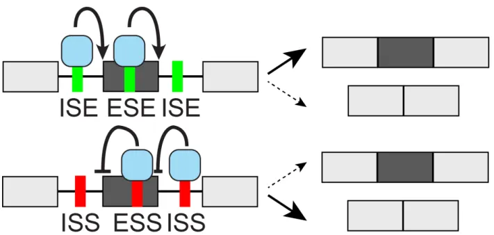
sites. These splicing regulatory elements (SREs) are broken down based on whether they are located in the Exon or Intron and whether they Enhance or Silence splicing from that location (ESE/ESS enhancing or silencing from exons, ISE/ISS from introns, Fig. 1-2). Although regulatory elements can in principle exert their action from anywhere within the pre-mRNA, most studies have focused on regulatory sequences within the exon or proximal flanking introns (∼200-300 nt adjacent to splice sites). Though some splicing factors are ubiquitously expressed, many act in a tissue- or developmental-specific manner to execute alternative splicing programs central to tissue and organ development (Baralle and Giudice
[2017]). Thus, one challenge in identifying potential SREs and their corresponding RBPs is that the same pre-mRNA sequences can be recognized differently in different cell types, partially due other RBPs expressed or the different RBP:RNA stoichiometries in each cell type. Additionally, the same cis-sequences, trans-factors, or combinations thereof can have different effects on splicing outcomes depending on the sequence context and their position within the intron or exon (Fu and Ares Jr[2014]). For example, G-runs can enhance splicing from intronic locations (McCullough and Berget[1997]) or repress splicing from exonic loca-tions (Chen et al.[1999]), and RBFOX2 and Nova typically suppress cassette exon inclusion from upstream introns but enhance it from downstream introns (Yeo et al.[2009], Ule et al.
[2006]). As it has been estimated that the three core human splice site motifs (5’ss, 3’ss, and BPS) only contain about half of the information needed to accurately define intron/exon boundaries (Lim and Burge [2001]), these cis-sequences and their trans-factors likely play a large role in ensuring the high fidelity of splicing.
Two key protein families that regulate pre-mRNA splicing and subsequent aspects of RNA metabolism are the SR (serine/arginine-rich) and HNRNP (heterogeneous nuclear ri-bonucleoprotein) proteins which often function by interacting with ESEs and ESSs/ISSs, respectively. The SR proteins (∼12 in human, depending on definition) are characterized by N-terminal RRM(s) which typically bind ESEs and C-terminal RS domains that participate in protein-protein interactions and facilitate spliceosome assembly (Busch and Hertel[2012],
Graveley and Maniatis[1998]), though the RS domains have also been shown to directly con-tact RNA splicing signals (see “Other RNA binding domains and low-complexity domains” above). The study of SR proteins originates in Drosophila screens that identified splicing
factors containing protein domains rich in arginine and serine dipeptides (Chou et al.[1987],
Moretti et al. [1987],Amrein et al. [1988]), with subsequent identification of human splicing factors SRSF1 and SRSF2 as proteins that also contain such RS-rich domains in addition to their RRM(s) (Ge and Manley [1990], Krainer et al. [1990], Fu and Maniatis [1992]). The diverse HNRNPs (∼37 in human, first detailed by the Dreyfuss lab,Piñol Roma et al.[1988]) contain one or more RBDs (typically RRMs, though five contain KH domains) and often RGG boxes (repeats of Arg-Gly-Gly tripeptides) and glycine-rich, acidic or, proline-rich do-mains that mediate protein-protein interactions and influence splicing outcomes through a wide variety of mechanisms (Busch and Hertel [2012], Geuens et al.[2016]).
A major goal of the field is the development of a ‘splicing code’ which could predict the splicing outcomes of any transcript from its primary sequence. One central feature in such a splicing code is a ‘parts list’ of splicing factors and the motif(s) they bind. Indeed, position-and tissue-specific effects of sequence elements that match the motifs of the FOX, NOVA, MBNL, CELF, TIA, PTB, and QKI protein(s) arose in an early inferred splicing code (Barash et al.[2010]). An updated splicing code model detected 2080 significant correlations between RNA-seq Ψ values of 10,689 exons and densities of 98 in vitro RBP binding motifs (Ray et al.
[2013]) in six intronic or exonic regions (Xiong et al.[2015]), and undoubtedly more will arise with an expanded catalog of RBP binding motifs. Such splicing codes and related efforts will not only provide greater mechanistic insight into the interplay between trans-factors and their cis-regulatory elements in splicing regulation but also may provide opportunities for understanding genetic determinants of human disease and support for casual variants acting at the level of pre-mRNA splicing.
Figure 1-2: cis-acting splicing regulatory elements and trans-acting splicing factors cis-acting splicing regulatory sequences in the pre-mRNA include ESE/ESS (Exonic Splicing Enhancer/Silencer) and ISE/ISS (Intronic Splicing Enhancer/Silencer) elements. Typically 4-6 nt in length, they are recognized by trans-acting proteins known as splicing factors, which promote or inhibit productive assembly of a catalytically active spliceosome that carries out the splicing reaction. The ‘context-dependent’ nature of cis-regulatory splicing sequences makes their activity highly reliant on location within the exon and flanking introns as well as the presence of other synergistic or antagonistic elements nearby. Additionally, different sets of splicing factors are expressed in different tissues and cell states, resulting in highly tissue-specific and dynamic alternative splicing programs.
3’ UTR cis-elements and regulation of mRNA stability and translation
3’ untranslated regions (3’ UTRs) are the noncoding parts of mRNAs following stop codons. Their length expansion in humans compared to yeast, as well as the increased prevalence of alternative 3’ UTR isoforms, suggests an important role for 3’ UTRs in the regulation of genes of higher organisms (Mayr [2017]). Best known to regulate the degradation, translation, and localization of mRNAs, 3’ UTRs function by recruiting RBPs that bind to cis-elements and recruit effector proteins such as deadenylases (Rissland [2017]). Though most 3’ UTR functions are carried out in the cytoplasm, some RBPs are loaded onto the mRNA in the nucleus and are exported with the message as an mRNP while others are added locally in the cytoplasm (Mayr [2017]). Co-transcriptional loading of RBPs at promoters can result in their deposition onto mRNAs with their remained association allowing them to play 3’ UTR regulatory roles in the cytoplasm, enabling crosstalk between the seemingly unrelated processes of mRNA synthesis in the nucleus and translation or decay in the cytoplasm (Bregman et al. [2011],Moore and Proudfoot [2009]).
3’ UTRs determine protein output by regulating mRNA stability and translation primar-ily through the activity of AU-rich elements and miRNAs. Though about half as conserved as coding sequences on average, 3’ UTRs often contain ‘islands’ that are conserved similarly to coding sequences and frequently contain binding sites for miRNAs or RBPs (Xie et al.
[2005], Friedman et al. [2009]). AU-rich elements (AREs), characterized by variants of the pentamer “AUUUA” occurring in variable length repetitions, were one of the first motifs dis-covered in 3’ UTRs, preferentially found in genes subject to tight expression level regulation such as immune-regulatory factors, cytokines, and proto-oncogenes (Barreau et al. [2006]). The mRNA half-lives of these genes are shorter than a half hour compared to a median ∼7 hour half-life across the entire mammalian transcriptome (Sharova et al. [2009]). Effects of AREs and miRNAs on protein abundance in cell lines is more modest (Baek et al. [2008],
Selbach et al.[2008],Spies et al.[2013]), possibly due to 3’ UTRs not substantially regulating protein abundance under steady-state growth conditions in cell culture with miRNAs and RBP-mediated repression more playing important roles in select biological contexts (Mayr
have different effects on mRNA depending on the protein(s) bound to them. Competition between ARE-BPs and which one(s) ultimately bind to messages can result in either mRNA stabilization and translational enhancement (commonly observed for the Hu/ELAV family RBPs) or in mRNA destabilization and translational repression (e.g., HNRNPD, ZFP36, and TIA1). Consistent with 3’ UTR repressive elements being more prevalent than activat-ing elements, 3’ UTR length is typically inversely correlated with mRNA stability and gene expression levels. Furthermore, among genes with alternative polyadenylation sites, those highly expressed typically prefer the proximal poly(A) site to produce a shorter 3’ UTR while genes with lower expression levels often use the distal poly(A) site to produce a longer 3’ UTR (Matoulkova et al. [2012]).
In addition to AU-rich elements, other 3’ UTR cis-elements are crucial for post-transcriptional gene regulation through their interplay with RBPs. GU-rich elements in arrangements of 2-5 overlapping pentamers are contained in at least 5% of human mRNAs. They are present in the 3’ UTRs of short-lived mRNAs in T-lymphocytes and contribute to additional post-transcriptional pathways such as deadenylation, mRNA decay, and mRNA splicing (Halees et al. [2011]). The CELF family of six RBPs have been identified as recognizing GU-rich elements, with two members almost identical in their RBDs having opposing effects on post-transcriptional regulation (CELF1 has destabilizing effects on mRNAs with subsequent in-creased translational efficiency, while CELF2 has stabilizing effects and inhibits translation,
Vlasova et al. [2008]). CA-rich elements, with A/C being the most common dinucleotide repeat found in the human genome, are located in both coding and noncoding regions. Thought to be predominantly recognized by HNRNPL, they typically exert stabilizing ef-fects on mRNA (Hui et al. [2003]). Other 3’ UTR cis-elements include CU-rich elements, iron responsive elements, and selenocysteine insertion sequence elements (Matoulkova et al.
[2012]).
In addition to being platforms for regulating mRNA stability, 3’ UTRs play a major role in regulating the translation of an mRNA molecule into protein, and indeed the mecha-nisms by which trans-acting factors regulate mRNA stability and translation can be coupled. Translation is initiated when eukaryotic translation initiation factors (eIFs) recruit the small ribosomal subunit to the 5’ end of the mRNA. The assembly of the eIF4F complex,
com-posed of the cap-binding protein eIF4E, the scaffold protein eIF4G, and the RNA helicase eIF4A, is rate-limiting in this process of translation initiation. eIF4G has binding sites for eIF4E, eIF4A, eIF3, and PABP (poly(A)-binding proteins), making it a hub for regulation of translation. The eIF4G/PABP interaction stimulates the formation of a closed-loop mRNA structure, activating cap-dependent translation and facilitating ribosome recruitment to the mRNA. mRNA translation is thus regulated by the formation of the eIF4F complex and mRNA circularization induced by eIF4G/PABP, with RBPs and miRNAs promoting or in-hibiting these processes to affect translational efficiency. Once the small ribosomal subunit is recruited to the mRNA, this 40S subunit and its associated factors then scan the 5’ UTR, recognize the start codon, and the large ribosomal subunit finally joins to form a full ribosome competent for elongation (Fukao and Fujiwara [2017]).
Many proteins, such as ARE-BPs, play roles in regulating translation via 3’ UTR or 5’ UTR binding in addition to possible other roles in regulating mRNA metabolism. For ex-ample, ZFP36 (also known as TTP) bound to AREs not only directly binds the deadenylase complex and recruits it to the mRNA (Fabian et al. [2013]), but ZFP36 also inhibits the translation of target mRNAs by directly interacting with a specific isoform of eIF4E, eIF4E2, that likely disrupts the assembly of the eIF4F complex (Tao and Gao [2015]). CPEB, the cytoplasmic polyadenylation element binding protein, binds a U-rich sequence in target 3’ UTRs and regulates the translation of maternally deposited mRNA during oocyte embryo-genesis as well as later cytoplasmic polyadenylation and activation of translation. CPEB maintains maternal mRNAs in a dormant state via binding of the protein Maskin, which contains an eIF4E-binding domain; the CPEB-Maskin complex thus competes with eIF4G for binding to eIF4E (Stebbins-Boaz et al. [1999]). The Hu family of proteins, composed of four highly conserved ARE-BPs in vertebrates with one (HuR/ELAVL1) ubiquitously expressed while the other three (HuB/ELAVL2, HuC/ELAVL3, HuD/ELAVL4) are primar-ily expressed in neurons, regulate numerous aspects of RNA metabolism including mRNA stability, poly(A)-tail length, and mRNA translation (Fukao and Fujiwara [2017]). Among the known translational regulatory roles of these proteins are HuR upregulating p53 protein levels after UV irradiation by binding the p53 3’ UTR (Mazan-Mamczarz et al.[2003]); HuR and HuD inhibiting p27 translation by binding to an internal ribosome entry site in the 5’
UTR (Kullmann et al. [2002]); HuD inhibiting translation of Ins2 mRNA in pancreatic 𝛽 cells by binding to a 22 nt sequence in its 5’ UTR (Lee et al. [2012]); and HuD enhancing cap-dependent translation by binding to eIF4A in a poly(A)-dependent manner that is re-quired for neurite outgrowth in PC12 cells (Fukao et al. [2009]). Together, reminiscent of the context-dependent RBP regulation of alternative splicing and mRNA stability, these findings underscore that Hu proteins can regulate translation in a positive or negative manner that partially depends on which messages and where within the mRNA (5’ or 3’ UTR) they are bound (Fukao and Fujiwara [2017]).
Although not RBPs themselves, miRNAs recruit RBPs such as Argonaute, the RISC complex, and deadenylases and TNRC6 to mediate mRNA degradation (discussed above) as well as an independent role in translational inhibition of initiation through displacement of PABP from the translation initiation complex to destroy the closed-loop structure (Moretti et al. [2012]). Yet other RBPs regulate translation at late-initiation or post-initiation steps (Gebauer and Hentze[2004]). For example, HNRNPK and HNRNPE1 inhibit the translation of LOX mRNA by binding a CU-rich element in 3’ UTRs known as the differentiation-control element (DICE), targeting initiation factors that prevent the large ribosomal subunit from joining the small subunit at the initiation codon (Ostareck et al. [2001]).
In sum, 3’ UTRs regulate gene expression, translation, and protein levels through a com-plex interplay of RBPs binding to UTR cis-elements to mediate functions via the recruitment of effector proteins in a dynamic cell state- and context-specific manner.
1.2
Approaches for the study of RNA binding proteins
The following sections contain a brief overview of approaches commonly used to profile RBP-RNA interactions at the biochemical level in vitro as well as at the systems level in vivo. Genetic studies of RNA profiling after RBP perturbation as well as the two most common structural methods to study RBPs and RBP-RNA interactions (X-ray crystallography and NMR spectroscopy) are also discussed.
1.2.1
In vitro RNA-RBP profiling techniques
Among the most commonly utilized assays that have been developed to characterize RBP-RNA biochemical interactions in vitro are:
∙ Systematic evolution of ligands by exponential selection (SELEX) identifies high-affinity ligands for a protein of interest through sequential cycles of ligand selection from a pool of variant sequences and amplification of the bound sequences (Tuerk and Gold
[1990]). Multiple rounds of enrichment selection result in the exponential increase of high-affinity ligands, which can then be clonally isolated and characterized through electrophoresis and Sanger sequencing. While SELEX typically identifies one or a few consensus sequences of an RNA binding protein de novo, it is not quantitative and doesn’t provide information about an RBP’s lower affinity sites.
∙ The RNA electrophoretic mobility shift (EMSA) or ‘gel-shift’ assay allows for the rapid detection, visualization, and quantification of protein-RNA interactions. In a gel-shift experiment, unlabeled protein is incubated with in vitro-generated RNA 5’ end-labeled with [𝛾-32P] ATP. Protein-RNA complexes are separated from unbound (free) RNA
by native, nondenaturing polyacrylamide gel electrophoresis (PAGE). The amount of bound RNA in the complex as well as the free RNA is measured via phosphorimaging, with the fraction of bound RNA plotted as a function of protein concentration. From this curve, the apparent equilibrium binding constant (𝐾𝑑), defined as the
concentra-tion of protein at which 50% of the RNA is bound, can be derived as a measure of the affinity that the protein has for the particular RNA assayed. An advantage of the gel-shift assay is that it provides an absolute 𝐾𝑑 for the protein-RNA interaction,
though previous knowledge of a potential RNA substrate for the RBP of interest is required (Yakhnin et al. [2012]).
∙ Surface plasmon resonance (SPR) is a real-time, label-free optical biosensing technol-ogy that provides kinetic information about the rates of association and dissociation of an RBP for an RNA ligand of interest (Katsamba et al. [2002]). The RNA is im-mobilized to a gold sensor surface and a solution containing the RBP is flowed over
the surface while a light source shines on the sensor chip and is reflected to a detector. As the RBP solution is injected into the flow cell and binds to the RNA ligand, a change in the refractive index causes some of the light to be reflected at a different angle, with measurement of this index throughout RBP injection and wash out over the course of minutes at multiple different protein concentrations allowing inference of the association (𝑘𝐴) and dissociation rates (𝑘𝐷), and thus the dissociation constant
𝐾𝐷 = 𝑘𝑘𝐷
𝐴. SPR was originally used to study two RRM-containing RBPs and mutants
and individual RBDs thereof (Katsamba et al. [2002]), and has more recently been utilized to provide absolute dissociation constants for RBFOX2 in a previous RNA Bind-n-Seq study (Lambert et al. [2014]). While SPR is a powerful method for mea-suring intermolecular interactions in real time, it requires specific instrumentation and expensive consumables, making it impractical for profiling hundreds of RBPs.
∙ In RNAcompete, a purified epitope-tagged RBP selects RNA sequences from an RNA pool of ∼240,000 designed mostly unstructured sequences up to 41 nt in length. Bound RNAs are identified via microarray hybridization and the 7mer binding profile of an RBP of interest is determined computationally (Ray et al. [2017]). Originally applied to nine yeast and human RBPs (Ray et al. [2009]), it has subsequently been applied to 205 RBPs from 24 diverse eukaryotic species (Ray et al. [2013]), and a more re-cent adaptation using a sequencing-based approach produced “Sequence and Structure Models" (SSMs) derived from 40mers for seven yeast and human RBPs performed at a single protein concentration (RNAcompeteS, Cook et al.[2017]).
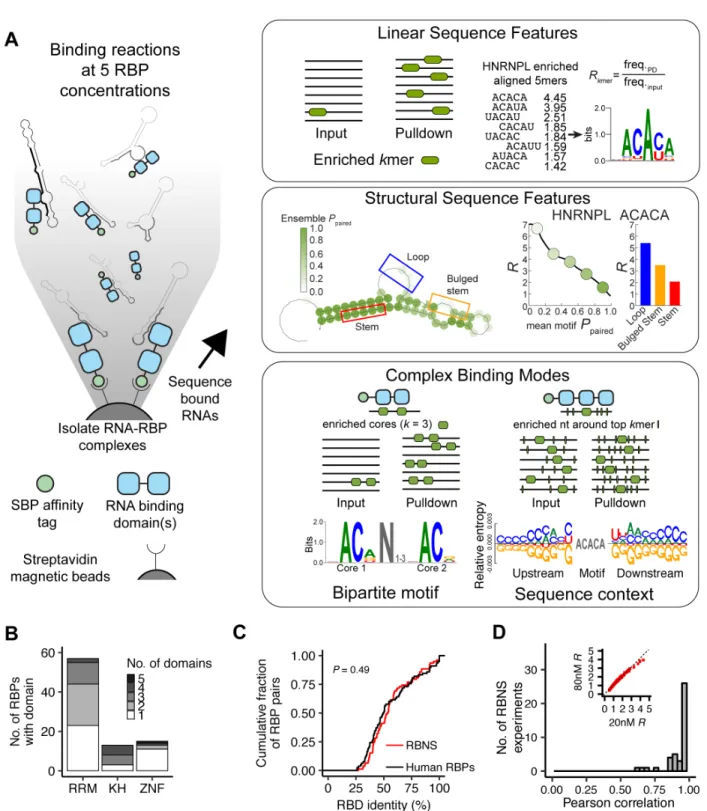
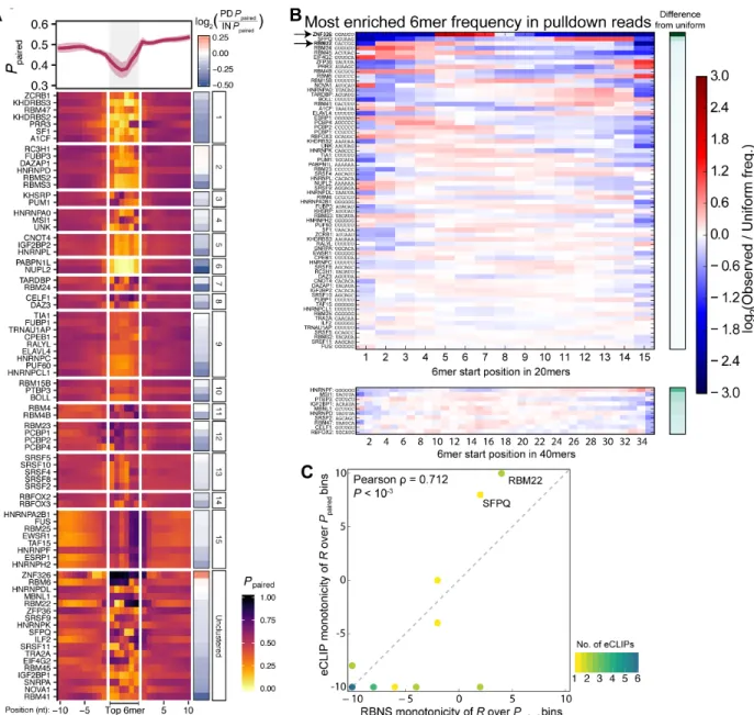
∙ In RNA Bind-n-Seq (RBNS), a purified epitope-tagged RBP (consisting minimally of the RNA binding domains plus 50 flanking amino acids on the N- and C-terminal ends) is incubated with a pool of random 20 or 40 nt oligonucleotides, and the pulled down RBP-bound RNA is subjected to high-throughput sequencing (Lambert et al.[2014]). Typically five separate incubation reactions are performed with differing quantities of the tagged RBP (5 - 1300 nM), with each of these five libraries as well as the input RNA sequenced to a depth of ∼15-20 million reads. Computational analysis of the pulldown reads compared to the input reads provides the full spectrum of bound motifs (including
high and moderate affinity RNA sequences) as well as their secondary structure and context preferences. Importantly, because the RBNS oligo pool is random in contrast to the designed pool of ∼250,000 oligos used in RNAcompete, the RBP is presented with motifs in a wide variety of sequence and secondary structure contexts, enabling the fine-tuned dissection of an RBP’s specificity and affinity landscape.
∙ Other in vitro techniques that have been used to profile the specificity and/or affinity of one or a few RBPs include: SEQRS (Selection, high-throughput sequencing of RNA and SSLs (sequence specificity landscapes),Campbell et al. [2012]); RNA-MaP (quan-titative analysis of RNA on a Massively Parallel array,Buenrostro et al.[2014]); HiTS-RAP (High-Throughput Sequencing - RNA Affinity Profiling,Tome et al.[2014]); and RNA-MITOMI (RNA-Mechanically Induced Trapping Of Molecular Interactions,
Martin et al. [2012]).
1.2.2
In vivo RNA-RBP profiling techniques
While in vitro techniques reveal the intrinsic specificity an RBP has for RNA sequence(s), complementary in vivo techniques have been developed to study RBP-RNA interactions in their endogenous cellular environments, including:
∙ The first genome-wide studies to profile the set of RNAs bound to an RBP of inter-est employed RNA ImmunoPrecipitation followed by microarray analysis (RIP-chip,
Tenenbaum et al. [2000]) or later high-throughput sequencing (RIP-seq, Zhao et al.
[2010]). These and other initial studies on dozens of RBPs revealed that RBP-RNA interactions are many-to-many; that is, each RBP typically binds hundreds to thou-sands of genes while each gene is typically bound by numerous different RBPs (Hogan et al. [2008]).
∙ Cross-Linking and ImmunoPrecipitation followed by high-throughput sequencing (CLIP-seq) is the state-of-the-art method to determine an RBP’s RNA targets and specific binding sites throughout the transcriptome. Though several CLIP-seq variants have been developed over the past years (see below), most share a general workflow of: sta-bilization of protein-RNA interactions via UV crosslinking to create covalent bonds
between amino acid residues and RNA bases in close proximity; RNA shearing; im-munoprecipitation of the RBP of interest; RNA adapter ligation; reverse transcription (RT); PCR amplification; high-throughput sequencing; and mapping of reads to the transcriptome and calling of significant regions of binding. Compared with previous methods to identify transcriptome-wide protein-RNA interactions such as RIP-chip or RIP-seq, the crosslinking step of CLIP enables more stringent purification of protein-RNA complexes, and the RNase digestion step provides binding-site resolution by creating short RBP-protected fragments of length 20-70 nucleotides (Wheeler et al.
[2017]). Though UV treatment at 254 nm is common due to its simplicity and ability to crosslink unmodified cells and tissues, this step does introduce known biases includ-ing: pyrimidines are more photoactivatable than purines (uracil most highly,Sugimoto et al.[2012], Hauer et al.[2015]); Cys, Lys, Phe, Trp, and Tyr residues crosslink more efficiently than other amino acids; and RBPs interacting with double-stranded RNA crosslink poorly due to the deep and narrow groove of A-form RNA helices making amino acid residues inaccessible to the nucleotides (Wheeler et al.[2017]).
Multiple variants of the CLIP-seq assay have been developed over the past decade, the most commonly used being:
– Photoactivatable Ribonucleoside CLIP (PAR-CLIP): Metabolic labeling in cell culture incorporates UV radioactive nucleoside analogs (4-thiouridine, 4sU, or 6-thioguanosine, 6sG) into RNA, which is subsequently crosslinked at 365 nm UV irradiation (Maatz et al. [2017]). RNA yield is increased due to the high reac-tivity compared to traditional UV crosslinking, and the mutation of T to C at the crosslinking site of 4sU after reverse transcription in up to 70% of reads pro-vides confidence in identified interaction sites (Hafner et al.[2010]). However, the method is only applicable to cell culture systems in which the modified nucleoside can be introduced, and the modified nucleoside may introduce stress responses (Burger et al. [2013]) and favor RNAs with short half-lives as the nucleoside is present in a higher proportion of those messages.
and 3’ RNA adapters were ligated to the immunoprecipitated RNA fragments prior to reverse transcription. As the UV-induced amino acid-RNA covalent adducts often terminate reverse transcriptase, up to 80% of the cDNA products did not contain the 5’ adapter and were not amplified. iCLIP addressed this by introducing a single RT primer that contained two cleavable adapter regions, with circularization after RT followed by digestion producing a linear cDNA molecule containing both sequencing adapters (König et al. [2010]). Because a number of reads terminate at the RT stop, a portion of the mapped iCLIP reads mark crosslink sites with individual nucleotide resolution. However, the circular ligation is very inefficient and has known biases of preferred nucleotides at the fragment ends, calling into question the quantitative nature of the assay (Baran-Gale et al.
[2015]).
– enhanced CLIP (eCLIP): modifications to the iCLIP protocol and inclusion of input controls in the eCLIP protocol has enabled large-scale, robust profiling of hundreds of diverse RBPs (Fig. 1-3, Van Nostrand et al. [2016]). These im-provements include omission of radiolabeling and autoradiographic visualization steps; improved enzymatic reaction efficiencies; ligation of a second 3’ adapter to the single-stranded DNA after reverse transcription instead of circular ligation to increase capture efficiency; inclusion of an in-line 5-10 nt randomer in the sec-ond adapter to distinguish unique molecules from PCR duplicates, making the number of reads more quantitative and enabling a higher percentage of usable, nonduplicated reads; and sequencing of a ‘size-matched’ input pre-IP sample that has the same crosslinking, fragmentation, ligation, and amplification biases as the IP sample to control for nonspecific background and inherent capture biases. These improvements reduce the PCR amplification needed by ∼1000-fold (e.g., 16 eCLIP vs. 28 iCLIP PCR cycles needed for RBFOX2), requiring fewer start-ing cells (less than a million) and resultstart-ing in a sequenced library with a higher percentage of usable reads and greater library complexity.
RNA binding proteins in two human cell lines through the Encyclopedia of DNA Elements (ENCODE) project, allowing a consistent comparison of the in vivo binding preferences of diverse RBPs that is explored in Chapter3.
Figure 1-3: The enhanced CLIP (eCLIP) assay
RBP-RNA interactions are covalently stabilized by UV-crosslinking, followed by RNase I digestion and IP with a validated antibody. After stringent washes, the 3’ adapter with an in-line random barcode is ligated to RNA, and a region 75 kDa (∼220 nt of RNA) above the protein size is excised and treated with proteinase K to isolate RNA. After RT and the second 3’ adapter ligation (this time to cDNA), a library is prepared for high-throughput sequencing. Reads that were truncated at the RT position result in sequencing reads with read 2 of the paired-end (PE) sequence beginning just 3’ of the crosslink site (Van Nostrand et al. [2016]).
∙ mRNA interactome capture
To assay the global scope of protein-RNA interactions in mammalian cells, ‘mRNA interactome capture’ methods have been developed that combine UV-crosslinking with highly stringent oligo(dT) affinity purification to enrich for proteins associated with polyadenylated RNA (Kastelic and Landthaler [2017]). These methods have uncov-ered vast repertoires of RBPs with hundreds of novel RBP candidates that were not predicted to bind RNA based on presence of well-defined, annotated RBDs or previous RNA-related roles reported in the literature. One pioneering study in HEK cells iden-tified close to 800 mRNA-bound proteins, nearly one-third of which were previously unannotated as binding mRNA and 15% of which were not computationally predicted to interact with RNA (Baltz et al. [2012]). Another study in HeLa cells identified 860 mRNA-bound proteins, only 233 of which contained a classical RBD with intrinsi-cally disordered regions being highly enriched in the mRNA-bound set (Castello et al.
[2012]). These interactome capture methods will allow changes in protein-mRNA inter-actions in response to stimuli and disease to be studied in addition to the identification of the RNA-bound proteome in diverse cell lines and organisms.
Variants of mRNA interactome capture to identify the proteins bound by a specific RNA of interest include ChIRP-MS (Comprehensive Identification of RNA binding Proteins by Mass Spectrometry, which utilizes cells grown in standard medium, Chu et al. [2015]) and RAP-MS (RNA Antisense Purification-Mass Spectrometry, which utilizes cells grown in SILAC medium,McHugh et al.[2015]), both of which were devel-oped to determine Xist-interacting proteins that mediate X-chromosome inactivation. After protein-RNA crosslinking, 20-90 nt long biotinylated DNAs complementary to the RNA of interest are used to capture RNA-protein complexes, and eluted proteins are identified through mass spectrometry. The high overlap of 9 out of 10 RAP-MS-identified RBPs also being pulled down in ChIRP-MS highlights the former’s specificity, with these two techniques enabling identification of the proteins that interact with any specific RNA sequence in vivo.
1.2.3
Genetic studies of RNA profiling after RBP perturbation
To better understand the role(s) than an RBP plays in RNA homeostasis, the RBP can be perturbed through genetic or other means with resulting effects on RNA processing measured in a systematic and genome-wide manner through any number of sequencing-based assays. RBP perturbation can be achieved through transient siRNA-, stably integrated shRNA-, or more recent CRISPR-Cas13a-mediated (Abudayyeh et al. [2017]) RBP knockdown (KD); CRISPR-mediated RBP knockout (KO, if the RBP is not essential) or deletion of particular protein domain(s); or RBP overexpression. The most common assay to profile changes in the transcriptome after RBP perturbation is traditional steady-state RNA sequencing, which can reveal changes in alternative splicing and promoter usage; gene expression levels (from which RBP regulation of mRNA stability can be inferred); and RNA editing in the RBP-perturbed cells compared to control cells. Alternative assays to probe different aspects of RNA metabolism after RBP perturbation include ribosome profiling to measure changes in translation (Ingolia et al. [2009]); RNA-seq on different subcellular fractions to measure changes in mRNA localization (Lefebvre et al. [2017]); techniques such as 3P-seq (Jan et al.
[2011]) to measure changes in poly(A) site usage and TAIL-seq (Chang et al. [2014]) or PAL-seq (Subtelny et al. [2014]) to measure changes in poly(A) tail length; and metabolic labeling or transcriptional inhibition followed by time course measurements to more directly measure mRNA half-lives (Tani and Akimitsu [2012]). A limitation of RBP perturbation studies is that it is often difficult to disambiguate direct from indirect changes (e.g., whether the RBP in question directly affects splice site choice or its perturbation changes the level of a splicing factor that is responsible for an observed splicing change), though confidence in identifying directly regulated events can be achieved by integrating binding (e.g., in vivo-based CLIP or in vitro-vivo-based RBNS) data and considering events that have evidence of direct RBP interaction.
1.2.4
Structural studies of RBPs and RBP-RNA interactions
The two most common methods for determining high-resolution atomic structures of pro-teins and protein-RNA complexes are nuclear magnetic resonance (NMR) spectroscopy and
X-ray crystallography. Since the first protein-RNA complex was solved using X-ray crystal-lography (Chen et al.[1989]), improvements in instrumentation and computational modeling techniques have led to these structural studies being incredibly influential in revealing infor-mation about RBP-RNA interactions. Overviews of the two techniques and and differences between them include:
∙ NMR structures represent an average over semi-random oriented molecules tumbling in solution over a nanosecond to second time scale (Brünger [1997]). Proteins ≤ 30 kDa (∼ 270 amino acids) are amenable to NMR spectroscopy. As they occur in solution, NMR studies of proteins are closer to their physiological state and allow identification of flexible portions of the protein including interdomain linker sequences commonly observed in multi-domain RBPs. NMR studies also allow conformational changes to be observed, such as those that may occur upon RNA binding to a multi-RBD protein (Afroz et al.[2015], Mackereth et al. [2011]).
∙ X-ray crystallography structures represent an average over molecules arranged in a regular crystal lattice over a seconds to hours time scale (Brünger [1997]). X-ray crystallography is also able to provide the position of water molecules in the structure, allowing prediction of water-mediated hydrogen bonds which may be important in protein-RNA specificity (Afroz et al.[2015]). It can be applied to proteins or complexes > 100 kDa, though obtaining a crystal is not guaranteed and is often time-consuming even when possible.
As of October 2017, there were 230 structures of human RRMs in the Protein Data Bank (140 solution NMR and 90 X-ray crystallography; 174 RRM(s) alone and 56 in complex with RNA). There were 58 structures of human KH domains in the PDB, 27 solution NMR and 31 X-ray crystallography, 13 of which are in complex with RNA or DNA.
1.3
RNA secondary structure in RBP-RNA interactions
and regulation
Deciphering the structures of complex three-dimensional biomolecules is essential to fully understand their regulatory capacity and biological function. In contrast to double-stranded DNA, RNA is single-stranded, permitting it to fold into complex secondary and tertiary structures that can directly influence RNA regulatory capacity or alter the ability of RBPs to bind cis-sequence elements. Single nucleotide polymorphisms (SNPs) in UTRs that induce RNA conformational changes have been associated with six genetic diseases, underscoring the importance of considering secondary structure in understanding RNA function (Halvorsen et al. [2010]).
RNA structures can be revealed through three complementary approaches: in silico fold-ing as well as in vitro or in vivo profilfold-ing studies. In silico-folded structures are typically predicted from dynamic programming algorithms that efficiently search the set of all pos-sible structures, except for those containing pseudoknots (Eddy [2004]). The prediction(s) for an RNA can include the minimum free energy (MFE) structure, which is the most prob-able structure at equilibrium, or a set of structures with associated probabilities based on the partition function that describes the ensemble thermodynamic properties of the system (Fig. 1-4). Commonalities at particular RNA bases over the latter set of structures can provide an estimate of the quality of the prediction and identify highly probable base pairs (Mathews et al. [2010]). Benchmarking of in silico RNA structure prediction from sequence alone for rRNAs and other well-studied catalytically active RNAs yielded an ∼70% base pair accuracy (Hajiaghayi et al. [2012]), with the accuracy dropping for sequences 1 kb or longer such as full-length mRNAs (Doshi et al.[2004]).
Figure 1-4: Examples of in silico-folded RNA oligos
(a) Example of an RNA oligo folded in silico with RNAfold (Lorenz et al.[2011]), with the probability of each RNA base being paired (𝑃paired) in the ensemble of predicted structures
according to their thermodynamic energies.
(b) Example of the RNA bases of the minimum free energy structure of an RNA oligo classified into five secondary structural elements.