In animals, fundamental clock mechanisms are highly conserved and are based on autoregulatory feedback loops that generate molecular oscillations in the transcript levels of approximately 5–10% of genes in essentially all tissues. In mammals, the transcription-factor heterodimer CLOCK–BMAL1 (or NPAS2–BMAL1) activates the transcription of Per1, Per2 and Per3 (denoted Period) and Cry1 and Cry2 (denoted Cryptochrome) genes from E-box enhancer elements, whose protein products form complexes inhibiting CLOCK–BMAL1 transactivational activity after a delay of several hours. In additional feedback loops, CLOCK–BMAL1 also supports the rhythmic expres- sion of Rev-ErbA and Rev-ErbB (official symbols Nr1d1 and Nr1d2, respectively), which modulate the expression of Bmal1 (official symbol Arntl), and D-site-binding protein (Dbp), which modulates the expression of Per2 (ref. 1).
Although circadian-clock regulation of many metabolic processes is well established, there is increasing evidence that metabolic signals feed back to the circadian oscillator and consequently adjust meta- bolic pathways within individual cells
2. Heme, an iron-containing porphyrin that serves as a prosthetic group in several hemoproteins, has also been described to be a ligand of REV-ERBA. Heme bind- ing to REV-ERBA supports co-repressor recruitment and transcrip- tional repression
3–5, thereby coordinating circadian and metabolic pathways. In addition, heme binding to NPAS2 in vitro mediates the CO-sensitive DNA binding of NPAS2–BMAL1 (ref. 6).
Heme synthesis is governed by the rate-limiting enzyme amino- levulinate synthase 1 (Alas1), which is negatively regulated by glucose
and heme concentrations
7. In addition, Alas1 transcription is circa- dian, through transcriptional regulation by NPAS2–BMAL1 (ref. 8).
Heme catabolism is mediated by heme oxygenases 1 and 2 (Ho-1 and Ho-2; official symbols Hmox1 and Hmox2, respectively), which generate biliverdin (and subsequently bilirubin), iron and carbon monoxide at the expense of nicotinamide adenine dinucleotide phosphate (NADPH)
9. Although heme can regulate the transcription of Period genes
8, and circadian heme proteins may sense the redox state and potentially dia- tomic gases such as oxygen, nitric oxide and CO, the relevance of heme catabolism to circadian transcription and dynamics is unknown.
RESULTS
Inhibition of heme degradation alters circadian dynamics To test whether cellular heme levels are critical for circadian dynam- ics, we treated human U2-OS reporter cells expressing firefly luci- ferase from a Bmal1 promoter fragment with various concentrations of either hemin (iron protoporphyrin) or cobalt protoporphyrin, a specific inhibitor of the heme-degrading heme oxygenases
10,11. Both compounds probably lead to increased intracellular heme levels either by providing additional heme or by inhibiting heme degradation. Whereas hemin treatment had no effect on the circadian period, the inhibition of heme degradation by cobalt pro- toporphyrin resulted in an inhibitor-dose-dependent lengthening of the circadian period of up to 1.5 h (Fig. 1). Thus, in this assay, heme-degradation products rather than the heme level itself were critical for circadian dynamics.
1Laboratory of Chronobiology, Charité Universitätsmedizin Berlin, Berlin, Germany. 2Institute of Pharmacology, Center for Cardiovascular Research CCR, Charité Universitätsmedizin Berlin, Berlin, Germany. 3Bernhard-Nocht-Institut, Hamburg, Germany. 4Department of Cell and Developmental Biology, University College London, London, UK. 5Division of Biochemistry, Department of Biology, University of Fribourg, Fribourg, Switzerland. 6Present address: Westfälische Wilhelms-Universität Münster, Münster, Germany. Correspondence should be addressed to A.K. ([email protected]).
Reciprocal regulation of carbon monoxide metabolism and the circadian clock
Roman Klemz 1 , Silke Reischl 1 , Thomas Wallach 1 , Nicole Witte 2 , Karsten Jürchott 1 , Sabrina Klemz 1 , Veronika Lang 1 , Stephan Lorenzen 3 , Miriam Knauer 2 , Steffi Heidenreich 2 , Min Xu 4 , Jürgen A Ripperger 5 , Michael Schupp 2 , Ralf Stanewsky 4,6 & Achim Kramer 1
Circadian clocks are cell-autonomous oscillators regulating daily rhythms in a wide range of physiological, metabolic and behavioral processes. Feedback of metabolic signals, such as redox state, NAD
+/NADH and AMP/ADP ratios, or heme, modulate circadian rhythms and thereby optimize energy utilization across the 24-h cycle. We show that rhythmic heme degradation, which generates the signaling molecule carbon monoxide (CO), is required for normal circadian rhythms as well as circadian metabolic outputs. CO suppresses circadian transcription by attenuating CLOCK–BMAL1 binding to target promoters.
Pharmacological inhibition or genetic depletion of CO-producing heme oxygenases abrogates normal daily cycles in mammalian cells and Drosophila . In mouse hepatocytes, suppression of CO production leads to a global upregulation of CLOCK–BMAL1- dependent circadian gene expression and dysregulated glucose metabolism. Together, our findings show that CO metabolism is an important link between the basic circadian-clock machinery, metabolism and behavior.
http://doc.rero.ch
Published in "Nature Structural & Molecular Biology 24(1): 15–22, 2017"
which should be cited to refer to this work.
Heme degradation is regulated by the circadian clock
The intricate connection between the circadian and metabolic sys- tems manifests itself in that metabolic feedbacks into the circadian oscillator often have a circadian component. To test whether heme degradation is regulated by the circadian clock, we analyzed tran- script and activity levels of Ho-1, the major heme-degrading enzyme in peripheral tissues
9. In regular 6-h intervals over the course of two consecutive days, we harvested livers from mice kept in constant dark- ness and determined Ho-1 transcript levels by quantitative RT–PCR.
Ho-1 expression showed a circadian rhythm with a peak at circadian time (CT) 12–15 and an approximately three-fold peak-to-trough ratio (Fig. 2a), results consistent with findings in peritoneal mac- rophages (Supplementary Fig. 1a) and with the diurnal variation observed by Xu and colleagues in liver tissue
12.
Transcription of Ho-1 is induced by heme via stress-response enhancer elements (StREs) in its promoter
13. To test whether rhyth- mic Ho-1 expression is regulated by potentially rhythmic heme levels, we applied constant high concentrations of heme or cobalt protopor- phyrin to synchronized primary hepatocytes. Whereas we observed the expected increase in Ho-1 transcription (and HO-1 activity, in the case of heme treatment), circadian oscillations persisted (Fig. 2b) but were absent in hepatocytes from Bmal1
−/−mice (Supplementary Fig. 1b). This finding indicates that rhythmic Ho-1 regulation is dif- ferent from rhythmic StRE activation and depends on the canonical circadian oscillator. Importantly, the activity of heme oxygenase was also rhythmic in the liver in both basal and heme-induced condi- tions (Fig. 2c), thus indicating that heme is rhythmically degraded.
Interestingly, expression of Alas1, which encodes the rate-limiting enzyme in the heme biosynthesis pathway, is also rhythmic with a similar phase
8to that of Ho-1, thus suggesting a potential mechanism for intracellular heme homeostasis.
To investigate whether Ho-1 is a direct output gene of the circadian clock, we searched for clock-controlled enhancer elements within the Ho-1 promoter and identified a highly conserved E box close to the transcription start site (Supplementary Fig. 1c). A 1,000-bp fragment of the Ho-1 promoter was activated by CLOCK–BMAL1 in HEK293 cells and repressed by CRY1, but this regulation was abolished by E-box mutation (Fig. 2d). In addition, chromatin immunoprecipi- tation (ChIP) experiments on liver nuclei revealed that the Ho-1 E box was occupied by endogenous BMAL1 in a manner depend- ent on the time of day (Fig. 2e). Moreover, transcript levels of Ho-1
were deceased in the hepatocytes of Bmal1
−/−mice (Supplementary Fig. 1d). Together, these results support the role of Ho-1 as a direct target gene of CLOCK–BMAL1 in vivo.
The heme-degradation product CO modulates circadian transcription
Rhythmic heme degradation mediated by HO-1 leads to rhythmic accu- mulation of heme-degradation products, i.e., biliverdin, iron and CO.
In vitro, CO inhibits binding of NPAS2–BMAL1 to DNA
6. If such an inhibitory effect also occurs in vivo, transcription of NPAS2–BMAL1- target genes should increase when endogenous CO levels are decreased.
Because heme oxygenase activity is the primary source of endogenous CO, and Ho-1 is the dominant paralog in the periphery
13, we measured circadian transcript levels of CLOCK(NPAS2)–BMAL1-target genes in primary fibroblasts from Ho-1-knockout mice
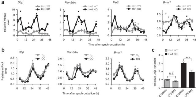
14. Indeed, transcript levels of clock genes containing functional promoter E boxes, such as Dbp, Rev-ErbA and Per2, were substantially upregulated, whereas transcription of Bmal1 was downregulated during periods in which levels of the repressor REV-ERBA was high (Fig. 3a). The extent of this upregulation (for example, for Dpb transcript levels) was somewhat variable, ranging from 1.5-fold up to six-fold (Supplementary Fig. 2).
It is likely that the effect of Ho-1 depletion on E-box-controlled mRNA levels was transcriptional rather than post-transcriptional, because precursor (pre)-mRNA levels of Dbp and Rev-ErbA were similarly upregulated in Ho-1-knockout cells (Supplementary Fig. 3a). Thus, decreasing CO levels by downregulating heme oxygenase activity has a major effect on circadian transcription.
If the upregulation of E-box-containing circadian transcripts in Ho-1-knockout animals were due a decrease in endogenous CO levels, exogenous application of CO should, at least in part, reverse this transcriptional effect. Continuous application of 6% gaseous CO (compared with 6% nitrogen as a control for any hypoxia-induced effects) to primary fibroblasts from wild-type mice led to a slight decrease in expression of the E-box-controlled transcripts Dbp and Rev-ErbA, but not Bmal1 (Fig. 3b). Similarly, acute CO application through use of CO-releasing molecules (CORMs) had only subtle effects on Dbp transcription in wild-type fibroblasts (discussion of the CO concentration in Supplementary Note 1). In contrast, acute or continuous CO treatment of Ho-1
−/−primary fibroblasts substan- tially, yet not always completely, rescued high Dbp transcript levels in Ho-1-knockout fibroblasts (Fig. 3c and Supplementary Fig. 2).
Importantly, this CO-mediated rescue was also present at the pre-mRNA level, a result consistent with the hypothesis that CO modulates transcription (Supplementary Fig. 3b).
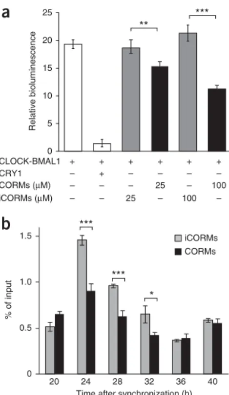
CO suppresses transactivation and target-gene binding of CLOCK–BMAL1
To test whether CO-mediated transcriptional inhibition of genes containing functional E boxes correlates with CO inhibition of CLOCK(NPAS2)–BMAL1, we measured the transactivation activity of CLOCK–BMAL1 from an E-box-driven luciferase reporter in HEK293 cells treated with either CORMs or inactive controls (iCORMs). We found a CO-dose-dependent decrease in CLOCK–
BMAL1-mediated transcription (Fig. 4a), thus indicating that CO directly regulates circadian transcriptional activity. This inhibitory effect is probably due to CO interfering with CLOCK(NPAS2)–
BMAL1 binding to DNA, because ChIP experiments revealed that CO treatment decreased BMAL1 binding to the Rev-ErbA promoter in synchronized U2-OS cells at times when BMAL1 is transcriptionally active (Fig. 4b), although overall BMAL1 levels remained unchanged (Supplementary Fig. 3c).
a b
26.0 25.5 25.0 24.5 24.0 23.5 0
Hemin CoPPIX 1.6
1.4 1.2 1.0 0.8 0.6 0.4
Hemin 0 CoPPIX
6 25 50 100
μM
**
***
***
Relative bioluminescence
24 48 72 96 120 24 48 72 96 120 0 6 25 50 100
Time after synchronization (h)
Period (h)
Concentration (μM)
Figure 1 Inhibition of heme degradation alters circadian dynamics in
human cells. (a) Circadian oscillation dynamics of dexamethasone- synchronized human osteosarcoma cells (U2-OS) bearing a Bmal1 promoter–luciferase reporter construct treated with the indicated concentrations of hemin or the heme oxygenase inhibitor cobalt protoporphyrin (CoPPIX). Shown are detrended average time series of six parallel treatments. (b) Circadian periods of time series shown in a (mean o s.d.; n = 6 independent cell cultures). Two-way ANOVA with Bonferroni post test revealed a significant difference between hemin and CoPPIX effects (P < 0.0001) and concentration (P < 0.0001). Three independent experiments yielded similar results. **P < 0.01; ***P < 0.001.
http://doc.rero.ch
Heme oxygenase–derived CO is essential for normal circadian dynamics in mammalian cells
The lengthening of the circadian period after pharmacological inhi- bition of heme oxygenases (Fig. 1a) suggested that heme oxygen- ase activity is an integral part of circadian-rhythm generation. To genetically test this phenomenon, we lentivirally transduced Ho-1
−/−primary fibroblasts with a Bmal1-luciferase reporter construct and measured circadian-rhythm dynamics, but we were unable to detect a change in circadian period when this heme oxygenase was
individually depleted (Supplementary Fig. 4a). We therefore further decreased heme oxygenase activity (Supplementary Fig. 4b), and thus probably endogenous CO levels, by RNA interference (RNAi)- mediated knockdown of the second active heme oxygenase paralog, Ho-2 (ref. 15), in Ho-1
−/−primary fibroblasts (genotype denoted Ho-1
−/−; Ho-2
KD). Circadian rhythms in these Ho-1
−/−; Ho-2
KDfibroblasts showed a substantial lengthening of the period by up to 2 h (Fig. 5a and Supplementary Fig. 5a), probably as a result of effects on circadian transcription, given that upregulation of the transcript
0 5 10 15
0 12 24 36 48 Untreated Hemin CoPPIX
0 50 100 150 200
0 12 24 36 48 Untreated Hemin
0.0 0.1 0.2 0.3 0.4
ZT6 ZT18 ZT6 ZT18 Bmal1 KO Ho-1 Rev-Erb Bmal1
Circadian time (h) 0
0.5 1.0 1.5 2.0
0 1 2 3 4 5
C–B CRY1
– + + – + +
Relative luciferase activity
– – + – – + WT Mut
a b c d e
Relative Ho1 mRNA Relative Ho1 mRNA
0 12 0 12 0
Time after medium change (h)
HO activity (nmol h–1 mg–1) % of input
Figure 2 Heme degradation is regulated by the circadian clock. (a) Heme oxygenase 1 (Ho-1) transcript levels (normalized to Hprt and relative to mean
levels) from mouse liver over 2 d in constant darkness (mean o s.e.m.; n = 4 livers). (b) HO activity rhythms in untreated and hemin (30 MM)-stimulated primary hepatocytes. Shown are mean levels (large symbols) of two independent hepatocyte samples (small symbols). (c) Ho-1 transcript rhythms (relative to Hprt) in primary hepatocytes treated with hemin or cobalt protoporphyrin (30 MM each). Data are normalized to mean levels of untreated control cells. Shown are mean levels (large symbols) of two independent hepatocyte samples (small symbols). (d) CLOCK–BMAL1 (C–B)-mediated transactivation and repression by CRY1 in HEK293 cells from a Ho-1 promoter luciferase reporter with (mut) or without (WT) a mutated E box (mean o s.e.m.; n = 4 independent cell cultures). (e) ChIP analysis of BMAL1 at the Ho-1, Rev-ErbA and Bmal1 genes at zeitgeber time (ZT) ZT6 and ZT18 from wild-type or Bmal1
−/−(Bmal1 KO) liver chromatin (mean o s.d.; n = 6 independent precipitations from liver samples from three mice per ZT and genotype). One-way ANOVA with a Bonferroni post test comparing all the different conditions indicated that the wild-type ZT6 data for the presence of BMAL1 at Rev-ErbA or Ho-1 were highly significantly different from all other data for the same gene, and there were no such significant changes observed for the negative control, Bmal1.
Bmal1
0 2 4 6 8
0 2 4 6 8 10
0 12 24 36 48
0 12 24 36 48
0 12 24 36 48 0 12 24 36 48
0 1 2 3 4
Dbp Per2
Relative mRNA level
Relative mRNA level 0.0 0.5 1.0 1.5 2.0 2.5
0.0 0.5 1.0 1.5 2.0
0 12 24 36 48
0.0 0.5 1.0 1.5 2.0 2.5
0 12 24 36 48
Time after synchronization (h)
Dbp Bmal1
Ho1 WT Ho1 KO Ho1 WT
Ho1 KO
Ho1 WT Ho1 KO
0.0 0.5 1.0 1.5
0 12 24 36 48
Ho1 WT Ho1 KO
N2 CO
N2 CO
N2 CO
4 3 2 1 0
***
Ho1 WT Ho1 KO Rev-Erb
Rev-Erb
Time after synchronization (h)
Relative Dbp transcript
N.S.
iCORMs CORMs iCORMs CORMs
a
b c
Figure 3 The heme-degradation product CO modulates circadian transcription. (a) Transcript rhythms of Bmal1, Per2, Rev-ErbA and Dbp in
dexamethasone-synchronized primary fibroblasts from Ho-1
−/−mice (Ho1 KO) or wild-type littermates (Ho1 WT). Data are normalized to Hprt expression and are presented relative to mean expression in wild-type cells. Shown are mean levels (large symbols) of two independent fibroblast samples from one mouse per genotype (small symbols). The amplitudes of sine fits (peak-to-trough ratios) for Ho1 WT versus Ho1 KO are 3.1 versus 2.9 for Dbp; 4.6 versus 6.1 for Rev-ErbA; 1.5 versus 3.2 for Per2; and 1.6 versus 3.4 for Bmal1. (b) Transcript rhythms of Dbp, Rev-ErbA and Bmal1 in dexamethasone- synchronized primary fibroblasts from wild-type mice continuously treated with 6% CO or N
2. Normalization and presentation are as in a. (c) Transcript levels of Dbp in embryonic fibroblasts from Ho-1
−/−mice (or wild-type littermates 24 h after dexamethasone synchronization, which were treated for 1 h with 100 MM CO-releasing molecules (CORMs) or inactive control molecules (iCORMs) before harvesting. Data are normalized to Gapdh expression and are presented relative to mean expression in wild-type cells treated with iCORMs. Data are shown as mean o s.d. of three independent samples from one mouse per genotype. Two-way ANOVA with Bonferroni post test revealed a significant difference between genotypes (P < 0.0001) and drugs (P < 0.0001). ***P < 0.001; N.S., not significant. Similar experiments with samples from different mice are shown in Supplementary Figure 2.
http://doc.rero.ch
levels of the E-box-controlled genes Dbp and Rev-ErbA was enhanced in the Ho-1
−/−; Ho-2
KDfibroblasts (Fig. 5b).
If depletion of endogenous CO in Ho-1
−/−; Ho-2
KDcells also con- tributes to the altered circadian dynamics, application of exogenous CO should be able to, at least in part, revert the period lengthening in Ho-1
−/−; Ho-2
KDcells. Indeed, continuous application of 6% CO, but not N
2,led to a reshortening of the circadian period in Ho-1
−/−; Ho-2
KDfibroblasts (Fig. 5c and Supplementary Fig. 5b) but had no effect on the circadian period in wild-type cells (Supplementary Fig. 5c), thus indicating that endogenous CO levels are essential for normal circadian-rhythm generation.
Heme oxygenase depletion globally alters clock-controlled transcription in hepatocytes
The strong effect of heme oxygenase depletion on circadian transcrip- tion and dynamics suggests a substantial effect on clock-controlled transcriptional output. To investigate this possibility globally, we per- formed microarray-based genome-wide transcriptional profiling of primary hepatocytes from wild-type or Ho-1
−/−mice with or without additional knockdown of Ho-2. As expected from results in primary fibroblasts (Fig. 3a), circadian transcript levels of the clock-controlled genes Dbp and Rev-ErbA were upregulated in Ho-1
−/−hepatocytes, even more so in Ho-2
KDhepatocytes and to the greatest extent in
Ho-1
−/−; Ho-2
KDcells (Fig. 6a). We identified 2,335 genes that were differentially expressed among these genotypes and grouped them into six clusters according to their gene-expression patterns (Fig. 6b and Supplementary Table 1).
If heme oxygenase depletion acts globally on circadian transcrip- tional output, genes that are rhythmically expressed in the liver should be highly represented among differentially expressed genes
16. This was indeed the case in cluster 1 (P < 10
−14) and to a lesser extent in cluster 2 (P < 10
−4) (Fig. 6b). Cluster 1 contained 473 genes whose transcript profiles were most similar to that of the E-box-containing clock- controlled gene Dbp (comparison with Fig. 6a). In addition, cluster 1 was highly enriched in genes reported to bind endogenous BMAL1 in ChIP experiments on liver chromatin (P < 10
−22(ref. 17)), and genes with promoter E-box motifs (both E1 and E2 E boxes) were overrep- resented only in cluster 1 (Fig. 6c). Although clusters 1 and 2 were
CLOCK-BMAL1 CRY1 CORMs (μM) iCORMs (μM)
+ + + + + +
– – – – + –
100 – 25 – – –
– 100 – 25 – –
Relative bioluminescence
25 20 15 10 5 0
***
**
Time after synchronization (h) 1.5
1.0
0.5
0
% of input
iCORMs CORMs
***
*
***
40 36 32 28 24 20
a
b
Figure 4 Carbon monoxide suppresses transactivation and target-gene
binding of CLOCK–BMAL1. (a) CLOCK–BMAL1-mediated transactivation from an E-box-containing artificial promoter in HEK293 cells treated with CO-releasing molecules (CORMs) or inactive control molecules (iCORMs). Data are shown as mean o s.d.; n = 3 independently transfected cell cultures. Two-way ANOVA with Bonferroni post test revealed a significant difference between iCORM and CORM treatments (P < 0.0001). ***P < 0.001; **P < 0.01. (b) ChIP analysis of BMAL1 bound to the Rev-ErbA gene in synchronized U2-OS cells treated with 100 MM CORMs or control iCORMs (mean o s.e.m. of four pairwise comparisons). Two-way ANOVA with Bonferroni post test revealed a significant difference between genotype (P < 0.0001) and time (P < 0.0001). ***P < 0.001; *P < 0.05.
0.6 0.7 0.8 0.9 1.0 1.1 1.2 1.3 1.4
0 24 48 72 96 120
NS 1 2 3 shRNA 26
25 24
96 90 98
Ho1 KO + shRNA1 shRNA2 shRNA3
5 4 3 2 1 0
3.0 2.5 2.0 1.5 1.0 0.5 0 Dbp
24 h 36 h
NS KD NS KD
Ho1 KO Ho1 KO
NS KD NS KD
0.7 0.8 0.9 1.0 1.1 1.2 1.3
CO 50 M 1 CO 50 M 2 24 h 36 h
Relative bioluminescence Period (h)
NS
Time after synchronization (h)
Rev-Erb
Relative mRNA expression Relative bioluminescence
24 48 72 96
Time after synchronization (h) N2 control
a
b
c
Figure 5 Heme oxygenases are essential for normal circadian dynamics
in mammalian cells. (a) Circadian oscillation dynamics of synchronized primary fibroblasts from Ho-1
−/−mice lentivirally transduced with (i) short hairpin RNA (shRNA) constructs targeting Ho-2 or a nonsilencing (NS) control and (ii) a Bmal1 promoter–luciferase reporter construct.
Shown are representative examples of detrended time series (raw data in
Supplementary Fig. 5a) and period quantification (inset; numbers abovebars show percentage Ho-2 mRNA knockdown). (b) Transcript levels of
Dbp and Rev-ErbA 24 h and 36 h after dexamethasone synchronizationof primary fibroblasts from Ho-1
−/−mice transduced with an shRNA construct targeting Ho-2 (KD) or a nonsilencing (NS) control. (c) Circadian dynamics of Ho-1
−/−; Ho-2
KDcells as in a, continuously treated with 6%
CO or N
2. Shown are two representative examples of detrended time-series (raw data in Supplementary Fig. 5b).
http://doc.rero.ch
also enriched in target genes of REV-ERBA and REV-ERBB (ref. 18), we believe that it is unlikely that the upregulation of these targets in Ho-1- and Ho-2-depleted hepatocytes was due to CO directly regulating the activity of REV-ERBA and REV-ERBB (discussion in Supplementary Note 2).
Heme oxygenase depletion alters glucose homeostasis
Gene ontology analysis revealed significant enrichment of genes involved in metabolic processes in cluster 1, whereas in the Ho-1- dominated cluster 4, the significant GO terms reflect known roles of Ho-1 in cytoprotection and immune response
13,14(Fig. 6b). Genes
5 4
4
3 Ho1 WT + NS
Ho1 KO + NS
Ho1 WT + Ho2 KD Ho1 KO + Ho2 KD Rev-Erb
E1 E2 2
Cluster 1 3
2 1 0
2
1
0
24 48 24 48
–4 –3 –2 –1
1
0
Density
Cluster Circadian transcripts
BMAL1 targets
Gene ontology terms
Time after synchronization (h)
0 P value (log10)
1
2
3
4
5
6
Small-molecule metabolic process Metabolic process
Lipid metabolic process Oxidation–reduction process Organic-acid metabolic process Cellular ketone metabolic process Macromolecule metabolic process Cellular-component organization Immune-system process Immune-system process
Regulation of immune-system process Cell activation
Metabolic process Reproductive process Reproduction Response to stress Metabolic process Primary metabolic process
–3 –2 –1 0 1 2 3
Relative mRNA level
Dbp
–5
WT + NS Ho1 KO + NSWT + Ho2 KDHo1 KO + Ho2 KD
4.1 × 10⁻⁵
1 1
1 1
0.2 0.57
1 1
2.9 × 10⁻⁶ 7.8 × 10⁻¹⁵ 8.4 × 10⁻²³
a b
c
Figure 6 Heme oxygenase depletion globally alters clock-controlled transcription in hepatocytes. (a) Circadian transcription of Dbp and Rev-ErbA in
synchronized primary hepatocytes of Ho-1
−/−or wild-type littermate mice with or without additional Ho-2 depletion by RNAi. Data are normalized to
Gapdh expression and are presented relative to mean expression in wild-type cells transduced with the nonsilencing (NS) control. Shown are meanlevels (large symbols) of two independent samples from two mice (small symbols). (b) Left, Heat diagram sorted in six clusters, showing changes in gene expression detected in a microarray experiment with hepatocytes, as described in a, 24 h after synchronization, the time of maximal CLOCK–BMAL1 binding to DNA (n = 2 mice per genotype). Increases (red) or decreases (blue) in transcript levels are shown. Middle, P values indicate significance for overrepresentation (Fisher’s test) of genes with a circadian transcript in liver
16or with BMAL1 binding in their promoters
17. Right, Most significant nonredundant gene ontology terms for genes enriched in the clusters. (c) Bioinformatics analysis of cluster 1 genes for overrepresentation of E-box motifs in regions from −1,000 bp to +1,000 bp with respect to the transcription start site. Shown are P-value distributions of TRAP scores (Online Methods) for depicted E-box motifs compared with background gene sets.
50
Relative mRNA levels
1 WT NS
2 NS
3 WT Ho2 KD
4 Ho2 KD
Pck1 Lpl Cyp7a1
5
1
1 2 3 4
Relative glucose synthesis (%)
0 50 100 150
*
Control siRNA +
Ho2 siRNA +
CLOCK(NPAS2)–BMAL1 Heme
oxy- genase
Glucose CO G6pc
Pck1 G6pc
Ho1 KO Ho1 KO
1 2 3 4 1 2 3 4
1 2 3 4
a b c
Figure 7 Interplay between carbon monoxide and glucose metabolism. (a) Transcript levels of Pck1, G6pc, Lpl and Cyp7a1 in primary hepatocytes
(24 h after dexamethasone synchronization) of Ho-1
−/−or wild-type littermate mice with or without additional Ho-2 depletion by RNAi. Data are normalized to Gapdh expression and are presented relative to mean expression in wild-type cells transduced with the nonsilencing (NS) control.
Shown are mean levels (bars) of two independent samples from two mice (small symbols). (b) Ho-2 depletion via siRNA transfection increases glucagon-induced glucose production in hepatocytes (normalized to total protein content; mean o s.d.; n = 3 independent transfections of hepatocyte cultures from the same mouse; *P < 0.05 by two-tailed t test). Two independent experiments yielded similar results. (c) Schematic model of the feedback interplay between endogenous CO and glucose homeostasis. The core of this model (dark gray) describes the activation of CO production by high glucose and oxidative-stress levels and the negative influence of CO on glucose production. The mechanisms by which these regulatory influences are exerted are depicted in the light-gray area of the scheme: endogenous CO is rhythmically produced by heme oxygenases, whose transcription is activated by CLOCK(NPAS2)–BMAL1 (among other factors) (Fig. 2). CO has a negative influence on CLOCK(NPAS2)–BMAL1-mediated transcriptional activity (Fig. 3 and Fig. 4), thereby also inhibiting genes important for glucose production (Fig. 7a). High glucose levels, however, lead to the induction of Ho-1 (ref. 46) and thereby support endogenous CO production.
http://doc.rero.ch
important in glucose and lipid metabolism, such as Pck1, G6pc, Lpl or Cyp7a1, were strongly upregulated in Ho-1
−/−; Ho-2
KDhepato- cytes and to a lesser extent in Ho-1
−/−and Ho-2
KDhepatocytes, as verified by quantitative RT–PCR (Fig. 7a). Again, heme oxygenase probably affects transcriptional rather than post-transcriptional proc- esses, because the pre-mRNA levels of Pck1, G6pc, Lpl and Cyp7a1 were similarly upregulated in heme oxygenase–depleted hepatocytes (Supplementary Fig. 6). Because Pck1 and G6pc are important players in gluconeogenesis, we investigated the effect of acute small interfer- ing RNA (siRNA)-mediated Ho-2 depletion on glucagon-stimulated glucose production in primary hepatocytes. Consistently with the upregulation of these enzymes in Ho-2-depleted cells, we found a significant increase in glucagon-stimulated glucose production, thus suggesting that heme oxygenase activity is required for normal glucose homeostasis (Fig. 7b).
Heme oxygenase is essential for normal daily activity patterns in Drosophila
Because the expression of Alas1 and heme oxygenase has also been reported to be circadian in Drosophila melanogaster heads
19,20, we investigated whether heme oxygenase (dHo (official symbol Ho), which is the only isoform of heme oxygenase in Drosophila and is essential for normal fly development
21) is also required for normal rhythms in this species. To this end, we examined behavioral rhythms in flies in which dHo expression was knocked down in various cell types by two independent UAS-dHo-RNAi lines by using the binary Gal4–UAS system
22. Under standard 12 h–12 h light–dark (LD) cycles, wild-type flies exhibit crepuscular behavior, with pronounced morning (M) and evening (E) activity peaks
23(Fig. 8a). When released into constant dark conditions (DD), wild-type flies maintain their E peak, whereas the M peak weakens or disappears (Fig. 8a). Knockdown of dHo in all neurons (by using the pan-neuronal Gal4 driver elav-gal4) resulted in a substantially advanced M peak, whereas the E peak was unaf- fected (Supplementary Fig. 7a). Next, we downregulated dHo with the timeless (tim)-gal4 driver, which is active in all clock neurons and peripheral clock cells throughout the body
24. Also, using Pdf-gal, we directed dHo knockdown to the subset of the clock neurons impor- tant for regulating M activity
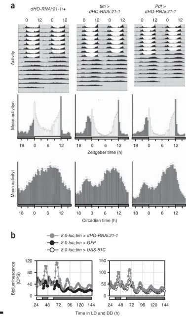
25–27. In both cases, we observed a phase advance of the M peak, which, interestingly, was also maintained in the subsequent DD period, thus indicating that dHo knockdown in clock neurons somehow enhances M activity (Fig. 8a and Supplementary Fig. 7a). Clock cell-specific dHo knockdown had no effect on E activ- ity or circadian period in DD, and dHo RNAi directed toward glia cells (by using the pan-glial driver repo-gal4) did not alter LD activity (Fig. 8a, Supplementary Fig. 7a, Supplementary Table 2 and ref. 28).
To test whether heme oxygenase depletion increases transcription from E-box-containing promoters, as observed in mammalian cells, we performed a real-time in vivo luciferase reporter assay for Period transcription in live flies
29. Flies in which dHo was downregulated in all clock cells (tim-gal4) showed normal expression levels and tempo- ral regulation of Period-promoter-driven luciferase expression under LD and DD conditions (Supplementary Fig. 7b). Similarly, clock cell-specific dHO RNAi had no effect on the expression of a Period- luciferase transgene reporter in central and peripheral clock cells
30(Supplementary Fig. 7b). On the basis of the behavioral phenotype of dHo RNAi flies, we used a reporter that directs PERIOD expres- sion in only a subset of the behavior-controlling clock neurons and that is not active in peripheral clock cells (8.0-luc
30). Strikingly, and consistently with the observed strengthening of behavioral rhythms, dHo knockdown resulted in a substantial increase in Period expression from the luciferase reporter during the normal peak expression times
tim >
dHO-RNAi:21-1
Pdf >
dHO-RNAi:21-1 dHO-RNAi:21-1/+
ActivityMean activityn
0 18
Zeitgeber time (h)
Mean activityt
Circadian time (h)
80 120
100 150 8.0-luc;tim > dHO-RNAi:21-1 8.0-luc;tim > GFP 8.0-luc;tim > UAS-51C
0 40
24
0 50
Time in LD and DD (h) Bioluminescence (CPS)
0 12 0 12 0 12 0 12 0 12 0 12
12
6 18 0 6 12 18 0 6 12
0
18 6 12 18 0 6 12 18 0 6 12
144 120 96 72
48 24 48 72 96 120 144
a
b
Figure 8 Heme oxygenase (dHo) is essential for normal daily activity
patterns in Drosophila. (a) Average locomotor activity of Drosophila males in 12 h light–12 h dark conditions for 5–8 d, followed by 7 d in constant darkness (DD). Exact genotypes from left to right: UAS-dHo-RNAi:21-1/+
(n = 15 flies); timeless-gal4:62/UAS-dHo-RNAi:21-1 (n = 16 flies); Pdf-
gal4/UAS-dHo-RNAi:21-1 (n = 16 flies). Top, double-plotted actogramsshowing average activity during the entire experiment. Gray and white areas indicate dark and light periods, respectively. The morning peak in flies with decreased dHo expression persists in DD but disappears rapidly in the controls. Bottom, histograms showing average activity within in 30 min (bars); dark bars, ‘lights off’; white bars, ‘lights on’, dots, s.e.m. The phase- advanced morning activity peak in flies with decreased dHo expression in clock cells. The same advance was observed in transgenic flies bearing a different dHo-RNAi construct (additional data and additional control genotypes in Supplementary Fig. 7a). (b) Real-time luciferase recordings of flies expressing a PERIOD-LUCIFERASE fusion protein in dorsal clock neurons encoded by the promoterless 8.0-luc transgene
30. Male flies were recorded in LD and DD, as indicated by the bars below the plots (white and black bars indicate lights on and lights off, respectively). Exact genotypes:
gray circles, 8.0-luc/+;tim-gal4:67/UAS-dHo-RNAi:21-1 (n = 14 flies);
black circles, 8.0-luc/UAS-GFP;tim-gal4:67/+ (n = 10 flies); open circles,
8.0-luc/UAS-attP-51C;tim-gal4:67/+ (n = 10 flies). The peak levels werehigher in LD and DD, and the amplitude of PER-LUC oscillations increased in dHo-RNAi flies. Similar results were obtained in two independent experiments and with the dHo-RNAi:21-8 line (Supplementary Fig. 7b).
http://doc.rero.ch
in LD and DD conditions, thus generating an increase in the overall amplitude of Period oscillations in LD (Fig. 8b and Supplementary Fig. 7b). Because the 8.0-luc construct lacks the Period promoter sequences (including the E boxes), the observed increase in PERIOD expression was probably post-transcriptionally regulated. Together, these results indicate that in Drosophila, as in mammals, the circa- dian system requires heme oxygenase for temporal coordination of behavioral activity and circadian accumulation of clock proteins, but in the Drosophila system, in contrast to the mammalian system, this coordination is not accomplished via increased transcription.
DISCUSSION
Here, we show that endogenous CO production by heme oxygenase activity is required not only for circadian oscillator function but also for balanced clock-gene and clock-target-gene expression, thereby modulating glucose homeostasis in hepatocytes. Mechanistically, we favor the hypothesis that, in mammals, heme oxygenase–derived CO attenuates CLOCK(NPAS2)–BMAL1 DNA binding, probably via coor- dination by a heme molecule bound to NPAS2 (ref. 6) and probably also to CLOCK
31, and thereby modulates expression of CLOCK(NPAS2)–
BMAL1-target genes. Although this scenario is not the only possible interpretation of our results (as described below), our preferred model is consistent with both past and present observations. (i) In vitro, micromolar concentrations of CO have been shown to impair DNA binding and BMAL-heterodimer formation of heme-bound NPAS2 but not apo-NPAS2 (ref. 6), and in the brain, these concentrations of CO are generated by heme oxygenases
32(detailed discussion of CO concentration in Supplementary Note 1). (ii) Mutation of heme-coor- dinating residues within NPAS2 also impairs heterodimer formation with BMAL1, thus decreasing both specific DNA binding to canoni- cal E boxes and NPAS2–BMAL1 transactivation activity, probably via conformational changes within the PAS-A domain of NPAS2 (ref. 33).
(iii) Although much less is known about whether CLOCK is also able to bind heme and whether CO can modulate CLOCK activity, recent biophysical data have confirmed that the PAS-A domain of CLOCK binds heme in vitro with spectroscopic properties that are consistent with a sensor of diatomic gases
31. (iv) We show here that depleting endogenous CO via heme oxygenase knockout leads to strong tran- scriptional upregulation of target genes of CLOCK(NPAS2)–BMAL1 in human and mouse hepatocytes. This observed modulation of cir- cadian transcription is probably a direct effect of CO, because appli- cation of exogenous CO, but not N
2, suppresses CLOCK–BMAL1 transcriptional activation through E-box elements, attenuates tran- scription of endogenous CLOCK(NPAS2)–BMAL1 targets, partially rescues transcriptional upregulation in heme oxygenase knockouts and suppresses BMAL1 binding to target promoters.
Together, these observations provide compelling evidence of a role of endogenous CO in modulating the transcriptional stimulatory activity of CLOCK(NPAS2)–BMAL1. However, additional potential mecha- nisms underlying the influence of heme oxygenases on circadian tran- scription and dynamics are not eliminated, because heme degradation also produces biliverdin and iron in addition to CO. Moreover, heme degradation consumes NADPH, whose intracellular concentration modulates circadian dynamics and expression of CLOCK–BMAL1- target genes
34. However, relevant to the present findings, we consider it unlikely that an increase in NADPH levels—as predicted to occur after heme oxygenase depletion—would generate long circadian periods and upregulation of CLOCK–BMAL1 targets, because such effects have previously been associated with decreased levels of NADPH
34.
It is also possible that CO indirectly influences circadian gene expression. The extent of CO’s effect on CLOCK(NPAS2)–BMAL1
transcriptional activity is difficult to estimate. Although CO was originally considered to be a metabolic waste product, today most of the described anti-inflammatory, antiapoptotic, antiproliferative and cytoprotective roles of heme oxygenases are ascribed to CO
35. Owing to the affinity of CO for metal ions, the few known CO sensors are heme-containing proteins including hemoglobin, myoglobin, soluble guanylyl cyclase (sGC), cytochrome c oxidase and the transcription factors Bach-1, Bach-2 and NPAS2. For example, heme oxygenase–
derived CO can, like nitric oxide, activate sGC, thereby leading to increased levels of cGMP. Such a mechanism has been proposed to play a role in resetting the cholinergic clock in the suprachiasmatic nucleus
36, where heme oxygenase activity is circadian
37. Moreover, increased cGMP levels can enhance AMP kinase (AMPK) activity
38, which in turn may act on the clock by promoting CRY-protein deg- radation
39. Activation of AMPK in the liver also represses expression of gluconeogenesis enzymes such as Pck1 and G6pc
40, results opposite from those observed in heme oxygenase–depleted hepatocytes. If the increases in Pck1 and G6pc transcript levels in HO-depleted hepato- cytes were due to decreased AMPK activity (perhaps mediated by increased oxidative stress
41), transcript levels of genes involved in other anabolic pathways would also be expected to be elevated
42. However, this was not the case: the expression of key genes involved in lipogenesis, such as sterol regulatory element–binding protein-1, car- bohydrate response element–binding protein, acetyl CoA carboxylase, fatty acid synthase, stearoyl-CoA desaturase, and glycerol-3-phosphate acyltransferase, was essentially unaltered. Thus, modulation of AMPK activity is unlikely to be the cause of altered clock function and increased gluconeogenesis in heme oxygenase–depleted cells.
Our results further establish that heme oxygenase also functions in the Drosophila circadian clock. Downregulation of dHo leads to an advanced morning peak and to increased PERIOD levels and higher-amplitude oscillations in clock neurons, presumably via post-transcriptional regulation. The enhanced molecular oscillations may explain the robustness of the morning activity peak and the per- sistence of bimodal behavior under constant conditions. Typically, the morning activity peak weakens or disappears under constant conditions, whereas the evening peak is sustained, thus leading to unimodal behavior
43. The enhanced molecular oscillations are remi- niscent of the effects of the Pdf
01mutation on the same 8.0-luc reporter used in the present study
44. This reporter construct is expressed in a small subset of dorsal clock neurons, and enhanced 8.0-luc oscil- lations in Pdf
01mutants are correlated with expression in additional dorsal clock neurons belonging to the evening oscillator, which show robust and synchronized PER oscillations in the absence of PDF
44. Because we observed advanced morning activity after downregula- tion of dHo in only the PDF cells, it is possible that heme oxygenase may affect PDF or the communication between the PDF cells and the dorsal clock neurons expressing 8.0-luc.
Interestingly, heme oxygenase activity has previously been linked to glucose homeostasis
15, and Ho-2
−/−mice develop symptoms of type 2 diabetes, including hyperglycemia
45. However, hepatocyte Ho-1
−/−mice are insulin hypersensitive
46, and high glucose levels lead to Ho-1 induction
47as well as to increased CO exhalation
48. Considering these results together with our present observation that heme oxygenase–
depleted hepatocytes show increased gluconeogenesis, we suggest a feedback model that links CO production to the expression of genes important in glucose homeostasis (Fig. 7c). In our model, rhythmic heme oxygenase activity causes rhythmic CO production that in turn generates time-of-day-dependent repression of CLOCK(NPAS2)–
BMAL1 transcriptional activation activity. CLOCK(NPAS2)–BMAL1- target genes supporting glucose production are thus inhibited by CO,
http://doc.rero.ch
whereas high glucose levels lead to counter-regulation via heme oxy- genase induction. Changes in redox state and heme levels not included in this feedback model are likely to have modulatory effects. It will be interesting to investigate whether CO, as a gaseous molecule, also con- tributes to intercellular circadian synchronization, thereby temporally coordinating metabolic signals within the organism.
METHODS
Methods, including statements of data availability and any associated accession codes and references, are available in the online version of the paper.
Note: Any Supplementary Information and Source Data files are available in the online version of the paper.
ACKNOWLEDGMENTS
We thank A. Grudziecki, B. Koller and U. Ungethüm for excellent technical support. We also thank M. Rauer and H. Herzel for bioinformatics help as well as A. Zenclusen (Otto von Guericke University Magdeburg), M. Brunner (Ruprecht-Karls-University Heidelberg), S. Taketani (Insect Biomedical Research Center, Kyoto Institute of Technology) and the NIG-Fly Stock Center (Genetic Strain Research Center, National Institute of Genetics Mishima) for materials.
This work was supported by the BBSRC (grant BB/J018589/1 to R.S.) and the German Research foundation (Emmy Noether grant SCHU 2546/1-1 to M.S.
and SFB 618/A4 and SFB 740/D2 to A.K.).
AUTHOR CONTRIBUTIONS
R.K., S.R., T.W., N.W., S.K., V.L., M.K., S.H., M.X. and J.A.R. performed
experiments; K.J. performed bioinformatics analyses; S.L. provided the ChronoStar software; R.K., S.R., T.W., K.J., S.K., V.L., J.A.R., M.S., R.S. and A.K. designed experiments and analyzed data; R.S. and A.K. wrote the paper; and A.K. oversaw the project.
COMPETING FINANCIAL INTERESTS
The authors declare no competing financial interests.
Reprints and permissions information is available online at http://www.nature.com/
reprints/index.html.
1. Buhr, E.D. & Takahashi, J.S. Molecular components of the mammalian circadian clock. Handb. Exp. Pharmacol. 217, 3–27 (2013).
2. Asher, G. & Schibler, U. Crosstalk between components of circadian and metabolic cycles in mammals. Cell Metab. 13, 125–137 (2011).
3. Yin, L., Wu, N. & Lazar, M.A. Nuclear receptor Rev-erbalpha: a heme receptor that coordinates circadian rhythm and metabolism. Nucl. Recept. Signal. 8, e001 (2010).
4. Yin, L. et al. Rev-erbalpha, a heme sensor that coordinates metabolic and circadian pathways. Science 318, 1786–1789 (2007).
5. Raghuram, S. et al. Identification of heme as the ligand for the orphan nuclear receptors REV-ERBA and REV-ERBB. Nat. Struct. Mol. Biol. 14, 1207–1213 (2007).
6. Dioum, E.M. et al. NPAS2: a gas-responsive transcription factor. Science 298, 2385–2387 (2002).
7. Oliveri, L.M., Davio, C., Batlle, A.M. & Gerez, E.N. ALAS1 gene expression is down-regulated by Akt-mediated phosphorylation and nuclear exclusion of FOXO1 by vanadate in diabetic mice. Biochem. J. 442, 303–310 (2012).
8. Kaasik, K. & Lee, C.C. Reciprocal regulation of haem biosynthesis and the circadian clock in mammals. Nature 430, 467–471 (2004).
9. Abraham, N.G. & Kappas, A. Pharmacological and clinical aspects of heme oxygenase. Pharmacol. Rev. 60, 79–127 (2008).
10. Martasek, P. et al. Properties of human kidney heme oxygenase: inhibition by synthetic heme analogues and metalloporphyrins. Biochem. Biophys. Res. Commun.
157, 480–487 (1988).
11. Sardana, M.K. & Kappas, A. Dual control mechanism for heme oxygenase: tin(IV)- protoporphyrin potently inhibits enzyme activity while markedly increasing content of enzyme protein in liver. Proc. Natl. Acad. Sci. USA 84, 2464–2468 (1987).
12. Xu, Y.Q. et al. Diurnal variation of hepatic antioxidant gene expression in mice.
PLoS One 7, e44237 (2012).
13. Ryter, S.W., Alam, J. & Choi, A.M. Heme oxygenase-1/carbon monoxide: from basic science to therapeutic applications. Physiol. Rev. 86, 583–650 (2006).
14. Yet, S.F. et al. Hypoxia induces severe right ventricular dilatation and infarction in heme oxygenase-1 null mice. J. Clin. Invest. 103, R23–R29 (1999).
15. Satarug, S. & Moore, M.R. Emerging roles of cadmium and heme oxygenase in type- 2 diabetes and cancer susceptibility. Tohoku J. Exp. Med. 228, 267–288 (2012).
16. Hughes, M.E. et al. Harmonics of circadian gene transcription in mammals.
PLoS Genet. 5, e1000442 (2009).
17. Rey, G. et al. Genome-wide and phase-specific DNA-binding rhythms of BMAL1 control circadian output functions in mouse liver. PLoS Biol. 9, e1000595 (2011).
18. Cho, H. et al. Regulation of circadian behaviour and metabolism by REV-ERB-A and REV-ERB-B. Nature 485, 123–127 (2012).
19. Ceriani, M.F. et al. Genome-wide expression analysis in Drosophila reveals genes controlling circadian behavior. J. Neurosci. 22, 9305–9319 (2002).
20. Ueda, H.R. et al. Genome-wide transcriptional orchestration of circadian rhythms in Drosophila. J. Biol. Chem. 277, 14048–14052 (2002).
21. Cui, L. et al. Relevant expression of Drosophila heme oxygenase is necessary for the normal development of insect tissues. Biochem. Biophys. Res. Commun. 377, 1156–1161 (2008).
22. Brand, A.H. & Perrimon, N. Targeted gene expression as a means of altering cell fates and generating dominant phenotypes. Development 118, 401–415 (1993).
23. Hamblen-Coyle, M.J., Wheeler, D.A., Rutila, J.E., Rosbash, M. & Hall, J.C. Behavior of period-altered circadian rhythm mutants of Drosophila in light:dark cycles.
J. Insect Behav. 5, 417–446 (1992).
24. Kaneko, M. & Hall, J.C. Neuroanatomy of cells expressing clock genes in Drosophila:
transgenic manipulation of the period and timeless genes to mark the perikarya of circadian pacemaker neurons and their projections. J. Comp. Neurol. 422, 66–94 (2000).
25. Grima, B., Chélot, E., Xia, R. & Rouyer, F. Morning and evening peaks of activity rely on different clock neurons of the Drosophila brain. Nature 431, 869–873 (2004).
26. Park, J.H. & Hall, J.C. Isolation and chronobiological analysis of a neuropeptide pigment-dispersing factor gene in Drosophila melanogaster. J. Biol. Rhythms 13, 219–228 (1998).
27. Stoleru, D., Peng, Y., Agosto, J. & Rosbash, M. Coupled oscillators control morning and evening locomotor behaviour of Drosophila. Nature 431, 862–868 (2004).
28. Mandilaras, K. & Missirlis, F. Genes for iron metabolism influence circadian rhythms in Drosophila melanogaster. Metallomics 4, 928–936 (2012).
29. Stanewsky, R., Jamison, C.F., Plautz, J.D., Kay, S.A. & Hall, J.C. Multiple circadian- regulated elements contribute to cycling period gene expression in Drosophila.
EMBO J. 16, 5006–5018 (1997).
30. Veleri, S., Brandes, C., Helfrich-Förster, C., Hall, J.C. & Stanewsky, R. A self- sustaining, light-entrainable circadian oscillator in the Drosophila brain. Curr. Biol.
13, 1758–1767 (2003).
31. Lukat-Rodgers, G.S., Correia, C., Botuyan, M.V., Mer, G. & Rodgers, K.R. Heme- based sensing by the mammalian circadian protein CLOCK. Inorg. Chem. 49, 6349–6365 (2010).
32. Ingi, T., Cheng, J. & Ronnett, G.V. Carbon monoxide: an endogenous modulator of the nitric oxide-cyclic GMP signaling system. Neuron 16, 835–842 (1996).
33. Ishida, M., Ueha, T. & Sagami, I. Effects of mutations in the heme domain on the transcriptional activity and DNA-binding activity of NPAS2. Biochem. Biophys. Res.
Commun. 368, 292–297 (2008).
34. Rey, G. et al. The pentose phosphate pathway regulates the circadian clock.
Cell Metab. 24, 462–473 (2016).
35. Kim, H.P., Ryter, S.W. & Choi, A.M. CO as a cellular signaling molecule.
Annu. Rev. Pharmacol. Toxicol. 46, 411–449 (2006).
36. Artinian, L.R., Ding, J.M. & Gillette, M.U. Carbon monoxide and nitric oxide:
interacting messengers in muscarinic signaling to the brain’s circadian clock.
Exp. Neurol. 171, 293–300 (2001).
37. Rubio, M.F., Agostino, P.V., Ferreyra, G.A. & Golombek, D.A. Circadian heme oxygenase activity in the hamster suprachiasmatic nuclei. Neurosci. Lett. 353, 9–12 (2003).
38. Lira, V.A. et al. Nitric oxide increases GLUT4 expression and regulates AMPK signaling in skeletal muscle. Am. J. Physiol. Endocrinol. Metab. 293, E1062–E1068 (2007).
39. Lamia, K.A. et al. AMPK regulates the circadian clock by cryptochrome phosphorylation and degradation. Science 326, 437–440 (2009).
40. Cool, B. et al. Identification and characterization of a small molecule AMPK activator that treats key components of type 2 diabetes and the metabolic syndrome.
Cell Metab. 3, 403–416 (2006).
41. Shao, D. et al. A redox-dependent mechanism for regulation of AMPK activation by Thioredoxin1 during energy starvation. Cell Metab. 19, 232–245 (2014).
42. Viollet, B. et al. AMPK: lessons from transgenic and knockout animals. Front. Biosci.
(Landmark Ed.) 14, 19–44 (2009).
43. Yoshii, T., Rieger, D. & Helfrich-Förster, C. Two clocks in the brain: an update of the morning and evening oscillator model in Drosophila. Prog. Brain. Res. 199, 59–82 (2012).
44. Yoshii, T. et al. The neuropeptide pigment-dispersing factor adjusts period and phase of Drosophila’s clock. J. Neurosci. 29, 2597–2610 (2009).
45. Sodhi, K. et al. Epoxyeicosatrienoic acid agonist rescues the metabolic syndrome phenotype of HO-2-null mice. J. Pharmacol. Exp. Ther. 331, 906–916 (2009).
46. Jais, A. et al. Heme oxygenase-1 drives metaflammation and insulin resistance in mouse and man. Cell 158, 25–40 (2014).
47. Chen, S., Khan, Z.A., Barbin, Y. & Chakrabarti, S. Pro-oxidant role of heme oxygenase in mediating glucose-induced endothelial cell damage. Free Radic. Res.
38, 1301–1310 (2004).
48. Paredi, P., Biernacki, W., Invernizzi, G., Kharitonov, S.A. & Barnes, P.J. Exhaled carbon monoxide levels elevated in diabetes and correlated with glucose concentration in blood: a new test for monitoring the disease? Chest 116, 1007–1011 (1999).