TWISTED DWARF1 Mediates the Action of Auxin Transport Inhibitors on Actin Cytoskeleton Dynamics
Jinsheng Zhu,a,1Aurelien Bailly,a,bMarta Zwiewka,cValpuri Sovero,bMartin Di Donato,aPei Ge,a,2
Jacqueline Oehri,a,dBibek Aryal,aPengchao Hao,aMiriam Linnert,e,3Noelia Inés Burgardt,e,fChristian Lücke,e Matthias Weiwad,e,gMax Michel,hOliver H. Weiergräber,hStephan Pollmann,iElisa Azzarello,jStefano Mancuso,j Noel Ferro,kYoichiro Fukao,l,4Céline Hoffmann,mRoland Wedlich-Söldner,nJirí Friml,oClément Thomas,m and Markus Geislera,b,5
aDepartment of Biology, University of Fribourg, CH-1700 Fribourg, Switzerland
bDepartment of Plant and Microbial Biology, University of Zurich, CH-8008 Zurich, Switzerland
cCEITEC-Central European Institute of Technology, Masaryk University, CZ-625 00 Brno, Czech Republic
dInstitute of Evolutionary Biology and Environmental Studies, University of Zurich, CH-8057 Zurich, Switzerland
eMax Planck Research Unit for Enzymology of Protein Folding, D-06099 Halle (Saale), Germany
fInstitute of Biochemistry and Biophysics (IQUIFIB), School of Pharmacy and Biochemistry, University of Buenos Aires, C1113AAD Buenos Aires, Argentina
gDepartment of Enzymology, Martin-Luther-University Halle-Wittenberg, Institute of Biochemistry and Biotechnology, D-06099 Halle, Germany
hInstitute of Complex Systems, ICS-6: Structural Biochemistry, D-52425 Jülich, Germany
iCentro de Biotecnología y Genómica de Plantas, 28223 Pozuelo de Alarcón, Madrid, Spain
jLINV-DIPSAA, Università di Firenze, 50019 Florence, Italy
kUniversity of Bonn, Mulliken Center for Theoretical Chemistry, Institute for Physical and Theoretical Chemistry, D-53115 Bonn, Germany
lPlant Global Educational Project, Graduate School of Biological Sciences, Nara Institute of Science and Technology, Ikoma 630-0192, Japan
mCytoskeleton and Cancer Progression, Laboratory of Experimental Cancer Research, Department of Oncology, Luxembourg Institute of Health, L-1526 Luxembourg, Luxembourg
nInstitute of Cell Dynamics and Imaging, University of Münster, D-48149 Münster, Germany
oInstitute of Science and Technology Austria, A-3400 Klosterneuburg, Austria
ORCID IDs: 0000-0002-8131-1876 (J.Z.); 0000-0002-5642-2523 (M.D.D.); 0000-0002-2981-9402 (J.O.); 0000-0003-1752-3986 (S.M.);
0000-0003-3121-2084 (N.F.); 0000-0002-8302-7596 (J.F.); 0000-0001-6720-5615 (C.T.); 0000-0002-6641-5810 (M.G.)
Plant growth and architecture is regulated by the polar distribution of the hormone auxin. Polarity andflexibility of this process is provided by constant cycling of auxin transporter vesicles along actinfilaments, coordinated by a positive auxin- actin feedback loop. Both polar auxin transport and vesicle cycling are inhibited by synthetic auxin transport inhibitors, such as 1-N- naphthylphthalamic acid (NPA), counteracting the effect of auxin; however, underlying targets and mechanisms are unclear. Using NMR, we map the NPA binding surface on theArabidopsis thalianaABCB chaperone TWISTED DWARF1 (TWD1). We identify ACTIN7 as a relevant, although likely indirect, TWD1 interactor, and show TWD1-dependent regulation of actinfilament organization and dynamics and that TWD1 is required for NPA-mediated actin cytoskeleton remodeling. The TWD1-ACTIN7 axis controls plasma membrane presence of efflux transporters, and as a consequenceact7andtwd1share developmental and physiological phenotypes indicative of defects in auxin transport. These can be phenocopied by NPA treatment or by chemical actin (de)stabilization. We provide evidence that TWD1 determines downstream locations of auxin efflux transporters by adjusting actinfilament debundling and dynamizing processes and mediating NPA action on the latter. This function appears to be evolutionary conserved since TWD1 expression in budding yeast alters actin polarization and cell polarity and provides NPA sensitivity.
INTRODUCTION
In land plants, virtually all developmental processes are de- pendent on the formation of local maxima and minima of the plant hormone auxin (Vanneste and Friml, 2009; Kania et al., 2014).
These auxin gradients are created by the cell-to-cell transport of auxin, designated as polar auxin transport (PAT; Vanneste and Friml, 2009). Due to the chemical properties of the main relevant auxin, indole-3-acetic acid (IAA), PAT is thought to be established and regulated mainly by the action of precisely tuned plasma membrane auxin exporters of the PIN-FORMED and ABCB/PGP
families (Geisler and Murphy, 2006; Vanneste and Friml, 2009;
Geisler et al., 2014). Both PINs and ABCBs are thought to constantly cycle between the plasma membrane (PM) and endosomal com- partments associated with the trans-Golgi network, which requires the brefeldin A (BFA)-sensitive ARF-GEF (exchange factors for ARF GTPases), GNOM (Geldner et al., 2001; Cho et al., 2007; Kleine-Vehn and Friml, 2008; Titapiwatanakun et al., 2009; Wang et al., 2013). In contrast to the mainly polarly expressed PINs, widely nonpolar ABCBs are less dynamic in PM trafficking (Titapiwatanakun et al., 2009; Cho et al., 2012). However, dynamics of both auxin exporter subclasses are dependent on actin filament (AF) organization
http://doc.rero.ch
Published in 7KH3ODQW&HOO±
which should be cited to refer to this work.
providing tracks for secretory vesicle delivery (Geldner et al., 2001;
Kleine-Vehn et al., 2006; Dhonukshe et al., 2008).
The plasma membrane presence of ABCBs is dependent on the FKBP42 (FK506 binding protein) TWISTED DWARF1 (TWD1) acting as a chaperone of endoplasmic reticulum (ER) to PM provision of ABCB1, ABCB4, and ABCB19 (Wu et al., 2010; Wang et al., 2013; Zhu and Geisler, 2015; Geisler et al., 2016). Therefore, these ABCBs, but not PIN1 or PIN2, are delocalized and degraded intwd1(Bouchard et al., 2006; Wu et al., 2010; Wang et al., 2013;
Bailly et al., 2014). As a result, polar auxin transport is drastically reduced inabcb1 abcb19andtwd1, leading to widely overlapping phenotypes, including dwarfism, disoriented growth, and helical rotation (twisting) of epidermal layers (Geisler et al., 2003; Wu et al., 2010; Wang et al., 2013). Epidermal twisting intwd1/fkbp42is in contrast to mutations of tubulin subunits, such as the rice (Oryza sativa) mutant twisted dwarf1 (Szymanski et al., 2015), non- handed. The chaperone function of TWD1/FKBP42 is in functional analogy with the closest mammalian ortholog, FKBP38, shown to chaperone ABCC7/CFTR to the PM (Banasavadi-Siddegowda et al., 2011), but underlying mechanisms are not clear.
Our knowledge on the mechanisms of PAT and auxin transporter trafficking has been expanded by the usage of synthetic auxin transport inhibitors, such as 1-N-naphthylphthalamic acid (NPA), a noncompetitive auxin efflux inhibitor (Cox and Muday, 1994;
Butler et al., 1998). At low concentrations (1 to 5mM), NPA efficiently inhibits the basal polar auxinflow required for plant development.
Moreover, growth ofArabidopsis thalianaon NPA [or its functional analog, 2-(4-diethylamino-2-hydroxybenzoyl)benzoic acid (BUM);
Kim et al., 2010] induces pin-formed inflorescences phenocopying the loss-of-function mutant of the auxin exporter,PIN-FORMED1 (PIN1). However, definitive data demonstrating NPA binding and direct transport inhibition for PIN proteins are lacking (Petrásek et al., 2006; Rojas-Pierce et al., 2007; Kim et al., 2010; Figure 1).
Therefore, it seems obvious that NPA acts on PIN-dependent auxin transport (Petrásek et al., 2003, 2006) but by an unknown mech- anism involving additional but so far not characterized regulators.
At high (>50mM) concentrations, NPA seems to block PAT by affecting trafficking components (Geldner et al., 2001; Gil et al., 2001;
Peer et al., 2009). One mode of NPA action likely affects the actin cytoskeleton, as NPA alters AF organization, especially the extent of actin bundling. By F-actin imaging, it was demonstrated that long- term treatment with NPA (10mM, 48 h) decreasedfilamentous actin and generated punctuated structures in root epidermal cells (Rahman et al., 2007; Zhu and Geisler, 2015). The opposite effects of auxins and auxin transport inhibitors on actin bundling raised the
question of whether auxin transport inhibitors act indirectly via local accumulation of auxin concentrations (Rahman et al., 2007).
However, this possibility is less likely as auxin transport inhibitors induce actin bundling in non-plant cells also (Dhonukshe et al., 2008).
Results of in vitro experiments have indicated that auxin transport inhibitors do not act on AF bundling directly (Dhonukshe et al., 2008) but rather require a not yet characterized NPA binding protein mediating the remodeling action of NPA on the actin cytoskeleton (Zhu and Geisler, 2015). Plant cells contain NPA binding proteins, although their exact number, identity, and modes of action remain surprisingly unclear (Luschnig, 2002; Zhu and Geisler, 2015). Based on biochemical in vitro assays, NPA binding proteins were reported to be PM associated, and NPA binding activity was localized to the cytoplasmic face of the membrane (Cox and Muday, 1994).
Moreover, a peripheral NPA binding protein was suggested to be associated with the cytoskeleton (Cox and Muday, 1994; Dixon et al., 1996; Butler et al., 1998). ABCB-type auxin transporters and TWD1 were originally identified by NPA chromatography and subsequently verified as NPA targets (Murphy et al., 2002; Geisler et al., 2003; Rojas-Pierce et al., 2007; Nagashima et al., 2008; Kim et al., 2010). Using radiochemistry, NPA was shown to bind to the putative FKBD (FK506 binding protein domain) of TWD1 that also serves as a binding surface for ABCB-TWD1 interaction (Granzin et al., 2006; Bailly et al., 2008). Interestingly, micromolar concen- trations of NPA disrupt ABCB1-TWD1 interaction, suggesting that NPA binds at the ABCB-TWD1 interface (Bailly et al., 2008).
The current picture that emerges is that auxin regulates its own transport byfine-tuning (unbundling) the organization of AFs (Holweg et al., 2004; Paciorek et al., 2005; Zhu and Geisler, 2015). Auxin transport inhibitors would counteract this effect by promoting AF bundling and subsequent auxin exporter vesicle trafficking defects and thus altered PAT (Kleine-Vehn et al., 2006; Dhonukshe et al., 2008). However, the mechanism by which auxin transport inhibitors modulate actin cytoskeleton organization remains to be explored.
Here, we searched for a functional role for NPA binding to the FKBP42, TWD1. Our data reveal that TWD1 regulates AF dyna- mizing and debundling processes and confers the modulatory effect of NPA on actin cytoskeleton remodeling. We precisely map the NPA binding surface on TWD1, which binds at the same time the vegetative actin subunit ACTIN7. Loss of function ofACT7results in mislocation of auxin exporters of PIN and ABCB subclasses and produces developmental phenotypes that are phenocopied pharmacologically by actin (de)stabilizing drugs or genetically by TWD1loss of function, respectively. These observations support a function for TWD1 in cytoskeleton-dependent auxin exporter trafficking in an action that seems to be evolutionary conserved and that is consistent with its previously suggested chaperone function (Wu et al., 2010; Wang et al., 2013; Zhu and Geisler, 2015).
RESULTS
The FK506 Binding Domain of TWD1 Binds NPA with Low Affinity, andTWD1Loss of Function Results in Reduced NPA Sensitivities
To identify residues of the FK506 binding protein (FKBP) domain (FKBD) of TWD1 implicated in NPA binding (Bailly et al., 2008), we
1Current address: Structural Plant Biology Laboratory, Department of Botany and Plant Biology, 1211 Geneva, Switzerland.
2Current address: Station Biologique de Roscoff, CNRS-UPMC, Place Georges Tessier, CS 90074, 29688 Roscoff, France.
3Current address: Fraunhofer Institute for Cell Therapy and Immunology IZI, Department of Drug Design and Target Validation, Halle, Germany.
4Current address: Department of Bioinfomatics, Ritsumeikan University, Shiga, Japan.
5Address correspondence to [email protected].
The author responsible for distribution of materials integral to thefindings presented in this article in accordance with the policy described in the Instructions for Authors (www.plantcell.org) is: Markus Geisler (markus.
www.plantcell.org/cgi/doi/10.1105/tpc.15.00726
http://doc.rero.ch
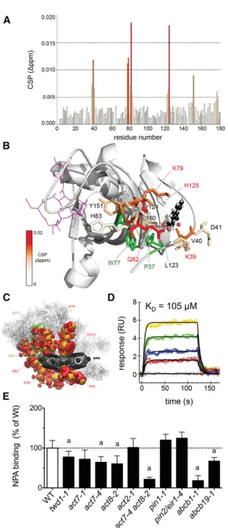
employed chemical shift perturbation (CSP) mapping based on the previously reported NMR resonance assignment of the TWD1 FKBD (Burgardt et al., 2012). Changes in the backbone amide signal positions of15N-labeled TWD11-180in the absence and in presence of NPA were analyzed (Figure 1A; Supplemental Figure 1) and mapped onto the x-ray structure of the TWD1 FKBD (PDB 2F4E; Weiergräber et al., 2006). The most pronounced shifts were observed for residues K39, V40, D41, T78, K79, S81, Q82, H83, A122, L123, V124, and Y151, indicating that the NPA contact region is located outside of the putative PPIase (cis-transpepti- dylprolyl isomerase) site of the FKBD at the convex face of the half b-barrel (Figure 1B). Quercetin, a putative auxin transport inhibitor shown not to bind to TWD1 (Bailly et al., 2008), revealed no major CSP shifts, thus indicating specificity of the binding for NPA (Supplemental Figure 1).
To corroborate the NMR data and to better understand the supramolecular NPA association with the FKBD structure, we employed in silico NPA docking on the entire TWD1 structure available (PDB 2IF4; Granzin et al., 2006) and on the FKBD (PDB 2F4E; Weiergräber et al., 2006) using PyMOL embedded in the AutoDock Vina toolset (Seeliger and de Groot, 2010; Bailly et al., 2011). Rigid in silico NPA docking followed byflexible side-chain optimization verified the FKBD as major NPA target and the predicted binding region correlates well with the one showing CSP value changes (Supplemental Figure 2).
Further, we used quantum chemical modeling based on density functional theory including dispersion correction terms (DFT-D;
Effendi et al., 2015) to optimize the binding geometry of the NPA interaction using the crystal structure of the TWD1 FKBD (PDB 2F4E; Weiergräber et al., 2006) as the basis. The statistical analyses of different components of the quantum chemical forces and the electron density distribution in the region of interaction pocket-NPA deformation suggest that Van der Waals forces (ΔEVdW) are primarily responsible for NPA binding to the TWD1 surface (Supplemental Figure 3), as has been recently proposed for other small molecule-protein interactions using thermody- namic data (Barratt et al., 2005). Furthermore, they suggest that a direct contact is provided by amino acid residues Q82, K79, H125, and D41 (Figure 1C). Strikingly, both in silico techniques suggest a nearly identical pocket for NPA binding and identify
Figure 1.TWD1 Is a Low-Affinity NPA Binding Protein.
(A)NMR CSP analysis of NPA binding to TWD11-180revealing combined1H and15N chemical shift changes. Relevant residues for the binding of NPA above 0.005 ppm are colored according to the legend in(B).
(B) Most stable quantum chemical modeling-derived NPA-bound conformations (balls and sticks) correlate with the mapping of CSP values on FKBP4234-180(PDB ID 2IF4, color code reflects CSP shifts).
Side chains of residues assumed to participate in the binding surface are depicted as sticks; relevant TWD1 mutations are in red. Theoretical FK506 binding to the noncanonical PPIase domain (PDB 1FKJ) is in- dicated in magenta; W77 and P37, thought to build a stacking in- teraction, are colored in green.
(C)Quantum chemical analysis of the electron density components re- sponsible for the Van der Waals forces between NPA and the surrounding amino acids. Electronic density deformation for NPA (dipole-type de- formation in gray and quadrupole-type deformation in black) and for the amino acid residues (dipole-type deformation in orange and quadrupole- type deformation in red; Supplemental Figure 3).
(D) Kinetic analysis of NPA binding to thiol-immobilized TWD11-339. Sensograms of injections of 15, 30, 60, and 90mM NPA in color and representative kineticfit models (1:1) indicated in black. Data are repre- sentative of three independent experiments with comparable results (Table 1). Residuals and goodness-of-fit are indicated in Supplemental Figure 5.
RU, normalized response units.
(E)Specific3H-NPA binding calculated as difference between total and unspecific NPA binding determined in the absence (total) and presence of a 1000-fold excess of nonradiolabeled NPA concentrations (unspecific).
Significant differences (unpairedttest with Welch’s correction, P < 0.05) from corresponding Ws and Col-0 wild types, respectively, are indicated by“a.”
http://doc.rero.ch
widely overlapping putative NPA contact residues, which are in good agreement with the NMR data (Figures 1B and 1C;
Supplemental Figure 2). Furthermore, both tools revealed highly reduced internal binding energies [DEave(NPA) =271.65 kJ/mol;
DE(ave)(BA) = 231.35 kJ/mol] and AutoDock Vina top-ranked docking pose scores (NPA =211 kcal/mol and BA =24 kcal/mol forflexible side chains mode) for benzoic acid (BA) in comparison to NPA. The former is commonly used as a negative control in auxin research and both methods exclude BA binding to TWD1 (Supplemental Figure 2).
These in vitro and in silico data allowed us to design point mutations in the putative NPA binding surface with the goal of generating a version of TWD1 that is NPA insensitive. Four out of five FKBD mutations, TWD1K79I, TWD1H125I, TWD1P37L, and TWDK39I, abolished NPA binding (Supplemental Figure 4), which, based on chemical modeling, is mainly caused by loss of van der Waals forces (ΔEVdW; Supplemental Figure 3) to the ligand, NPA. In TWD1P37L, NPA binding seems to be affected additionally by massive protein deformation (ΔEPdef) resulting in positiveDDE and DDEave based on constrained geometry optimization and full geometry optimization, respectively (Supplemental Figure 3). In- terestingly, TWD1Q82Arevealed higher NPA binding but with lower specificity shown by BA binding assayed in parallel (Supplemental Figure 4). The latterfinding is in agreement with lower calculated binding energies (DDEave=221.43 kJ/mol for the full geometry optimization) and the presence of very similar chemical forces in comparison to the wild-type protein according to the quantum chemical modeling (Supplemental Figure 3). TWD1 K79 was identified by NMR and both in silico dockings involved in NPA binding and TWD1K79Iexhibited reduced NPA binding (in line with reducedDDEave= 6.92 kJ/mol for the constrained geometry op- timization). TWD1K79Iwas one of the most distant mutants in the multivariable analysis of the calculated chemical forces of the binding energies (Supplemental Figure 3) and retained its capacity to regulate ABCB1-mediated auxin transport (Supplemental Figure 4) and was therefore selected for further studies.
To test other auxin transport inhibitors for TWD1 binding, we employed surface plasmon resonance (SPR) analyses of re- combinant Arabidopsis TWD11-339protein cross-linked to sensor chips by thiol coupling. We chose this immobilization strategy over the classical amine coupling because K79 and K39 were part of the putative NPA binding surface, while TWD1 contains four cysteine residues that are outside of this domain. NPA exhibited concentration-dependent SPR responses (Figure 1D), verifying previous binding studies using radiochemistry (Bailly et al., 2008;
Kim et al., 2010). Kinetic binding constants were approximated using a 1:1 Langmuir fit model, although sensograms did not always strictly follow pseudo-first-order kinetics, which was most obvious for TIBA (triiodobenzoic acid) (Supplemental Figure 5).
However, dissociation constants (Kd) obtained from both kinetic and equilibrium analyses [Kd(kin)= 105612mM,Kd(eq)= 1336 20mM; Table 1; Supplemental Figure 5] qualify the TWD11-339 protein as a low-affinity NPA binding protein in vitro, which is in agreement with small NMR shifts (Figure 1). Amine coupling of TWD11-339enhanced higher order kinetic behavior of interactions and thus required a 1:2 surface heterogenicityfit model resulting in dissociation constants for NPA of 135 and 910 mM (data not shown). This supports an inactivation of the binding surface in
a portion of the immobilized TWD1 protein, most likely by K79 and K39 coupling.
The electronic binding energy (DE) and the average of the in- ternal energies (DEave) for NPA calculated by quantum chemical modeling (DE =292.56 kJ/mol;DEave=271.65 kJ/mol) cannot be precisely converted intoΔG values. However, taking into account the dominating influence of Van der Waals interactions and therefore excluding the entropy as major binding driver, a direct transformation (see Methods for details) suggested aDG between 220.92 and229.29 kJ/mol. This is in agreement with the ex- perimental SPR data, as aKdof 105mM gives a theoreticalDG of 222.614 kJ/mol (Table 1).
As expected, other auxin efflux inhibitors, like TIBA, and BUM (Kim et al., 2010) bound to TWD1, although with lower affinities (Table 1), while the unspecific diffusion control, BA, did not (Table 1;
Supplemental Figure 5), verifying experimentally the in silico data.
To complement these in vitro studies, we quantified specific NPA binding to microsomes prepared from TWD1 and auxin exporter loss-of-function mutants. In agreement with low-affinity NPA binding to TWD1 and low expression ofTWD1, we found slightly but significantly reduced NPA binding fortwd1micro- somes (Figure 1E). Reduced binding was also found forabcb1and abcb19material (Rojas-Pierce et al., 2007; Kim et al., 2010) but not forpin1andpin2/eir1-4, verifying the idea that direct NPA binding primarily affects the TWD1-ABCB complex (Petrásek et al., 2006;
Rojas-Pierce et al., 2007; Kim et al., 2010).
In light of the above, we tested the in planta sensitivity of thetwd1 mutant toward NPA in auxin-regulated developmental processes (Bailly et al., 2008). Quantification of apical hook opening (see below), root gravitropism, and hypocotyl elongation (Supplemental Figures 6 and 7) revealed thattwd1is widely NPA insensitive, while overexpression of HA-TWD1 intwd1-3complemented the NPA insensitivity of the mutant (Supplemental Figure 6).
TWD1 Indirectly Interacts with ACTIN7
To identify downstream targets of a low-affinity, NPA-mediated TWD1 action, we employed a coimmunoprecipitation approach followed by tandem mass spectrometry (MS/MS) analyses similar to Henrichs et al. (2012) but using whole TWD1:TWD1-CFP roots as starting material. Three independent coimmunoprecipitation/
mass spectrometry analyses identified 51 common, putative TWD1 interacting proteins (Supplemental Table 1). These showed a remarkable enrichment of PM proteins with a few suggested molecular functions, such as transporter activity (20%), protein binding (23%), and GTPase activity (8%; Supplemental Figure 7).
Interestingly, we preferentially pulled down proteins (such as ABCB4, HSP90, ABCC1, ABCC2, and calmodulin) that either have already been shown to interact with TWD1 (Kamphausen et al., 2002; Geisler et al., 2003, 2004; Wu et al., 2010; Wang et al., 2013) or to putatively function in protein trafficking (RAB GTPases, DYNAMIN-LIKE3, CLATHRIN, and ACTIN7 [ACT7]). We selected ACT7 (score, 278; number of sequences, 9; emPAI, 1.3) for further analyses based on the following: First, ACT7 and ACT8 were previously pulled down with 35S:TAP1-TWD1 (Henrichs et al., 2012) and HA-TWD1 (data not shown) but also with ABCB1:
ABCB1-MYC (data not shown), suggesting that ACT7 (and possibly also ACT8) is part of the TWD1-ABCB1 efflux complex.
http://doc.rero.ch
Second,ACT7is strongly expressed in young plant tissues and is induced by auxin, andact7alleles cause a reduction in root growth and increased root slanting and waving, resembling thetwd1 mutant phenotype (Kandasamy et al., 2009).
Identification of ACT7 as a TWD1 interactor does not neces- sarily imply direct physical interaction. To assess the ability of TWD1 to autonomously bind AFs and to map the potential actin binding region, high-speed cosedimentation assays were con- ducted with TWD11-180(FKBD) and TWD11-339and rabbit muscle actin in various conditions of calcium and pH (Papuga et al., 2010).
As exemplified for FKBD (Supplemental Figure 9A), both the truncated and full-length proteins mostly remained in the su- pernatant fraction, and only a faint amount (<5%) was detected in the pellet fraction together with AFs. Because a similar portion of TWD11-180and TWD11-339 also sedimented in control experi- ments (i.e., without actin), we considered it as being unspecific. As an additional indication that TWD1 does not interact with AFs in a direct manner, it had no effect on the actin polymerization rate in pyrene-actin assays conducted in the absence or presence of NPA (Supplemental Figure 9B).
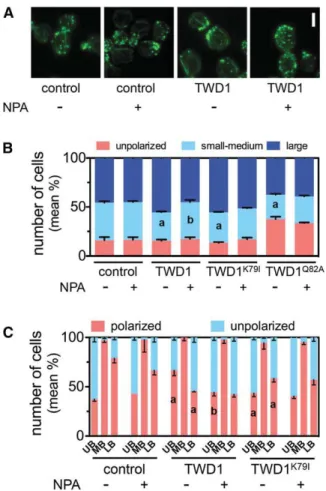
TWD1 Regulates Actin Cytoskeleton Organization and Dynamics and Mediates the Effect of NPA on AF Turnover To gain insight into a putative functional link between TWD1 and the actin cytoskeleton, we carefully examined actinfilament or- ganization in wild-type andtwd1-1seedlings in which we in- troduced the actin reporter GFP-fABD2 (Sheahan et al., 2004).
Employing variable-angle epifluorescence microscopy (VAEM), we noticed that, compared with wild-type hypocotyls,twd1-1 exhibited fewer thick and brightfilamentous structures corre- sponding to actin bundles (Figure 2). To confirm and extend these data, actinfilament bundling (skewness) and density (percentage of occupancy) were quantified in the hypocotyl epidermal cells from dark-grown seedlings following established protocols (Higaki et al., 2010). Average skewness values of 2.8460.05 and 2.2960.06 were calculated for wild-type andtwd1-1hypocotyls, respectively (Figure 2B), confirming that loss ofTWD1lowers the overall cellular level of actin bundling. In addition, and most likely
as a result from reduced actin bundling, actinfilament density was increased intwd1-1(Figures 2A and 2C). Interestingly, long-term treatment with NPA (10mM, 5 d after germination [dag]) induced prominent cytoskeletal changes in wild-type hypocotyls with a 40%
increase in actin bundling and 35% decrease in actin filament density (Figures 2A to 2C), similar to what has been reported before for the auxin transport inhibitors 2-(1-pyrenoyl) benzoic acid (PBA) and TIBA (Dhonukshe et al., 2008). The absence of an effect of NPA on actin stability in previous studies might be due to shorter treatments and/or a lower microscopic resolution compared with our VAEM analyses (Petrásek et al., 2003; Dhonukshe et al., 2008).
In striking contrast to the wild type, NPA had no significant effect on actin filament bundling and density in twd1-1 hypocotyls (Figures 2A to 2C), indicating that NPA-mediated actin bundling requires TWD1. Interestingly, act7-4 hypocotyls exhibited ex- cessive bundling compared with the wild type. However, like in twd1-1, the extent of actin bundling was insensitive to NPA, further supporting a functional TWD1-ACT7 interaction. The NPA re- sistance oftwd1-1andact7-4hypocotyls was confirmed using standard confocal microscopy analyses of identical GFP-fABD2 lines (Supplemental Figure 10 versus Figure 2) using described methods (Higaki et al., 2010). We found thattwd1-1andact7-4 were insensitive to NPA in tested concentrations up to 100mM, while bundling reached already a saturation at 10mM NPA in the hypocotyls of wild-type andtwd1-1hypocotyls complemented by TWD1:TWD1-CFP (Supplemental Figure 11). The bundling de- fects caused by low micromolar NPA concentrations in wild-type hypocotyls might atfirst view seem contradictory to the relatively high Kd value (;105mM) that we obtained by SPR analyses.
However, it is likely that NPA accumulates within tissues and can locally reach high concentrations, especially after long-term treatments as those used in our culture conditions. Moreover, binding affinities generated in vitro might considerably differ from those in planta.
In addition to the static analyses above, we quantified several parameters of single actinfilament dynamics (Staiger et al., 2009;
Henty-Ridilla et al., 2013; Hoffmann et al., 2014b). In the wild type, cortical AFs exhibited typical dynamics including a relatively short lifetime (31 6 7.4 s), and high elongation rate and severing Table 1.Kinetic Parameters Deduced from SPR Analyses Using TWD11-339
Compound ka(1/M * s) kd(1/s) Kd(mM) DG° (kJ/mol)
NPA 1,4806238 0.15560.013 105612 (kin) 222.7060.28 (kin)
133620 (eq) 222.1460.40 (eq)
BUM 645635 0.17560.018 272628 (kin) 220.3660.25 (kin)
252639 (eq) 220.5660.37 (eq)
TIBA 290629 0.12860.007 442627 (kin) 219.1560.16 (kin)
449634 (eq) 219.1060.19 (eq)
BAa 5436501 14.318612.270 33,097611,077 (kin) 28.5560.87 (kin)
1.4 101964.4 1018(eq) 75.0060.73 (eq)
Double referencing and data analysis were performed using Scrubber2 (BioLoglic Software) and TraceDrawer (Ridgeview Instruments) analysis software. Affinity binding constants (Kd) were obtained by equilibrium analysis [Kd(eq)] and by least-squares nonlinearfit of the obtained sensograms using a 1:1 Langmuir binding model [Kd(kin)], which additionally allowed for the evaluation of dissociation (kd) and association (ka) rate constants. Values represent means and standard deviations of kinetic constants obtained from at least three experiments on independent sensor chips. Corresponding sensograms andfit models are shown in Supplemental Figure 5.
aSensograms obtained for BA did not allow for a meaningfulfit, indicating that TWD1-339did not bind BA.
http://doc.rero.ch
frequency (2.961.0mm/s and 0.01160.004 breaks/mm/s, re- spectively; Table 2) that are in good agreement with previous data (Henty et al., 2011). Intwd1-1, actinfilament lifetime was signif- icantly higher than in the wild type with an average of 45.1610.3 s.
This increase in actin filament stability was consistent with a reduced depolymerization rate and severing frequency (Supplemental Movies 1 to 3). NPA treatment altered virtually all parameters of actinfilament dynamics in wild-type hypocotyls.
Consistent with the fact that NPA increases the overall level of actin bundling (Figure 2), it lowered actinfilament dynamics as shown by an increase in actinfilament lifetime and length and a decrease in actinfilament severing (Supplemental Movies 4 to 6). Most remarkably, and in agreement with the inability of NPA to promote actin bundling intwd1-1hypocotyls, none of the single actinfilament dynamic parameters analyzed was affected by NPA treatment intwd1-1(Table 2).
Our data revealed that bundling frequencies of AFs were similar in wild-type andtwd1-1hypocotyls (Table 2). By contrast, de- bundling frequency was much (nearly 4 times) higher intwd1-1, which seems to account for the reduced overall bundling level observed intwd1-1hypocotyls (Figure 2). Thus, it appears that TWD1 triggers a dual action on actin cytoskeleton organization and dynamics in hypocotyls: On one hand, it stimulates single AF dynamics, e.g., by increasing AF depolymerization and severing, and on the other, it downregulates the process of debundling.
Surprisingly, NPA reduced actin bundling and debundling fre- quencies by ;50% in both wild-type and twd1-1 hypocotyls (Table 2). This unexpected result suggests that, in addition to its
TWD1-dependent action on single AF dynamics, NPA may modulate the processes of actin bundling/debundling in a TWD1- independent manner. Consistent with the fact that actin bundling and debundling frequencies were similarly reduced by NPA, NPA treatment had no apparent effect on the overall actin bundling level and AF density in twd1-1 (Figures 2B and 2C; Supplemental Figures 10B and 11B).
ACT7 Regulates Expression and Location of ABCB- and PIN-Type Auxin Exporters as well as TWD1
The known role of the actin cytoskeleton in auxin transporter cycling (Geldner et al., 2001; Kleine-Vehn et al., 2006; Dhonukshe et al., 2008) and delocalization of ABCBs intwd1(Wu et al., 2010;
Wang et al., 2013) prompted us to investigate the locations of auxin transporters inact7-4. We found that expression of ABCB1- GFP and ABCB19-GFP and, most drastically, ABCB4-GFP was significantly downregulated inact7-4and that all tested ABCBs were partially retained on intracellular structures (Figures 3 and 4).
PIN1-GFP and PIN2-GFP were likewise found on similar struc- tures and their polarity was reduced (Figures 3 and 4). However, in contrast to ABCBs, expression of PIN2 was less affected, while expression of PIN1 was even slightly upregulated (Figure 3C). To our surprise, TWD1-CFP was also partially delocalized from ER/
PM locations inact7-4(Figure 3B), which is in line with the pro- posed chaperone function of TWD1 during ER-to-PM secretion of ABCBs (Wu et al., 2010; Wang et al., 2013). Moreover, TWD1 lost lateral PM polarity (Wang et al., 2013) in immunostained roots of act7 single and double loss-of-function mutant material (Supplemental Figure 12). We noticed that both endosomal structures and transporter delocalizations inact7-4were often found in misshaped cells (Figures 3 and 4). A thorough in- vestigation employing PIN2-GFP and propidium iodide staining of cell walls revealed that these defects indeed are found in mis- shaped cells but that they do not strictly correlate with cell wall defects (Figures 3 and 4). Delocalizations for PIN1,2-GFP and ABCB4-GFP were also not caused by nonisogenic ecotype backgrounds (see Methods for details) as revealed by im- munolocalization of PIN1,2 in act7-4 and crossings between ABCB4-GFP and act7-6lines that are both in the Col-0 back- ground (Supplemental Figure 13).
Short treatments with endocytic tracer FM4-64 (2mM, 15 min) resulted in FM4-64 accumulation into internal (probably TGN or early endosomal) structures inact7-4and, more severely, in actin double mutants, but not in twd1 (Supplemental Figure 12A), suggesting a role for ACT7 in endosomal balance. An early endosomal/TGN marker, ARF1, showed a similarly defective dis- tribution inact7-4andact7-4 twd1-1but not intwd1-1, verifying the endosomal identity of defective endomembranes (Supplemental Figure 12B). LysoTracker red (LTR) and long-term FM4-64 (4mM, 3 h) applications revealed additionally aberrant vacuolar mor- phologies (Supplemental Figures 12C and 12D), while crossings of act7-4 with lines expressing endosomal/prevacuolar markers RabF1/ARA6, Syp22, and Syp61 revealed intracellular localization defects (Supplemental Figure 14).
Thesefindings encouraged us to colocalize ABCB4/ABCB19 and PIN1/PIN2 with different endosomal and vacuolar markers inact7-4. ABCB4, ABCB19, and PIN2 (but not PIN1) showed Figure 2.Actin Architecture and NPA Sensitivity Is Altered intwd1.
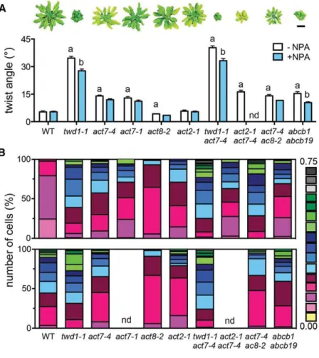
Time-lapse VAEM analyses of cortical actin of dark-grown hypocotyl expressing fABD2-GFP treated with 1mM NPA. Representative epidermal cells(A)and quantification of actin bundling (skewness;[B]) and per- centage of occupancy (density;[C]). Significant differences (unpairedttest with Welch’s correction, P < 0.05) between wild-type and mutant alleles are indicated by“a”and significant differences to solvent controls by“b” (mean6SE;n$100). Bar = 10mm.
http://doc.rero.ch
increased colocalization with FM4-64 after long-term treatments (Figures 4A and 4B) and with the vacuolar marker LTR inact7-4 (Figures 4C and 4D), while PIN1 revealed already a high coloc- alization factor in the isogenic wild-type background. These data suggest a mislocalization of both classes of auxin exporters on compartments of vacuolar/late endosomal origin in act7-4.To address further the nature of these structures, we performed short BFA treatments (25mM, 1 h) of PIN1-GFP and ABCB19-GFP lines and found that both colocalize with PM marker, FM4-64, on BFA compartments in the wild-type background andact7-4. Inact7-4, PIN1-GFP and ABCB19-GFP were retained on BFA bodies on additional compartments with putative late endosomal-like nature (Supplemental Figure 15). These data support further retention of both classes of auxin exporters on compartments of endosomal/
vacuolar origin inact7-4.
twd1andact7Show Overlapping Developmental Phenotypes That Are Caused by Defects in Polar Auxin Transport
Arabidopsis contains eightACTINgenes that are grouped into reproductive and vegetative classes; the latter class contains ACT2,ACT7, andACT8(Kandasamy et al., 2009).ACT7mutant combinations are dwarfed, with altered cell and organ morphol- ogy, suggesting that ACT7 is involved in root growth, epidermal cell specification, cell division, and root architecture (Kandasamy et al., 2009). The actin defects in twd1 and auxin transporter delocalizations inact7stimulated us to compare in detail auxin- regulated phenotypes intwd1andact7, with special focus on tissues and processes related to actin-dependent growth.
A hallmark of the twd1phenotype is a non-handed, helical rotation (twisting) of epidermal layers that can be (like inabcb1 abcb19) partially rescued by NPA treatment (Geisler et al., 2003;
Wu et al., 2010; Bailly et al., 2014; Figure 5A). Interestingly, we found that bothact7alleles and mutant combinations withact7, but notact2-1oract8-2, also show substantial epidermal twisting that was, however, NPA insensitive (Figure 5A). Next, we analyzed the planar polarity of root hair positioning, which is determined by a concentration gradient of auxin distribution in the root tip (Grebe, 2004). This gradient itself is regulated by upstream events, such as polar placement of ROP (Rho-of-plant) GTPases or auxin trans- porter activity (Masucci and Schiefelbein, 1994; Grebe, 2004).
Surprisingly,twd1-1and bothact7alleles showed a striking apical shift in root hair positioning that was strongest inact7-4 twd1-1 andact7-4 act8-2(Figure 5B). Defects in planar root hair polarity have also been reported very recently for two newACT7alleles shown to influence ROP positioning (Kiefer et al., 2015). In analogy to epidermal twisting, root hair positioning was only mildly dis- turbed inact2-1 andact8-2. NPA treatment of wild-type phe- nocopiedtwd1andactinalleles, but againtwd1was found to be widely NPA insensitive, whileactinmutants were even partially rescued (Figure 5B).
Figure 3. ACT7 Regulates ABCB- and PIN-Type Auxin Transporter as well as TWD1 Expression and Location.
(A)and(B)Auxin transporters, ABCB1,4,19-GFP(A)and PIN1,2-GFP, as well as the ABCB chaperon, TWD1-CFP(B), are delocalized from the root PM and ER, respectively, retained on punctuated structures (arrows), and significantly downregulated inact7-4in comparison to the corresponding wild-type lines. Bars = 10mm.
(C)Quantification of(A)and(B). Significant differences (unpairedttest with Welch’s correction, P < 0.05) between the wild type andact7-4are in- dicated by“a”(mean6SE;n$50 images). Note that GFP quantifications by confocal imaging were performed over identically defined areas of the entire root tip and therefore quantified intensities in(C)might not match
pictures in(A)and(B). Furthermore, PIN1,2-GFP and ABCB4-GFP non- isogenic lines were used for analyses inact7-4but artifacts caused by ecotype mixes were excluded by immunolocalizations or isogenic controls (Supplemental Figure 13).
http://doc.rero.ch
Finally, we analyzed leaf trichome branching, which usually displays a variation between one and four branches in wild-type plants (Hülskamp et al., 1994). Bothact7alleles exhibited a signif- icant shift toward dibranched trichomes (Figure 5C), as previously reported foract2 act7double mutants (Kandasamy et al., 2009).
Strikingly,act7-4andtwd1-1showed largely identical trichome branching patterns, which were even more radical inact7 twd1and act2 act7, again resembling NPA treatments (Grebe, 2004).
To link auxin-related phenotypes to actin function, we employed the actin destabilizing and stabilizing drugs, latrunculin B and jasplakinolide, shown to reduce the elongation rate and to induce actin polymerization, respectively (Staiger et al., 2009). Like NPA, latrunculin B and jasplakinolide treatments were able to block apical hook formation in etiolated wild-type seedlings (Figure 5D) known to be dependent of a local accumulation of auxin at the inner side of the hook (Zádníková et al., 2010). Interestingly,twd1hooks were less sensitive to NPA and latrunculin B treatments than the wild type but not to jasplakinolide, most probably due to already reduced actin bundling (Figure 2B).act7-4was fully responsive to all treatments, further underlining functional redundancy between vegetative actin isoforms (Kandasamy et al., 2009).
Latrunculin B and jasplakinolide had a similar inhibitory effect on hypocotyl elongation as NPA in the wild type, and like for hook opening, twd1-1 but not act7-4 was greatly insensitive (Supplemental Figure 7A). Interestingly, paclitaxel/taxol and ory- zalin, well-established stabilizers and inhibitors of microtubule polymerization, similarly affected hypocotyl length intwd1-1(and act7-4) in comparison to the wild type (Supplemental Figure 7B), making an involvement of TWD1 (and ACT7) in microtubule- related cell elongation unlikely.
Overlapping auxin-related growth phenotypes between act7 andtwd1mutants support a tight mechanistic link between actin dynamics and auxin efflux (Muday, 2000; Blancaflor et al., 2006;
Dhonukshe et al., 2008). This prompted us to quantify auxin responses and transport in act7 mutants. Activation of the
auxin-responsive DR5rev:GFP reporter (Friml et al., 2003) was slightly reduced in theact7-1allele but more drastically in the act7-4root tip, as has been reported before fortwd1-1(Figures 6A and 6B; Bouchard et al., 2006). Interestingly, latrunculin B and jasplakinolide treatments phenocopiedTWD1andACT7loss of function, implying that these drugs also cause PAT defects. In agreement, free IAA levels were significantly elevated inact7-4 andtwd1-1roots (Figure 6C; Bouchard et al., 2006).
Finally, we employed a self-referencing IAA-specific micro- electrode that permits continuous, noninvasive recordings of distinct IAAflux peaks in the epidermis of the apex of Arabidopsis roots (Mancuso et al., 2005; Henrichs et al., 2012), correlating with a PIN-dependent auxin“reflux loop”from peripheral root cells toward central vascular cells (Blilou et al., 2005). Latrunculin B and jasplakinolide treatments (each 5mM) blocked root PAT as effi- ciently as NPA (Bailly et al., 2008), which is genetically copied by loss ofACT7function (Figures 6D and 6E). Interestingly, root auxin fluxes intoact7-4roots show reduced sensitivities to latrunculin B and jasplakinolide (Supplemental Figure 16), resembling twd1, which is insensitive to NPA (Bailly et al., 2008).
In summary, these data sets support the concept that either destabilization or stabilization of the actin cytoskeleton causes PAT defects to a similar magnitude as reported for NPA treat- ments. Overlapping growth phenotypes and PAT defects caused either by chemical actin (de)stabilization or by TWD1orACT7 mutations suggest that abnormal actin cytoskeleton function in twd1is the primary reason for PAT defects, which are likely to be the cause of previously overlooked developmental defects intwd1.
Expression of TWD1 in Yeast Alters Budding and Actin Polarity and Confers NPA Sensitivity
With the apparent conserved FKBP functionality in mind, we phenotypically characterized wild-type budding yeast expressing Table 2.Parameters of Actin Dynamics Deduced from Time-Lapse VAEM Analyses of Single, Cortical Actin Filaments of Arabidopsis Hypocotyls Expressing fABD2-GFP
Parameter
Wild Type twd1-1
2NPAa +NPAb 2NPAc +NPAd
Max.filament lifetime (s) 31.167.4 47.0615.7** 45.1610.3* 44.3611.3
Max.filament length (mm) 27.969.9 33.2614.5** 28.2610 28.569.1
Elongation rate (mm/s) 2.961.0 3.461.3** 2.960.9 3.061.0
Depolymerization rate (mm/s)
1.460.7 1.160.8 0.9*60.4 0.960.3
Severing frequency (breaks/mm/s)
0.01160.004 0.00860.003** 0.009*60.003 0.00860.003
Bundling frequency (events/mm2/s)
1.640 102461.324 1026 0.883 102460.645 1026** 1.890 102461.314 1026 0.872 102460.999 1026**
Debundling frequency (events/mm2/s)
0.590 102460.2445 1025 0.280 102460.194 1025** 1.910 102460.414 1025* 0.910 102460.264 1025**
Shown are means6SEof 127filaments from 34 cells.
aNinety-ninefilaments from 31 cells.
bNinety-sixfilaments from 41 cells.
cNinety-sixfilaments from 46 cells.
dSignificant differences (unpairedttest with Welch’s correction; P value < 0.01) to wild-type or solvent controls are indicated by * and **, respectively.