Blastocyst development from supernumerary embryos
after intracytoplasmic sperm injection: a paternal
influence?
Youssef Shoukir, Didier Chardonnens,
Aldo Campana and Denny Sakkas
1Clinic of Infertility and Gynaecological Endocrinology–WHO Collaborating Centre in Human Reproduction, Department of Obstetrics and Gynaecology, University Hospital of Geneva, 1211 Geneva 14, Switzerland
1To whom correspondence should be addressed at: Assisted Conception Unit, Birmingham Women’s Hospital, Edgbaston, Birmingham B15 2TG, UK
The success of intracytoplasmic sperm injection (ICSI) warrants further study on the role of paternal factors in early human embryogenesis. To investigate whether poor sperm parameters can influence embryo development, we examined the development of ICSI-fertilized embryos to the blastocyst stage. We present results of blastocyst development from supernumerary ICSI embryos after co-culture on monkey kidney epithelial cells. In addition, we compare the development of supernumerary embryos to the blastocyst stage after ICSI and in-vitro fertilization (IVF). Of 168 supernumerary ICSI embryos, 45 (26.8%) developed to blastocysts. Sperm concentration and morpho-logy did not influence blastocyst development. In contrast, blastocysts arose from spermatozoa that had a significantly higher (PJ 0.015) forward progressive motility compared with spermatozoa from those patients who failed to produce blastocysts (42.7% versus 28.2%, respectively). Overall the rate of embryo development to the blastocyst stage after ICSI was lower (26.8%) than that after IVF (47.3%). When the rate of blastocyst development was calculated for patients with three or more supernumerary embryos, it remained significantly higher for the IVF patients than for the ICSI patients (45.6% versus 30.0%). There was no significant difference in the mean cell number and quality of the supernumerary embryos between the IVF and ICSI patients. This study confirms previous reports that have postulated that abnormal spermatozoa may manifest a negative paternal effect on preimplantation embryo development.
Key words: human blastocysts/ICSI/in-vitro fertilization/ paternal influence
Introduction
The role of a paternal factor in early human embryogenesis is gaining more attention because of the development of new techniques for the treatment of men with poor spermatozoa, as indicated by the success of intracytoplasmic sperm injection (ICSI), and because of the improvement in our knowledge of
sperm biology, for example through the characterization of the DAZ gene (Reijo et al., 1995). It can be hypothesized that a paternal effect on embryonic development would occur only after the appearance of the first paternal transcriptional products in the embryo, i.e. after embryonic genome activation. In fact, a negative paternal effect need not necessarily be at the genetic level but can also be observed in components that are transmitted by the spermatozoa. For example, incorrect formation of the sperm aster due to a centrosome defect can lead to failure as early as fertilization or in subsequent cell cycles (Asch et al., 1995; Simerly et al., 1997). A negative paternal influence on embryo development would therefore be expected to be more prominent in men with poor semen quality. In a study examining patients undergoing routine in-vitro fertilization (IVF) treatment, Janny and Me´ne´zo (1994) found that, when the usual parameters of sperm quality were good, there was a strict linear relationship between cleavage and blastocyst formation rates. Their results presented evidence of a strong paternal effect on human preimplantation embryo development and blastocyst formation.
ICSI bypasses many upstream events associated with clas-sical sperm–egg interactions, including plasma membrane and cortical region interactions. It is unclear whether bypassing these steps in the pathway of sperm transformation after cytoplasmic penetration leads to inherent problems, arising in either nuclear decondensation, pronuclear migration and/or mitosis. Furthermore, it is expected that sperm defects in microtubule function or centrosomal reconstitution will not be resolved by ICSI-assisted fertilization.
Recently, a number of studies have reported the successful culture of human embryos to the blastocyst stage, with and without feeder cells, and improved pregnancy rates after the transfer of fresh (Olivennes et al., 1994) and frozen blastocysts (Kaufmann et al., 1995). Transfer of embryos at the blastocyst stage provides a better synchrony between the uterine endo-metrium and embryo. The other advantages of such a procedure include the possibility of the selection of better-quality embryos with a higher implantation potential (Nakayama et al., 1995), supporting the concept that some embryos with chromosomal or genetic abnormalities may cleave, but fail to reach the blastocyst stage (Edwards and Hollands, 1988).
In a previous study (Shoukir et al., 1998), we have presented our results on the development of supernumerary embryos to the blastocyst stage after conventional IVF. We found that, out of 423 supernumerary embryos, 200 developed to the blastocyst stage (47.3%). Reports in the literature of blastocyst develop-ment from supernumerary embryos after ICSI are scant. Barnes et al. (1995) reported a case of blastocyst development and birth after in-vitro maturation of primary oocytes, ICSI and
assisted hatching. The aim of this study is to present the results of blastocyst development from supernumerary embryos after ICSI and to compare them with those after routine IVF, in order to determine whether there is a paternal effect on blastocyst development.
Materials and methods
The present study was performed on patients entering the IVF programme at the Clinic of Infertility and Gynaecologic Endo-crinology, Department of Obstetrics and Gynaecology, University Hospital of Geneva, Geneva, Switzerland between April 1992 and August 1997. In total, 90 and 60 cycles were assessed where the patient underwent a routine IVF treatment or ICSI, respectively, with embryo transfer and development of supernumerary embryos. All patients without supernumerary embryos were excluded. The stimula-tion protocol and patient selecstimula-tion procedure adopted by our group have been previously described (Sakkas et al., 1994; 1996).
Collected oocytes (day 0) were fertilized in our standard culture medium Whittingham’s T6 (Quinn et al., 1982) supplemented with 10% maternal serum. Serum was prepared from blood taken on the first day of stimulation. Oocyte retrieval took place between 0830 and 1030 h. For normal IVF, insemination was performed between 1530 and 1630 (~1600) h with 53104motile spermatozoa obtained by either swim-up or mini-Percoll gradient centrifugation. In cases where the normal sperm morphology of the patient was 15–20 or ,15%, insemination was performed with 153104or 33105motile spermatozoa, respectively.
ICSI was used for couples in whom the male partner had very poor semen quality. Of the 60 ICSI cycles, 39 and 9 cycles were performed because patients had less than 10 and between 10 and 203106sperm per ml, respectively, while in the remaining 12 cycles the male had more than 203106sperm per ml but had poor motility or poor morphology and routine IVF was not considered as an option. No cases of repeated fertilization were included in this study.
ICSI was performed between 1300 and 1500 (~1400) h. For the ICSI procedure the injection pipette was prepared with an internal diameter of 5–7µm with a 30° bevel and no spike was placed on the tip. The cumulus cells were removed ~2–3 h after oocyte aspiration by pipetting the oocytes in HEPES-buffered T6 medium containing 1 mg/ml type IV hyaluronidase (Sigma, Buchs, Switzerland). All manipulations were carried out on a warming stage and under oil. Oocytes were placed on a glass slide with a depression in HEPES-buffered T6 medium during the procedure. Spermatozoa were selected from a droplet of culture medium containing 5% polyvinylpyrrolidone (PVP-360; Sigma). Following immobilization by stroking the mid-piece-tail region, the spermatozoa were injected into the cytoplasm of the oocyte, which was held with the polar body in the 12 o’clock or 6 o’clock position. After injection, the oocytes were examined for any damage, washed carefully and placed in culture medium.
The following morning (day 1) the oocytes were removed from the medium, washed and placed in 20-µl culture drops under oil (light white mineral oil; Sigma) in petri dishes, and the presence of two pronuclei (2PN) was assessed. Embryo transfer was routinely performed on day 2. However, when a patient had three or more previous implantation failures, the embryos were co-cultured on African green monkey kidney epithelial (Vero) cells from the 2PN stage and the transfer was performed on day 3. Routinely, a maximum of three embryos was transferred to the patient, normally between 1000 and 1500 h.
On the day of transfer, the number of cells was assessed to determine the cleavage rate, and each was assigned a quality score
based on the presence of fragments and clarity of the cytoplasm of the blastomeres. The ratings given to embryos for cell number were 1 for 1-cell, 2 for 2-cells, 3 for 3-cells, 4 for 4-cells, 6 for between 4- and 8-cells and 8 for.8-cells. The ratings given to embryos for quality were the same as those used by Cummins et al. (1986) except that the values of 0 for poorest and 3 for best embryo were given. Patient serum human chorionic gonadotrophin (HCG) was measured 14 days after embryo transfer (Total hCG: IMX, Abbot, Abbot Park, USA). Patients in whom the intrauterine fetus or fetuses displayed a heart beat at ultrasound examination 4–5 weeks after transfer were considered to have achieved a clinical pregnancy.
Supernumerary embryos were co-cultured on Vero cells using T6 supplemented with vitamins and amino acids until the blastocyst stage as previously described (Sakkas et al., 1994). The blastocysts were then frozen using the protocol described by Me´ne´zo et al. (1992) except that T6 medium was used in place of Me´ne´zo’s B2. Briefly, the blastocyst was immersed in a 5% glycerol solution (Sigma) in T6 medium1 10% serum for 10 min, followed by 10 min in a solution of 9% glycerol in T61 10% serum containing 0.2 M sucrose. The programmed freezing curve was –1°C/min from 23°C to 5.5°C. Automatic seeding was performed, and the embryo was cooled slowly from –5.5°C to –30°C at a rate of –0.3°C/min and then stored in liquid nitrogen.
At the time of transfer, the blastocysts were thawed rapidly at room temperature. The cryoprotectant was removed using seven steps, with decreasing concentrations of glycerol and each step lasting 5 min. In all steps, the blastocysts were processed at room temperature under a stream of gas (5% CO2, 5% O2and 90% N2). After thawing, the blastocysts were allowed to recover for 3 to 4 h in co-culture before transfer. In normally ovulating patients, the transfer was performed during a natural cycle monitored by ultrasound echography and hormone assay. Transfer was timed according to the urinary luteinizing hormone (LH) peak. In patients with anovulation, ovarian stimulation was performed using human menopausal gonadotrophin and ovulation was triggered by HCG. An HCG determination was conducted 7 days after transfer.
The statistical evaluations used were analysis of variance followed by Scheffe´’s F-test for comparisons of mean values, and Fisher’s exact test orχ2analysis with continuity correction for comparison of pregnancy rates.
Results
The development of supernumerary embryos to the blastocyst stage after ICSI
The development of supernumerary embryos to the blastocyst stage was assessed in 60 cycles after ICSI. In fresh cycles, cleavage-stage embryos were transferred on either day 2 or 3, with only one cycle in which blastocyst transfer was performed on day 6. Of 168 supernumerary embryos, 45 developed to blastocysts (26.8%). Figure 1 represents the number of frozen expanded and early blastocysts in relation to their development in culture prior to cryopreservation. Of 42 frozen blastocysts, 52.4% had reached the blastocyst stage on day 6, and were frozen on that day, compared with 28.6% on day 7.
The clinical characteristics of patients grouped according to whether supernumerary embryos developed to blastocysts are presented in Table I. Owing to the small number of day-3 transfer embryos (5 cycles in patients with and 8 cycles in patients without blastocyst development), we combined these cycles with the corresponding day-2 transfers. There were no
Figure 1. The percentage of expanded and early blastocysts following ICSI in relation to the number of days in culture. Embryos were frozen on the day they reached blastocyst stage. ICSI5 intracytoplasmic sperm injection.
Table I. Development of supernumerary embryos to the blastocyst stage
following ICSI for patients grouped according to whether blastocysts were produced or not
No blastocysts Blastocysts P-value
No. of cycles 38 22 – No. of patients 28 18 – Age of female 34.66 4.6 32.96 4.0 0.15 Age of male 37.96 5.5 38.46 6.1 0.99 No. of oocytes 429 255 (per cycle) (11.36 5.2) (11.66 3.4) 0.81
No. of oocytes injected 400 236 –
No. of 2PN embryos 248 153
(per cycle) (6.56 2.2) (7.06 2.1) 0.46
Fertilization rate 62.0 64.8
No. of embryos on day 2 203 144
(per cycle) (5.36 1.7) (6.56 1.8) 0.01
No. of embryos transferred 119 60
(per cycle) (3.16 0.3) (2.76 0.3) 0.005
No. of supernumerary embryos 84 84
(per cycle) (2.26 1.6) (3.86 2.0) 0.002
No. of blastocysts 0 45
(per cycle) (2.06 1.3) –
Results are expressed as mean6 SD.
ICSI5 intracytoplasmic sperm injection; PN 5 pronuclear.
significant differences in the ages of the couples, number of oocytes and number of 2PN embryos between patients with and without blastocysts. However, there was a significant difference in the number of total and supernumerary embryos on day 2. When examining whether there was an influence of the husband’s sperm parameters on blastocyst development, we found that there was no significant difference in sperm concentration and morphology between the groups. However, patients whose blastocysts developed after ICSI had spermato-zoa that had a significantly higher (P 5 0.015) forward progressive motility (mean6 SD) than those patients without blastocysts (42.76 21.3 versus 28.2 6 20.8, respectively).
The clinical pregnancy and implantation rates for both groups of patients is shown in Table II. Although the patients in the group who failed to produce blastocysts had a significantly higher number of embryos transferred, as shown in Table I, the clinical pregnancy rates were not significantly different between the groups. However, the implantation rates in the
Table II. Clinical pregnancy per transfer for patients with and without
blastocysts after ICSI
No blastocysts Blastocysts No. of transfers 38 21 Pregnancies following: Day-2 transfers 7/30a 7/16a (%) (23.3) (43.7) Day-3 transfers 1/8 1/4 (%) (12.5) (25.0) Day-6 transfers – 1/1 (%) (100) Total 8/38a 9/21a (%) (21.1) (42.9)
Implantation rate per embryo transferred 9/119b 14/60b
(%) (7.6) (23.3)
aNot significantly different. bSignificantly different P5 0.01. ICSI5 intracytoplasmic sperm injection.
Table III. Mean cell number and quality of all embryos on day 2 of
transferred embryos and of supernumerary embryos
Day-2 transfer Day-2 transfer P-value with no blastocyst with blastocyst
development development
Cell number of:
All embryos on the day 3.26 1.2 3.76 1.2 0.002
of transfer (157) (101) Transferred embryos 3.56 1.1 4.26 1.0 0.0004 (93) (46) Supernumerary embryos 2.76 1.2 3.26 1.2 0.02 (64) (55) Quality of:
All embryos on the day 2.36 0.8 2.760.6 0.0001
of transfer (157) (101)
Transferred embryos 2.56 0.6 2.86 0.4 0.002
(93) (46)
Supernumerary embryos 1.860.9 2.66 0.7 0.0001
(64) (55)
The results are expressed as mean6 SD.
The number of embryos for each group is in the parentheses.
patients with blastocyst development were significantly higher than those in patients with no blastocysts. Only a limited number of transfers of thawed blastocysts have been obtained following ICSI. Of these, two clinical pregnancies in 9 cycles (22.2%) were established with a mean number of 1.8 blastocysts transferred per patient.
To evaluate whether the quality of embryos transferred can affect the development of the supernumerary embryos to the blastocyst stage, we calculated the mean cell number and quality for all embryos, transferred embryos and supernumerary embryos on the day of transfer (Table III). When comparing the supernumerary embryos of day-2 transfer patients, who did or did not have blastocysts, we found that there was a significant difference in the mean cell number and the mean quality/embryo.
Comparison of the development of supernumerary embryos to the blastocyst stage after ICSI and IVF
The results of blastocyst development after routine IVF have previously been published by our group (Shoukir et al., 1998).
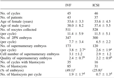
Table IV. Comparison of the development of all supernumerary embryos to
the blastocyst stage in patients who had transfer on day 2 following IVF and ICSI
IVF ICSI
No. of cycles 45 46
No. of patients 43 37
Age of female (years) 33.66 3.3 33.66 4.5
Age of male (years) 38.06 8.2 37.46 5.5
No. of oocytes collected 511 528
(per cycle) 11.46 5.9 11.56 5.1
No. of 2PN embryos 347 300
(per cycle) 7.76 3.4 6.56 2.2
No. of supernumerary embryos 173 120
(per cycle) 3.86 2.7a 2.66 1.9a
Cell number of supernumerary embryos 3.161.2 2.96 1.2 Quality of supernumerary embryos 2.46 0.7b 2.26 0.9b
No. of cycles with blastocysts 35 16
No. of blastocysts 85 31
(% of embryos) (49.1)c (25.8)c
No. of blastocysts per cycle 1.96 1.7d 0.76 1.3d aP5 0.01;bP5 0.02;c,dPø 0.0001.
Results are expressed as mean6 SD.
ICSI5 intracytoplasmic sperm injection; IVF 5 in-vitro fertilization; PN 5 pronuclear.
The rate of blastocyst development of embryos after ICSI is lower (26.8%) than that after IVF (47.3%). The rate of blastocyst development was found to be significantly higher in IVF patients (49.1%) than that in ICSI patients (25.8%) (Table IV), if only patients who had transfer on day 2 were considered. The mean number of supernumerary embryos was however significantly higher for IVF patients. Of the supernumerary embryos, the mean cell number did not differ between the two groups. However, the supernumerary embryos obtained following IVF were of significantly better quality. It could be argued that IVF patients had more supernumerary embryos available to develop to the blastocyst stage. When patients with three or more supernumerary embryos in the IVF and ICSI groups were considered, the rate of blastocyst development remained higher for the IVF patients even though there was no significant difference in the number of super-numerary embryos between the groups (Table V). More import-antly, in this sub-group of patients there was also no significant difference in the mean cell number and quality of the super-numerary day-2 embryos between the IVF and ICSI patients (Table V).
Discussion
The introduction of ICSI has revolutionized the treatment of severe forms of male infertility (Palermo et al., 1992; Van Steirteghem et al., 1993). However, following the successful treatment of these male factor patients, a number of questions have arisen. One relates to whether the arbitrary selection of spermatozoa from male factor patients to achieve fertilization by ICSI will lead to a negative paternal impact on embryo-genesis.
In this study we have shown a lower overall rate of blastocyst development from embryos following ICSI than following IVF (26.8% versus 47.3%). This lower percentage of development might be due to numerous factors, ranging from the fact that
Table V. Comparison of blastocyst development following IVF or ICSI
patients with three or more supernumerary embryos who underwent transfer on day 2
IVF ICSI
No. of cycles 28 18
No. of supernumerary embryos 149 80
(per cycle) 5.36 2.4a 4.46 1.9a
Cell number of supernumerary embryos 3.36 1.2a 3.06 1.2a Quality of supernumerary embryos 2.46 0.7a 2.46 0.7a
No. of blastocysts 68 24
(%) (45.6)b (30.0)b
No. of blastocysts per cycle 2.46 1.9c 1.36 1.7c Comparisons were made between IVF and ICSI data as follows:anot significantly different;bP5 0.03;cP5 0.01.
Results are expressed as mean6 SD.
ICSI5 intracytoplasmic sperm injection; IVF 5 in-vitro fertilization.
ICSI patients have poorer sperm parameters to technical problems associated with the ICSI procedure itself. The invasiveness of the ICSI procedure may cause subtle changes to the inherent structure of the oocyte and the resulting embryo. Indeed, in the monkey, Sutovsky et al. (1996) have shown that ICSI results in unusual chromatin, cytoskeletal and membrane events compared with those of normal fertilization, but eventu-ally leads to pronuclear development. Motoishi et al. (1996) have however shown that bovine oocytes develop at the same rate to the blastocyst stage after ICSI as compared with that of control oocytes, indicating that the ICSI procedure is not detrimental to embryo development. ICSI embryos may also have a greater susceptiblity to certain in-vitro culture systems, notably co-culture which was used in this study. Recently, a number of sequential culture systems have been described using defined media which lead to improved blastocyst development (Gardner and Lane, 1997). The data presented in this study need to be verified by other groups performing ICSI and in different culture systems before we can obtain a final answer as to a whether there is a technical influence on the development of ICSI embryos. Although technical aspects may play a role in influencing ICSI embryo development, the main difference between the IVF and ICSI groups is the fact that ICSI patients have poorer sperm parameters. This would therefore point to a strong possibility of a paternal effect on embryo development.
The IVF and ICSI groups also had differences in the number of supernumerary embryos and in their cleavage rates and quality. The argument that the higher the number of super-numerary embryos in a cycle, the better the chance for the patient to have blastocyst development was however negated when the number of supernumerary embryos for IVF and ICSI patients was adjusted by omitting those cycles with two or fewer supernumerary embryos. However, there remains a significant difference in the rate of blastocyst development in favour of IVF embryos. Furthermore, when only patients with three or more supernumerary embryos were assessed in either group, the mean cell number and quality were not different between the supernumerary IVF and ICSI embryos. It has been suggested that ICSI produces a significantly higher proportion of morphologically superior embryos than conven-tional ones (Yang et al., 1996) and high insemination
concentra-tion IVF (Oehninger et al., 1996). In contrast, our results show that, whereas the mean cell number of day-2 supernumerary embryos did not differ, the quality of those embryos was better in IVF cycles than in ICSI cycles. Overall, the number of supernumerary embryos and the type (cell stage and quality) after IVF and ICSI did not appear to have a major influence on the outcome of blastocyst development.
Comparison of the groups of patients whose supernumerary embryos did or did not develop to the blastocyst stage after ICSI could therefore throw some light on the different factors which might affect blastocyst development. Since ICSI is used in patients with severe male factor infertility, a major influence by female factors would not be expected. This is supported by the finding that there was no difference in the female age, number of oocytes and number of MII stage-injected oocytes between the two groups. A female factor however cannot be completely excluded. This might be present when ICSI is used in cases where the sperm parameters are considered to be normal but a repeated failure of fertilization after routine IVF has occurred. The major differences between the ICSI patients with and without blastocysts were significantly more embryos on day 2 and more supernumerary embryos after embryo transfer, even though there was no difference in the number of 2PN embryos between both groups. Subsequently, the quality and cell number of all, transferred and supernumerary embryos were always superior in the group of patients that had blastocysts.
In examining the semen parameters of patients with and without blastocysts we found a significant difference only in sperm motility. Semen parameters, however, do not seem to correlate with the outcome of ICSI treatment (Palermo et al., 1993; Nagy et al., 1995). The motility of spermatozoa may be impaired by a number of factors, for example, free radicals, a wide variety of ultrastructural abnormalities in the sperm tail or defects in the pathways of ionic control (Ryder et al., 1990; Aitken, 1995; Fraser, 1995). Although healthy births after ICSI using immotile testicular spermatozoa (Barros et al., 1997; Kahraman et al., 1997), and ejaculated and testicular spermatids (Antinori et al., 1997; Fishel et al., 1997) have been reported, the embryos tended to be of lower quality when totally immotile spermatozoa of any origin, i.e. ejaculate, epididymis or testis were used (Nijs et al., 1996). In fact, Nijs et al. (1996) reported only one biochemical pregnancy (from totally immotile ejaculated sperm), the absence of any preg-nancy from totally immotile epididymal spermatozoa and a relatively high miscarriage rate (50%) from initially immotile ejaculated spermatozoa. Interestingly, no supernumerary embryos could be cryopreserved for patients with totally immotile spermatozoa from ejaculate or epididymis in that study. This may suggest the importance of sperm motility in the prediction of ICSI outcome and blastocyst development from supernumerary embryos.
The effect of a defect in sperm motility on early human embryogenesis and blastocyst development is not clearly known. The first epigenetic contribution of the spermatozoon is the centrosome, the microtubule organizing centre of the cell. It is evident that the sperm centrosome is the functional active centrosome in the human, while the female is inactive
but may contribute some centrosomal material to the zygote centrosome (Sathananthan et al., 1996). It is very likely that the paternal centriole is the ancestor of the centrioles in fetal and adult somatic cells. Preliminary studies on washed sperm pellets from men with immotile spermatozoa with no forward progression, poorly motile and highly motile spermatozoa show that there are more centriolar defects in immotile spermatozoa than in motile samples (Sathananthan, 1994). If a defective sperm centriole/centrosome is inherited by an embryo at fertilization, it may lead to abnormal cleavage and produce aberrant embryos (Sathananthan et al., 1996). If embryo development is retarded or arrested, or if embryo cleavage is irregular, this might be a good indicator of mitotic spindle disturbances, very likely involving centriole/ centrosomal dysfunction. The block in the development of supernumerary embryos in the group of patients who produced no blastocysts could therefore be related to a sperm motility defect reflected in mitotic spindle disturbances.
More importantly, the role of paternal DNA in early human embryogenesis cannot be ignored. Development of the human embryo after the 4-cell stage is controlled by the embryonic genome, in contrast to earlier stages, which are controlled by maternally inherited mRNA (Braude et al., 1988). When comparing embryos at the day-2 stage, it appears that the overall morphology and cell number following IVF does not greatly exceed those following ICSI (current study; Staessen et al., 1995). In this study the differences between IVF and ICSI supernumerary embryos manifested themselves between the day-2 stage and the time of blastocyst formation; ICSI embryos had a significantly lower potential to form blastocysts. This inherent deficiency may be linked to a post-embryonic genome activation event where the influence of the paternal genome appears. Spermatozoa that have the ability to fertilize may not necessarily be able to contribute to further embryonic development.
The influence of poor quality spermatozoa on the increased developmental arrest between embryonic genome activation and the blastocyst stage has also been discussed by Me´ne´zo and Janny (1997). Apart from conditions including oligo-, astheno- and/or teratozoospermia, numerous other anomalies are evident in the spermatozoa of male factor patients. For example, the conventionally used semen parameters do not provide information on the quality of the DNA which is present in the sperm head. Numerous studies have indicated that mature spermatozoa of ICSI patients possess anomalies in their DNA (Sakkas et al., 1997). Ageing of spermatozoa can result in DNA strand breaks and, although fertilization and first cleavage stages can occur, the genome of the spermato-zoon may not be capable of completing embryogenesis and blastocyst development (Nijs et al., 1996). Defective protamine packaging in sperm DNA and their incorrect replacement by histones during fertilization may also create problems such as asynchronous cleavage and delays in the cell cycle after fertilization. The lower rates of development to the blastocyst stage shown by embryos after ICSI compared with those after routine IVF indicate that a negative selection may occur during the preimplantation stage of embryos fertilized by abnormal sperm. The abnormalities in certain sperm that result in an
inability of embryos to reach the blastocyst stage remain largely unknown. Blastocyst culture following IVF may be a potential diagnostic tool.
This study has confirmed previous reports that have postu-lated that abnormal spermatozoa may manifest a negative paternal effect on preimplantation embryo development. A greater understanding of the molecular basis of male infertility is therefore needed to broaden our knowledge concerning the effect of abnormal spermatozoa on fertilization and embryo development and to avoid the inappropriate use of ICSI.
References
Aitken, R.J. (1995) Free radicals, lipid peroxidation and sperm function. Reprod. Fertil. Dev., 7, 659–668.
Antinori, S., Versaci, C., Dani, G. et al. (1997) Fertilization with human testicular spermatids: four successful pregnancies. Hum. Reprod., 12, 286–291.
Asch, R., Simerly, C., Ord, T. and Schatten, G. (1995) The stages at which fertilization arrests in humans: defective sperm centrosomes and sperm asters as causes of human infertility. Hum. Reprod., 10, 1897–1906. Barnes, F.L., Crombie, A., Gardner, D.K. et al. (1995) Blastocyst development
and birth after in-vitro maturation of human primary oocytes, intracytoplasmic sperm injection and assisted hatching. Hum. Reprod., 10, 3242-3247.
Barros, A., Sousa, M., Oliveira, C. et al. (1997) Pregnancy and birth after intracytoplasmic sperm injection with totally immotile sperm recovered from the ejaculate. Fertil. Steril., 67, 1091–1094.
Braude, P., Bolton, V. and Moore, S. (1988) Human gene expression first occurs between the four- and eight-cell stages of preimplantation development. Nature, 332, 459–461.
Cummins, J.M., Breen, T.M., Harrison, K.L. et al. (1986) A formula for scoring human embryo growth rates in in vitro fertilization: its value in predicting pregnancy and in comparison with visual estimates of embryo quality. J. In Vitro Fert. Embryo Transf., 3, 284–295.
Edwards, R.G. and Hollands, P. (1988) New advances in human embryology: implications of the preimplantation diagnosis of genetic disease. Hum. Reprod., 3, 549–556.
Fishel, S., Steve, G., Hunter, A. et al. (1997) Human fertilization with round and elongated spermatids. Hum. Reprod., 12, 336–340.
Fraser, L.R. (1995) Ionic control of sperm function. Reprod. Fertil. Dev., 7, 905–925.
Gardner, D.K. and Lane, M. (1997) Culture and selection of viable blastocysts: a feasible proposition for human IVF? Hum. Reprod. Update, 3, 367–382. Janny, L. and Me´ne´zo,Y.J.R. (1994) Evidence for a strong paternal effect on human preimplantation embryo development and blastocyst formation. Mol. Reprod. Dev., 38, 36–42.
Kahraman, S., Isik, A.Z., Kubilay, V. et al. (1997) A healthy birth after intracytoplasmic sperm injection by using immotile testicular spermatozoa in a case with totally immotile ejaculated spermatozoa before and after percoll gradients. Hum. Reprod., 12, 292–293.
Kaufmann, R.A., Me´ne´zo, Y., Hazout, A. et al. (1995) Cocultured blastocyst cryopreservation: experience of more than 500 transfer cycles. Fertil. Steril.,
64, 1125–1129.
Me´ne´zo, Y.J.R. and Janny, L. (1997) Influence of paternal effects in early embryogenesis. In Barratt, C., De Jonge, C., Mortimer, D. and Parinaud, J. (eds), Genetics of Human Male Fertility. EDK Press, Paris, pp. 246–257. Me´ne´zo, Y., Nicollet, B., Herbaut, N. and Andre, D. (1992) Freezing cocultured
human blastocysts. Fertil. Steril., 58, 977–980.
Motoishi, M., Goto, K., Tomita, K. et al. (1996) Examination of the safety of intracytoplasmic injection procedures by using bovine zygotes. Hum. Reprod., 11, 618–620.
Nagy, Z., Liu, J., Joris, H. et al. (1995) The result of intracytoplasmic sperm injection is not related to any of the three basic sperm parameters. Hum. Reprod., 10, 1123–1129.
Nakayama, T., Goto, Y., Kanzaki, H. et al. (1995) Developmental potential of frozen-thawed human blastocysts. J. Assist. Reprod. Genet., 12, 239–243. Nijs, M., Vanderzwalmen, P., Vandamme, B. et al. (1996) Fertilizing ability of immotile spermatozoa after intracytoplasmic sperm injection. Hum. Reprod., 11, 2180–2185.
Oehninger, S., Kruger, T.F., Simon, T. et al. (1996) A comparative analysis
of embryo implantation potential in patients with severe teratozoospermia undergoing in-vitro fertilization with a high insemination concentration or intracytoplasmic sperm injection. Hum. Reprod., 11, 1086–1089. Olivennes, F., Hazout, A., Lelaidier, C. et al. (1994) Four indications for
embryo transfer at the blastocyst stage. Hum. Reprod., 9, 2367–2373. Palermo, G., Joris, H., Derde, M.-P. et al. (1993) Sperm characteristics and
outcome of human assisted fertilization by subzonal insemination and intracytoplasmic sperm injection. Fertil Steril, 59, 826–835.
Palermo, G., Joris, H., Devroey, P. and Van Steirteghem, A. (1992) Pregnancies after intracytoplasmic injection of single spermatozoon into an oocyte. Lancet, 17, 340.
Quinn, P., Barros, C. and Whittingham, D.G. (1982) Preservation of hamster oocytes to assay the fertilizing capacity of human spermatozoa. J. Reprod. Fertil., 66, 161–168.
Reijo, R., Lee, T.Y., Salo, P. et al. (1995) Diverse spermatogenic defects in human caused by Y chromosome deletions encompassing a novel RNA-binding protein gene. Nature Genet., 10, 383–393.
Ryder, T., Mobberley, M., Hughes, L. et al. (1990) A survey of the ultrastructure defects associated with absent or impaired human sperm motility. Fertil. Steril., 53, 550–560.
Sakkas, D., Bianchi, P.G., Manicardi, G.C. et al. (1997) Chromatin packaging anomalies and DNA damage in human sperm: their possible implications in the treatment of male factor infertility. In Barratt, C., De Jonge, C., Mortimer, D. and Parinaud, J. (eds), Genetics of Human Male Fertility. EDK Press, Paris, pp. 205–221.
Sakkas, D., Jaquenoud, N., Leppens, G. and Campana, A. (1994) Comparison of results after in vitro fertilized human embryos are cultured in routine medium and in coculture on Vero cells: a randomised study. Fertil. Steril.,
61, 521–525.
Sakkas, D., Urner, F., Bianchi, P.G. et al. (1996) Sperm chromatin anomalies can influence decondensation after intracytoplasmic sperm injection. Hum. Reprod., 11, 837–843.
Sathananthan, A.H. (1994) Functional competence of abnormal spermatozoa. In Fishel, S. (ed), Baillie`re’s Clinical Obstetrics and Gynaecology— Micromanipulation Techniques. Baillie`re Tindall, London, pp. 141–156. Sathananthan, A.H., Ratnam, S.S., Ng, S.C. et al. (1996) The sperm centriole:
its inheritance, replication and perpetuation in early human embryos. Hum. Reprod., 11, 345–356.
Shoukir, Y., Chardonnens, D., Campana, A. et al. (1998) The rate of development and time of transfer play different roles in influencing the viability of human blastocysts. Hum. Reprod., 13, 676–681.
Simerly, C., Hewitson, L.C., Sutovsky, P. and Schatten, G. (1997) The inheritance, molecular dissection and reconstitution of the human centrosome during fertilization: consequences for infertility. In Barratt, C., De Jonge, C., Mortimer, D. and Parinaud, J. (eds), Genetics of Human Male Fertility. EDK Press, Paris, pp. 258–286.
Staessen, C., Nagy, Z.P., Liu, J. et al. (1995) One year’s experience with elective transfer of two good quality embryos in the human in-vitro fertilization and intracytoplasmic sperm injection. Hum. Reprod., 10, 3305–3312.
Sutovsky, P., Hewitson, L., Simerly, C.R. et al. (1996) Intracytoplasmic sperm injection for rhesus monkey fertilization results in unusual chromatin, cytoskeletal, and membrane events, but eventually leads to pronuclear development and sperm aster assembly. Hum. Reprod., 11, 1703–1712. Van Steirteghem, A., Nagy, Z., Joris, H. et al. (1993) High fertilization and
implantation rates after intracytoplasmic sperm injection. Hum. Reprod., 8, 1061–1066.
Yang, D., Shahata, M.A., Al-Bader, M. et al. (1996) Intracytoplasmic sperm injection improving embryo quality: comparison of the sibling oocytes of non-male factor couples. J. Assist. Reprod. Genet., 13, 351–355. Received on November 17, 1997; accepted on March 11, 1998