HAL Id: tel-01921480
https://tel.archives-ouvertes.fr/tel-01921480
Submitted on 13 Nov 2018
HAL is a multi-disciplinary open access
archive for the deposit and dissemination of
sci-entific research documents, whether they are
pub-lished or not. The documents may come from
teaching and research institutions in France or
abroad, or from public or private research centers.
L’archive ouverte pluridisciplinaire HAL, est
destinée au dépôt et à la diffusion de documents
scientifiques de niveau recherche, publiés ou non,
émanant des établissements d’enseignement et de
recherche français ou étrangers, des laboratoires
publics ou privés.
Design and development of a universal handheld probe
for optoacoustic-ultrasonic 3D imaging
Mohammad Azizian Kalkhoran
To cite this version:
Mohammad Azizian Kalkhoran.
Design and development of a universal handheld probe for
optoacoustic-ultrasonic 3D imaging. Imaging. Université de Lyon, 2017. English. �NNT :
2017LY-SEI027�. �tel-01921480�
THÈSE de DOCTORAT DE L’UNIVERSITÉ DE LYON
Opérée au sein de
L’Institut National des Sciences Appliquées de Lyon
ÉCOLE DOCTORALE No ED 162
Mécanique - Energétique - Génie Civil - Acoustique
Spécialité : Acoustique
Soutenue publiquement le 5 avril 2017, par
Mohammad
Azizian Kalkhoran
Design and development of a universal
handheld probe for optoacoustic-ultrasonic
3D imaging
Devant le jury composé de :
Georg Schmitz Professeur, Ruhr-Universität Bochum Rapporteur
Anabela Da Silva Chargée de Recherche CNRS, Institut FRESNEL Rapporteur
Sylvain Gigan Professeur, Université Pierre et Marie Curie, Paris Examinateur Alessandro Stuart Savoia Maître de Conférences, Università degli Studi Roma Tre Examinateur
Didier Vray Professeur, INSA de Lyon Directeur de thèse
Département FEDORA – INSA Lyon - Ecoles Doctorales – Quinquennal 2016-2020
SIGLE ECOLE DOCTORALE NOM ET COORDONNEES DU RESPONSABLE
CHIMIE
CHIMIE DE LYON
http://www.edchimie-lyon.fr Sec : Renée EL MELHEM Bat Blaise Pascal 3e etage
Insa : R. GOURDON
M. Stéphane DANIELE
Institut de Recherches sur la Catalyse et l'Environnement de Lyon IRCELYON-UMR 5256
Équipe CDFA
2 avenue Albert Einstein 69626 Villeurbanne cedex
E.E.A. ELECTRONIQUE, ELECTROTECHNIQUE, AUTOMATIQUE http://edeea.ec-lyon.fr
Sec : M.C. HAVGOUDOUKIAN
M. Gérard SCORLETTI
Ecole Centrale de Lyon 36 avenue Guy de Collongue 69134 ECULLY
Tél : 04.72.18 60.97 Fax : 04 78 43 37 17
E2M2 EVOLUTION, ECOSYSTEME, MICROBIOLOGIE, MODELISATION http://e2m2.universite-lyon.fr Sec : Safia AIT CHALAL Bat Darwin - UCB Lyon 1 04.72.43.28.91
Insa : H. CHARLES
Mme Gudrun BORNETTE
CNRS UMR 5023 LEHNA Université Claude Bernard Lyon 1 Bât Forel 43 bd du 11 novembre 1918 69622 VILLEURBANNE Cédex Tél : 06.07.53.89.13 e2m2@ univ-lyon1.fr EDISS INTERDISCIPLINAIRE SCIENCES-SANTE http://www.ediss-lyon.fr Sec : Safia AIT CHALAL Hôpital Louis Pradel - Bron 04 72 68 49 09
Insa : M. LAGARDE
Mme Emmanuelle CANET-SOULAS
INSERM U1060, CarMeN lab, Univ. Lyon 1 Bâtiment IMBL
11 avenue Jean Capelle INSA de Lyon 696621 Villeurbanne
Tél : 04.72.68.49.09 Fax :04 72 68 49 16
INFOMATHS INFORMATIQUE ET MATHEMATIQUES
http://infomaths.univ-lyon1.fr Sec :Renée EL MELHEM Bat Blaise Pascal 3e etage
Mme Sylvie CALABRETTO
LIRIS – INSA de Lyon Bat Blaise Pascal 7 avenue Jean Capelle 69622 VILLEURBANNE Cedex Tél : 04.72. 43. 80. 46 Fax 04 72 43 16 87 [email protected] Matériaux MATERIAUX DE LYON http://ed34.universite-lyon.fr Sec : M. LABOUNE PM : 71.70 –Fax : 87.12 Bat. Saint Exupéry
M. Jean-Yves BUFFIERE
INSA de Lyon MATEIS
Bâtiment Saint Exupéry 7 avenue Jean Capelle 69621 VILLEURBANNE Cedex
Tél : 04.72.43 71.70 Fax 04 72 43 85 28
MEGA
MECANIQUE, ENERGETIQUE, GENIE CIVIL, ACOUSTIQUE
http://mega.universite-lyon.fr Sec : M. LABOUNE
PM : 71.70 –Fax : 87.12 Bat. Saint Exupéry
M. Philippe BOISSE
INSA de Lyon Laboratoire LAMCOS Bâtiment Jacquard 25 bis avenue Jean Capelle 69621 VILLEURBANNE Cedex
Tél : 04.72 .43.71.70 Fax : 04 72 43 72 37
ScSo ScSo* http://recherche.univ-lyon2.fr/scso/ Sec : Viviane POLSINELLI
Brigitte DUBOIS Insa : J.Y. TOUSSAINT
Mme Isabelle VON BUELTZINGLOEWEN
Université Lyon 2 86 rue Pasteur 69365 LYON Cedex 07
Tél : 04.78.77.23.86 Fax : 04.37.28.04.48
To my mom Shahin Sanee ;
Harmonic source of life’s music,
To my dad Mohammadreza Azizian Kalkhoran ;
Perpetual coherence in the turbid world,
To Lin Cheng ;
I have heard articulate
speech
produced
by
sunlight, I have heard
a ray of the sun laugh
and cough and sing !
...
I have been able to
hear a shadow, and I
have even perceived by
ear the passage of a
cloud across the sun’s
disk.
Abstract
When the interest is in multiscale and multipurpose imaging, there exists such a will in inte-grating multi-modalilties into a synergistic paradigm in order to leverage the diagnostic values of the interrogating agents. Employing multiple wavelengths radiation, optoacoustic imaging benefits from the optical contrast to specifically resolve molecular structure of tissue in a non-invasive man-ner. Hybridizing optoacoustic and ultrasound imaging comes with the promises of delivering the complementary morphological, functional and metabolic information of the tissue. This disserta-tion is mainly devoted to the design and characterizadisserta-tion of a hybridized universal handheld probe for optoacoustic ultrasound volumetric imaging and developing adaptive reconstruction algorithms toward the physical requirements of the designed system.
The distinguishing features of this dissertation are the introduction of a new geometry for optoacoustic ultrasonic handheld probe and systematic assessments based on pre and post recons-truction methods. To avoid the biased interpretation, a de facto performance assessment being capable of evaluating the potentials of the designed probe in an unbiased manner must be practi-ced. The aforementioned features establish a framework for characterization of the imaging system performance in an accurate manner. Moreover, it allows further task performance optimization as well.
Correspondingly, two advanced reconstruction algorithms have been elaborated towards the re-quirement of the designed optoacoustic-ultrasound (OPUS) imaging system in order to maximize its ability to produce images with homogeneous contrast and resolution over the entire volume of interest. This interest is mainly due to the fact that the medical data analysis pipeline is often carried out in challenging conditions, since one has to deal with noise, low contrast, limited pro-jections and undesirable transformations operated by the acquisition system. The presented thesis shows how reconstruction artifacts can be reduced by compensating for the detecting aperture properties and alleviate artifacts due to sparse angular sampling.
In pursuit of transferring this methodology to clinic and validating the theoretical results, a synthetic imaging platform was developed. Using the measurement system, the evolution of a no-vel sparse annular geometry and its dynamics has been investigated and a proof of concept was demonstrated via experimental measurement with the intention of benchmarking progress. Keywords : optoacoustic imaging, ultrasound imaging, synthetic aperture focusing technique, sparse 2D array, volumetric imaging, bimodality, 3D reconstruction.
Résumé en français
Lorsqu’on s’intérèsse à l’imagerie multi-échelle et multi-fonction, on a souvent envie d’utiliser un système d’imagerie multi-modal et faire en sorte que ce dernier crée un effet synergique per-mettant d’exploiter les diagnostics fournis par chacune des modalités. Basée sur des rayonnements de longueurs d’onde multiples, l’imagerie optoacoustique utilise le contraste optique pour imager, spécifiquement, la structure moléculaire des tissus d’une manière non-invasive. Ainsi, l’hybridation de l’imagerie optoacoustique et de l’imagerie ultrasonore pourrait fournir en plus, des informations morphologiques, fonctionnelles et métaboliques des tissus. La présente thèse est principalement consacrée à la conception et à la caractérisation d’une sonde universelle pour l’imagerie volumé-trique ultrasons-optoacoustique et le développement d’un algorithme de reconstruction adaptaté aux exigences physiques pour la conception du système.
Les traits distinctifs de cette thèse sont l’introduction d’une nouvelle géométrie pour les sondes manuelles ultrasons-optoacoustique et des évaluations systématiques basées sur des méthodes de pré-reconstruction et post-reconstruction. Pour éviter l’interprétation biaisée, une évaluation ca-pable d’évaluer le potentiel de la sonde doit être faite. Les caractéristiques mentionnées établissent un cadre pour l’évaluation des performances du système d’imagerie d’une manière précise. En outre, elle permet d’optimiser les performances suivant l’objectif fixé.
Ainsi, deux algorithmes de reconstruction ont été élaborés pour la conception du système OPUS (optoacoustique ultrasons) capables de produire des images avec un contraste et une résolution homogènes sur tout le volume d’intérêt. L’intérêt d’avoir de tels algorithmes est principalement dû au fait que l’analyse des données médicales est souvent faite dans des conditions difficiles, car on est face au bruit, au faible contraste, aux projections limités et à des transformations indésirables opérées par les systèmes d’acquisition. Cette thèse montre, aussi, comment les artefacts de reconstruction peuvent être réduits en compensant les propriétés d’ouverture et en atténuant les artefacts dus à l’échantillonnage angulaire parcimonieux.
Afin de transférer cette méthodologie à la clinique et de valider les résultats théoriques, une plate-forme d’imagerie expérimentale a été développée. En utilisant le système de mesure dé-veloppé, l’évolution d’une nouvelle géométrie annulaire parcimonieuse et sa dynamique ont été étudiées et une preuve de concept a été démontrée à travers des mesures expérimentales dans le but d’évaluer les progrès réalisés.
Mots clés : Imagerie optoacoustique, échographie, capteurs 2D parcimonieux, synthèse d’ou-verture focalisée, imagerie volumétrique, bimodalitè, reconstruction 3D.
Table des matières
1 Introduction 1
1.1 Incentive of the work . . . 1
1.2 Current OPUS systems, applications and limitations . . . 3
1.3 Questions to be addressed in this dissertation (contributions) . . . 4
1.4 Overview of the dissertation . . . 4
2 The Basic Principles of OPUS 7 2.1 Introduction. . . 7
2.2 Basic of Ultrasound imaging. . . 9
2.2.1 Ultrasonic wave interaction with the tissue . . . 9
Attenuation . . . 9
Speckle . . . 11
2.2.2 Ultrasonic transducers . . . 11
Piezoelectric transducer . . . 11
Micromachined Ultrasonic Transducers. . . 12
CMUT . . . 13
PMUT . . . 13
Laser ultrasound and optical detectors . . . 14
2.2.3 Array imaging . . . 15
Image formation . . . 16
2.3 Basic of Optoacoustic imaging. . . 17
2.3.1 Light propagation in tissue . . . 17
2.3.2 Photon to phonon translation, origin of optical contrast . . . 19
Time domain . . . 19
Boundary build-up effect . . . 20
2.3.3 Optoacoustic image formation. . . 21
2.4 Conclusion . . . 23
3 A Discrete Imaging Model For OPUS 25 3.1 Introduction. . . 25
3.2 Green’s functions and acoustic wave equation . . . 26
3.3 Numerical solutions for transducers impulse response . . . 28
3.3.1 Numerical solution for subdiced planar aperture . . . 28
3.4 Discrete-domain model based on Impulse Response . . . 31
3.5 IR model matrix otherwise interpretation . . . 33
3.5.1 Transient field . . . 33
3.5.2 Weighting factor . . . 34
3.5.3 The aperture limited view . . . 35
4 Design and Characterization of OPUS 39
4.1 Introduction. . . 39
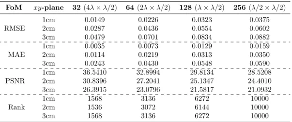
4.2 Pre-reconstruction generic assessment . . . 42
4.2.1 Voxel Crosstalk matrix. . . 42
Signal fidelity metric . . . 43
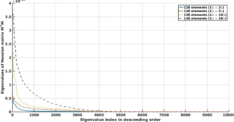
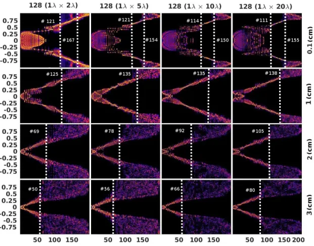
4.2.2 Eigenanalysis . . . 44
Singular value decomposition . . . 44
Rank of matrix . . . 45
Eigenspectrum . . . 46
Singular vector analysis . . . 46
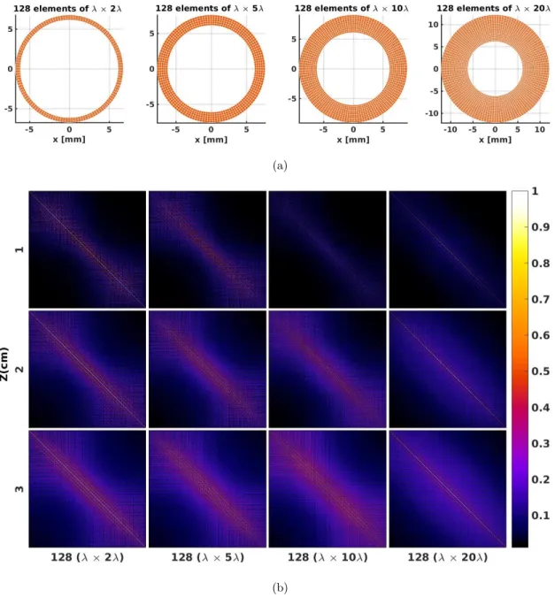
4.3 Array design . . . 48
4.3.1 Frequency response. . . 49
4.3.2 Light delivery system . . . 49
4.3.3 Design strategy . . . 51
Inter-element spacing . . . 51
The aspect ratio . . . 56
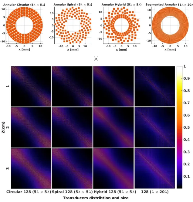
Geometrical distribution . . . 61
4.4 Conclusion . . . 67
4.4.1 Perspective . . . 67
5 Post processing evaluation 69 5.1 Introduction. . . 69
5.2 Deconvolution of acousto-electric impulse response . . . 70
Simulation test data . . . 72
5.3 Method based on the Delay and Sum Back Projection . . . 74
5.3.1 Weighted Synthetic Aperture . . . 77
Coherence factors. . . 77
Spatial impulse response. . . 79
5.3.2 Comparison of weighting factors and their effect in DSBP . . . 79
5.3.3 Virtual element . . . 82
5.3.4 Matrix representation of DSBP . . . 84
5.3.5 Matched filter interpretation . . . 84
5.4 Focusing as an inverse problem . . . 85
5.5 Post-reconstruction evaluation. . . 88
5.5.1 Optoacoustic . . . 88
5.5.2 Ultrasound . . . 90
5.6 Conclusion . . . 93
6 Experimental validation of OPUS 95 6.1 Introduction. . . 95 6.2 Experimental setup. . . 96 6.2.1 Illumination system . . . 96 6.2.2 Acquisition system . . . 97 6.2.3 Scanning system . . . 97 6.3 Measurement . . . 98 6.3.1 Transducer characterization . . . 98 6.3.2 Acquisition . . . 99 6.3.3 Phantom . . . 99 6.4 Results. . . 100 6.5 Conclusion . . . 101
7 Conclusion and Perspective 103 7.1 Summary and concluding remarks . . . 104
CHAPITRE
1
Introduction
After a certain high level of technical skill is achieved, science and art tend to coalesce in aesthetics, plasticity and form. The greatest scientists are always artist as well.
Albert Einstein
1.1
Incentive of the work
I
n the past decades, the continuous development in medical imaging systems paved the way for variety of applications and opportunities in clinical and preclinical research and studies. The significant diagnostic value and the level of confidence in the efficacy and safety of some of these agents make them a reliable tool for routine clinical trials. Others, while still advancing, have been shown to hold great promises of transition from bench to bed side and going beyond principle to practice. In fact, when the interest is in multiscale and multipurpose imaging, there exists such a will in integrating multi-modalilties into a synergistic paradigm in order to leverage the diagnostic values of the interrogating agents. Additionally, hybridizing prompts the clinical translation of the new approach and further, opens a new door for the currently established device and augments their functionality. For instance, if the clinically routine system is targeting only the structural deformation, integration with an imaging system that aims at the functional information may assist the early stage diagnosis, to track the disease development progress and improve the treatment monitoring.Throughout the years, ultrasound has shown great potentials and impacts as a diagnostic agent. It’s safety records, portability, rich and non-ionizing interaction with the anatomy of the tissue and capability for real time display of the tissue dynamics in 2D and 3D, makes ultrasound a viable tool in diagnostic imaging. The recent endeavors to improve US 3D dynamic acquisition is particularly important where the temporal information of the entire volume is critical for ac-curate measurement in multiple planes, as is the case for neurosonography and echocardiography. The aforementioned competence and some of ultrasonic wave characteristics including the acous-tic speed and attenuation are still being investigated for the tissue characterization. However, ultrasound has limited capacity in providing sensitivity to molecular and functional contrast and chemical specificity, as often the mechanical contrast in neighboring tissues are not high enough for pathological purposes. Therefore, despite promises [2], the accurately delineation of the tissue integrity using ultrasound with no additional contrast enhancing is left to be exploited.
It is a well known fact that the electromagnetic radiation interaction with biological tissue is highly selective with respect to the electronic structure and chemical bonding of tissue itself.
This is specially true for the visible and infrared spectrum of the light, thereby making light an ideal technological ally for further physiological interrogation of the tissue. Yet, high resolution pure optical imaging deeper than one transport mean free path remains challenging. The so called optoacoustic imaging technique has been practiced over the last few years with promises in ex-tending the depth of optical imaging by pairing the ultrasonic wave properties with the merits of optical imaging. In this technique, the optical absorption properties of the tissue are encoded into the broadband ultrasonic pulse which is a function of shape, photoacoustic efficiency and optical absorption coefficient of the chromophore and the fluence of the light. Detecting these induced acoustic waves on the surface of the medium can reveal information associated with the structure of the absorbers, and in multispectral systems, physiological properties such as angiogenesis and blood oxygenation that helps in early stage diagnosis as major indication for cancer.
Since the detection is taking place with the same ultrasonic sensors, and due to the conceptual similarities between optoacoustic (OA) and ultrasound (US), hybridizing these imaging modalities into one compound measurement system is intrinsic and only requires the integration of the light delivery system to the ultrasonic probe. Such a combination allows to realize the potential benefits of optoacoustic-ultrasound (OPUS) as a promising multimodality approach in obtaining the highly complementary diagnostic information. This would thereby enables the concurrent measurement, co-registration and assimilation of distinct features of the investigating tissue. Consequently the limitation of individual modality can be surmounted for example via mapping the volumetric functional and anatomical information of the targeted tissue.
Yet, until the beginning of this work at 2014, OPUS integration has not been fully evolved to ad-dress the physical requirements of both imaging systems. The existing implementations presented in the literature are either based on ultrasound probe or systematically optimized for optoacoustic acquisition. The detection array, even though take place by the ultrasonic transducers, demands different set of characterizations for either of modalities which might not be consistent.
Indeed, designing an imaging system is a matter of mutual concession among the transmission of the interrogating waves and detection qualities of the measurement system in terms of sensitivity and geometry. In other words, the utmost determinant of imaging performance is the overall properties of the aperture, the interplay between the transmission and detection capability. Withal, the reconstruction algorithm remains fairly an important indication of the system performance and plays a major role in accurate estimation of the investigating object properties.
The main goal of the present dissertation therefore is to address the current challenges and limitations exist in hybridizing the optoacoustic (OA) and ultrasound (US) imaging system. To do so, the optimal properties for designing a bimodal 2D handheld probe has been investigated in de-tails in order to offer an integrated solution. Correspondingly an advance reconstruction algorithm has been elaborated toward the requirement of designed OPUS imaging system. Considering the ill-posed nature of the reconstruction [6], characterization of the design system solely based on the quality of final image accompanies with considerable level of uncertainty. To avoid the biased interpretation, a de facto performance assessment being capable of evaluating the potentials of the designed OPUS probe in an unbiased manner must be practiced.
The distinguishing features of this dissertation are introducing a new geometry for OPUS handheld probe and a systematic assessments based on pre and post reconstruction methods. The majority of our investigation is taking place over the aperture model matrix which is developed in the context of wave propagation in volume, emanated and sensed by the active surface of the aperture. The aforementioned features establish a framework for characterization of the perfor-mance of imaging system in an accurate manner. Moreover, it allows further task perforperfor-mance optimization.
1.2
Current OPUS systems, applications and limitations
Today US is recognized as a mature technology yet remaining as a developing imaging method. The amount of effort devoted to advance the approach whether improving the technological side or expanding its medical and biological applications corroborates the impact. Yet to further push the boundaries and relax the imposed restrictions of purely acoustic systems, other allied technologies are being explored. Among them, optoacoustic imaging has been of particular interest, owing to its merits of employing the optical contrast in an ultrasonic manner. Unlike US imaging, majority of the OA systems, including the commercially available ones are single purpose instruments de-signed for a particular task. Nevertheless, handheld probes tend to be exempt from this principle, along facilitating the dual modality combination of OA and US for clinical studies. This trend has been practiced over a decade by tagging the optical delivery system, to the linear handheld ultrasonic probe. Variety of applications have been proposed and developed with respect to the frequency response of the transducers and the wavelength of the utilized laser light. The range is not limited to diagnosis and may extend to staging, guiding therapy or even drug delivery [9] and theranostics [183,67]. One particular target with high diagnostic value is angiography, which is a hallmark for cancer. Zemp et al. [182] examined the capability of high frequency transducer (30 MHz) in visualization of superficial microvascular networks to the depth of 3 mm. To visualize deeper vessels such as human legs, forearms vessel [111,83] and carotid artery [86], lower frequency response linear array transducer (7.5 MHz) have been employed. Moreover, targeting specific chro-mophore by tuning the laser spectrum, whether endogenous or exogenous, allows to investigate the functionality and molecular composition of the underlying tissue. Multispectral acquisition of the vessel’s wall [72,28] with the purpose of staging atherosclerosis or plaque composition, monitoring the blood oxygenation which is related to oncology [88] or contrast agent [110,167] for a better detection of tumor is of this kind. Additionally, 3D acquisition is also possible by mechanically translating the probe over the surface of the sample [47] or using a 1.75D [3] and 2D matrix array US probe [167] with application in oncology. The 3D acquisition allows to widen the limited view angle of the detection aperture and have a better estimation of the three dimensional structure of the targeted tissue. However, these probes are essentially optimized for ultrasound imaging. They are only able to ensure high resolution morphological data for US images by employing a proper beam forming technique [73,160, 107], basically through increasing the number of projection in transmission. If utilized for optoacoustic acquisition, images may suffer from lack of accuracy in localization and imposed reconstruction artifact, because of lesser number of achievable projection and limited angle of view. Furthermore, the integration of light delivery system is another chal-lenge for OPUS systems. The associate problems are optically induced clutters and reflection of the light at the irradiation side and are closely related to the skin pigmentation and the diameter and angle of the incident beam. For example the perpendicular illumination maximize the fluence [23] which is not achievable by utilizing the classical US probes. Albeit, clutters induced at superficial layers [128] can be dealt with either by adjusting the distance between irradiation side and US probe [61] or sophisticated post processing techniques [141,71].
Recently, Dean Ben [29] reported the design of an universal handheld probe for multispectral real time volumetric OA imaging. Shortly, the probe houses a bundle fiber for illumination purpose in the center of the hemispherical shell where the staring passive transducers are situated in a multi-ring structure. While the perpendicular illumination is reserved, the spherical detection geometry provides allegedly enough tomographic view required for OA imaging. The potency of the probe have been evaluated in number of applications including angiography and monitoring therapy. In an effort, Fehm et al. [43] examined the efficiency of the probe for US pulse echo imaging, in a fashion similar to the laser ultrasound. Nevertheless, the results were not compelling compare with conventional ultrasonography in terms of contrast and resolution. On that account a perfect hybridization remains challenging and is left for a more adaptive design if favor of both imaging systems.
1.3
Questions to be addressed in this dissertation
(contributions)
This dissertation is mainly devoted to the design and characterization of a hybridized universal handheld probe for OPUS imaging and developing an adaptive reconstruction algorithm toward the physical requirement of the designed system. Therefore the questions to be addressed in this thesis are in the following context :
• With respect to the handheld probe design
— What is the optimum compromise between number and size of transceivers required for the successful OPUS handheld probe.
— What would be the appropriate array geometry to enable dual modality acquisition. — What are the necessary parameters in order to characterize the performance of the
designed system.
• With respect to the reconstruction algorithm
— What is the optimal reconstruction algorithm in order to mitigate the artifacts arose from the nonideality in the designed system.
— How far the adaptive weighting factors are relaxing the impose constraints by FBP/DAS algorithms.
— How accurate the performance of model based reconstruction algorithm can be, by incorporating the transceiver response.
— How responsive the handheld probe is to the acoustic source within the medium as a function of spatial location.
1.4
Overview of the dissertation
The organization of this work is as follow. Chapter 2 opens with the discussion on the basic physics of both imaging systems individually, by providing a brief background and the theory behind the image formation. It also reviews the postulated ideal instrumentation for both imaging systems and the consequences of non-idealized parameters. The purpose of this chapter is to underpin the building-block necessary for the upcoming chapters.
In Chapter 3, the development of imaging system forward model and its implementation for both OA and US is described. The models are based on acoustic wave propagation and the behavior of the transceivers in transmitting or sensing the emanated/reflected acoustic waves from the medium. The models will be employed for further evaluation and characterization of the imaging system performance and further, as imaging operator for model based image reconstruction in the subsequent chapters.
Chapter 4 proposes a framework for performance characterization of the imaging system based on the model explained in Chapter 3. It outlines the steps required for the pre-reconstruction evaluation of the imaging system. The performance assessment takes place mainly by two sets of analysis in quantitative and qualitative manner, namely voxel crosstalk matrix [136,130,13] and eigen-based analysis. The implementation of voxel crosstalk matrix for photoacoustic tomography are developed by Wong et al. [171] and is adapted for OPUS system. Necessarily it addresses the following concerns, spatial sensitivity and spatial aliasing. On the other hand eigen-based analysis reflects the overall performance of the imaging system in achievable amount of the resolvable data. Based up on these analysis, the ideal geometry for integration of OA and US will be discussed in details.
Chapter 5 is allocated to the volumetric image reconstruction algorithms and is divided into three sections. The first is an attempt to explore the optimum DAS based algorithm for US and extend the approach to OA. It aims to address the restrictions imposed by DAS based approaches and the possibility of relaxing them to the favor of integration purpose. The second part, deals
with the model based reconstruction approach based on the model generated in Chapter 3 that is incorporating the shape, geometry and the characterization of ultrasonic transducers. As the crucial step in the reconstruction approach is inverting the forward model, the focus will be on the regularization to deal with the ill-posed nature of this inverse problem. Last but not least the third part is devoted to the post-reconstruction evaluation of the design imaging system based on the final image.
As the proof of concepts, the experimental results are highlighted in Chapter 6. A single element has been scanned in the same fashion as the array to mimic the acquisition performance of the proposed geometry in chapter 5. The imaging performance of the proposed geometry for handheld probe, i.e segmented annular array, is evaluated and the feasibility of developing the array is discussed.
Finally chapter 7 provides a summary of the dissertation and prospective outlooks for the future works are suggested.
CHAPITRE
2
The Basic Principles of Optoacoustic Ultrasound Imaging
It is my inner conviction that the development of science seeks in the main to satisfy the longing pure knowledge.
Albert Einstein
Abstract
The underpinning step in designing a bimodal system is to genuinely understand and gain insight about the physical principles of both parties, the optical and acoustic wave interaction with tissue and image formation. The similarities and differences between the modalities and the key-factors that affect the performance of each system must be realized in an etiological manner. As Alexander Graham Bell once puts it « before anything else, preparation is the key to success ». Thus the goal of this chapter is to provide a fundamental background with respect to the principle of two imaging systems and their inherent similarities and differences.
2.1
Introduction
A
coustic imaging in general consists of revealing the underlying information about the investigating tissue, encoded in the form of acoustic pulses, hence seeing the sound. Although these information are initially in the form of acoustic waves, but later are being transformed to the electrical signals for further recording and post processing purposes. Acoustic imaging to many respects is obeying the same physical phenomenon that are ruling optical (wave) domain, which leads to developing elements with the similar functionality (e.g. refractive or reflective) such as acoustic lens and mirror ; or developing same techniques such as holography which is obeying the interference, diffraction and phase conjugation phenomena. However, it would be naive to postulate that the paradigm in acoustic imaging is always following optics. Generally, there are three phases of acoustic imaging namely, inducing, collecting and processing. The last two phases are taking place in a similar manner among all of the acoustic imaging devices. Acoustic transducers are collecting the encrypted acoustic waves emanated from the induced sources within the medium, followed by translation to the electrical signals. Normally, this is followed by further post processing actions with deciphering purposes, for example the localization of the sources based on time of flight, also known as image formation. The fundamental differences, however is stemmed from the mechanism of acoustic source generation inside the medium in order to study the features and characterize the objects of interest.Following the same scenario, two modes of acoustic imaging1 that can be mentioned with medical and biological applications are medical ultrasound (US) and its younger half-brother optoacoustic (OA) imaging. Ultrasonic pulse-echo (PE) is arguably one of the most efficient diag-nostic tools in order to estimate the anatomical abnormalities and localization of the lesions. Just like its forefather, the supersonic reflectoscope [45], it is based on the idea of echo-locationing that estimates the depth of reflecting objects from the transducer by calculating the round trip time of flight trt. The desired pulse of sound can be generated by deriving the ultrasonic transducer as
the transmitter and detecting the partially back-reflected energy (echoed wave) with the same (or neighboring) transducer functioning in receive mode. Echo-locationing is a straightforward process as the speed of sound found to be similar in majority of soft tissues and that is close to the speed of sound in water, due to their high water content. However, the reflection in biological tissue is happening due to the acoustic impedance (Z) mismatches at the boundaries of soft tissues. The acoustic impedance is defined as Z = ρν, with ν denoting speed of sound and ρ the density of the medium. The speed of sound is a complex value itself. The real part is related to the elasticity K and density ρ by the following equation ν =qKρ. The imaginary part explains the dispersion and frequency dependent attenuation via Kramers-Kronig relationship [115]2. Subsequently, the
reflectivity of the reflecting surface for the incident wave at the angle of θi can be calculated from
the following formula :
R = Z2cos θi− Z1cos θt Z2cos θi+ Z1cos θt
, (2.1)
where θt is the angle of refraction and that the following relationship "sin(θi)/ sin(θt) = ν1/ν2"
holds at the boundary. Given the conservation of energy, the transmitted and most probably refracted (bent), energy can be found by T = 1 − R.
Therefore in PE technique, object localization and characterization is fundamentally subjected to the mechanical contrasts. The array of transceivers facilitates the aforementioned tasks by providing extra rooms for diversifying the transmitting energy and direction for generated wave and extra view angles for receiving the echoed energy, thus improving the estimation about the original object. The properties of generated wave is, to many extend, defined by the physical properties of the ultrasonic transducer. These features are including the bandwidth and wave field propagation w.r.t. the transducer size. By further considering the backscattering (or reflecting) objects as the source of emanating wave from the medium, the characteristics of the incoming wave in US-PE is foretold.
In essence, optoacoustic imaging is an extended variety of ultrasonic technique in which the acoustic waves are induced inside the investigating sample upon the deposition of optical energy. The transition of photons to broadband acoustic pulses is a complex interplay of several physical phenomena. Yet, the contrast in OA is inherent to the optical wavelength selectivity of the ab-sorbents in the tissue rather than mechanical contrast. Unlike US, the acoustic properties of the generated wave, such as the amplitude and frequency spectrum, are highly dependent to the opti-cal properties of absorbents (sources). Withal, the image formation is following the same time of flight concept and theretofore same algorithm can be employed for both, with minimal adaptation. The presented thesis is concerned with designing a high throughput array for acoustic 3D imaging to address the physical requirement of both parties, OA and US techniques. Our interest is in hybridyzing the two modalities in order to pursue the promises of delivering the complementary morphological, functional, anatomical and metabolic information of the tissue. To do so, we start with providing theoretical background on the physical phenomena associated with both modalities and their requirements, in a separate manner.
1. To our concerns, the definitions of acoustic wave are limited to longitudinal waves. 2. The speed dispersion is often ignored as the effect is as insignificant as 0.01%MHz2
2.2
Basic of Ultrasound imaging
2.2.1
Ultrasonic wave interaction with the tissue
In the first glance, the concept of US pulse echo (PE) technique seems fairly easy, a set of ultrasonic short pulses generated by the transducers are eliciting the mechanical discontinuities (reflector) of the soft tissues, along their path. The reflected or backscattered echos are collected by the same or neighboring transducers (in-case of array imaging) and discontinuities are located in the formed image by the stated echo-locationing concept. Depending on the relative size of reflector (Ψ) to the wavelength (λemit) of the transmitted wave and also angle of incidence, the nature of
reflected wave would differ. Therefore the reflector can be considered as an acoustic source itself, with acoustic properties to be found by the imaging system. For example, the so called Rayleigh scattering is taking place when λemit Ψ, as is the case for red blood cells ( ≈ 7µm), which
causes uniform and concentric reflection of the energy in all directions. This type of backscattering is strongly depending on the frequency such that the scattering cross section S = κ4Ψ6, and κ = 2π/λemit is the wave number. For the object of the similar size as the wavelength Ψ ' λemit,
diffuse reflection with redirected wavelets is the dominant effect. Even though the backscattered wave is of the shape of spherical wave, the energy of the wave is more concentrated toward the angle of incidence. The reflected energy is getting more directional with increase in Ψ. This type of backscattering is responsible for the visible texture of the soft tissue in the ultrasound image. The properties of the reflected waves are analogous to the generation of acoustic wave using the λ/2 and λ size transducer, being used in phased and linear arrays respectively. Such an analogy allows to treat each reflectors as an acoustic source. The third case is when Ψ λemit, then the so called
specular reflection will take place, an effect that explains the bright appearance of boundaries, for example between tendons, fat and muscles, bones etc. The intensity of the reflected waves are conveying information about the compressibility and density gradient at the interface of the two tissues and can be found from the equation 2.1. However if the angle of incident violates 90◦, it can affect the shape and amplitude of the received wave, or the echoed wave might not meet the aperture at all, depending on the aperture covering angle of view, thus the encoded information about the medium would be partially or totally lost.
Attenuation
In the US imaging, the resolution is subjected to the so called resolution cell associated with the wavelength of the transmitted and consequently reflected wave. The wavelength and frequency (f ) of the acoustic waves are related by the speed of sound such that f = ν/λ. The choice of ul-trasound frequency for transducer is however, a trade off between the resolution and penetration (attenuation). The high frequency ultrasonic wave is encountering relatively more diffusion events, more rarefaction and compression over the course of propagation, thence smaller resolution cell can be provided at the cost of higher attenuation. The main phenomena associated with attenua-tion are absorpattenua-tion, refracattenua-tion, reflecattenua-tion, scattering and diffracattenua-tion. The first one is attributed to the viscoelastic forces between neighboring particles, relaxation mechanism of protein [12]3
and temperature gradient within or between soft tissues [122]. The associated absorption with the viscosity is following a square law dependence relation, in contrast to the linear frequency dependence via relaxation process. Refraction and reflection are caused by the variation in density of the medium and depend on the angle of incidence taking place at the tissue interface, which in general are obeying the equation2.1. However, their impact on the energy of traveling wave in soft tissues is low, in compare with the backscattering. Last but not least, the diffraction describes the divergence of the wave as it migrates away from the source, but also the convergence of the wave behind the reflecting (specular and diffusive) object and further the shape of the reflected wavefront. Diffraction and refraction are both detouring the wavelets of the wavefront, but unlike 3. The attenuation of acoustic wave is phenomenally a different concept in porous structure such as bone. Depending on the bone structure multiple effect such as viscous friction, scattering due to heterogeneity and mode conversion are playing major roles of which none may happen in soft tissues.
refraction, diffraction is a bending wave phenomenon that does not associated with the change in speed of sound of the medium4.
The diffraction, refraction and interference can be explained by Huygen’s wavelets which la-ter augmented employed by Fresnel and further work of Kirchhoff [50], yet widely recognized as Huygens-Fresnel principle. Huygen’s wavelets are imaginary sources of spherical waves populating the entire surface of primary wavefront. At every time instance, the interference between these secondary wavelets define the direction of propagation and the ensemble wavefront. The Kirch-hoff rectifies this postulation by adding obliquity factor K(θ) to explain the direction dependent behavior of wavelet which originally was assumed to be uniformly spherical.
K(θ) = 1
2(1 + cos(θ)) (2.2)
If the Ψ (size of the obstruction, or reflector) be smaller than λ, all wavelets are in-phase by default, thus the interference would be coherent and constructive. Therefore the wave will spread out at large angle, or even can be considered as fully spherical in case of λ/2 or smaller. This is similar to the Fourier analysis perspective, the infinitesimal source can produce an infinitely rich in spatial frequency [60]. However, when the reflector size is Ψ λ, the region where the wavelets are constructively interfering would be limited to the region extending in front of the reflector, with the same diameter. Outside the region, the destructive interference between wavelets is dominant. Therefore, diffraction effect can explain the backscattering and reflection. Not to mention that Huygen’s principle can be used to derive the Snell’s low in order to explain the reflection and refraction [60].
The diffraction is attributed to the fluctuation between the acoustic particles in the vicinity of the source and consequently set of wavelets constructive and destructive interference appears. This behavior can be explained by Fresnel diffraction. The robust behavior begins where the coherence movement between the particles causes a relatively uniform yet spreading out wavefront, known as the far field and governed by Fraunhofer diffraction. Depending on the geometrical appearance of the source relative to the wavelength of propagating wave, the pattern of divergence would differ accordingly. A practical rule of thumb is separating near field from far field, given by :
z > ψ2/λ (2.3)
According to the Babinet’s principle [143], diffraction can explain the convergence of the wave beyond the opaque (reflecting) object, in a similar fashion. For example the disturbance introduced by an object smaller than λ to the migrating wave is negligible and the traveling wavefront remains unaffected, as if the thin object is transparent. Meanwhile for the Ψ λ, convergence of the edge diffracted waves happens at "Poisson spot" in the transition zone between near field and far field. This effect is similar to the flat transducer natural focus, which happens in the transition stage between near field and far field and has the maximum intensity value.
znaturalf ocus=
4ψ2− λ2
4λ (2.4)
given ψ as the size of transducer, or reflecting object. Also, for the disturbed wave by the obstruc-tion, it can estimate the post obstruction behavior of traveling wavefront and in particular, the distance the wavefront needs to converge such that the obstruction never happened.
Over the course of propagation, if the incident wave is not canceled due to the reflection, it would further weakened due to absorption and diffraction, until the energy ultimately transfers to the medium. The reciprocal relationship between the size of the source and the angle of divergence causes faster attenuation of the wave energy. The rule of thumb is the 6 dB loss per doubling migration distance.
Last but not least, if the acoustic wave is broadband, due to the frequency selectivity of at-tenuation, the shape of the wave would change as well. Additionally it can be shown that when 4. The literal meaning of diffraction can be extracted from the Latin word diffringere, "breaking up into different direction".
the attenuation is related to the frequency of the wave, the acoustic velocity is also frequency dependent [115]. Soft tissues have their own attenuation and scattering properties and those pro-perties are distributed randomly in inhomogeneous samples. Additionally, orientation of the object is another consideration factor. Muscle as an example, has illustrated lower attenuation for the wave passing along the fiber than across the fibers, almost as half the value[168]. Therefore, the selection of frequency components for the transmitting wave in PE imaging is of-course biased in the favor of application.
Speckle
The insonified objects by a highly coherent wave are observed to acquire a peculiar granu-lar structure. These ubiquitous structure are called speckle, are mainly considered undesirable, stemmed from the individual scatterer varying in strength and position w.r.t. the aperture and the wavelength of ultrasonic wave. The complex pattern bears relationship to the microscopic properties of the insonified parenchymal tissue. Rather it appears chaotic and unordered and is best described quantitatively by the methods of probability and statistics. Despite their random appearance, the pattern remains the same for the relative stationary geometry between aperture and object, an indication of their coherent level. Yet, slight change in the conditions will result to a new pattern, to the extent that the strength for the same point can be totally different, suggesting a dubious link to the tissue structure.
Speckles can be explained by the interference between wavelets of multiple scatterers with particular phase angle, occupying one resolution cell. Depending on the nature of interference, whether constructive or destructive, the gray value (strength) of the resolution cell is set. Overall speckle is not desirable in many conditions and is considered as artifact adversely affecting the quality of image, detectability of the small targets and deteriorating the resolution and contrast. As a matter of fact, there are various approaches to reduce the speckle level, such as compounding multiple acquisition varying in frequency, pulse length or scanning geometry [152] or applying adaptive coherent factors [21,63]. However, their level must be kept, even in lower scale, in order to realize the boundaries of anechoic regions. Additionally, the appearance of speckle pattern is not limited to the Rayleigh scatterers, leaving some room for tissue stratification [142] based on their texture and adding further diagnostic value.
2.2.2
Ultrasonic transducers
The core of any acoustic imaging system is the transducer, a key element that allows the ultrasonic wave being applied on the sample and record the interacted wave containing information. Ultrasonic transducers perform the conversion of electrical energy to mechanical (ultrasound) and vice versa, therefore are widely used as transmitters, receivers or transceivers (both) of ultrasonic waves, ultimately defining the image quality. Although, the frontier of ultrasonic imaging has been expanded owing to the advances in the ultrasonic transducer, yet paradoxically it remains as a bottleneck for further advancing the application. For instance, the frequency of which the US transducers are working effectively is limited to a certain range. Before broadband transducers, non-linear and harmonic imaging were quite challenging. Also, the broad bandwidth comes with lesser distortion and higher resolution for the final image.
Since 1927 that ultrasound has been introduced to the medical field [172], variety of forms, size, materials and approaches are being practiced in order to optimize the generation or detection of acoustic wave with desired properties. Among them, three major techniques are piezoelectric transducers (PZT), micromachined ultrasound technique (MUT) and all-optical transducer, using photoacoustic effect.
Piezoelectric transducer
As of today, the dominant technology for ultrasonic transducer is by the help of piezoelectric materials. These include piezoelectric crystals (e.g quartz, LiNbO3), ceramics (e.g PbZrTiO3) and
polymer films like polyvinylidene fluoride (PVDF). Indeed the preference is altering with applica-tion, and that requires technical compromise. Taking ceramics PVDF, the acoustic impedance of similar to human tissues allows a better mechanical coupling and more signal to noise ratio (SNR), at the cost of low electromechanical coupling efficiency of polymer material. Meanwhile, ceramic may not suffer from low electromechanical coupling but high dielectric constant and inflexible surface are the draw backs.
A typical PZT transducer is composed of five layers, respectively a backing block, active pie-zoelectric material, matching layer, lens and protective layer (wear plate). The characteristic of PZT transducers namely, resonant frequency, electromechanical coupling coefficient, quality fac-tor, impedance, beam profile are controlled by the properties of aforementioned composition. The piezoelectric material is sandwiched by two electrodes and the combination allows the acousto-electric translation. There exists a rule of thumb associating the first resonant frequency (fresonant)
of the material to its thickness (d) via speed of sound (ν) such that : fresonant = ν/2d. At this
frequency the material has the maximum deformation, leading to strong ultrasonic wave. While resonating, the material produces range of frequencies in adjacent to resonating frequency drop-ping in amplitude as it changes from fresonant. This range with amplitude of at least half of the
fresonant is called bandwidth (BW). The quality factor (Q) of the resonating system is related to
the generated bandwidth via following equation :
Q = fresonant
BW (2.5)
demonstrating the reciprocal relationship between BW and high Q system. High Q system is a well isolated system from external forces inducing damping effect. Such a system is efficient in terms of required energy for keep resonating at fresonant.
The backing block is made of a high attenuating material in order to damp the ringing of the housing that causes extra vibration, therefore shortens the acousto-electric impulse, increasing the bandwidth of the transducer. Alternatively, the bandwidth is shown to be increased by improving the electromechanical coupling efficiency using multiple matching layers. In fact, the electrome-chanical coupling efficiency is a measure of energy conversion efficiency and can be interpreted as a measure of sensitivity as well. Thus the advantage of multiple matching layers is two fold. A good example is PbZrTiO3 which is conventionally used in ceramic PZT, for its high
piezoelec-tric efficiency. Yet, its high acoustic impedance is associated with high reflection at the two ends, leading to ringing effect and spurious reverberated echoes in the tissue and lower sensitivity. As a remedy, the low acoustic impedance materials is added to ameliorate the mismatches effects and increase the bandwidth as the matching layer operates at different frequencies [66].
The nature of protective layer is determined by the either of two modes of transducer, immersion or contact. To maximize the transfer of acoustic wave, the quarter of wavelength thickness is considered but sometimes deliberately thickened for wedging purposes. The lens is used to diffract the generated acoustic wave, with focusing or defocusing purpose. However if the PZT materials are flexible, the same effect might be achieved with no loss by bending the surface of transducer. Yet, this bending is limited to certain degree.
Micromachined Ultrasonic Transducers
Micromachined ultrasonic transducers (MUTs) are moderately a new family of transducer ba-sed on microelectromechanical systems (MEMS) technology. The technology is attractive from the processing perspective where the micrometer precision is effectively improving the performance of transducer, compact form factor and in case of array imaging, the consistency among the elements behavior. There are two types of MUT transducers, capacitive MUT (CMUT) and piezoelectric MUT (PMUT) being widely developed with great promises for medical ultrasound.
CMUT
Since the invention in mid 90s [54], CMUT technology grabbed a lot of attentions specifically in the medical ultrasound, owing to its moderately easier and more precise fabrication and broad bandwidth response. As the name suggests, the transduction of energy is taking place by Coulomb force between the capacitor plates. Suppose that only one plate is mobile, the basic principle can be simplified as follows. The two plates are charged by a DC voltage (aka. bias voltage), creating an electrostatic attraction force in between, which is being opposed by the stiffness of the mobile plate resisting the attraction. Applying the electrical signal (AC) induces vibration over the surface of mobile plate hence ultrasonic wave is being generated. Similarly, when the mobile plate is subjected to the acoustic wave, the capacitance is being modulated relative to the frequency of incident acoustic wave and that, an electronic signals will be generated knowing the relationship between the capacitance (C), voltage (V ) and charge (q) hold as such C = q/V . The generated current is a function of three variables, the bias voltage (DC voltage), the capacitance and characteristic of incident wave.
The idea of using capacitive transducers5 is dated back to 19th. The hurdle, however was the
required high electric field for a thin gap of capacitor in order to obtain the large coupling coefficient in the same range as of PZTs. As it has been foreseen by Paul Langevin [68], it must be in order of MV/m. According to the Paschen’s law, in such a condition the spark voltage can be avoided by a very thin gap. This is achieved thanks to MEMs technologies which has allowed this centennial idea to develop and being practiced. CMUT’s building blocks are cells made of micron size metal plate (or membrane, top electrode) suspended over the vacuum gap of sub micros size and a fixed plate (substrate, bottom electrode) normally made of silicon, together acting as a capacitor. For the safety aspect, in medical application the transducer is being electrically insulated by coating a layer of polymer on top of the metallic membrane [95].
The operating frequency of the system is a function of geometrical and mechanical properties of the membrane. Analogously, actuation of CMUT is following the same principle as PZT, except for the DC bias voltage that regulates the operation of CMUT, according to the aforementioned formula. On transmit, the deriving current is inducing the attraction force on the electrodes, deflecting the mobile electrode to the fixed one and upon release the membrane vibrates, creating the acoustic wave. Since the cell size is very small, the desired acoustic wave is a result of multiple cells populated and connected in parallel, operating in a synchronized manner. It is worthy to mention that the generated acoustic frequency is corresponding to the frequency of the applied current multiply by two. This is due to the unipolar property of the induced attraction force [39]. On reception, upon the incidence of the acoustic wave, a harmonic vibration of membrane changes the C and as a result q, transducing acoustic energy into the electric signal.
In compare to the PZT, CMUTs are miniaturized capacitors that demonstrated a superior performance w.r.t. the acoustic matching, size reduction, broader bandwidth, front-end electronics and more flexibility for complex geometries. However, there are some challenging areas left to be addressed such as transmit/receive sensitivity, acoustic crosstalk, high electrical impedance and high bias voltage around 200 V [134].
PMUT
To many aspects, PMUTs are performing in the similar fashion as the CMUTs, except for the fact that in PMUT fabrication piezoelectric material of micron size is being employed, instead of the vaccum gap. The piezoelectric effect induces electric fields across the layers, which in turn causes the actuation. Therefore, the bias voltage and small capacitance gap is no more required for their operation [77]. They do this by offering higher capacitance and lower electrical impedance, leading to lower parasitic capacitance and consecutively higher sensitivity and SNR [65]. The thickness of piezoelectric film is between nanometer to 3 microns. It can be said that the resonant frequency of PMUTs is not a function of piezoelectric layer thickness, but similar to CMUT,
geometrical and mechanical properties of the membrane such as stiffness and intrinsic stress. The challenge is placing a well defined thin yet piezoelectric materials, and that requires significant effort and cost. For example, one particular challenge could be the fact that resonant frequency is highly subjected to the membrane residual stress [53]. Therefore the applied stress T during fabrication will dramatically alters the resonant frequency :
fresonant∝ 1 2πr2 s DE ρh (2.6)
where r is the radius of membrane, DE is the flexular rigidity, ρ is the effective density and h
is the membrane thickness. T can dominate DE and alters the resonant frequency. As of today
many research groups are investigating the optimum fabrication techniques and materials for PMUTs. Nonetheless, in compare to CMUT, PMUT is a younger technology and requires more development.
Laser ultrasound and optical detectors
An attractive alternative to the MUT techniques is the all optical transducers in which photoa-coustic effect has been employed for transmission of broadband signal and interferometric detectors in reception. Instead of electronic signals, normally two laser are being input as a set of input and output.
Over the years, variety of methodologies have been explored in order to ameliorate the trans-duction of optical energy to tone-burst-like acoustic waves (i.e. laser ultrasound), among them thermoelastic expansion [52] is recognized as a reliable mechanism. This transduction normally happens by photo-exciting metals with ultra-short laser pulses [153] that is followed by photon absorption, heat conversion ans thermoacoustic relaxation, leading to generation of coherent pho-nons. Typically the irradiating light is focused with a focal spot size affecting the characteristic of the generated wave. Despite the fact that metal sheets are alleviating difficulties associated with fabrication, they are suffering from the low transduction efficiency, turn them to improper choice for medical ultrasound. For this very reason, a mixture of polydimethylsiloxane (PDMS) (high transduction efficiency) with black carbon (absorbent) is of favorite choices, obtaining over 20 dB in transduction efficiency [19]. The thickness of material is yet another parameter affecting the properties of generated acoustic wave. The aforementioned composite is acoustically attenuating thus thinner material is of interest which in turn tailors the frequency properties of the genera-ted wave. However, the major determining factor for frequency content of the acoustic generagenera-ted wave is the laser pulse duration, the shorter the duration of laser pulse, the higher frequencies components will be induced. Normally, for ultrasonic imaging where the desired frequency is not violating the range of 10 MHz, laser pulse in the order of 10 nsec is seemingly enough [84].
On the other side, for detection of an incoming acoustic wave, a common paradigm is the so called active optical detection employing a thin polymer Fabry Pérot etalon [106]. The concept is fairly easy, a pair of optically reflecting facing each other sandwiching the polymer is used as the interface between the optical field and ultrasonic wave. The incident light from a continuous wave (CW) laser goes through multiple reflection, producing interference pattern that depends on the laser wavelength and optical path length. Upon incidence, the strain associated with ultrasonic wave induces vibration on one of the mirrors, alters the thickness, changes the optical path, modulate the optical pattern and finally the resonance condition, proportional to the amount of displacement. The key to the sensitive system is the optical wavelength w.r.t. the surface displacement, such that the altered phase can be measured. The resonance in the cavity happens when the optical path length is equal to the integer multiple of wavelength. Etalon based detectors are promising broader bandwidth of detection [10], omni-directional response and competitively more sensitve response, much more compact. In fact the array of transmit/receive can be minimized to micron size. As both measurement and excitation occurs optically, electronic errors are alleviated as well. The limiting drawback of these system, are their laser dependent high cost.
2.2.3
Array imaging
Otherwise focused transducers are normally used in macroscopic imaging, such as US-PE. While the behavior of transmitted wave is solely determined by the geometrical shape of the transmitting aperture (i.e. element/array), single element transducer is not able to sweep the beam. Earlier in this chapter, it has been stated that array imaging is facilitating the volume interrogation of the sample toward real time, by electronic steering the transmitted beam in the desired location. The steering is possible by phase adjusting the array toward the specific location or direction, such that the constructive interference only occurs at particular angle, destined for the traveling wavefront. The dynamic directivity and steerability of the array in return, imposes certain limit in the geometric positioning of the array "the λ/2 interelement spacing"6. If the
transceivers are small enough to allow this happens, they act as a point source and the individual near field would be too short that propagating wave are by their nature spherical. The interference between the generated spherical waves allows only the ensemble wavefront survives in the focused (or angled) direction. The rest of energy is going through the destructive interference, leading to interferometric nulling, except for the wave from marginal elements (diffraction rays). In the far field of the aperture (or at the focusing plane) the residual energy forms the diffraction lobes, also known as side lobes. This can be rectified by apodization, a weighting technique that tailors the amplitudes of generated wave source (elements) w.r.t. their location from the center of aperture, at the expense of directivity and perhaps the achievable resolution.
From Fraunhofer approximation, far field pattern can be approximated by Fourier Transform of the aperture. Intuitively it can be interpreted that interelement spacing larger than λ/2 violates the Nyquist sampling criterion, and that yields spatial aliasing in the constructive interference of the wavefront, The consequence is out of scope energy formations, known as grating lobe (1st diffraction order). This has been discussed in detail by Johnson and Dudgeon [76]. If the desired steering angle is not too aggressive, λ interelement spacing is a good compromise between the generated energy, number of element and steerability. Considering a rectangle function for each direction of rectangular element showcasing a uniform behavior over the entire surface, Π(x)Π(y) represents one element behavior. This follows that the far field pattern of the element can be approximated by sinc(u)sinc(v). Conceptually for transmission, it represents the beam patterns while for receive, the spatial/angular sensitivity. A convolution by the comb function (III) can extend this way of representation to an array with uniform interelement spacing. It is shown that the periodicity in the array, introduces the periodicity in the beam pattern, creating off-axis replica (grating lobe) of on-axis energy with lower amplitude. In polar coordinate, the grating lobes occurs at 90◦ for array with λ interelement spacing and almost non for λ/2.
The array that contains the λ/2 characteristic is called phased array and can be classified into three categories, linear, planar and annular. Linear array is composed of small elements ar-ranged equidistantly in a simple line assembly7. Thus steering/focusing is only take place in two
dimensions. Planar array is extending the properties of linear array into three dimension by re-peating the element positioning in the second dimension, a matrix structure over the surface of aperture. Annular array is categorized into two geometries. The first consist of a set of concentric rings enabling the spherical focusing but the steerability remains fruitless. Other is called sectorial (or segmented) annular [4], which is basically a set of elements distributed around one or mul-tiple rings, has a higher capability of steering and focusing yet remained theoretical due to the complexity in manufacturing.
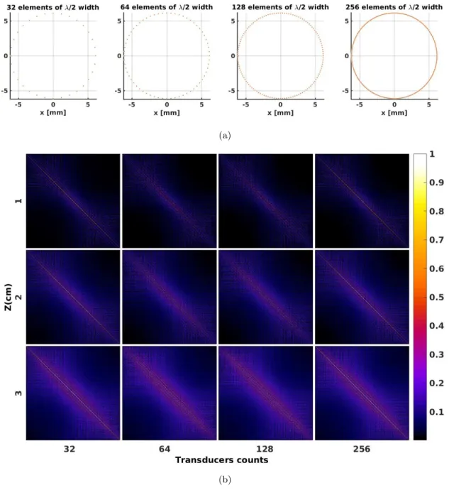
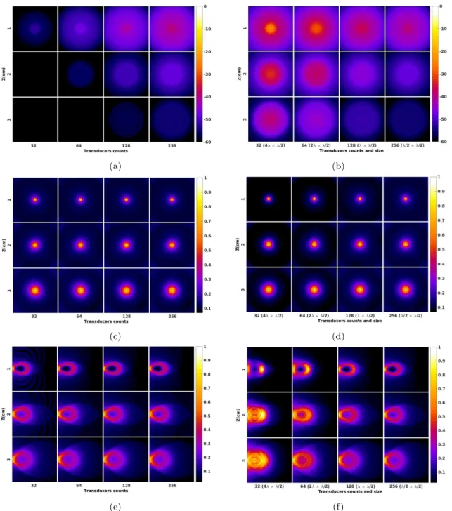
Conventional linear arrays are mainly composed of 128 (also 192 and 256) elements along their length, which is a reasonable compromise between aperture size, achievable resolution and number of channel required to actively and individually control the elements. Planar arrays however, might not benefit such an agreement as manufacturing a probe containing 128 × 128 = 16384 elements is a formidable challenge in many regards, including electomechanical hinders, amount of data to be processed and acquisition time. What commercial probes are offering does not violate 2880
6. This can be also attributed to the Huygens principle
elements8and that, not all the elements are accessible simultaneously, mainly due to the limitation in the available digital acquisition systems (DAQ) with limited accessible channel. At best, the customized 1024 channels systems allow to derive 32 × 32 elements of a matrix array9.
The volume acquisition is possible synthetically, by mechanically rotating/tilting scan the 1D linear array such that the pyramid-like volume can be reconstructed out of set of 2D images. The offered resolution is generally higher than what is achievable with the 2D matrix probes due to the number of elements for the synthetic aperture and aperture size. Nonetheless, such a technique costs the real time acquisition and is limited to 40 volumes per second [33]. Therefore pursuing dynamic acquisition for extracting functional information such as the brain functional imaging [32], Doppler cardiography, cardiac electrophysiology and tumor mechanical properties is left to 2D probes. The so called beamforming techniques allow to scan the volume by electronic steering the beam over the volume via adjusting the time between the transmitting elements. Beamforming can be used at both the transmitting and receiving ends in order to achieve spatial selectivity, by selecting certain elements in a random fashion. The selection or sub-sampling of the elements aims to avoid or minimize the periodicity in the position of elements [64]. In US PE, this is done by monitoring or calculating the transmit-receive radiation pattern [144] and that must null the cross-talk between the grating lobes in transmit energy and main lobe in receive and vice versa. This rule is also followed for sparse arrays in order to find the optimum location for the elements. The reciprocal relationship between the lateral resolution and aperture size and on top of that avoiding the grating lobes might be met by random configuration of elements over the surface of aperture. Often mentioned as sparse arrays [11], such an arrangement promises the utilization of lesser but larger elements, achieving reasonable level of signal to noise ratio (SNR), decrease computation load and mitigate the font-end complexity. Withal, the development of a sparse 2D probe for 3D ultrasonic imaging remains as an attractive challenge and the optimum configurations are still being investigated [131,137] in order to achieve uniform insonification and sensitivity pattern over the desired field of view and keeping the dynamic range between main lobe to side lobe in a reasonable level.
Image formation
The image formation in US PE is basically performs by scanning the ultrasonic beam generated by set of elements over the volume of interest, and recording the backscattered wave. The volume can be reconstructed by relating the position of transmitting and receiving elements to the echoed wave via time of flight. The spatial resolution in ultrasonic imaging is subjected to the quality of the trains of produced beams, sequentially insonifying the sample. Evidently, the optimum resolution provided by the aperture is achievable when the collected data by all transmit-receive combinations are available, i.e. full matrix capture (FMC) synthetic aperture, enabling the total focusing method (TFM). In this way, the investigating object is being insonified at all the available angles provided by the elements of the aperture in a synthetic manner (hence, synthetic aperture). The image reconstructed for every transmit-receive events is referred to as a projection. Therefore, coherent compounding of the back projections will allow to reconstruct the final image10.
The FMC or synthetic aperture focusing technique (SAFT) allows for focusing at every single points inside the volume of interest in a simultaneous manner. For an aperture composed of N elements, the FMC provides N × N projections to be processed. Therefore the calculation time is increased with the increase in the number of elements, thus frame-rate is decreased. Another drawbacks is the acoustic power transmitted by single element, degrading the SNR. To cope with, sparse matrix capture (SMC) might be beneficial approach in a sense that the multiple employed elements in the transmission, would increase the SNR yet reduce the acquisition time. In each transmission, the employed elements are phase adjusted in a way to mimic the wave transmitted from a virtual source behind the transducers.
8. Philips 9. Vermon