HAL Id: lirmm-01828409
https://hal-lirmm.ccsd.cnrs.fr/lirmm-01828409
Submitted on 4 Apr 2021HAL is a multi-disciplinary open access archive for the deposit and dissemination of sci-entific research documents, whether they are pub-lished or not. The documents may come from teaching and research institutions in France or abroad, or from public or private research centers.
L’archive ouverte pluridisciplinaire HAL, est destinée au dépôt et à la diffusion de documents scientifiques de niveau recherche, publiés ou non, émanant des établissements d’enseignement et de recherche français ou étrangers, des laboratoires publics ou privés.
Intraoperative Ultrasonography-based Augmented
Reality For Application In Image Guided Robotic
Surgery
Jun Shen, Nabil Zemiti, Agnès Viquesnel, Oscar Caravaca Mora, Auguste
Courtin, Renaud Garrel, Jean-Louis Dillenseger, Philippe Poignet
To cite this version:
Jun Shen, Nabil Zemiti, Agnès Viquesnel, Oscar Caravaca Mora, Auguste Courtin, et al.. Intraoper-ative Ultrasonography-based Augmented Reality For Application In Image Guided Robotic Surgery. 32nd International Congress and Exhibition on Computer Assisted Radiology and Surgery (CARS 2018), Jun 2018, Berlin, Germany. pp.S45-S46, �10.1007/s11548-018-1766-y�. �lirmm-01828409�
Intraoperative Ultrasonography-based Augmented Reality For Application In Image Guided Robotic Surgery
J. Shen1,2, N. Zemiti1, A. Viquesnel1,3, O. Caravaca-Mora1, A. Courtin1, R. Garrel3, J.-L. Dillenseger2, P. Poignet1 1 LIRMM, University of Montpellier, CNRS, Montpellier, France
2 INSERM UMR 1099, Rennes, France; University of Rennes 1, LTSI, Rennes, France 3 ENT Department, Centres Hospitaliers Universitaires Gui de Chauliac, Montpellier, France.
Keywords: Intraoperative ultrasonography, 3D ultrasound calibration, augmented reality,
image guidance, transoral robotic surgery
Purpose
Accurate Tumor delineation in the operating room (OR) is a big challenge for surgical oncologist. For instance, the preoperative images used in the transoral robotic surgery (TORS) for tongue base tumor resection cannot reflect the deformation of the soft tissue during the surgery. Furthermore, due to the camera’s small field of view and the loss of cross-modality landmarks in the tongue base, it is difficult to register the preoperative imaging modality to the intraoperative stereo camera with deformable registration. We propose an intraoperative ultrasonography (IOUS)-based augmented reality (AR) framework, which is able to accurately delimit the tumor boundaries and provide them to the surgeon’s view during the surgery. Instead of some works requiring manual registration [1], additional fiducial markers attached on the organ [2], or intraoperative imaging modalities which is highly ionizing [2] [3], our solution uses a safe and cheap US imaging modality and does not use additional fiducial markers disturbing the TORS workflow.
Methods
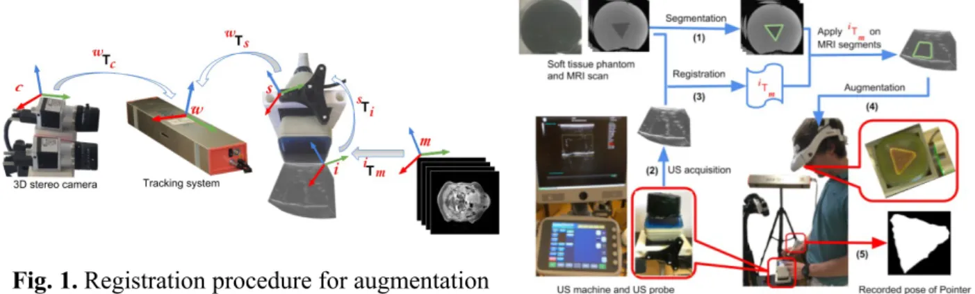
Fig.1 shows the registration pipeline used to achieve AR on stereo camera. A tracking system was defined as the world coordinate system (WCS) w. The transformation between the coordinate system (CS) of the camera c and the WCS was found by calibrating the camera with the WCS based on the mathematical framework given by wT
c = wTs sTc, where s
represents the CS of an active marker. The tracking system computed the transformation wT s
from the CS of the marker s to the WCS, meanwhile, the marker was localized in the 3D camera and its position was used to compute the transformation sT
c.
After calibrating the stereo camera in the WCS, the preoperative MRI image had to be registered to the WCS, in order to augment the camera view with the information extracted from the MRI. The corresponding mathematical registration framework can be presented as
wT
m = wTs sTi iTm, where m, i represent the CS of MRI image and US image. The
transformation iT
m was achieved in our experiment using Elastix toolbox performing a
deformable registration between MRI image and US image. B-spline transformation model and Mutual Information similarity measure were used for the registration. The transformation
sT
i was obtained by US probe calibration. We developed a fast and automatic calibration
method based on a custom-made 3D printed phantom and an untracked marker for 3D US probe calibration. Finally, the tracking system measured the position of the marker mounted on the probe and computed the transformation wT
s.
The performance of the setup was evaluated with 3 soft tissue phantoms made of soft polyvinyl chloride plastic. There was a triangle-shaped object embedded inside of each phantom. The invisible triangle-shaped objects could be distinguished under palpation and US imaging. Fig.2 shows the experimental setup. The participants were asked to use a calibrated Pointer to localize the boundaries of the invisible targets according to the augmented view projected in a head-mounted display system.
Results
The errors of the US-based AR framework are mainly from MRI/US registration and US probe calibration. The target registration error (TRE) was used to compute the distance, after registration, between corresponding points. MRI and US image registration were performed
on the three phantoms. 6 vertexes of the triangle-shaped targets in the MRI and US images were used as landmarks for computing TRE. The root mean square (RMS) of TRE for the three phantoms were 0.313mm, 0.19mm and 0.41mm, respectively. The US probe calibration was evaluated by point reconstruction tests and obtained the RMS of point reconstruction errors of 1.39mm.
The presented approach was developed for our clinical application in TORS for tongue base tumor resection. Therefore, we compared our AR framework with conventional procedure where preoperative MRI and manual palpation were used to localize the tumor. With these two approaches, 6 participants estimated the boundaries of the invisible targets inside of the 3 phantoms. The localization accuracy was computed by measuring the overlap between the boundaries estimated by the participants and the ground truth using Dice scores. We found the Dice scores from the experiment based on our AR framework remained clearly higher than those from the experiment based on conventional procedures. We achieved all of the Dice ≥ 0.86 and Hausdorff distance ≤ 3.49mm in the experiment based on AR guidance.
Conclusion
We present a US-based AR framework for accurate tumor delineation in soft tissues. The performance was evaluated in an experiment delineating the boundaries of invisible targets in soft tissue phantoms. With our setup, the participants achieved higher accuracy of boundary delineation than with the conventional procedure. This framework was developed for the clinical application in TORS for tongue base tumor.
Acknowledgement
This work was supported in part by the French ANR within the Investissements d'Avenir Program (Labex CAMI, ANR-11-LABX0004, Labex NUMEV, ANR-10-LABX-20, and the Equipex ROBOTEX, ANR-10-EQPX-44-01), the ARC Foundation and by the Région Bretagne.
References
[1] Su, L.M., et al.: Augmented reality during robot-assisted laparoscopic partial nephrectomy: toward real-time 3DCT to stereoscopic video registration. Urology, 73(4), 896-900 (2009).
[2] Liu, W.P., et al.: Toward intraoperative image-guided transoral robotic surgery. Journal of Robotic Surgery, 7(3), 217-225 (2013).
[3] Mountney, P., et al: An augmented reality framework for soft tissue surgery. In International Conference on Medical Image Computing and Computer-Assisted Intervention, 423-431. Springer, 2014.
Fig. 1. Registration procedure for augmentation
of stereo camera with MRI image
Fig. 2. Implementation of IOUS-based
AR framework for the experiment with soft tissue phantoms