HAL Id: hal-02000829
https://hal-amu.archives-ouvertes.fr/hal-02000829
Submitted on 31 Jan 2019HAL is a multi-disciplinary open access
archive for the deposit and dissemination of sci-entific research documents, whether they are pub-lished or not. The documents may come from teaching and research institutions in France or abroad, or from public or private research centers.
L’archive ouverte pluridisciplinaire HAL, est destinée au dépôt et à la diffusion de documents scientifiques de niveau recherche, publiés ou non, émanant des établissements d’enseignement et de recherche français ou étrangers, des laboratoires publics ou privés.
Dysferlin Exon 32 Skipping in Patient Cells
Florian Barthelemy, Sebastien Courrier, Nicolas Lévy, Martin Krahn, Marc
Bartoli
To cite this version:
Florian Barthelemy, Sebastien Courrier, Nicolas Lévy, Martin Krahn, Marc Bartoli. Dysferlin Exon 32 Skipping in Patient Cells. Exon Skipping and Inclusion Therapies, 1828, pp.489-496, 2018, �10.1007/978-1-4939-8651-4_31�. �hal-02000829�
489
Toshifumi Yokota and Rika Maruyama (eds.), Exon Skipping and Inclusion Therapies: Methods and Protocols, Methods in Molecular Biology, vol. 1828, https://doi.org/10.1007/978-1-4939-8651-4_31,
© Springer Science+Business Media, LLC, part of Springer Nature 2018
Chapter 31
Dysferlin Exon 32 Skipping in Patient Cells
Florian Barthélémy, Sébastien Courrier, Nicolas Lévy, Martin Krahn,
and Marc Bartoli
Abstract
Dysferlinopathies are rare genetic diseases affecting muscles due to mutations in DYSF. Exon 32 of DYSF has been shown to be dispensable for dysferlin functions. Here we present a method to visualize the skip-ping of exon 32 at the RNA and protein levels using an antisense oligonucleotide on cells derived from a dysferlinopathy-affected patient.
Key words Dysferlin, Exon skipping, Muscular dystrophies, Therapies, RNA editing
1 Introduction
Exon skipping has been a tremendous breakthrough in the field of RNA therapeutics. First use for beta-thalassemia, exon skipping has rapidly been tested in the field of rare disease where no treatment is currently available. Therefore, it has been firstly tested on lym-phoblastoid cell lines from a boy affected with Duchenne muscular dystrophy [1, 2] and as the technique seemed promising, numer-ous studies have been published using this technology in varinumer-ous cell types and in a large cohort of diseases including other neuro-muscular diseases, cystic fibrosis or cancer (for review see [3]). Exon skipping is mainly investigated for removing or including one or more exons to restore the reading frame in a context of a deletion or duplication. Other applications are also explored but are limited by the number of patients that can benefit from it (point mutations, mutations leading to the activation of a cryptic splice site, insertion of a pseudo exon, …) and will unlikely be translated to the clinic. Here we give an overview of the strategy employed to restore functionality in dysferlin-deficient cell lines. We based our work on the use of 2’OMethyl-Phosphorothioate modified antisense oligonucleotides (AON) since this chemistry is more easily uptakes by the cells in comparison to phosphorodiamidate
490
morpholino oligomer. The choice of dysferlin exon 32 was based on the report from Sinnreich and colleagues of a patient with a very mild and late onset phenotype associated to a natural skipping of exon 32 [4]. We demonstrated an efficient exon 32 skipping using AON in myotubes generated from MyoD-transduced fibroblasts cell lines derived from a control and a patient [5]. In the dysferlin context, the levels of exon skipping at the messenger RNA (mRNA) level do not necessarily correlate with the amount of protein visualized by western blot making harder to analyze the efficiency of the strategy. However, despite the relatively low abundance of protein in a given population of cells, it appears evident now that some cells are able to produce proteins to a physiologically relevant level that partially restore functionality [6]. Based on a repeated administration of the AON, three times a week, we can visualize a restoration of dysferlin at the protein level [6]. After treatment, the ability of the cells to respond to different kinds of membrane stresses is of particular interest since one of the main roles of dysferlin is promoting membrane repair mechanisms [7]. The importance of demonstrating that a partial protein rescue is efficient to restore specific functions of dysferlin is therefore crucial. Here we present a step-by-step process to induce DYSF exon 32 skipping using a specific AON and its validation at mRNA and protein levels.
2 Materials
1. Skeletal Muscle Cell Growth Medium (Promocell #C23060) (see Note 1).
2. Skeletal Muscle Cell Differentiation Medium (Promocell #C23061).
3. DPBS, no calcium, no magnesium.
4. Antibiotic antimycotic 100× solution (Ge Healthcare ref. P11-002).
5. Trypsin–EDTA (0.05%) (ThermoFisher Scientific# 25300054). 6. Fetal bovine serum.
1. Oligofectamine (ThermoFisher Scientific #12252-011) (see
Note 2).
2. DMEM, high glucose, GlutaMAX™ Supplement
(ThermoFisher Scientific # 61965026).
3. Resuspend the 2′O-methyl antisense oligonucleotides (AON) in TE pH 7.0. Aliquot and place at −20 °c (see Note 3). 4. Purelink RNA extraction kit (ThermoFisher Scientific #
12183020).
2.1 Myoblasts Cell Culture
2.2 Exon Skipping
5. DNAse I (RNase free) (ThermoFisher Scientific. #AM1906).
6. Primers 1F 5′- ATGGAGCCACTGGAGAAGAC -3′
3R 5′ AGGGTGTTCTTCACCACCAC -3′.
7. AON: 5′-GCGUAGAUGGUAGCGGUUCCC-3′.
8. Scrambled AON: 5′-GGAUCCCGCUCAGGAGUGCUG-3′.
9. SuperScript™ One-Step RT-PCR System with Platinum™ Taq DNAPolymerase (ThermoFisher Scientific #10928-034). 10. Ultrapure water.
11. NanoDrop1000 (ThermoFisher Scientific).
1. Lysis Buffer 2×: 4% SDS, 0.02% bromophenol blue, 20% glyc-erol–tris pH 6.8, 100 mM EDTA.
2. 2-Mercaptoethanol.
3. NuPAGE reducing agent 10× (ThermoFisher Scientific #NP0009).
4. Transfer buffer: 4.84 g of tris base, 22.52 g of glycine, 400 mL of ethanol 200 proof, 0.02% SDS filled to 2 L with water (see Note 4).
5. Blocking solution: TBS + 1% of nonfat milk. 6. Wash solution; TBS + 0.1% Tween 20.
7. Hamlet Dysferlin [Ham1/7B6] (Abcam #ab75571).
8. Peroxidase AffiniPure F(ab’), Fragment Donkey Anti-Mouse IgG (H + L) (Jackson Immuno Research #715-036-15). 9. Luminata forte (Millipore #WBLUF0100).
3 Methods
1. Cultivate cells in Skeletal Muscle Cell Growth Medium until confluent (see Note 1).
2. When cells have reached confluence, switch to Skeletal Muscle Cell Differentiation Medium, after a wash in PBS.
3. Fourty eight hours after differentiation, proceed to transfec-tion using Oligofectamine as followed (for a T 75 cm2 flask)
(see Note 5):
– Mix in a 1.5 mL eppendorfs 64 μL of Oligofectamine and 64 μL of DMEM. Briefly vortex. Wait 5 min at room temperature (see Note 1).
– Mix 64 μL of AON (at 100 μM) with 720 μL of
DMEM. Briefly vortex and spin.
– Combine both mix in one tube. Briefly vortex and spin. Wait 20 min at room temperature.
2.3 Western Blot
3.1 Antisense Oligonucleotide Transfection
492
– During the incubation time, wash cells with PBS prior to adding 6.4 mL of DMEM per flask.
– After the incubation time, dispatch the mix by dropping it gently in the flask. Place in the CO2 incubator at 37 °C
for 4 h.
– After 4 h, add 8 mL of Skeletal Muscle Cell Differentiation Medium and place back into the CO2 incubator at 37 °C.
4. Twenty four hours after transfection, replace the media with 10 mL of fresh Skeletal Muscle Cell differentiation Medium. 5. Fourty eight hours after transfection, harvest cells for RNA
extraction and Western Blot. Cells are first washed with PBS and then incubated for 5 min in the CO2 incubator at 37 °C
with EDTA-trypsin. After 5 min add a mix of PBS-FBS (7/1). Collect the cells into a 15 mL falcon tube and centrifuge for 5 min at 200 × g.
6. After centrifugation, cells are resuspended in 10 mL PBS and split in three tubes as followed: 1 mL for RNA extraction and 2 tubes containing 4.5 mL of cell suspension for protein extrac-tion. After splitting, cells are spin at 200 × g for 5 min and kept in −80 °C as a dry cell pellet (see Note 6).
1. Transfer the frozen cell pellet to an RNase-free tube, then add 500 μL of TRIzol 5 min at room temperature. Pass the suspen-sion through a syringe (see Note 7).
2. Add 100 μL of chloroform solution directly in the TRIzol suspension; keep 2–3 min at room temperature.
3. Centrifuge at 12,000 × g for 15 min at 4 °C.
4. Transfer the aqueous phase to a clean RNase-free tube. Add one volume 70% ethanol to each volume of cell homogenate to have a final concentration of 35% ethanol. Then vortex and invert 2–3 times.
5. Transfer up to 700 μL of the sample (including any remaining precipitate) to the Spin Cartridge.
6. Centrifuge at 12,000 × g for 15 s at room temperature. 7. Discard the flow-through and reinsert the Spin Cartridge into
the same tube.
8. Add 700 μL Wash Buffer I to the Spin Cartridge. 9. Centrifuge at 12,000 × g for 15 s at room temperature. 10. Discard the flow-through and tubes. Place the Spin Cartridge
into a new tube (see Note 8).
11. Add 500 μL Wash Buffer II with ethanol to the Spin Cartridge. 12. Centrifuge at 12,000 × g for 15 s at room temperature.
3.2 RNA Extraction and Reverse Transcription
13. Discard the flow-through and reinsert the Spin Cartridge into the same tube.
14. Repeat the last step once.
15. Centrifuge the Spin Cartridge at 12,000 × g for 1 min to dry the membrane with attached RNA.
16. Discard the tube and insert the Spin Cartridge into a new tube. 17. Add 30 μL RNase–Free Water to the center of the Spin Cartridge,
incubate at room temperature for 1 min (see Note 9).
18. Centrifuge the Spin Cartridge for 2 min at ≥12,000 × g at room temperature to elute the RNA from the membrane into the tube.
19. Repeat the last step once with the same water (see Note 10).
20. Incubate at room temperature for 5 min and then dose RNA concentration with the NanoDrop1000 (see Notes 11 and 12).
21. Add 5 μL 10× DNase I Buffer and 1 μL rDNase I to the RNA, and mix gently add 30 μL of RNA adjusted with ultrapure water to a final volume of 50 μL, incubate at 37 °C for 20–30 min.
22. Add 5 μL of DNase inactivation reagent; incubate for 2 min at room temperature (see Note 13).
23. Centrifuge at 10.000 × g for 1.5 min and transfer the RNA to a fresh RNase-free tube.
24. After DNAse treatment, we perform a one step procedure including the Reverse transcription and the PCR using our primers 1F-3R.
(a) Buffer2× 25 μL. (b) Template RNA 500 ng.
(c) Sense primer 1F (10 μM) 1 μL. (d) Anti-sense Primer 3R (10 μM) 1 μL. (e) RT/Platinum Taq mix 1 μL.
(f) Ultrapure water up to a final volume of 50 μL.
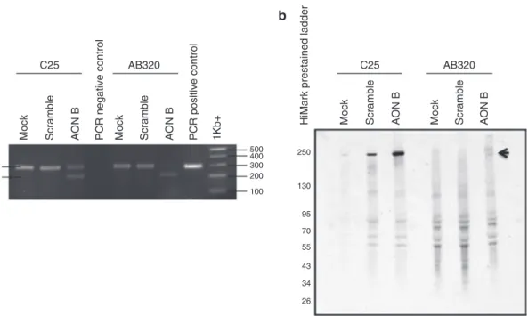
25. The program used is as followed: one step at 50 °C for 30 s, followed by a quick denaturation at 94 °C for 2 min. Then 40 cycles of a three steps amplification procedure are performed (94 °C 15 s. 59 °C 30 s and 72 °C 1 min). A final amplification step of 7 min is added. After completion, PCR products are visualized on a 2% agarose gel and visualized using alpha Innotech imager (see Note 14) (see Fig. 1 panel a).
494
1. The cell pellet is extemporaneously resuspended in 50 μL of lysis buffer 2× in which we add 5 μL of beta-mercaptoethanol and 5 μL of NUPAGE reducing agent (see Note 15).
2. After a 10 min incubation at 100 °C cells are centrifuged 5 s at 12,000 × g to bring down the condensate.
3. 20 μL of samples are then loaded on a 4–12% MOPS precast gels and run for 30 min at 80 V and then 100 V until the dye front (from the BPB dye in the samples) reached the bottom of the gel.
4. After electrophoresis, open the cast using a spatula, with the gel remaining on one of the parts. Rinse the gel with distilled water and transfer carefully to a container with western blot transfer buffer.
5. A PVDF membrane of the gel size is then activated in metha-nol, rinse twice in ultrapure water and place in the container with western blot transfer buffer.
6. Flip the gel into two Whatman # 3-filter cut to the size of the gel. Place the PVDF membrane on top of the gel and followed by two Whatman # 3 filter paper cut similarly. Place this assem-blage in a prewarmed humidified plastic container with a sponge in each side of the sandwich. Place the sandwich in the tank and run for 3 h at 100 V at 4 °C.
3.3 Western Blot 250 130 95 70 55 43 34 26 100 200 300 400 500 AB320 Mock Mock Mock Scramble Scramble Scramble 1Kb+ PCR negative control PCR positive control
AON
B
AON
B
Mock Scramble AON
B
AON
B
HiMark prestained ladder
AB320
C25 C25
U
a b
S
Fig. 1 Validation of exon skipping by RT-PCR and western blot control cell line (C25) and patient-derived cell
line were transfected with a specific AON, a scrambled AON or without AON (Mock) before analysis by RT-PCR (panel a) for the presence of skipped (S) and unskipped (U) transcripts. The same conditions were analyzed by
western blot using Hamlet antibody for the detection of dysferlin (panel b)
7. After transfer, the membrane is then readjusted to the size of the gel edges. The membrane is then placed in blocking solution for half an hour. Hamlet antibody (1:200 dilution) (see Note 16) is then incubated on the membrane for 2 h with
constant motion.
8. Then wash 3× with TBS 1× + 0.1% Tween 20 and incubate with secondary antibody (1:2000 dilution) in blocking buf-fer + 0.1% Tween-20 for 45 min with constant motion. Then wash 3× with TBS 1× + 0.1% tween20 prior to visualization with luminata forte reagent using Biorad Imager (see Note 17)
(see Fig. 1 panel b).
4 Notes
1. Other media may be used but after testing, it appears that this media was the only one giving us a stable undifferentiated cell culture.
2. Other transfection reagents/methods may be used but the use of Oligofectamine gives us the best results
3. Antisense oligonucleotides can be resuspended in ultrapure water if stored for a short-term period, but it is preferable to use TE pH 7.0 for a long-term storage. To avoid multiple thawing–freezing cycles consider small aliquots.
4. All solutions should be prepared using ultrapure and analytical grade reagents. All reagents are made and stored at room temperature (unless indicated otherwise).
5. Here we can optimize the protocol by trying Opti-MEM I Reduced Serum Medium (ThermoFisher Scientific #31985070) instead of DMEM.
6. Here, the cell pellets can be used right away for the subsequent procedures but should be kept at −80 °C for a long- term storage.
7. The cell pellet is usually passed through the syringe up and down several times (up to 3 times) in order to break down the cells.
8. At this step, you can also treat the pellet with the on-column DNAseI (ThermoFisher Scientific #12185010).
9. The total volume of water can be adjusted depending on the amount of starting material. We found that our best results were obtained using volumes comprise between 15 and 30 μL of ultrapure water.
10. We found that we increase our RNA concentration by eluting the sample a second time using the same water.
496
11. We let the samples for 5 min to allow sample to homogenize at room temperature and they are always kept on ice after that. 12. We use the Nanodrop1000 but alternative options exist to
measure the concentration of RNA like the Qubit3.0 (ThermoFisher Scientific).
13. This procedure should only be used if you do not do DNAse I column treatment (see Note 8).
14. The ratio of skipping could be calculated by quantifying the ratio of integrated density (obtained via ImageJ software) of the skipped band versus skipped + unskipped bands.
15. 2-mercaptoethanol needs to be manipulated under a chemical hood according to your laboratory regulations.
16. Hamlet antibody is a mouse monoclonal IgG1 serotype directed against a synthetic peptide: exon 53 (1999–2016) of human dysferlin.
17. The exon 32 deleted dysferlin band is shorter (26 amino acid less) than the wild type form of dysferlin (expected size 230 kDa).
Acknowledgment
The work was supported by grant from INSERM, Aix Marseille Univ, Association française contre les myopathies, Neuromics FP7 program E.U. (2012-305121). F.B. has received a postdoc fellow-ship grant from the UCLA Center for Duchenne Muscular Dystrophy-NIH NIAMS U54 AR052646 Wellstone Center of Excellence Training Fellowship respectively.
References
1. Dominski Z, Kole R (1993) Restoration of correct splicing in thalassemic pre-mRNA by antisense oligonucleotides. Proc Natl Acad Sci U S A 90(18):8673–8677
2. Pramono ZA, Takeshima Y, Alimsardjono H et al (1996) Induction of exon skipping of the dystrophin transcript in lymphoblastoid cells by transfecting an antisense oligodeoxynucleotide complementary to an exon recognition sequence. Biochem Biophys Res Commun 226(2):445–449
3. van Roon-Mom WM, Aartsma-Rus A (2012) Overview on applications of antisense- mediated exon skipping. Methods Mol Biol 867:79–96.
"https://doi.org/10.1007/978-1-61779- 767-5_6"
4. Sinnreich M, Therrien C, Karpati G (2006) Lariat branch point mutation in the dysferlin
gene with mild limb-girdle muscular dystro-phy. Neurology 66(7):1114–1116. "https:// doi.org/10.1212/01.wnl.0000204358. 89303.81"
5. Wein N, Avril A, Bartoli M et al (2010) Efficient bypass of mutations in dysferlin deficient patient cells by antisense-induced exon skipping. Hum Mutat 31(2):136–142
6. Barthélémy F, Blouin C, Wein N et al (2015) Exon 32 skipping of dysferlin rescues mem-brane repair in patients’ cells. J Neuromuscul Dis 2(3):281–290. "https://doi.org/ 10.3233/JND-150109"
7. Bansal D, Miyake K, Vogel SS et al (2003) Defective membrane repair in dysferlin- deficient muscular dystrophy. Nature 423(6936): 168–172