L E T T E R T O T H E E D I T O R
Open Access
DNA methylation signature of interleukin 1
receptor type II in asthma
Valérie Gagné-Ouellet
1, Simon-Pierre Guay
2,3, Anne-Marie Boucher-Lafleur
1, Luigi Bouchard
2,3and Catherine Laprise
1*Abstract
Interleukin 1 and its receptors are associated with allergic diseases such as asthma. In the present study, we
measured DNA methylation at the
IL1R1 and IL1R2 gene loci and assessed for associations with asthma-related
phenotypes and gene expressions. We found that asthmatic and atopic individuals have higher
IL1R2 promoter
DNA methylation than control subjects. Additionally, we observed a negative correlation between DNA methylation
at the
IL1R2 promoter and IL1R2 mRNA expression. These results suggest for the first time that IL1R2 promoter DNA
methylation is associated with its gene repression in allergic diseases such as asthma.
Keywords: Epigenetics, Methylation, IL1, IL1R1, IL1R2, Asthma, Atopy
Introduction
Interleukin 1 (IL1) plays a key role in the inflammatory
process of asthma [1]. We reported the association of
polymorphisms within the IL1 receptors type I (IL1R1)
and type II (IL1R2) gene loci with asthma and atopy in
the French Canadian Saguenay–Lac-Saint-Jean (SLSJ)
asthma study [2, 3]. The
IL1R2 gene expression
signa-ture in allergic asthma has also been described [4–6].
Epigenetics has received tremendous attention, and
vari-ations in DNA methylation (DNA-Me) in candidate
genes have been reported associated with asthma and
al-lergic related disorders [7–12]. These findings underline
the relevance of genetic and epigenetic profiling to
iden-tify pathways associated with allergic diseases. Such a
combined approach will facilitate the understanding of
the functional impacts of genetic and epigenetic
varia-tions on transcription and molecular mechanisms
in-volved in allergic diseases. In this study, we hypothesized
that DNA-Me in the promoters of
IL1R1 and IL1R2 is
associated with asthma and/or atopy.
Patients and methods
Clinical characteristics of the 93 individuals (21
non-atopic asthmatic, 26 non-atopic asthmatic and 21 non-atopic
individuals, and 25 non-asthmatic non-atopic controls)
from the Saguenay
–Lac-Saint-Jean asthma familial
col-lection [13] and included in the analysis are shown in
Table 1. Ethics committee approved the study, and all
subjects gave informed consent. Based on previous
gen-etic [14] and epigengen-etic analyses [15], methylation at 1
CpG in promoter and 3 CpGs in exon 1 of
IL1R1
(Additional file 1: Figure S1) and 5 CpGs in promoter
of
IL1R2 (Fig. 1a) was measured. DNA-Me differences
(Δβ) between affected (individuals with asthma, atopy,
or both) and non-affected individuals were assessed
and DNA-Me was correlated with gene expression for
each CpG. DNA-Me was measured on DNA extracted
from blood (blood and cell culture Midi kit, Qiagen,
Canada) using bis-pyrosequencing (EpiTech Bisulfite Kits,
Pyromark PCR Kit, Pyromark Gold Q24 Reagents,
Qiagen, Canada). PCR primers were designed using
Pyro-Mark Assay Design software (v2.0.1.15). Total RNA was
extracted from whole blood (RNeasy Plus Mini Kit,
Qia-gen, Canada) using a subset of affected and non-affected
individuals (n = 30). For each sample, RNA was converted
into cDNA (qScript™ cDNA SuperMix, Quanta
Biosci-ences, USA), and mRNA quantification was determined
(PerfeCTa® qPCR FastMix®, Quanta Biosciences, USA)
using the two standard curves method with
RPLP0 as a
reference gene [16].
The association between
IL1R1 and IL1R2 DNA-Me
levels and asthma and/or atopy at each CpG was
* Correspondence:Catherine_Laprise@uqac.ca
1
Département des sciences fondamentales, Université du Québec à Chicoutimi, Chicoutimi, QC, Canada
Full list of author information is available at the end of the article
© 2015 Gagné-Ouellet et al.Open Access This article is distributed under the terms of the Creative Commons Attribution 4.0 International License (http://creativecommons.org/licenses/by/4.0), which permits unrestricted use, distribution, and reproduction in any medium, provided you give appropriate credit to the original author(s) and the source, provide a link to the Creative Commons license, and indicate if changes were made. The Creative Commons Public Domain Dedication waiver (http://creativecommons.org/publicdomain/zero/1.0/) applies to the data made available in this article, unless otherwise stated.
Gagné-Ouellet et al. Clinical Epigenetics (2015) 7:80 DOI 10.1186/s13148-015-0114-0
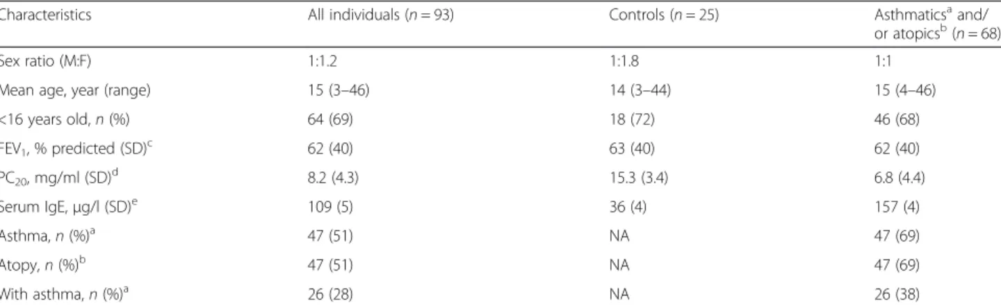
Table 1 Clinical characteristics of individuals from the Saguenay
―Lac-Saint-Jean asthma familial collection
Characteristics All individuals (n = 93) Controls (n = 25) Asthmaticsaand/
or atopicsb(
n = 68)
Sex ratio (M:F) 1:1.2 1:1.8 1:1
Mean age, year (range) 15 (3–46) 14 (3–44) 15 (4–46)
<16 years old,n (%) 64 (69) 18 (72) 46 (68) FEV1, % predicted (SD)c 62 (40) 63 (40) 62 (40) PC20, mg/ml (SD)d 8.2 (4.3) 15.3 (3.4) 6.8 (4.4) Serum IgE,μg/l (SD)e 109 (5) 36 (4) 157 (4) Asthma,n (%)a 47 (51) NA 47 (69) Atopy,n (%)b 47 (51) NA 47 (69) With asthma,n (%)a 26 (28) NA 26 (38) a
Present asthma or past documented clinical history of asthma. Data available for all individuals
b
Defined as having at least one positive response on the skin prick test (wheal diameter≥3 mm at 10 min). Data available for all individuals
c
FEV1= mean and standard deviation (SD) calculated for forced expiratory volume in 1 s for 67 individuals (16 controls, 51 asthmatic and/or atopic individuals) d
PC20= geometric mean and SD of provocative methacholine concentration inducing 20 % decline in FEV1calculated for 58 individuals (14 controls, 44 asthmatic
and/or atopic individuals)
e
IgE = geometric mean and SD of serum immunoglobulin (Ig) E level concentration calculated for 80 individuals (20 controls, 60 asthmatic and/or atopic individuals)
Fig. 1 Association between CpGs’ DNA methylation levels for IL1R2 and gene expression in asthma and atopy. a Schematic representation of IL1R2, location of epigenotyped CpG sites, and pairwise correlations between CpG sites. b Mean DNA-Me levels for CpG2 and CpG3-4 of IL1R2 in control and affected subjects (individuals with asthma, atopy, or both). c Correlation between DNA-Me level ofIL1R2-CpG2 and mRNA level. d Correlation between mean DNA-Me level ofIL1R2-CpG3 and 4 and mRNA level
analyzed by logistic regression considering age and sex as
covariates [9]. Gene expression analysis by phenotype was
not performed as control group sample size was
insuffi-cient (n = 4). The association between DNA-Me and
mRNA levels was assessed by Spearman correlation. CpG
dinucleotides with
r > 0.6 were combined before they were
tested for associations with asthma and/or atopy and for
correlation with gene expressions.
Δβ with p value < 0.05
was considered statistically significant. Statistical analyses
were conducted using the statistical software SPSS
(v11.5.0, USA).
Results
In this study, we detected higher levels of DNA-Me at
IL1R2 among affected individuals (i.e., with asthma,
atopy, or both) as compared to non-affected controls
(Δβ = 8.02 %, p value = 0.013, and Δβ = 3.72 %, p value =
0.012 for
IL1R2-CpG2 and the mean for CpG3 and
CpG4, respectively (Table 2, Fig. 1b)). Atopic and
non-atopic asthma were associated with DNA-Me at
IL1R2
but not atopy alone (data not shown). We also observed
that DNA-Me at
IL1R2-CpG2 was negatively correlated
with its mRNA levels (r = −0.511, p value = 0.004)
(Fig. 1c), but it was not correlated for CpG3 and CpG4
(Fig. 1d).
Discussion
An epigenetic signature has also been identified for
IL1R2 promoter in systemic lupus erythematosus (SLE)
[15]. The risk of allergic disorders was significantly
in-creased in SLE patients, which suggests that these
condi-tions share some common biomarkers [17]. The
negative correlation we observed between DNA-Me and
gene expression levels for
IL1R2 may be due to
stoichi-ometry. Methylation may limit access of a transcription
factor to DNA and hinders transcriptions [18]. We
iden-tified potential binding sites for transcription factors
relevant to asthma near the CpG dinucleotide sites of
IL1R2 analyzed (Additional file 2: Figure S2) which could
explain the inverse correlation between methylation and
gene expression [19]. Noteworthy is the potential
bind-ing site for nuclear factor kappa B/c-rel (NFKB) at the
IL1R2 promoter; it is involved in inflammation through
several pathways, including IL1 signalization [20]. Given
that IL1R2 acts as a decoy receptor to antagonize the
bound ligand [21], our data prompted the speculation
that hypermethylation of
IL1R2 in asthma and atopy
negatively regulates
IL1R2 expression and less decoy
receptors are available to reduce the downstream
pro-inflammatory response of IL1 in the presence of
un-changed IL1R1 level [22, 23]. Unlike IL1R1, IL1R2 does
not have an intracellular domain and the formation of
IL1-IL1R2 complex inactivates the IL1 downstream
sig-naling cascade; hence, silences the role of IL1 in
inflam-mation. Functional study will be needed to investigate
the impact of observed epi-variations on the production
of expressed receptors. This hypothesis could be
attrib-uted to both asthma and atopy as IL1R2 non-signaling
receptor is suspected to influence Th2 imbalance [24],
and both disorders are driven by Th2 allergic lung
in-flammation [25, 26].
To our knowledge, this is the first report of (1) a
hypermethylation signature of
IL1R2 promoter in
asthma with or without atopy and (2) an inverse
correl-ation between methylcorrel-ation at
IL1R2 promoter and its
gene expression. Together, they underline the relevance
of IL1R2 as a potential biomarker of asthma and atopy.
Further work is needed to understand the interactions
between environmental exposures and epigenetic
modi-fications like the ones identified in this study. Such
un-derstanding will aid the discovery of disease mechanisms
associated and development of more effective therapies.
Additional files
Additional file 1: Figure S1. Schematic representation of IL1R1 and location of epigenotyped CpG sites. This figure illustrates a simplified schematic representation ofIL1R1 and location of selected CpG dinucleotide sites and pairwise correlations between each CpG. Additional file 2: Figure S2. Potential binding sites for transcription factors inIL1R1and IL1R2. This figure shows a segment of the primary sequences ofIL1R1 and IL1R2. Both sequences lie in the respective gene promoter (UCSC Genome Browser assembly GRCh38/hg38).
Epigenotyped CpG sites are shown in blue and are numbered from the distal part of the promoter. Exons are shown in red. Potential binding sites for transcription factors in differentially methylated loci are shown by underlined sequences, and the name of the transcription factors are indicated underneath.
Abbreviations
CpG:cytosine-phosphate-guanine; DNA-Me: DNA methylation; IL1: interleukin 1; IL1R1: interleukin 1 receptor type 1; IL1R2: interleukin 1 receptor type 2; RPLP0: ribosomal protein, large, P0; SLE: systemic lupus erythematosus; SLSJ: Saguenay–Lac-Saint-Jean; Δβ: difference of methylation.
Competing interests
The authors declare that they have no competing interests.
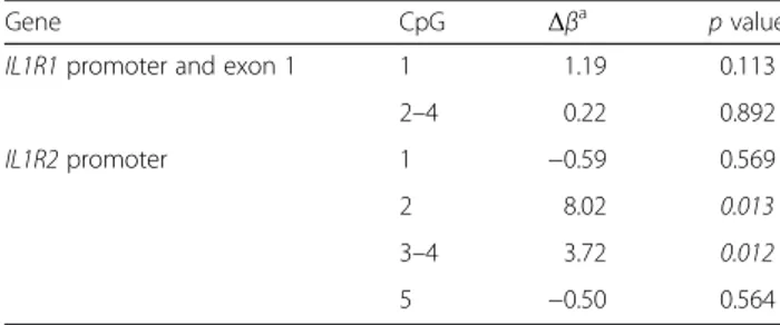
Table 2 Summary of DNA methylation analysis on promoter of
two interleukin 1 receptors in whole blood samples from
Saguenay
―Lac-Saint-Jean asthma familial collection
Gene CpG Δβa p value
IL1R1 promoter and exon 1 1 1.19 0.113
2–4 0.22 0.892
IL1R2 promoter 1 −0.59 0.569
2 8.02 0.013
3–4 3.72 0.012
5 −0.50 0.564
Significant p values are shown in italics
a
Δβ are calculated with mean methylation ratio for asthmatic and/or atopic individuals on control individuals
Authors’ contributions
VGO carried out the pyrosequencing and gene expression studies, performed the statistical analysis, and drafted the manuscript. SPG carried out the pyrosequencing primers design. AMBL participated in the pyrosequencing process. LB participated in the study design and revised the manuscript. CL led each step of the current study including the asthma familial collection build and management, study design, laboratory works, statistical analysis, as well as manuscript redaction and revision. All authors read and approved the final manuscript.
Acknowledgements
The authors thank all families for their valuable participation and all funding organizations. This study was supported by the Canadian Institutes of Health Research (CIHR) Catalyst Grant: Environments, Genes, and Chronic Disease. Catherine Laprise is the chairholder of the Canada Research Chair in Environment and Genetics of Respiratory Disorders and Allergy and Director of the Asthma Strategic Group of the Respiratory Health Network (RHN) of Fonds de recherche du Québec—Santé (FRQS) and researcher of the AllerGen NCE. Valérie Gagné-Ouellet received a Summer Student Research Training Award from the AllerGen NCE Inc. and a master degree studentship award from the RHN. Anne-Marie Boucher-Lafleur received a Summer Student Research Training Award from the AllerGen NCE Inc. Simon-Pierre Guay is the recipient of a Doctoral Research Award from the CIHR. Luigi Bouchard is a Junior Research Scholar from the FRQS and member of the FRQS-funded Centre de recherche clinique Étienne-Le Bel (affiliated with Centre hospitalier de l’Université de Sherbrooke).
Author details
1Département des sciences fondamentales, Université du Québec à
Chicoutimi, Chicoutimi, QC, Canada.2Department of Biochemistry, Université
de Sherbrooke, Sherbrooke, QC, Canada.3ECOGENE-21 and Lipid Clinic,
Hôpital de Chicoutimi, Saguenay, QC, Canada.
Received: 30 April 2015 Accepted: 13 July 2015
References
1. Dinarello CA. Biologic basis for interleukin-1 in disease. Blood. 1996;87:2095–147. 2. Daley D, Lemire M, Akhabir L, Chan-Yeung M, He JQ, McDonald T, et al.
Analyses of associations with asthma in four asthma population samples from Canada and Australia. Hum Genet. 2009;125:445–59.
3. Daley D, Park JE, He JQ, Yan J, Akhabir L, Stefanowicz D, et al. Associations and interactions of genetic polymorphisms in innate immunity genes with early viral infections and susceptibility to asthma and asthma-related phenotypes. J Allergy Clin Immunol. 2012;130:1284–93.
4. Laprise C, Sladek R, Ponton A, Bernier MC, Hudson TJ, Laviolette M. Functional classes of bronchial mucosa genes that are differentially expressed in asthma. BMC Genomics. 2004;5:21.
5. Chamberland A, Madore A-M, Tremblay K, Laviolette M, Laprise C. A comparison of two sets of microarray experiments to define allergic asthma expression pattern. Exp Lung Res. 2009;35:399–410.
6. Pociot F, Molvig J, Wogensen L, Worsaae H, Nerup J. A TaqI polymorphism in the human interleukin-1 beta (IL-1 beta) gene correlates with IL-1 beta secretion in vitro. Eur J Clin Invest. 1992;22:396–402.
7. Morales E, Bustamante M, Vilahur N, Escaramis G, Montfort M, de Cid R, et al. DNA hypomethylation at ALOX12 is associated with persistent wheezing in childhood. Am J Respir Crit Care Med. 2012;185:937–43. 8. Reinius LE, Gref A, Saaf A, Acevedo N, Joerink M, Kupczyk M, et al. DNA
methylation in the neuropeptide S receptor 1 (NPSR1) promoter in relation to asthma and environmental factors. PLoS One. 2013;8:e53877. 9. Naumova AK, Al Tuwaijri A, Morin A, Vaillancourt VT, Madore AM, Berlivet S,
et al. Sex- and age-dependent DNA methylation at the 17q12-q21 locus associated with childhood asthma. Hum Genet. 2013;132:811–22. 10. Wang IJ, Karmaus WJ, Chen SL, Holloway JW, Ewart S. Effects of phthalate
exposure on asthma may be mediated through alterations in DNA methylation. Clin Epigenetics. 2015;7:27.
11. Seumois G, Chavez L, Gerasimova A, Lienhard M, Omran N, Kalinke L, et al. Epigenomic analysis of primary human T cells reveals enhancers associated with TH2 memory cell differentiation and asthma susceptibility. Nat Immunol. 2014;15:777–88.
12. Yang IV, Pedersen BS, Liu A, O’Connor GT, Teach SJ, Kattan M, et al. DNA methylation and childhood asthma in the inner city. J Allergy Clin Immunol. 2015;136:69–80.
13. Laprise C. The Saguenay-Lac-Saint-Jean asthma familial collection: the genetics of asthma in a young founder population. Genes Immun. 2014;15:247–55.
14. Smith AJ, Keen LJ, Billingham MJ, Perry MJ, Elson CJ, Kirwan JR, et al. Extended haplotypes and linkage disequilibrium in the IL1R1-IL1A-IL1B-IL1RN gene cluster: association with knee osteoarthritis. Genes Immun. 2004;5:451–60.
15. Lin SY, Hsieh SC, Lin YC, Lee CN, Tsai MH, Lai LC, et al. A whole genome methylation analysis of systemic lupus erythematosus: hypomethylation of the IL10 and IL1R2 promoters is associated with disease activity. Genes Immun. 2012;13:214–20.
16. Wang T, Liang ZA, Sandford AJ, Xiong XY, Yang YY, Ji YL, et al. Selection of suitable housekeeping genes for real-time quantitative PCR in CD4(+) lymphocytes from asthmatics with or without depression. PLoS One. 2012;7:e48367.
17. Shen TC, Tu CY, Lin CL, Wei CC, Li YF. Increased risk of asthma in patients with systemic lupus erythematosus. Am J Respir Crit Care Med. 2014;189:496–9.
18. Blattler A, Farnham PJ. Cross-talk between site-specific transcription factors and DNA methylation states. J Biological Chemistry. 2013;288:34287–94. 19. Turker MS. Gene silencing in mammalian cells and the spread of DNA
methylation. Oncogene. 2002;21:5388–93.
20. Acuner Ozbabacan SE, Gursoy A, Nussinov R, Keskin O. The structural pathway of interleukin 1 (IL-1) initiated signaling reveals mechanisms of oncogenic mutations and SNPs in inflammation and cancer. PLoS Comput Biol. 2014;10:e1003470.
21. Colotta F, Re F, Muzio M, Bertini R, Polentarutti N, Sironi M, et al. Interleukin-1 type II receptor: a decoy target for IL-Interleukin-1 that is regulated by IL-4. Science. 1993;261:472–5.
22. Colotta F, Dower SK, Sims JE, Mantovani A. The type II‘decoy’ receptor: a novel regulatory pathway for interleukin 1. Immunol Today. 1994;15:562–6. 23. Garlanda C, Dinarello CA, Mantovani A. The interleukin-1 family: back to the
future. Immunity. 2013;39:1003–18.
24. Sims JE, Gayle MA, Slack JL, Alderson MR, Bird TA, Giri JG, et al. Interleukin 1 signaling occurs exclusively via the type I receptor. Proc Natl Acad Sci U S A. 1993;90:6155–9.
25. Islam SA, Luster AD. T cell homing to epithelial barriers in allergic disease. Nat Med. 2012;18:705–15.
26. Paul WE. History of interleukin-4. Cytokine 2015. doi:10.1016/j.cyto.2015.01.038.
Submit your next manuscript to BioMed Central
and take full advantage of:
• Convenient online submission • Thorough peer review
• No space constraints or color figure charges • Immediate publication on acceptance
• Inclusion in PubMed, CAS, Scopus and Google Scholar • Research which is freely available for redistribution
Submit your manuscript at www.biomedcentral.com/submit