HAL Id: tel-00698579
https://tel.archives-ouvertes.fr/tel-00698579
Submitted on 16 May 2012HAL is a multi-disciplinary open access archive for the deposit and dissemination of sci-entific research documents, whether they are pub-lished or not. The documents may come from teaching and research institutions in France or abroad, or from public or private research centers.
L’archive ouverte pluridisciplinaire HAL, est destinée au dépôt et à la diffusion de documents scientifiques de niveau recherche, publiés ou non, émanant des établissements d’enseignement et de recherche français ou étrangers, des laboratoires publics ou privés.
Therapeutic effects of TGT-β induced regulatory T cells
on the established autoimmune and inflammatory
diseases
Song Guo Zheng
To cite this version:
Song Guo Zheng. Therapeutic effects of TGT-β induced regulatory T cells on the established autoim-mune and inflammatory diseases. Agricultural sciences. Université d’Orléans, 2011. English. �NNT : 2011ORLE2061�. �tel-00698579�
UNIVERSITÉ D’ORLÉANS
ÉCOLE DOCTORALE SCIENCES ET TECHNOLOGIES
Laboratoire d’Immunologie et Embryologie Moléculaires – IEM UMR6218
-CNRS – Université d’Orléans
THÈSE
présentée par :Song Guo Zheng
soutenue le : 5 décembre 2011pour obtenir le grade de : Docteur de l’université d’Orléans Discipline : Science du vivant / Immunologie
Therapeutic effects of TGF- -induced
regulatory T cells on the established
autoimmune and inflammatory diseases
THÈSE dirigée par :
Bernhard Ryffel Directeur de Recherche, HDR, CNRS Orléans
RAPPORTEURS :
Nathalie Moiré Docteur UMR Université-INRA 0483, Tours
Julien C. Marie Docteur, Centre de Recherche sur le Cancer, Lyon
____________________________________________________________________
JURY
Nathalie Moiré Docteur, UMR Université-INRA 0483, Tours
Julien C. Marie Docteur, Centre Recherche Cancer, INSERM – CNRS, Lyon
Chantal Pichon Professeur, Université d’Orléans (Président du jury)
Jacques Van Snick Professeur, Ludwig Institute for Cancer Res., Bruxelles, Belgique Catherine Uyttenhove Professeur, Ludwig Institute for Cancer Res., Bruxelles, Belgique Makoto Miyara Docteur, Hôpital Pitié-Salpêtrière, INSERM, Paris
LIST OF CONTENTS
ACKNOWLEDGEMENT 3
I. ABBREVIATIONS 4
II. ABSTRACTS 5
III. INTRODUCTION, RATIONALE AND OBJECTIVES 7 III.1 Phenotypic and Functional Characteristic of Regulatory T Cells 7 III.2 Constitution and Types of Regulatory T Cells 9 III.3 Molecular Mechanisms Underlying the iTreg Cell Differentiation 15 III.4 Preventive and Therapeutic Roles of Regulatory T Cell Subsets 19 III.5 Objective of the PhD Project 20 IV. ARTICLE ONE: Adoptive Transfer of TGF- -Induced Regulatory T cells
Effectively Attenuates Murine Airway Allergic Inflammation 22 V. ARTICLE TWO: Antigen-Specific TGF- -Induced CD4+ Regulatory T Cells
but not Expanded nTregs Ameliorate Established Autoimmune Arthritis by
Shifting the Balance between Tregs and Th17 Cells in vivo 35 VI. ARTICLE THREE: Polyclonal CD4+Foxp3+T Cells Generated ex-vivo with
TGF- Suppress the Development of Lupus via Induction of Tolerogenic
Dendritic Cells 53
VII. ARTICLE FOUR: All-Trans Retinoic Acid Promotes TGF- -Induced Tregs
via Histone Modification but Not DNA Demethylation on Foxp3 Gene Locus 74 VIII. OVERALL DISCUSSION ANDPERSPECTIVE 88
VIII.1 iTregs Are Stable and Functional in the Inflammatory Condition 88 VIII.2 iTregs but not nTregs Ameliorate the Established Autoimmune and
Th17-Mediated Diseases 89 VIII.3 iTregs Suppress Disease via TGF- !"#$!%&-10 Dependent Mechanisms 91 VIII.4 iTregs Induce the Formation of Tolerogenic DCs in the Inflammatory
Condition 91 VIII.5Molecular Mechanisms Underlying the Promotion of iTreg Development 92 VIII.6 Future Plans 94
Remerciements or Acknowledgment
I greatly thank Professor Bernhard Ryffel for the opportunity to finalize this thesis project. His enthusiasm, scientific knowledge and inspiration all contribute to the success of this thesis. Although many roads lead to Rome, I have to say I have been taught to use the strictest standard in judging the values of scientific findings. One simply could not wish for a better or friendlier supervisor.
I also offer my sincerest gratitude to other members of my advisor committee, such as Professor Valerie Quesniaux and Professor Francois Erard who have supported me throughout my thesis with his patience and knowledge whilst allowing me the room to work in my own way. I am thankful for other professors including Dr. David A Horwitz and Dr. William Stohl for their supports. Their discussion and constructive suggestions have greatly enhanced the thesis’s quality. I also thank the team of Dr Valerie Quesniaux at UMR6218, Molecular Immunology, University and CNRS for the support and the work was supported by the “Fondation pour la Recherche Médicale” (FRM allergy DAL 2007 0822007), the “Fond européen de développement regional” (FEDER Asthme 1575-32168) and Le Studium Orleans,CNRS, Orléans, France.
In the various laboratories and workshops I have been aided in running the equipment and in conducting immunological experiments by Ju-Hua Wang, a super technician. I appreciate other colleagues and collaborators, Dr. David Brand, Dr. Wei Shi, Dr. Pawel Kiela and others for their collaboration and efforts. I also thank Jennifer Palomo, a PhD student, for her selfless assistance. I greatly appreciated the time and attention given to the examination of this work by the thesis committee: Dr. Chantal Pichon, Dr. Nathalie Moiré, Dr. Julien Marie, Dr. Makoto Miyara, Dr. Catherine Uyttenhove, Dr. Jacques Van Snick, and Dr. Francois Erard.
Finally, I would like to thank my wife Julie Wang who gives me a warm family and love and my daughter, Tina Zheng, who is a sophomore in Arcadia High School in California. Her outstanding achievements and leadership ability have always encouraged me to move forward to next step in my scientific career.
I. ABBREVIATIONS
'-SMA Alpha smooth muscle actin atRA Allo-trans retinoid acid AHR Airway hyperresponsiveness BAL Bronchoalveolar lavage
BFA Brefeldin A
CFA Complete Freund's adjuvant
CFSE Carboxyfluorescein succinimidyl ester ChIP Chromatin immunoprecipitations
CHX Cycloheximide
CKO Conditional knock-out
CNS Conserved non-coding DNA sequence CIA Collagen-induced arthritis
CII Collagen II
DC Dendritic cells DMSO Dimethyl sulfoxide
EAE Experimental autoimmune encephalomyelitis ELISA Enzyme-linked immunosorbent assay
GFP Green fluorescent protein GVHD Graft-vs-host disease H&E Hematoxylin and eosin
HPRT Hypoxanthine guanine phosphoribosyl transferase IHC Immunohistochemistry
i.n. Intranasal i.p. Intraperitoneal i.v. Intravenous
iTregs CD4+CD25+Foxp3+cells generated ex vivo with IL-2 and TGF-y
LN Lymph node
nTregs Naturally-occurring CD4+CD25+Foxp3+cells MCh Methacholine MS Multiple sclerosis IFN Interferon KO Knock out OVA Ovalbumin PAS Periodic-Acid-Schiff PBS Phosphate buffered saline PMA Phorbol 12-myristate 13-acetate RA Rheumatoid arthritis
SLE Systemic lupus erythematosus SMC Smooth muscle cell
Tregs Regulatory T cells
TGF- Transforming growth factor- ( ) TGF- !*+,+-./*!
TSS Transcription start site
II. ABSTRACT
Therapeutic effects of TGF- -induced regulatory T cells on the established autoimmune and inflammatory diseases
By
Song Guo Zheng
Advisor of thesis: Professor Bernhard Ryffel
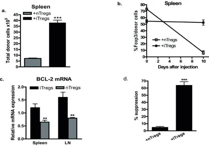
While it has been well recognized that both natural Foxp3+regulatory T (nTreg) cells and TGF -induced Treg (iTreg) cells can prevent autoimmune diseases in animal models, recent studies revealed that injection of nTregs has less therapeutic effects on established autoimmune diseases. It is less clear if iTregs can treat the established autoimmune diseases. We now provide evidence that unlike nTregs, transfer of iTreg cells markedly ameliorate established autoimmune diseases such as allergic asthma, autoimmune arthritis, and chronic GVHD with a lupus like syndrome.
In allergic asthma we observed that adoptive transfer of iTreg significantly suppressed airway and peri-vascular inflammation. iTreg infusion also markedly reduced airway résistance, eosinophil recruitment, mucus hyper-production, airway remodeling and IgE levels. This therapeutic effect was associated with increase of Treg cells (CD4+Foxp3+) in the draining lymph nodes, and with reduction of Th1, Th2, and Th17 cell responses as compared to untreated and non-Treg cell treated controls.
In collagen-induced arthritis (CIA) both antigen-specific iTregs and expanded nTregs prevented appearance and development of disease. However, only iTregs transfer suppressed established CIA. CIA mice given iTregs have a significantly lower incidence of disease and lower clinic scores than mice given nTregs, Teff cells or no cells. We found while nTregs were converted into Th1/Th17 cells in vitro and in vivo in the inflammatory milieu, iTregs were resistant to T effector cell conversion in the similar condition. Injection of iTregs to naïve mice displayed similar levels of Foxp3 stability as comparing with nTregs. Of note, the stability of Foxp3 expression was only found in iTreg cells during established CIA. iTregs suppressed Th17 cell differentiation that paralleled with improved clinical scores, collagen II (CII)-specific IgG production and bone erosion. In the chronic GVHD model mimicking lupus the transfer of iTregs to the established lupus disease significantly decreased the levels of anti-dsDNA and proteinuria, and markedly prolonged the survival of lupus. Blocking of TGF- 0(12- )! -".34"5! 678#9! "#.8-TGF- ! "#.8:/$5! /*! (12- )%!
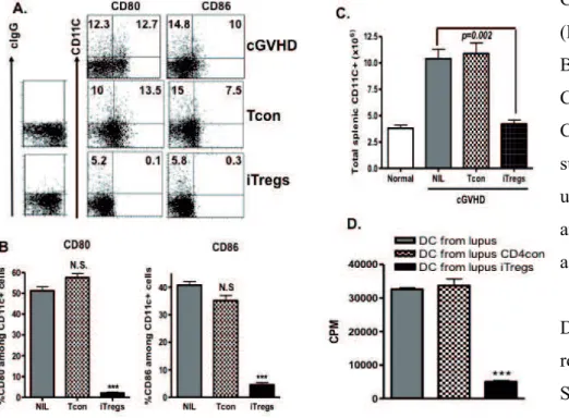
(ALK5) inhibitor, or anti-IL-10R antibody almost completely abolished the therapeutic effects of iTregs on lupus-like syndromes, suggesting that TGF- ! "#$0/*! %&-10 secreted by iTregs play a crucial role in the cell therapy. iTregs can induce the formation of tolerogenic DCs through TGF- ! signaling on DCs but not IL-10 signaling. We further observed that DCs isolated from cGVHD with a typical lupus syndrome receiving iTregs but not control cells expressed lower levels of CD80 and CD86 and adoptive transfer of these DCs to another lupus-like disease mouse can suppress the disease development through TGF- ! *".3+*! .3"#! %&-10 signal pathway. We therefore suggest that iTregs are stable and able to target DCs in the inflammatory milieu. These DCs then have become tolerogenic DCs and further suppress disease progression through its direct or indirect effect (inducing new iTregs) in autoimmune and inflammatory disease settings and may result in a long-term protective effect of iTregs in autoimmune diseases. Moreover, we also demonstrated that all-trans retinoic acid (atRA) promotes and sustains the Foxp3+ regulatory T cells, and identified that atRA significantly increased histone methylation and acetylation within the promoter and conserved non-coding DNA sequence (CNS) elements at the Foxp3 gene locus and the recruitment of phosphor-RNA polymerase II, while DNA methylation in the CNS3 was not significantly altered. These results will further help to enhance the quantity and quality of development of iTregs and may provide novel insights into clinical cell therapy for patients with autoimmune diseases and those needing organ transplantation.
III. INTRODUCTION, RATIONALE AND OBJECTIVES
III.1 Phenotypic and Functional Characteristic of Regulatory T Cells
It is now well accepted that a cell population called “CD4+CD25+regulatory or suppressor cells” are critically involved in immune tolerance and homeostasis. In the early 1970s, Gershon and colleagues initially reported that thymocytes from his experimental animal model included a population of suppressor T cells (Gershon and Kondo, 1970). This suggestion was not appreciated until Sakaguchi et al found that a population of CD4+CD25+ cells did indeed possess immunosuppressive activity that is now referred to as “regulatory T cells or natural regulatory T cells, nTregs (Sakaguchi et al., 1995).
CD4+CD25+cell populations also exist in humans, although only the CD4+CD25brightcell population appears to display an immune suppressive effect. A better approach for the identification of human Treg cells is to target the CD4+CD25+CD127-/lowpopulation (Seddiki et al., 2006).
CD25 is also an activation marker for lymphocytes. Thus, the utility of CD25 expression as a Treg marker is limited since it does not discriminate between activated T effector cells and Tregs. Fortunately, the nuclear transcription factor Foxp3 has been identified as a much more specific marker for Treg cells. Foxp3 is critically involved in the development and function of Treg cells(Fontenot et al., 2003). In mice, the lack of functional Foxp3 expression results in a fatal autoimmune and lymphoproliferative disorder known as scurfy and mutations of the human FOXP3 gene results in a human syndrome known as IPEX (immune dysregulation, polyendocrinopathy, enteropathy, X-linked), which is characterized by autoimmune disease expression in multiple endocrine organs (Wildin et al., 2001).
Despite the fact that Foxp3-GFP "knock-in" studies clearly demonstrate that there is a very broad spectrum of CD25 expression on Treg cells and that the intranuclear location of Foxp3 makes it difficult to use this protein for immunoaffinity-based purification methods although we have recently identified a new technique to improve the isolation of the live Treg cells (Zhou et al., 2010b), CD4+CD25+cells are still widely used in the field of the biology of Treg cells without using genetically modified tissues, particular in human studies. Although Foxp3 is considered as a specific marker for Tregs in mouse, this may not be the case for human Tregs. Recent data demonstrate that FOXP3 (FOXP3 for human cells and Foxp3 for mouse cells) may be upregulated in rapidly proliferating human T cells and might be viewed as an activation marker for human T cells (Allan et
al., 2007). More studies are needed to determine how FOXP3 might also be expressed on rapidly proliferating human T effector cells and more specific molecular markers to identify human Tregs are also desirable.
Many studies have revealed that the numbers of CD4+CD25+ cells and CD4+FOXP3+ cells in patients with various autoimmune diseases are diminished and that this Treg deficit is associated with disease activity (Tritt et al., 2008). This peripheral Treg deficit in patients with autoimmune diseases is not resultant from their redistribution to different organs (Miyara et al., 2005). Diminishment of Tregs in the face of autoimmunity is not a universal finding. Other groups have actually observed the converse; that the numbers of human CD4+CD25+cells can be increased under these circumstances (Yan et al., 2008). Since CD25 and FOXP3 can also be classified as activation markers, this aspect may reflect the disparity between these findings. Miyara et al have further classified human FOXP3+cells into three cell subsets: CD45RA+FOXP3low, CD45RA-FOXP3hi and CD45RA-FOXP3low. Functional assay demonstrated that the CD45RA-FOXP3low subset contains non suppressor cells, that the CD45RA+FOXP3low subset contains resting Tregs and that active Tregs are found in the CD45RA-FOXP3hi subset. Using these criteria, they found that Treg cell numbers were indeed diminished in patients with active autoimmune disease (Miyara et al., 2009).
In addition to Treg frequency, others have also reported that autoimmune disease can alter the functional activity of Tregs. For example, the suppressive activity of CD4+CD25+cells isolated from active rheumatoid arthritis patients was significantly decreased (Valencia et al., 2006). It is likely that some intrinsic defect in CD4+CD25+ cells in active rheumatoid arthritis patients accounts for their decreased functional activity. Similarly, the frequency of CD4+CD25+ cells in patients with multiple sclerosis (MS) is unaltered, however, the functional activity of these cells to suppress T cell immune responses including antigen-specific or non-specific stimulation is also decreased (Haas et al., 2005; Kumar et al., 2006; Viglietta et al., 2004). These results suggest that the manipulation of nTregs to restore their numbers and function may be therapeutic.
Although most people claim that CD4+CD25+ in peripheral blood belong to natural Treg cells, we and others would suggest that CD4+CD25+cells in PBMCs consist of a mixture of both thymic nTregs and those induced in the periphery (induced Tregs, iTregs) (Horwitz et al., 2008; Zhou et al., 2011, Lan Q et al; Zheng S.G.). There is no specific marker that can distinguish nTregs from iTregs so far. Although Shevach’s group recently reported that Helios, an Ikaros family transcription factor, may be helpful for distinguishing nTregs from iTregs (Thornton et al., 2010), others reported that
Helios is also highly expressed on Th2 and T follicular helper cells and may be associated with the differentiation of these cells (Serre et al., 2011).
It has been well known that the adoptive transfer of nTregs can prevent the appearance and development of autoimmune diseases in many animal models. Conversely, there are also considerable numbers of studies demonstrating that the therapeutic effect of nTregs on established diseases is fairly unsatisfactory. For example, the efficacy of adoptive transfer of nTregs to established collagen-induced arthritis (CIA) is poor for controlling the disease progression (Zhou et al., 2010a). Injection of nTregs to established lupus had mild protective effects and it failed to suppress lupus glomerulonephritis and sialoadenitis (Bagavant and Tung, 2005; Scalapino et al., 2006). Moreover, adoptive transfer of nTregs was unable to suppress other Th17-mediated autoimmune disease (Huter et al., 2008).
There are several possibilities that could explain the inability of nTregs to treat CIA and other autoimmune diseases. First, pro-inflammatory cytokines may hamper their suppressive activity. Pasare et al have reported that nTreg suppressive activity can be abolished by IL-6 (Pasare and Medzhitov, 2003). Valencia et al also revealed that elevated TNF-'! ;"5! 8#.+*<+*+! 48.3! .3+! suppressive capacity of nTregs (Valencia et al., 2006). There is no question that these pro-inflammatory cytokines are elevated in RA patients (Wakkach et al., 2003). Secondly, Th17 cells may be resistant to the suppressive effects exerted by nTregs. This could explain how nTregs are able to prevent development of disease before Th17 cells become established, while demonstrating ineffective suppression after disease expression is evident. Third, nTregs are inherently unstable and can be converted to Th1, Th2, Th17 and Tfh effector cells when they encounter an inflammatory milieu (Lu et al., 2010b; Tsuji et al., 2009; Wan and Flavell, 2007; Xu et al., 2007; Zhou et al., 2010a).
There are still other reasons that could hamper the utilization of nTregs as therapeutics. First, the intranuclear location of Foxp3 makes it difficult to purify nTregs for functional study. Second, nTregs constitute only 1-2% of human CD4+ T cells. nTregs must be expanded ex vivo to gain sufficient numbers for therapy. Although several groups have claimed that expansion in vitro can overcome this problem (Hippen et al., 2011), other laboratories have reported that repeated expansion alters Treg phenotype and function (Hoffmann et al., 2009). Third, the expansion of nTregs from patients with RA and MS for therapeutic purposes may be problematic due to potential other intrinsic defects in RA and MS Tregs. nTreg instability, Teff cell resistance and the influence
of an inflammatory milieu may individually or collectively account for the inability of nTregs to control established autoimmune diseases.
Of interest, the plasticity of nTregs under inflammatory conditions could be fixed with cytokines or other compounds. Our group recently reported that while nTregs become Th17 cells in the presence of IL-6, these cells also lost their suppressive role in suppressing progression of the lupus-like syndromes and CIA. We also documented that pretreatment of nTregs with IL-2 combined with TGF- , or all-trans retinoic acid (atRA), a vitamin A metabolite, can render these nTregs resistant to Teff cell conversion and allow them to begin to suppress lupus and CIA progression (Zheng et al., 2008; Zhou et al., 2010a). This indicates that the manipulation of nTregs still holds a promise in the treatment of autoimmune diseases.
III.2 Constitution and Types of Regulatory T Cells
Current studies have demonstrated that Treg cells are a heterogeneous set of cells that consist of CD4+CD25+Foxp3+cells, IL-10-producing CD4+Tr1 cells, TGF- -producing Th3 cells, CD8+cells, NK T cells, CD4-CD8- (! ,+==7! "#$! >?! (! ,+==7! (Horwitz et al., 2004; Tang and Bluestone, 2008). CD4+ Treg subsets can be further classified into three main populations, thymus-derived, naturally occurring CD4+CD25+Foxp3+cells (nTregs) described as above, endogenous induced Tregs in vivo and those that can be induced ex vivo from CD25-precursors in peripheral lymphoid organs (iTregs) (Zheng et al., 2002). Although IL-10-induced Tr1 cells represent another cell population of iTregs, they do not express Foxp3 and produce considerable levels of IL-10 (Pot et al., 2011). As IL-10 may promote autoimmune response through stimulating B cell activation and its level is highly increased in active systemic lupus erythematosus (SLE) patients (Yu et al., 2011), Tr1 may not be suitable for the treatment of SLE and other autoimmune diseases. TGF- -induced Tregs will be defined as iTregs in this thesis.
While Yamagiwa et al reported that TGF- !-*/;/.+7 endogenous CD4+CD25+ cells (Yamagiwa et al., 2001), our group first reported that TGF- ! $/+7! 3"@+! "#! ":8=8.5! ./! 8#$6,+! ABC+CD25- cells to become CD4+CD25+Treg cells in vitro (Zheng et al., 2002). When Foxp3 was identified as Treg marker in Tregs, several groups including us immediately found that TGF- ! ,"#! 8#$6,+! 2/D-E! expression in iTregs (Chen et al., 2003; Fantini et al., 2004; Zheng et al., 2004a). Additionally, other studies have also clearly demonstrated the capacity of Foxp3+ Tregs in vivo through TGF -dependent mechanism (Liang et al., 2005).
Phenotypically, both nTregs and iTregs express similar molecules such as CD25, CTLA-4, GITR, CCR4, CD62L and Foxp3, and express CD45RBlow in mice and CD45RO in humans. CD4+CD25+Foxp3+cells in the periphery have been considered as a mixed population comprised of nTregs and iTregs. Although Helios might possibly help to distinguish nTregs from iTregs (Thornton et al., 2010), more specific molecular markers are needed to distinguish both Treg cell populations.
Although both nTreg and iTreg subsets share similar phenotypes and display comparable suppressive activity, several factors distinctly affect their development, stability and function (Table 1). First, nTregs develop in the thymus through recognition of self antigens. A high and medium affinity cognate interaction between self-peptide:MHC complex and T cell receptor is required for this process. They also require CD28 co-stimulation because they do not develop in CD28 deficient mice (Salomon et al., 2000). Although IL-2 and TGF- !-="5!"#!8;-/*."#.!*/=+!8#!.3+!;"8#.+#"#,+!/<!.3+!-//=!78F+!/<!#(*+97 (Marie et al., 2005), both cytokines are redundant for their development since both IL-2 and TGF- ! G#/,G-out mice contain CD4+CD25+Foxp3+ regulatory T cells in the thymus (Piccirillo et al., 2002). By contrast, the generation of iTregs is dependent upon the presence of both TGF- !"#$!(12- !*+,+-./*! signals since the absence of TGF- ! / *! (12- ! *+,+-./*7! /*! :=/,G8#9! .3+! (12- ! *+,+-./*! 789#"=! prevents the induction of Foxp3 expression and the subsequent functional suppressive capacity (Lu et al., 2010a; Lu et al., 2010b). Similarly, IL-2 plays an essential role in the differentiation of Foxp3+ iTregs. TGF- !<"8=7!./!8#$6,+!2/D-E+iTregs from naïve CD4+CD25-precursor cells in IL-2 deficient
mice (Zheng et al., 2007). The conversion of CD4+CD25- cells in the periphery to CD25+ iTregs requires a suboptimal TCR stimulation and thus environmental antigens may sufficiently trigger iTreg development. The absence of CD28 co-stimulatory molecules does not affect the differentiation of iTregs (Lan Q and Zheng SG, unpublished data), but inhibitory CTLA-4 costimulatory molecule and CTLA-4/B7.1 signaling is crucially required for the generation of iTregs (Zheng et al., 2006b). This conclusion is further documented by an observation that the blocking of CTLA-4/B7.1 signal abolished the capacity of TGF- !./!8#$6,+!8(*+97!8#!48=$!.5-+!;8,+! (Read et al., 2006). OX40/OX40L, an alternate CD28/B7-independent co-stimulatory pathway, also negatively regulates the development and function of both nTregs and iTregs. While stimulation of mature nTregs by OX40 results in the loss of suppression of T cell proliferation and cytokine production, the generation of iTregs is completely abolished by OX40 although OX40 does not affect the generation of nTregs (So and Croft, 2007).
Recently, Housley et al reported that while the TNF-R2 expression is essential for nTregs-mediated suppression of colitis, its expression is not required for iTreg-mediated suppression (Housley et al., 2011). Differing IL-2 and co-stimulatory molecule requirements for Treg cell development, and TNFRII expression requirements for the suppressive function of both nTregs and iTregs suggests that nTregs and iTregs are possibly heterogeneous populations and that integration of both Treg subsets is required for the maintenance of normal immune homeostasis. It is also likely that both nTreg and iTreg subsets can either act in concert or separately on different targets. In addition, as anti-TNF-'!.3+*"-5!3"7!:++#!48$+=5!67+$!8#!.*+".8#9!-".8+#.!48.3!*3+6;"./8$!"*.3*8.87, further studies are required to understand whether this therapy differentially regulates nTregs and/or iTregs development in individual diseases.
Fig. 1. Multi effects of TGF- on regulatory and effector T cells. TGF- inhibits the differentiation, proliferation and function of various immune cells including Th1, Th2 and Tfh cells. TGF- also promotes iTreg, Th17 and Th9 cell differentiation depending upon the cytokine environment. Additionally, TGF- inhibits maturation and function of other immune cells such as CD8+CTL, NK cell, DC
and macrophages.
As TGF- !+8.3+*!-*/;/.+7!2/D-E+iTregs, Th9 or 17 cells depending upon other cytokines involved (Figure 1), and as nTreg cells express a membrane-bound form of TGF- ! "#$! this TGF- ! 3"7! <6#,.8/#"=! activities, it is reasonable to assume that IL-6 can convert nTregs to become Th17 and other T helper cells (Xu et al., 2007). To demonstrate this, Xu et al used purified nTregs from Foxp3 GFP knock in-mice to exclude the possibility that CD4+CD25+Foxp3- non-Tregs made this conversion. We used both wild type and Foxp3 GFP knock-in mice to confirm this observation (Zheng et al., 2008). Endogenous TGF- !-*/$6,+$!:5!#(*+97!87!,*8.8,"==5! required for this conversion since blocking TGF- ! *+,+-./*! %! 789#"=! /*! 678#9! #(*+97! <*/;! (12- ! receptor II dominant mice resulted in the failure of Th17 conversion (Lu et al., 2010b; Zheng et al., 2008). Moreover, activation of nTregs with IL-6 resulted in decreased Foxp3 expression and suppressive activity both in vitro and in vivo. Furthermore, adoptive transfer experiments revealed that nTregs treated with IL-6 ex vivo lost their ability to protect mice from a lupus-like disease (Zheng et al., 2008). Thus, in an IL-6 rich inflammatory milieu, nTregs may be unstable and lose the functional activity. In the current study, we will further investigate whether nTregs can be converted into Th17 cells in an in vivo model.
In sharp contrast, TGF- -induced iTregs were found to be completely resistant to the Th17 conversion by IL-6. This difference cannot be explained by insufficient production of TGF- ! :5! iTregs since both nTregs and iTregs expressed similar levels of membrane-bound TGF- !HIJ-25%). Furthermore, the resistance of iTregs to Th17 conversion also may not be explained by alterations in TCR stimulation since anti-CD3/CD28 activated nTregs can still differentiate into Th17 cells upon
IL-6 stimulation. To account for this difference between nTregs and iTregs, we found that the combination of IL-2 and TGF- !$/4#-regulated IL-6 receptor expression and function in activated T cells. We have observed that both cytokines markedly decreased IL-6 receptor alpha-chain (CD126) and beta-chain (CD132) expression on CD4+ cells and these cells expressed significantly lower level of phosphorylated STAT3 expression when stimulated by IL-6 (Zheng et al., 2008). All-trans retinoic acid (atRA) has a similar effect on nTreg stability under inflammatory conditions as well (Zhou et al., 2010a). In the current study, we will compare the stabilities and functionalities of both Treg cell subsets in autoimmune inflammatory disease in vivo.
Nonetheless, others have reported that TGF- -induced iTregs were unstable in vitro (Floess et al., 2007) and in vivo following antigen-stimulation (Chen et al., 2011), and lack protective activity to prevent lethal graft-versus-host disease (GVHD) (Floess et al., 2007; Koenecke et al., 2009). It has been claimed that the Foxp3 promoter on TGF- -induced iTregs but not nTregs is methylated and accounts for their instability (Floess et al., 2007). However, we have recently observed that the methylation status in Foxp3 gene loci does not affect Foxp3 stability. Moreover, addition of atRA to TGF- !-*/;/.+$!8(*+9!cell stability and maintenance in vitro and in vivo and this effect is unrelated to CpG methylation in Foxp3 promoter but related to acetylation of Foxp3 histone (Lu et al., 2011). Others have also observed protective human TGF- -induced Tregs that exhibit methylated Foxp3 (Hippen et al., 2011). To explain these controversial results, we consider the technical reasons are possibly responsible for the generation of unstable, ineffective TGF- -induced iTregs in these groups. They have used high concentrations of plate-bound anti-CD3 with TGF- K! 43+*+"7! /6*! group has used suboptimal concentrations of anti-CD3 and anti-CD28 coated beads with IL-2 and TGF- L!%.!3"7!:++#!G#/4#!.3".!7.*/#9K!767."8#+$!(A)!7.8;6=".8/#!",.8@".+7!.3+!;(M)0Akt signaling pathway which facilitates Teff cell differentiation and inhibits Foxp3 expression and Treg differentiation (Sauer et al., 2008). Treg generation is best established with suboptimal TCR stimulation that facilitates Foxp3expression (Horwitz et al., 2008).
These studies also raise the possibility that nTregs and iTregs may have distinct roles in the adaptive immune response. In response to microbial infections nTregs could possibly serve as a first line of host defense by differentiation to IL-17-producing cells, which contribute to neutrophil mobilization and have other pro-inflammatory effects. After eradication of invading pathogens, the late appearance of TGF- -induced iTregs would not only terminate the antigen-specific response, but also prevent the emergence of non-specifically stimulated or cross-reactive self-reactive T cells. Accordingly, failure of this mechanism could result in an immune-mediated disease.
III.3 Molecular Mechanisms Underlying the iTreg Cell Differentiation
Current studies have demonstrated that several signaling pathways, such as the TGF- 0N;"$K IL-2/IL-2R/STAT, T cell receptor (TCR) and costimulatory signaling pathways are crucial for the induction of Foxp3 transcription although the TGF- ! *+,+-./*! HT R) signaling pathway is considered to be the key one.
As the lack of either T RI or T RII will terminate the cellular response to TGF- (Wrana et al., 1992), it is understandable that transcription factor Foxp3 cannot be induced by TGF- ! 8#! ( )! deficient T cells (Lu et al., 2010b). In lymphocytes, TGF- binds to its cognate receptor complex composed of type I (ALK5) and type II receptors. TGF- type I receptor (T RI) and type II receptor (T RII) associate as interdependent components of a heteromeric complex. ( RII is required to activate ( RI in the ligand–receptor complex, and activated T RI Ser/Thu kinases phosphorylate downstream specific SMAD2 and SMAD3. Upon phosphorylation, these two SMADS bind to their common partner, SMAD4, to formSMAD2/4 and SMAD3/4 complexes. These complexes then translocate to the nucleus and modulate target gene expression (Lagna et al., 1996; Rubtsov and Rudensky, 2007). Unlike Smad2 and Smad4 null mice, Smad3 null mice are viable and survive to adulthood (Datto et al., 1999). Accumulating evidence has revealed that Smad3 is essential for the suppressive effect of TGF- on IL-2 production and T cell proliferation (McKarns et al., 2004). Smad3 is also required for the suppressive effects of TGF- on Th2 type cytokine production and Th2 type diseases in the skin (Anthoni et al., 2008).
Recent studies have also begun to explore the roles of Smad pathways in iTreg differentiation. Anthoni and colleagues examined the role of Smad3 in a Smad3-/- murine model of contact hypersensitivity and found that the lack of intact TGF- signaling via Smad3 resulted in an increased pro-inflammatory, Th2 and Th17 type response in the skin with reduced Foxp3 mRNA in the lymph nodes (Anthoni et al., 2008). These data implicate that the Smad3 may be involved in iTreg cell differentiation. Using an in vitro culture system, several groups found that Smad2 or Smad3 plays a significant role in TGF- -iTreg generation (Jana et al., 2009; Xu et al., 2010).
Tone and colleagues identified an evolutionarily conserved enhancer site in Foxp3 gene that bindsSmad3 and nuclear factor of activated T cells (NFAT), suggesting that TGF- regulates Foxp3 transcription through a Smad3 dependent pathway. Smad3 and NFAT functioned in a coordinated fashion and were essential for histone acetylation in the enhancer region and induction of Foxp3
transcription(Tone et al., 2008). Xu et al also found that retinoic acid (RA) augmentation of TCR-and TGF- -induced Foxp3 transcription were related processes involving modifications of baseline (TGF- -induced) phosphorylated Smad3 (pSmad3) binding to a conserved enhancer region (enhancer I). This led to increased histone acetylation in the region of the Smad3 binding site and increased binding of pSmad3 (Xu et al., 2010).
Nonetheless, we and others have recently reported that Smad2 or Smad3 plays a partial role in iTreg differentiation in vitro but plays no roles in iTreg differentiation in vivo (Lu et al., 2010b; Takimoto et al., 2010). Although it is possible the either Smad2 or Smad3 can compensate for each other, we also observed that TGF- O7! $/4#7.*+";! non-Smad pathways actually play an important role in iTreg development. TGF- ! ,"# activate SMAD-independent pathways such as MAPKs, in T cells(Zhang, 2009). In fact, TGF- !8#38:8.8/#!/<!%2P-> induced signaling and Th1 gene expression in CD4+ T cells is Smad3 independent but MAPK dependent mechanism (Park et al., 2007). Among MAPKs, we further observed that ERK and JNK but not P38 activation is involved in TGF -mediated Foxp3 induction (Lu et al., 2010b). ERK and JNK may activate AP1 that will coordinate with NTAT to regulate Foxp3 expression. In this project, one of goals is to determine how atRA affects Smad or non-Smad pathways during iTreg cell differentiation.
Although TCR activation is needed for the TGF- -mediated Foxp3 induction, its role in this process is complicated. Antigen stimulation TCR activates the transcription factor NFAT, a key regulator in T cell activation and anergy. NFAT forms cooperative complexes with the AP-1 family of transcription factors and regulates T cell activation-associated genes. Treg function is mediated by an analogous cooperative complex of NFAT with the forkhead transcription factor Foxp3 (Wu et al., 2006). TCR engagement also activates nuclear factor (NF)-QR (NF-QRS!"#$!phosphatidylinositol 3-kinase (PI3K)/Akt/mTOR (mammalian target of rapamycin) axis. Most of these transcription factors conversely play a negative role in the iTreg differentiation although cRel and p65, two out of five NF- QRfamily members: c-Rel, RelA-p65, RelB, NF-kB1 (p50-p105), and NF-kB2 (p52-p100) play a positive role in iTreg cell differentiation (Ruan et al., 2009). Thus, rapamycin suppresses PI3K/Akt/mTOR axis and promotes iTreg differentiation (Ruan et al., 2009). This is likely that different signaling intensities of TCR activation will result in the expression of different transcription factors and subsequently affect the development of different T effector cell subsets (Zhou et al., 2011).
reduced in IL-2-deficient T cells and addition of exogenous IL-2 promotes and sustains Foxp3 expression (Davidson et al., 2007; Zheng et al., 2007). In fact, IL-2- and IL-2R-deficient mice have a low frequency of Foxp3+cells (Malek and Bayer, 2004). CD28/B7 costimulatory signaling may promote Treg cell production and maintenance through IL-2 production. As IL-4, IL-7, IL-15, and IL-IT!"=7/!73"*+!,/;;/#!>!,3"8# with IL-2R, these cytokines may also affect iTreg differentiation. It has been reported that IL-2-/-IL-15-/-double KO mice have a much lower Treg frequency than IL-2-/-mice, suggesting that IL-15 also affects iTreg cell differentiation and maintenance (Burchill et al., 2007). IL-7 also plays an important role in the maintenance of long-lived memory iTreg phenotype and function (Li et al., 2011). Conversely, IL-4, IL-21, IL-27 as well as IL-6 suppress iTreg cell differentiation.
Signaling downstream of the IL-2R can act through the Janus kinase (JAK)/STAT pathway. STATs comprise a family of several transcription factors that are activated by a variety of cytokines, hormones, and growth factors. STATs are activated through tyrosine phosphorylation, mainly by JAK kinases, which lead to their dimerization, nuclear translocation and regulation of target gene transcriptions. STAT5 molecule is a key component of the IL-2 signaling pathway, the deficiency of which often results in autoimmune pathology due to reduced number of Treg cells (Burchill et al., 2007).
Fig.2. Multi effects of TGF- , TCR and cytokine signaling pathway-related transcription factors in the regulation of Foxp3 expression and maintenance.
Activated STAT5 is translocated into the nucleus where it binds to highly conserved tandem consensus STAT binding motifs located in the promoter region and/or first intron of the Foxp3 gene and promotes Foxp3 expression (Burchill et al., 2007; Zorn et al., 2006). STAT5a/b is required for optimal induction of Foxp3 in vitro and binds directly to the Foxp3 gene (Yao et al., 2007). Conversely, IL-6, IL-21 and IL-27 activate STAT-3 and p-STAT-3 then bind a gene silencer in a second conserved enhancer region (enhancer II) downstream from enhancer I; this leads to a loss of pSmad3-binding to enhancer I and eventually suppresses Foxp3 expression (Xu et al., 2010). IL-4 activates STAT6, and p-STAT6 binds to a promoter just before exon I of the Foxp3 gene and suppresses Foxp3 gene transcription. Retinoic acid can suppress p-STAT6 binding to the Foxp3 promoter and promotes iTreg differentiation. Thus, control of accessibility and binding of different transcription factors provides a common framework for positive and negative regulation of TGF- -induced Foxp3 transcription.
These transcription factors could also work together to regulate Foxp3 expression. Ruan et al propose a c-Rel-dependent enhanceosome model, which may apply to explain the regulation of iTreg cell differentiation. They suggest that antigen-presenting cells (APC) carrying specific peptides engage precursor T cells by TCR and CD28, in the presence of TGF- !" #$%&'$()" (*" +,-!" CD28, and TGF- "./0/1'(.2"#/&32"'("'4/"&0'$5&'$()"(*"6778!"94$04"14(214(.:#&'/2"6;<&"=$)4$8$'(." of kBa), releasing c-Rel and p65. The freed c-Rel-p65 dimer then migrates into the nucleus, binds to the Foxp3 promoter, and induces the formation of a Treg cell-specific multifactorial transcriptional complex including NFAF/AP1/Smad/STAT called ‘‘enhanceosome’’ which comprises transcription factors that bind not only to the promoter but also to the distal enhancers. Current studies have demonstrated that the Foxp3 gene is controlled by a core promoter and at least three distal enhancers
(Mantel et al., 2006; Tone et al., 2008). More recently, researchers have identified that Foxp3 differentiation and maintenance is controlled by three non-coding DNA sequence (CNS1-3) elements (Zheng et al., 2010). While CNS1 containing a TGF- -NFAT response element and CNS3 containing cRel response element are associated with Treg differentiation, CNS2 containing Cbf -Runx1, Stat and CpG DNA mostly regulates Foxp3 maintenance. Thus, Treg cell-specific enhanceosome in Foxp3 CNS regions in turn serves as the ‘‘on-and-off’’ switch of the Foxp3 gene and the Treg cell differentiation and maintenance program. A schematic representative of various signaling pathway related transcription factors that regulate Foxp3 transcription and expression has been demonstrated in Figure 2.
III.4 Preventive and Therapeutic Roles of Regulatory T Cell Subsets
Both iTregs and nTregs share similar functional characteristics. Adoptive transfer of iTregs generated ex-vivo also can prevent the development of autoimmune diseases. For example, Wahl group has demonstrated that adoptive transfer of TGF- -converted/induced iTregs prevented house dust mite–induced allergic pathogenesis and inflammation in lungs in an asthmatic mouse model (Chen et al., 2003). In lupus model, our study has demonstrated that iTreg prevented disease development (Zheng et al., 2004b). Weber et al observed that injection of murine islet-specific CD4+ iTregs prevented spontaneous development of type 1 diabetes and inhibited development of pancreatic infiltrates and disease onset orchestrated by Th1 effectors in NOD mice (Weber et al., 2006). Dipaolo et al reported similar preventive role of iTregs in a murine model of autoimmune gastritis (Dipaolo et al., 2007). Similarly, iTreg cells also significantly prevented Th1-mediated colitis on CD4+CD62L+T cell transfer in vivo (Fantini et al., 2006). Selvaraj et al demonstrated that adoptively transferred iTregs were as potent as natural Foxp3+Treg in preventing EAE development (Selvaraj and Geiger, 2008). It seems both antigen-specific and non-specific iTregs prevent autoimmune diseases although the former is more efficacious than the latter.
In addition to their use in a preventative role, adoptive transfer of these cells to ongoing diseases still suppressed disease development in a lupus-like syndrome model (Zheng et al., 2004b). We have developed a chronic graft-vs-host disease model characterized by rapid and vigorous formation of SLE-like autoantibodies and the formation of severe immune-complex glomerulonephritis. DBA/2 (D2) mouse T cells induce this syndrome when injected into (DBA/2 x C57BL/6) F1 mice. We found TGF- -treated DBA/2 T cells not only lost their ability to induce graft-vs-host disease but also prevented other parental T cells from inducing lymphoid hyperplasia, B cell activation, and an immune complex glomerulonephritis. Moreover, a single transfer of TGF- -conditioned T cells to
animals that had already developed anti-dsDNA Abs decreased the antibody titer, suppressed proteinuria, and doubled survival (Zheng et al., 2004b). This result was further confirmed by a study from Su et al (Su et al., 2008). Selvaraj et al observed that iTregs were still efficacious in ongoing experimental autoimmune encephalomyelitis (EAE), animal model of multiple sclerosis, and Godebu et al also reported that iTregs can revise type I diabetes in animal (Godebu et al., 2008; Selvaraj and Geiger, 2008). Similarly, in autoimmune gastritis model, Nguyen et al found that antigen-specific iTregs also still suppressed inflammation and associated pathology when administered late in the process of ongoing disease(Nguyen et al., 2011).
As nTregs seem to be unable to control the progress of established collagen-induced arthritis (CIA), we will plan to conduct a head-to-head comparison of therapeutic effects of antigen specific thymus-derived nTregs and TGF- -induced iTregs on the established CIA in the current study. We chose antigen-specific Tregs since these are more protective than polyclonal Tregs in autoimmune diseases (Penaranda and Bluestone, 2009). It has been known that polyclonal nTregs can prevent disease but are ineffective in established CIA disease (Morgan et al., 2003; Zhou et al., 2010a). We will test the hypothesis that antigen-specific iTregs are superior to nTregs in ameliorating established CIA. We will determine whether iTregs remained stable and fully functional following transfer. The recent studies of Nugyen et al indicated that chemokines secreted by antigen-specific TGF- -induced iTregs regulated T cell trafficking and thereby suppressed ongoing autoimmune. They reported that these iTregs were therapeutic in an ongoing autoimmune gastritis model (Nguyen et al., 2011). Others have also reported that only TGF- -induced iTregs but not nTregs suppressed Th17-mediated diseases (Huter et al., 2008). These studies all implicate that iTregs may have a therapeutic potential in suppressing the established autoimmune diseases.
III.5 Objective of the PhD Project
Previous studies have revealed that Tregs play an important role in the prevention of autoimmune diseases. However, the adoptive transfer of nTregs to the established autoimmune diseases is less therapeutic and these cells are unstable and can convert to T effector cells and lose the suppressive activity. Interestingly, the iTregs are resistant to T effector cell conversion and stable in vitro in the inflammation-like environment. In this study, we will use an in vivo model, en established collagen-induced arthritis (CIA), to learn whether both Treg subsets have a different stabilities and functionalities in vivo. We will make a head-to-head comparison to determine the preventive and therapeutic roles of both Treg subsets in the ongoing and established CIA. We will learn whether
infusion of iTregs also suppress the ongoing allergic asthma and chronic GVHD with a lupus-like disease in the project.
To explain the long-term effect of single infusion of iTregs on autoimmune diseases, although others have claimed that long-term survival of selected antigen-specific iTregs can account for this phenomenon (Godebu et al., 2008), “infectious tolerance” may be another main reason for immune tolerance effect of iTregs (Andersson et al., 2008; Zheng et al., 2004a). To determine whether infectious tolerance contributes to long-term protective effect of iTregs on autoimmune diseases, we will investigate the interaction of iTregs and DC in the chronic graft-vs-host disease with a typical lupus syndrome in the current study. We will test whether infusion of iTregs to lupus mice can induce the formation of tolerogenic DCs. We will further determine underlying mechanisms if it is a case. In this study, we will use DC-specific T RII conditional KO mice to address this issue. We will also determine molecular mechanisms by which atRA promotes iTreg generation, maintenance and function. These studies will further disclose the mystery of development and function of iTregs in autoimmunity.
IV. ARTICLE 1
Adoptive transfer of TGF- -induced-regulatory T cells effectively attenuates murine airway allergic inflammation
Song Guo Zheng1,5, Wei Xu2, Qin Lan1,3, Hui Chen2, Ning Zhu4, Xiaohui Zhou1,3, Julie Wang1, Huimin Fan5, David Warburton2, Dieudonnée Togbe5, Valerie Quesniaux5, Wei Shi2
and Bernhard Ryffel5
1Division of Rheumatology and Immunology, Department of Medicine, The Keck School of
Medicine, University of Southern California, Los Angeles, CA 90033; 2Developmental Biology and Regenerative Medicine Program, Department of Surgery, The Saban Rrsearch Institute of Children’s
Hospital Los Angeles, 4650 Sunset Blvd., Los Angeles, CA 90027; 3Institute of Immunology, Shanghai East Hospital, Shanghai, China; 4Institute of Immunology, Fujian Medical University, Fuzhou, Fujian Province, China; 5UMR6218, Molecular Immunology, University and CNRS, 3b rue
de la Ferollerie, F-45071Orleans, France
Abstract
Both nature (nTreg) and induced-regulatory T lymphocytes (iTreg) are potent regulator of autoimmune and allergic disorders. Defects in Treg cells have been reported in patients with allergic asthma, and therefore replenishment of Treg cells might attenuate asthma. Here we report that adoptive transfer of iTreg cells generated ex-vivo with IL-2 and TGF- effectively attenuated lung and airway allergic inflammation in a murine model of asthma. Immunized mice given 5x10⁶iTreg cells just before antigen challenge displayed markedly reduced airway résistance, eosinophil recruitment, mucus hyper-production, airway remodeling and IgE levels. This therapeutic effect was associated with increase of Treg cells (CD4+Foxp3+) in the draining lymph nodes, and with reduction of Th1, Th2, and Th17 cell response as compared to untreated and non-Treg cell treated controls. Therefore, adoptive transfer of iTreg reduces the allergic response, which might be a novel and promising therapeutic approach to treat severe asthma.
Introduction
Chronic allergic airway inflammation and airway hyperresponsiveness (AHR) are characteristic of atopic asthma pathophysiology. More than 7% of Americans suffer from asthma (Moorman et al., 2007), and annual expenditure for health and lost productivity due to asthma is estimated at nearly $20 billion. The currently available therapeutic approaches for asthma usually include quick symptomatic relief measures directed to relaxation of airway smooth muscle (bronchodilator) and long-term control with suppression of airway inflammation (Fanta, 2009). However, these existing standard asthma therapies have several caveats and remain inadequate. For example, inhaled anti-inflammatory corticosteroids only suppress but do not cure asthmatic inflammation, and long-term use of corticosteroids causes many pleiotropic side effects. Other more recently developed therapies, including inhibitors of leukotriene production and leukotriene receptor blockade, and anti-IgE monoclonal antibody (omalizumab), are used as alternative treatments for persistent asthma. However, limited efficacy, high cost, and lack of responsiveness in some asthma patients are the major drawbacks. Thus, novel and more effective therapeutic approaches for asthma are still needed.
Recent studies have found that immune function dysregulation is one of the key pathogenic mechanisms underlying chronic asthma(Doherty and Broide, 2007). Reduction and/or defects in regulatory T (Treg) cells, which function as negative regulators to suppress excessive immune response and maintain immunological tolerance have been detected in asthma patients(Apostolou and von Boehmer, 2004). Therefore, replenishment of Treg cells is thought to be a promising cell therapeutic approach. However, the use of thymus-derived naturally occurring regulatory T (nTreg) cells has several caveats that may significantly diminish their practical application for asthma treatment. These include limited availability, susceptibility to inflammation-triggered apoptosis, inability in suppressing pro-inflammatory Th17 cells, and self-conversion to Th17 and/or other T effector cells in the milieu of inflammation. In contrast, Treg cells that are induced by TGF- and IL-2 in combination with low dose antigen exposure have similar phenotypic and functional characteristics to nTreg cells, without the caveats of nTreg cells mentioned above. Herein, we report that adoptive transfer of the induced-Treg (iTreg) cells to Ovalbumin (OVA)-sensitized mice effectively attenuates OVA-induced airway allergic inflammation, airway hyperresponsiveness, and other asthma-like lung pathology by modulating the systemic immune system.
Materials and Methods Animal care
C57BL/6 mice were purchased from the Jackson Laboratory and bred at the specific pathogen-free animal facility at Keck School of Medicine at University of Southern California. All experiments were approved by the Institutional Animal Care and Use Committee at University of Southern California.
OVA-sensitized mouse asthma model and exogenous cell infusion
6 to 8-week-old female mice weighing 20-25g were used for the experiments. Mice were sensitized by intraperitoneal (i.p.) injections of 25ug OVA (Grade V, Sigma Chemical Co.) mixed with aluminum hydroxide (Pierce) at day 1, and followed by another booster i.p. injection at day 14. These sensitized mice then were challenged with 20>g of OVA or saline control through an intranasal (i.n.) route for three consecutive days (days 25, 26, and 27)to generate an acute allergic asthma model.5x106of iTreg cells or control cells generated as described below were intravenously injected into mice on day 22, three days before antigen challenge.
Generation of in vitro TGF- -induced regulatory T (iTreg) cells
Splenic CD4+CD25-CD62L+CD44lownaive T cells were isolated by autoMACS (Miltenyi Biotech) from C57BL/6 female mice, which were littermates to the mice used for generating the OVA-asthma model. iTreg cells were then prepared using the methods described in our previous publication(Zheng et al., 2007). Briefly, naïve CD4+T cells were first isolated by negative selection, in which naïve CD4+ T cells were labeled with FITC-conjugated anti-CD25 mAb and sorted for CD4+CD25-T cells. The CD4+CD25-T cells were then labeled with PE-conjugated anti-CD62L and Cyc-conjugated CD44, and positively selected by anti-PE magnetic beads (CD4+CD62L+CD44lowCD25-cells). These isolated naïve T cells were stimulated with anti-CD3/28 coated beads (1:5 ratio, five cells to one bead) in the presence of IL-2 (40 U/ml) and TGF- ?" =@" ng/ml) for 4-6 days to generate iTreg cells; controls cells were treated with anti-CD3/CD28 beads with IL-2 only. The iTreg cell phenotypes were verified by related molecular marker expression (CD4+CD25+Foxp3+) detected by Flow Cytometery. Also the immune suppressive activity of iTreg cells was verified using a standard in vitro suppressive assay as previously reported (Zhou et al., 2010b).
On day 28 (24 hours following the last i.n. administration of either normal saline or OVA challenge), the mice were anesthetized with intraperitoneal injections of sodium pentobarbital (90mg/kg). When an appropriate depth of anesthesia was achieved, a tracheostomy was performed, in which a standard 20G × 32 mm Abbocath®-T cannula (Abbott, Sligo, Ireland) was gently inserted into the trachea and secured with suture. The mice were then connected to a computer controlled small animal ventilator (FlexiVent, Scireq, Canada) and ventilated at 150 breath/min with a tidal volume of 10ml/Kg and a positive end-expiratory pressure of 3 cmH2O. Methacholine (MCh, 40 mg/ml in
PBS) was then delivered to the subject by nebulized aerosol. The frequency-independent airway resistance (Raw) in mouse lung was then automatically measured by FlexiVent/SciReq software, and the MCh challenge experiments are repeated at least three times.
Bronchoalveolar lavage (BAL)
The bronchoalveolar lavage was performed three times with 0.8 ml of normal saline in each time, and pooled together. The number of cells in BAL was counted using a hemocytometer. The remaining samples were then centrifuged at 4ºC for 4 min at 400xg to separate cell-free supernatant and cells. An aliquot of supernatant was used to measure BAL protein concentration by Bradford method (Bio-Rad).
Lung histopathology and immunohistochemistry
The lungs were inflated with PBS under 25 cm H2O, and fixed in 4% paraformaldehyde overnight at
40C. The fixed tissues were then processed for paraffin embedding. 5->A"#B)%"'$22B/"2/0'$()2"9/./" stained with Hematoxylin and Eosin (H&E) for lung morphological analyses. Eosinophils in the lung tissue were stained with Discombe’s Solution (Discombe, 1946). Periodic-Acid-Schiff (PAS) staining was used to characterize glycoproteins using a kit purchased from Sigma. Briefly, slides were deparaffinized and hydrated, then immersed in Periodic Acid Solution for 5mins. After washing with several changes of distilled water, the slides were incubated in Schiff's reagent for 15mins and counterstained with hematoxylin. The numbers of airways with PAS-positive epithelial cells versus PAS-negative epithelia were then determined in each lung section. Lung inflammatory histopathology was evaluated using a semi-quantitative method reported by other group(Richards et al., 1996). Briefly, at least two sections of each mouse lung were examined using the following criteria. Alveolar inflammation foci were scored 0-5 (0=no foci, 1=CD!"@EF -15, 3=16-25, 4=26-35, 5=GHDIJ" +4/" 1/.$-vascular and peribronchiolar inflammation was also scored separately on a 0-5 scale (0=no inflammation, 1=minimal, 2=mild, 3=moderate, 4=marked, 5=severe). Thus, the total inflammation score is the average of alveolar and peri-vascular/bronchiolar inflammation. At least
five mice in each group were selected for this analysis. Immunohistochemical studies were performed using Zymed Histostain-Plus system (Zymed). Alpha smooth muscle actin (K-SMA) antibody was purchased from Sigma (St Louise, MO). Normal serum was used as negative control.
Serum cytokine analysis
Serum was prepared from blood collected after measuring AHR. Samples were frozen at L@MN," until analysis. The levels of cytokines (IL-4, IL-5 and IL-13) and IgE in sera were measured using ELISA kits purchasing from Invitrogen or BioLegend, following the manufacturer’s instructions.
Intracellular cytokine staining and Flow Cytometry analysis
Lymphocytes in axillary draining lymph nodes and spleen were collected and stained for surface or intracellular markers with combinations of fluorochrome-conjugated mAb specific for CD3, CD4, CD8a, CD62, CD44, IL-17A (BioLegend, San Diego, CA), as well as IL-O!"6PQR"=<S"<$(20$/)0/2" Pharmingen, San Diego, CA). In the case of intracellular IL-4, IL-?T" &)3" 6PQR!" 0/##2" 9/./" 2'$AB#&'/3"9$'4"MJ@D">%UA#"VWX"&)3"MJ@D">%UA#"$()(A:0$)"=,$(04/A!"Y&"Z(##&!",XI"*(."?"4(B.!" &)3" *(##(9/3" 8:" &33$'$()" (*" 8./*/#3$)" X" =D" >%UA#[" ,$(04/AI" &)3" $)0B8&'$()" *(." &33$'$()&#" O" hours prior to processing for intracellular staining. The phenotypes and intracellular cytokine expression were analyzed using a LSRII Flow Cytometry (BD Biosciences, San Diego, CA).
Statistical analysis
The data are presented as the mean. A Student’s two-tailed t test was used to compare data between the different experimental groups. Differences were considered statistically significant for P values<0.05.
Fig.4. Attenuated allergic inflammation by iTreg cells. (a) Total proteins in BAL fluids from mice with different treatments were quantified; (b) Eosinophil, detected by Discombe’s staining (red intracellular granules), was the major type of cells that were infiltrated in small airways and adjacent vasculature; (c) IgE level in serum from different groups of mice was quantified by an ELISA. Increased IgE level in serum by OVA-induced allergic reaction was significantly reduced with iTreg treatment. *P<0.05.
Fig.3. Reduced inflammation in lung tissues by adoptive transferring of iTreg cells prior to OVA challenge. (a) Lung tissue sections from the mice with indicated treatments were stained with H&E. Mice with i.n. instillation of saline were used as “Normal” control. iTreg-OVA and T cell-iTreg-OVA represent mice were treated with adoptive transfer of iTreg cells versus control T cells prior to OVA challenge, respectively. (b) The intensities of lung inflammation were graded with scores 0 to 5 (none to severe inflammation, see Materials and Methods for details). *P<0.05, **P<0.01, n=5.
Results
Adoptive transfer of in vitro TGF- -induced Treg (iTreg) cells attenuated OVA-induced allergic inflammation in mouse respiratory airways and alveoli.
In OVA-sensitized mice, repeated i.n. OVA challenges at day 25-27 resulted in severe small airway and alveolar inflammation, indicated by excessive inflammatory cell infiltration surrounding small airways and vasculature, as well as alveolar septa (Fig.3).
The serum level of IgE was significantly increased, and infiltration of eosinophil around airway was also verified by Discombed staining (Fig. 4). Consistent with the lung histological changes, the total amount of proteins in BAL fluid was significantly increased (Fig. 4). The number of cells in BAL also increased more than 10-fold than the control groups (data not shown). Moreover, epithelial cell hypertrophy with increased mucin expression in small airways, thickened airway smooth muscle cell (SMC) layer, and resultant smaller lumen with rippled epithelial surface of small airways were also observed in OVA-challenged mouse lungs (Fig. 5). Therefore, a typical OVA-allergic airway inflammatory model was verified.
Using this established OVA-allergic mouse model, the anti-inflammatory effect of adoptive transfer of iTreg cells was then examined. Three days before OVA challenge (Day 22), a single transfer of 5x10⁶ iTreg cells was given to the mice via tail vein injection. T cells cultured without TGF- addition
was
used as an additional specificity control. After three-day i.n. OVA challenge, lung specimens were harvested for detailed analyses. iTreg cells, but not control T cells, significantly attenuated OVA-induced allergic inflammation including reduced infiltration of inflammatory cells, particularly eosinophils, in airway and alveolar septa, decreased levels of serum IgE and BAL proteins (Fig. 3-4), as well as reduction in the number of cells in BAL by 2-fold (p<0.05). Alterations of airway walls subsequent to allergic inflammation, including epithelial hypertrophy and increased mucin production (PAS-positive staining), as well as thickened smooth muscle cells in small airways (Fig. 5), were likewise significantly attenuated. These results indicate that adoptive
Fig.5. Abnormal airway wall remodeling was subsided with iTreg cell treatment. (a) Excessive mucin expression in small airway epithelial cells was detected by PAS staining (red color). (b) The numbers of airways with PAS-positive epithelial cells per lung tissue section were quantified in different experimental groups (n=5), suggesting overproduction of mucin. (c) Clara cells in small airway epithelia were stained by CCSP immunofluorescence staining (green) and the surrounding airway smooth muscle cells were immunostained by SMA (red). **P<0.01.
Fig. 6. Adoptive transfer of iTreg cells significantly reduced AHR. Airway resistance was measured upon Mch (40mg/ml) aerosol delivery. Although the airway resistance was still significantly higher in iTreg cell-treated group than that in normal control group, significant reduction of airway resistance was achieved in iTreg cell-treated group compared to non-treated (OVA) or control T cell-treated group. (*P<0.05, **P<0.01).
transfer of iTreg cells prior to OVA allergic challenge can effectively prevent lung inflammation and abnormal airway remodeling.
Adoptive transfer of iTreg cells also effectively inhibited airway hyperresponsiveness (AHR) in OVA-challenged mice.
OVA-sensitized mice with repetitive i.n. administration of OVA developed significant AHR to methacholine (MCh) challenge compared to normal control mice (Fig. 6). However, adoptive transfer of iTreg cells prior to repetitive challenge of OVA significantly inhibited AHR, although increased AHR was still detected. In contrast, adoptive transfer of T control cells did not significantly reduce AHR, although slight reduction in AHR was detected in some mice.
Thus, adoptive transfer of iTreg cells, but not control T cells, prior to allergen exposure can also effectively reduce AHR in addition to the reduction in airway inflammation described above.
Adoptive transfer of iTreg cells modulated systemic immune response to OVA-allergic challenge.
Numerous studies have found that asthmatic inflammation is related to abnormal cellular immunity, including defective Treg cells, inappropriate ratio of Th2 over Th1 cell population, and excessive Th17 cells. Thus, we have evaluated these immune cells and their related cytokine production.
Fig.7. Adoptive transfer of iTreg cells significantly diminished Th1/Th2/Th17 cell frequencies in draining lymph nodes in asthmatic mice. Cells from axillary lymph nodes were isolated and stimulated with PMA and Ionomycin (5 hours), and BFA (4 hours). Intracellular expression of IFN- , 5, and IL-17A in CD3+T cells were determined by FACS. (a)
A representative of 9 mice in each group. Cells were gated on CD3+cells. (b) Results were mean
± SEM of values of 9 mice in each group. *P<0.05, **P<0.01, ***P<0.001.
A 2-fold increase of Treg cells (CD4+Foxp3+) was detected in the draining lymph nodes and the spleen of the mice receiving adoptive transfer of iTreg cells prior to OVA challenge, while there were no significant changes in Treg cells in the comparison to OVA or T control cell-OVA groups. More interestingly, increased Th1, Th2, and Th17
cells caused by repetitive OVA challenge was significantly attenuated by adoptive transfer of iTreg cells prior to OVA administration (Fig. 7), but not by control T cells. Consistent with this, serum Th2 cytokines including IL-5 and IL-13 were significantly increased upon OVA challenge (Fig. 8). Adoptive transfer of iTreg cells, but not T control cells, was able to partially block these increases in mice. Therefore, adoptive transfer of iTreg cells may modulate both local and systemic immunity, and thus prevent excessive cellular response against OVA-induced allergic reaction and inflammation.
Fig.8. Adoptive transfer of iTreg cells inhibited OVA-induced increase of Th2 cytokines. IL5 and IL13 in mouse serum were quantified by specific ELISA. Significant reduction of IL5 and IL13 in the group receiving iTreg treatment was detected compared to OVA challenge only control group. **P<0.01.