Study of the oxidation process of crystalline powder of In 2 S 3 and thin films obtained by Dr Blade method
23
0
0
Texte intégral
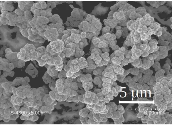
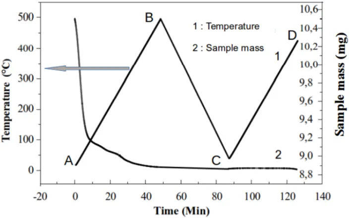
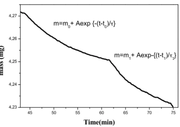
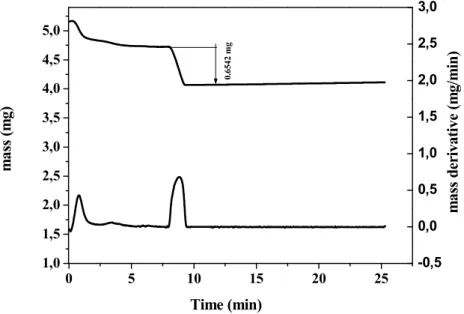
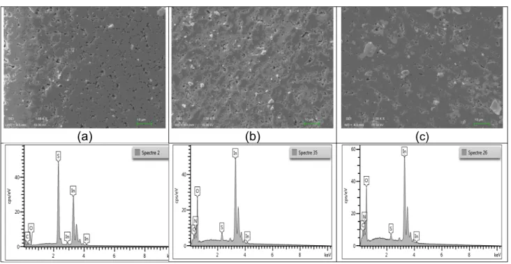
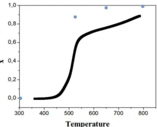
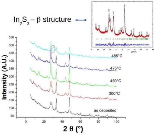
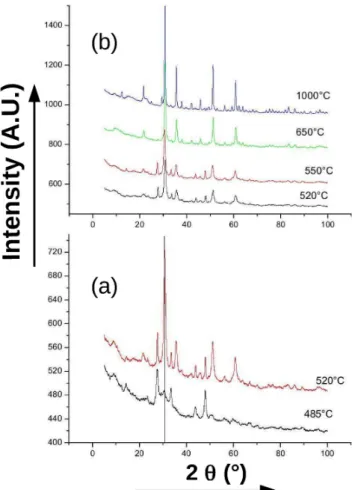
Figure


+7
Documents relatifs