HAL Id: hal-01960172
https://hal.umontpellier.fr/hal-01960172
Submitted on 19 Dec 2018
HAL is a multi-disciplinary open access
archive for the deposit and dissemination of sci-entific research documents, whether they are pub-lished or not. The documents may come from teaching and research institutions in France or abroad, or from public or private research centers.
L’archive ouverte pluridisciplinaire HAL, est destinée au dépôt et à la diffusion de documents scientifiques de niveau recherche, publiés ou non, émanant des établissements d’enseignement et de recherche français ou étrangers, des laboratoires publics ou privés.
vertebrates from Gabon, Central Africa
Larson Boundenga, Boris Makanga, Benjamin Ollomo, Aude Gilabert,
Virginie Rougeron, Bertrand Mve-Ondo, Céline Arnathau, Durand Patrick,
Nancy Moukodoum, Alain-Prince Okouga, et al.
To cite this version:
Larson Boundenga, Boris Makanga, Benjamin Ollomo, Aude Gilabert, Virginie Rougeron, et al.. Haemosporidian parasites of Antelopes and other vertebrates from Gabon, Central Africa. PLoS ONE, Public Library of Science, 2016, 11 (2), pp.e0148958. �10.1371/journal.pone.0148958�. �hal-01960172�
Haemosporidian Parasites of Antelopes and
Other Vertebrates from Gabon, Central Africa
Larson Boundenga1,2☯*, Boris Makanga1,3,4☯, Benjamin Ollomo1, Aude Gilabert3,Virginie Rougeron1,3, Bertrand Mve-Ondo1, Céline Arnathau3, Patrick Durand3, Nancy
Diamella Moukodoum1, Alain-Prince Okouga1, Lucresse Delicat-Loembet1, Lauriane Yacka-Mouele1, Nil Rahola1,3, Eric Leroy1,3, Cheikh Tidiane BA2,
Francois Renaud3, Franck Prugnolle1,3‡*, Christophe Paupy3‡
1 Centre International de Recherches Médicales de Franceville (CIRMF), Franceville, Gabon BP 769 Franceville, Gabon, 2 Laboratory of Evolutionary Biology, Ecology and Management of Ecosystems, Faculty of Sciences and Techniques, Cheikh Anta Diop University of Dakar, Dakar, Senegal, 3 Laboratoire
MIVEGEC, UMR 224–5290 IRD-CNRS-UM, Centre IRD de Montpellier, 34295 Montpellier, France, 4 Institut de Recherche en Ecologie Tropicale, Libreville, Gabon
☯ These authors contributed equally to this work. ‡ These authors also contributed equally to this work. *larsonamedeo@yahoo.fr(LB);franck.prugnolle@ird.fr(FP)
Abstract
Re-examination, using molecular tools, of the diversity of haemosporidian parasites (among which the agents of human malaria are the best known) has generally led to rear-rangements of traditional classifications. In this study, we explored the diversity of haemos-poridian parasites infecting vertebrate species (particularly mammals, birds and reptiles) living in the forests of Gabon (Central Africa), by analyzing a collection of 492 bushmeat samples. We found that samples from five mammalian species (four duiker and one pango-lin species), one bird and one turtle species were infected by haemosporidian parasites. In duikers (from which most of the infected specimens were obtained), we demonstrated the existence of at least two distinct parasite lineages related to Polychromophilus species (i.e., bat haemosporidian parasites) and to sauropsid Plasmodium (from birds and lizards). Molecular screening of sylvatic mosquitoes captured during a longitudinal survey revealed the presence of these haemosporidian parasite lineages also in several Anopheles species, suggesting a potential role in their transmission. Our results show that, differently from what was previously thought, several independent clades of haemosporidian parasites (family Plasmodiidae) infect mammals and are transmitted by anopheline mosquitoes.
Introduction
The order Haemosporida (also called Haemosporidia, Haemospororida or Haemospororina) includes many protozoan parasites among which the best known are the agents of human malaria, a disease affecting every year several millions of people in tropical regions and particu-larly in sub-Saharan Africa [1]. Within the Haemosporida order, the large family of
OPEN ACCESS
Citation: Boundenga L, Makanga B, Ollomo B, Gilabert A, Rougeron V, Mve-Ondo B, et al. (2016) Haemosporidian Parasites of Antelopes and Other Vertebrates from Gabon, Central Africa. PLoS ONE 11(2): e0148958. doi:10.1371/journal.pone.0148958 Editor: Richard Culleton, Institute of Tropical Medicine, JAPAN
Received: May 8, 2015 Accepted: January 25, 2016 Published: February 10, 2016
Copyright: © 2016 Boundenga et al. This is an open access article distributed under the terms of the
Creative Commons Attribution License, which permits unrestricted use, distribution, and reproduction in any medium, provided the original author and source are credited.
Data Availability Statement: All sequences produced in our study were deposited in Genbank (accession numbers KT367817 to KT367865). Funding: The study was funded by the Centre International de Recherches Médicales de Franceville (CIRMF, Gabon), the Centre National de la Recherche Scientifique (CNRS, France), the Institut de Recherche pour le Développement (IRD, France) and the Agence Nationale de la Recherche (ANR, France, grant ORIGIN JCJC 012). Larson Boundenga was financed by an ANBG scholarship. The funders had no role in study design, data
Plasmodiidae comprises several genera and species that parasitize a wide range of vertebrates, such as fish, reptiles, birds and mammals [2]. All the parasite species that infect mammals belong to this family and are restricted to nine genera (Biguetiella, Bioccala, Dionisia, Nycteria, Hepatocystis, Plasmodium Polychromophilus, Rayella and the fossil genus Vetufebrus). Although most of these parasites were described during the second half of the twentieth cen-tury, many new taxa have been discovered in wildlife during the last decade [2,3].
The first taxonomic descriptions of haemosporidian parasites were based on morphological characteristics [4,5] and relied mainly on the microscopic observations of biological features [6], such as the schizont and gametocyte forms [7–9], and on their life history traits as well as the nature of the host species. This approach was very useful; however, it is not very reliable [10]. Indeed, over time, the morphology of an organism can be modified by the influence of environmental factors [10]. Moreover, the morphological features and life history traits of a parasite species can vary from one host species to another [11,12]. In addition, some species can display the same morphological features, although they are genetically very different (cryp-tic species) [10]. Therefore, most taxonomic identifications and classifications are now based on the species genetic characteristics obtained using molecular tools [10]. Particularly, molecu-lar analyses of haemosporidian species have led to major rearrangements of the traditional clas-sifications. For instance, molecular studies on avian malaria parasites revealed much more diversity than previously assumed based on morphological analyses [13,14]. Molecular tools also revealed the existence of an abundant diversity of Plasmodium species that infect African great apes [15–18]. This diversity was largely underestimated by microscopic examinations [19].
Recently, following a longitudinal survey of sylvan anopheline mosquitoes in Gabon (Cen-tral Africa), we discovered several phylogenetic lineages of haemosporidian parasites (based on the sequence characterization of a cytochrome B fragment in the mitochondrial genome) for which the closest reference sequence in Genbank was a sequence obtained from an African monkey (isolate S2138 [20].). Besides this sequence, the closest reference sequences were from parasites infecting birds (e.g. Plasmodium sp_GD2_GD201). Despite significant screening of malaria parasites in central African monkeys, we could not find any other sequence related to S2138 in monkeys [20,21], suggesting that monkeys are accidental hosts. Therefore, to identify the natural vertebrate hosts of these different lineages, we decided to explore or re-explore, with molecular tools, the diversity of haemosporidian parasites in the different groups of syl-vatic vertebrates (from reptiles to birds and mammals) present in Gabon (excluding non-human primates and bats, the parasite diversity of which was recently revised in Central Africa; see for instance [6,15,20–23]). We could identify several haemosporidian lineages that infected different groups of vertebrates, especially ungulates, and that did not correspond to any of the previously described parasite species. Most of them also infected different sylvan anopheline mosquito species. Their discovery and the phylogenic position of these new line-ages bring new information on the evolution of haemosporidian parasites in general and par-ticularly on those infecting mammals. Indeed, our results show that, contrary to what
previously thought (e.g., [24–26]), extant malaria parasites colonized and radiated in mammals and anopheles several times independently.
Results and Discussion
In this study, we investigated the diversity of haemosporidian parasites that circulate among wild vertebrates in Gabon, Central Africa, using molecular tools to analyze the parasite content of bushmeat samples collected in different areas of Gabon (Fig 1) and stored in a biobank. This biobank included bushmeat samples from 13 species of mammals, four species of reptiles and
collection and analysis, decision to publish, or preparation of the manuscript.
Competing Interests: The authors have declared that no competing interests exist.
three species of birds that represented mainly species hunted and consumed in Gabon (Table 1). Molecular analyses revealed that different species of mammals, reptiles and birds
Fig 1. Location of the Gabon provinces where bushmeat samples were collected (OL: Ogooue-Lolo; OM: Ogooue-Maritime, OI: Ogooue-Ivindo, NG: Ngounie; NY: Nyanga; HO: Haut-Ogooue; MO: Moyen-Ogooue; WN: Woleu-Ntem) and of the two wildlife reserves where the longitudinal survey of sylvan Anopheles was carried out (LOP: La Lopé and LEK: La Lékédi).
were infected by haemosporidian parasites (Table 1). Among these specimens, ungulates from the genus Cephalophus were infected by parasites belonging to at least two main and distinct phylogenetic Haemosporida lineages (Fig 2) (hereafter, referred as lineage A and B). We detected lineage A in three ungulate species [Cephalophus monticola (blue duiker), Cephalo-phus calliphygus (Peter’s Duiker) and Cephalophus nigrifrons (black fronted duiker)] and line-age B in two species of duikers [C. monticola and Cephalophus dorsalis (Bay duiker)] and one pangolin sample (Phataginus tricuspis). Lineage B also included one reference sequence obtained from an African monkey of the genus Cercopithecus (S2138) [20].
Historically, infections by haemosporidian parasites in ungulates were considered to be uncommon [27]; nevertheless, two haemosporidian species were previously described in Afri-can antelopes. Both were classified within the genus Plasmodium (Plasmodium cephalophi and Plasmodium brucei) and were first detected in Grim’s duikers (Sylvicapra grimmia) [28]. Therefore, the parasites identified in the present study could correspond, or could be related to these two previously described species. However, because of the absence of molecular data on
Table 1. Host species screened for the presence of haemosporidian parasites, number of tested vertebrate samples and number of specimens harboring a parasitic cytochrome B (Cyt-b) gene sequence and parasite lineage.
Vertebrate class
Common Name Host species Tested specimens
Cyt-b-positive specimens sequences obtained
Haemosporidian lineage (n. of positive samples) Mammals Peters’s duiker Cephalophus
callipygus
19 1 Lineage A (1)
Black-fronted duiker Cephalophus nigrifrons
6 1 Lineage A (1)
Blue duiker Cephalophus monticola
170 13 Lineage A (7); Lineage B (6)
Bay duiker Cephalophus dorsalis
59 12 Lineage B (12)
Pangolin Phataginus tricuspis 38 1 LineageB (1)
Sitatunga Tragelaphus spekei 10
-Red river hog Potamochoerus porcus
10 -
-African brush-tailed porcupine
Atherurus africanus 68 -
-African civet Civettictis civetta 8 -
-African palm civet Nandinia binotata 25 -
-Water chevrotain Hyemoschus aquaticus 9 - -African bush squirrel Paraxerus poensis 6 - -Gambian pouched rat Cricetomys gambianus 10 -
-Birds Black guineafowl Agelastes niger 10 -
-Black-casqued hornbill
Ceratogymna atrata 9 1 Haemoproteus-like (1)
African collared dove
Streptopelia roseogrisea
2 -
-Reptiles Forest hinge-back tortoise
Kinixys erosa 14 2 Haemocystidium-like (2)
Nile monitor Varanus niloticus 4 -
-Dwarf crocodile Osteolaemus tetraspis
7 -
-African rock python Python sebae 6 -
these two previously described species and the lack of morphological descriptions in our study, it is impossible to draw any conclusion.
Analysis of the molecular data from anopheline specimens collected in two forest areas of Gabon during a longitudinal survey (Fig 1andFig 3) indicated that they also were infected by parasites from lineages A and B (Fig 2). Specifically, we identified lineage A parasites in the DNA from the abdomen, but not from the salivary glands, of one female mosquito specimen
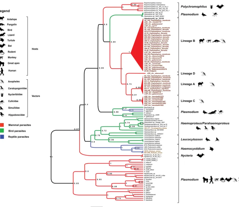
Fig 2. Phylogenetic relationships between the Cyt-b sequences of haemosporidian parasites obtained in our study and reference Cyt-b sequences (names in black) from existing databases. The tree was constructed using a Bayesian outgroup-free method with relaxed molecular clock assumptions. The names of our isolates include: 1) the abbreviation of the sampling site (LOP: La Lopé and LEK: Lékédi) or province (OL: Ogooue-Lolo; OM: Ogooue-Maritime, OI: Ogooue-Ivindo, NG: Ngounie; NY: Nyanga; HO: Haut-Ogooue; MO: Moyen-Ogooue; WN: Woleu-Ntem), 2) the sample number, 3) the name of the host species in which it was found (vertebrate host or anopheles) (for instance, OL115-Cepholophus monticola), 4) SG if the parasite was detected in the salivary glands (for anopheles only). The tree was built based on 757 bp-long Cyt-b sequences. Branch colors indicate different groups of vertebrates. Posterior probabilities are given at each node. More details on the different reference sequences can be found in Table A inS1 File. doi:10.1371/journal.pone.0148958.g002
belonging to Anopheles gabonensis (a species recently discovered in the Gabonese forest [29]) and in one Anopheles vinckei sample (Fig 2). We found parasites from lineage B most fre-quently (11 times) in mosquitoes belonging to six Anopheles species [Anopheles carnevalei (one specimen), Anopheles coustani (one specimen), An. gabonensis (four specimens), Anopheles obscurus (three specimens), An. vinckei (one specimen) and An. sp. (one specimen)]. Two spec-imens (identified as An. gabonensis and An. obscurus) harbored the parasites in the salivary glands (Fig 2). This strongly suggests that lineage B parasites can complete their life cycle within An. gabonensis and An. obscurus and produce infective forms (i.e., sporozoites that can be inoculated in a mammalian host with the saliva during blood feeding). Although we do not have any evidence about the viability of such sporozoites, we hypothesize that these two mos-quito species might contribute to the transmission of lineage B parasites. Moreover, anopheline mosquitoes, but not vertebrate specimens, were infected also by two other Haemosporida line-ages (C and D) that are phylogenetically related to lineline-ages A and B (Fig 2). We found lineage
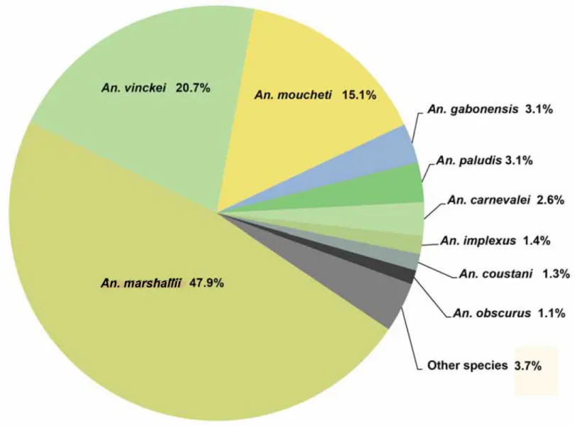
Fig 3. Diversity of Anopheles mosquitoes collected in the two wildlife reserves (La Lopé and La Lékédi) in Gabon from October 2012 to December 2013. Only species representing more than 1% of the entire population are indicated. The other species are grouped in the“other species” category. doi:10.1371/journal.pone.0148958.g003
C six times in Anopheles specimens belonging to three species: An. coustani (one specimen), Anopheles marshallii (three specimens) and Anopheles moucheti (two specimens). The identifi-cation of lineage C parasites in the salivary glands of An. marshallii (one specimen) and An. moucheti (one specimen) suggests a potential role as vectors. Conversely, we detected lineage D only in one An. obscurus specimen. For lineages C and D, the nature of their vertebrate host remains to be discovered. However, the parasites were recovered in Anopheles species that are known to have mainly a mammalophilic feeding behavior [30]. This suggests that mosquitoes might have acquired such parasites from mammalian hosts.
Our findings highlight several features of the ecology of these lineages. First, these parasites are not host-specific because they can infect a variety of hosts, including different species of antelopes. The propensity of ungulate haemosporidian parasites to infect different hosts was previously described [27,31,32] and led to the hypothesis [27] that P. cephalophi and P. brucei could be more widely distributed and possibly present also in other African antelopes. More-over, their host range might not be restricted only to antelopes because lineage B includes para-sites detected also in other orders, such as Pholidotas (pangolin) or Primates (Cercophitecus sp.) [20]. Whether these non-ungulate species are frequent hosts of these lineages or only acci-dental hosts remains to be clarified. Previous studies on African monkeys rather support the second hypothesis because only one monkey, among the several hundred individuals studied, was found to be infected by parasites belonging to lineage B [20,21].
Second, our results suggest that some sylvan Anopheles species could serve as vector for these lineages. Indeed, we found that at least four Anopheles species (two species for lineage B and two for lineage C) might support the transmission of this group of parasites in forests. Other Anopheles species also were infected by these lineages, but we currently have no evidence that they serve as vectors (we detected the parasite Cyt-b gene sequence only in the whole body and not in salivary glands). Very little is known about the biting behavior of sylvan Anopheles and about the vertebrate hosts that constitute their preferred source of blood [33]. Zoophilic species, such as An. vinckei and An. gabonensis, were previously shown to transmit zoonotic Plasmodium parasites, including those circulating among great apes and rodents [29,33,34]. Our study suggests that these species also bite ungulates. This indicates that, concerning their blood meal, An. vinckei and An. gabonensis are opportunistic rather than specialized. Such a propensity to bite a wide range of hosts is probably an adaptive trait in response to temporal fluctuations of host diversity and density in forest environments. This feature could enhance the possibility for cross-species transfer of parasites and could explain the parasite propensity to infect different host species. In addition, the panel of Anopheles species infected by these newly described haemosporidian lineages encompassed mosquito species with a well-known anthropophilic feeding behavior (sometimes very pronounced, such as in An. marshallii or An. mouchetii [33]).
Finally phylogenetic analysis of the relationship between the Cyt-b sequences of the haemos-poridian parasites obtained in our study and reference sequences (S1 File) indicated that the phylogenetic position of the four newly described haemosporidian lineages was closer to saur-opsid Plasmodium and Polychromophilus (bat parasites) than to other mammalian parasites (Fig 2, Figure A inS1 File, and Figure B inS1 File). According to the current Haemosporida classification, all species infecting mammals belong to different genera within the Plasmodiidae family [2]. However, molecular data are lacking for several genera identified in bats or flying squirrels (e.g., Biguetiella, Dionisia, Bioccala and Rayella) and they were never included in modern phylogenies. Previous molecular-based studies proposed that most Plasmodiidae mammalian parasites (i.e., the genera Plasmodium and Hepatocystis) belong to a single mono-phyletic clade and are transmitted by anopheles (e.g., [24–26]), with the exception of some bat parasites (genera Polychromophilus and Nycteria). It was suggested that parasites of the genus
Polychromophilus in bats could be the result of a secondary invasion of mammals and that they derived from avian or reptile parasites (sauropsid Plasmodium) and were transmitted by bat flies [35]. The origin of Nycteria parasites is less clear and needs to be further investigated. Par-ticularly, it is unclear whether they represent another case of host switch or an ancient mam-malian Plasmodium lineage. Similarly, the origin of the four clades described in this study is uncertain. Indeed, depending on the phylogenetic analyses (e.g., different rooting strategies) and reference sequences we used, the position of the four clades relatively to sauropsid Plasmo-dium or to Polychromophilus parasites changed (i.e., closer to sauropsid PlasmoPlasmo-dium or to Polychromophilus species; data not shown). Therefore, it remains to be determined whether these four clades are the result of a host switch from sauropsid Plasmodium or whether they are more related to Polychromophilus parasites. Another remaining question concerns the roles played by the vertebrate hosts and their vectors in their evolution and diversification.
Besides these four lineages discovered in antelopes and/or sylvan mosquitoes, we identified two additional lineages in bushmeat samples: one in a bird sample and the other in two tortoise specimens. The bird lineage was related to parasites of the Haemoproteus genus [36] and the turtle lineage was related to parasites of the Haemocystidium genus [2,36]. More data on these groups of vertebrates are required to determine whether these last two lineages are new [2,25,
36]. Indeed, the current data and those available in the literature are too scarce to conclude with confidence.
Conclusion
In this study, we identified four new haemosporidian molecular lineages that belong to the Plasmodiidae family and that might be transmitted by Anopheles mosquitoes. Concerning their vertebrate hosts, two of the lineages (A and B) can infect several mammalian species. The verte-brate hosts of the other two lineages (C and D) remain to be identified. None of these four line-ages could be matched against reference sequences, although several species of haemosporidian parasites were previously described in African antelopes, but only based on their morphological features. As a consequence, and in agreement with other authors (e.g., [2]), we think that it is now crucial to rapidly undertake a complete re-evaluation of the taxonomy of these protozoa in the different groups of vertebrates with a particular attention to the genus Plasmodium. Indeed, this genus includes some species that are more genetically distant between them than they are with species belonging to other genera. Ideally, this re-evaluation should be done using the different criteria of phenotypic similarities, the concept of biological species and, most importantly, the concept of phylogenetic species [2,10]. Such a re-evaluation is critical to our understanding of host-pathogen evolution in this group of parasites.
Material and Methods
Wildlife screening
To analyze the diversity of haemosporidian parasites circulating among wild vertebrates in Gabon (Central Africa), we screened several bushmeat samples from wild animal species col-lected by the CIRMF (Centre International de Recherches Médicales de Franceville) in eight provinces of Gabon (Fig 1) between 2009 and 2013. Most bushmeat samples (n = 347) were obtained from officers of the provincial direction of the Gabon Ministry of Water Affairs and Forestry after their seizure because of illegal hunting and 145 from bushmeat salesmen during hunting periods. Species identification was performed by visual inspection according to the Kingdom Field Guide to African Mammals [37] and the“Guide des Reptiles du Gabon” [38]. Biological samples of interest (whole blood, liver or spleen) were taken and kept in liquid nitro-gen until delivery to the CIRMF where they were stored at -80°C for molecular analyses. For
each selected animal, total DNA was extracted from approximately 200μl of blood or 100mg of liver/spleen according to the procedures described in [21] and [39], respectively.
Ethic statement. The study was conducted in Gabon outside protected areas. All samples were collected from dead animals only (bushmeat). Some animal carcasses confiscated from poachers by Gabonese authorities (Fauna and Hunting Department, Ministry of Water Affairs and Forestry, Environment and Sustainable development of Gabon) belonged to protected spe-cies (e.g., water chevrotain). All samples obtained from bushmeat salesmen were collected only during the authorized hunting season of non-protected species and only in public markets. No money was given to the salesmen in exchange of the pieces of tissues from the animals. All sam-ples were collected for a CIRMF project aiming at identifying the natural reservoir of Ebola virus. New samples were not collected for this study. All work was carried out with the authorization from the Gabonese Ministry of Water Affairs and Forestry (Département de la Faune et de la Chasse—Authorization N°005/MEFEDD /SG/DGEF/PEFWN, N°001/MEFEDD/SG/DGEF/ PEENY, N°108/MEFEDD/SG/DGEF/ PEFOL and N°016/MEFEDD/SG/DGEF/PEFMO) and the Gabonese Ministry of Higher Education, Scientific Research and Innovation (Centre National de la Recherche Scientifique et Technique—Authorization N°AR0031/09 /MENESRESI/ CEN-AREST/CG/CST/CSAR).
Mosquito sampling
Mosquitoes were sampled in two wildlife reserves in Gabon: the Lopé National Park (Ogooué-Ivindo province) and the private park of La Lékédi, near Bakoumba (Haut-Ogooué province). At both sites (Fig 1), a longitudinal survey of sylvan Anopheles was carried out using CDC light traps placed in several sites of the forest between 5pm to 7am from October 2012 to December 2013. Mosquitoes were morphologically identified using identification keys [40]. Salivary glands of females Anopheles were separated from the rest of the body. All samples were stored in liquid nitrogen and transferred to the CIRMF where they were kept at -80°C. In total, 2,184 females of Anopheles were collected that belonged to 17 species. An. marshallii (47.9%), An. vinckei (20.7%) and An. moucheti (15.1%) mosquitoes constituted most of the captures. The other Anopheles (14%) belonged to other species and only 2.3% of Anopheles could not be iden-tified (Fig 3).
Molecular analyses
Total DNA was extracted from bushmeat tissues, mosquito bodies and salivary glands with the DNeasy Blood and Tissue Kit (Qiagen) and used as template for the detection of haemospori-dian parasites according to a previously described protocol [16] based on PCR amplification and sequencing of a portion of the parasite Cyt-b gene. All amplified products (10μl) were run on 1.5% agarose gels in TBE buffer. Amplicons were then sequenced by Eurofins MWG (Ger-many). The sequences reported in this study were deposited in GenBank under the following accession numbers KT367817 to KT367865.
For species identification, each sequence was compared to a list of reference sequences obtained from GenBank (http://www.ncbi.nlm.gov/) (S1 File). Because amplicons with chimeric sequences can form during PCR amplification of DNA from samples with multiple infections, similarity plot analyses were performed on the nucleotide alignments generated with the new and all reference sequences using the SIMPLOT package version 2.5 [41] and a sliding window of 80 nucleotides (nt) moved in steps of 10 nt. Once verified that the amplicons did not contain chimeric sequences, phylogenetic analyses were done after multiple alignments of the obtained partial Cyt-b sequences (757 nucleotides) and of the Genbank reference sequences using Clus-talW (v 1.8.1 in BioEdit v.7.0.9.0. Software) [42]. It was previously shown that enough
phylogenetic data can be extracted from Cyt-b sequences to study the phylogenetic relationships between haemosporidian parasites and to recover major clades [43,44]. Maximum Likelihood (ML) methods were used for tree construction [16]. The best-fitting ML model based on the Akaike Information Criterion was GTR (General Time Reversible) + Gamma + I (invariant sites), as determined using ModelTest [45]. The highest-likelihood DNA tree and the corre-sponding bootstrap support values were obtained by using PhyML [46,47] (freely available at the ATGC bioinformatics platformhttp://www.atgc-montpellier.fr/) using NNI (Nearest Neighbor Interchange) + SPR (Subtree Pruning Regrafting) branch swapping and 100 bootstrap replicates. Recently, the choice of outgroup to use to root the tree of haemosporidian species has been a mat-ter of debate. In our study, ML trees were rooted: (i) using Toxoplasma spp. and Eimeria spp.; and (ii) using Leucocytozoon spp (S1 File). Finally, BEAST [46] was used to implement a Bayesian outgroup-free method to construct the tree under a Yule tree process and by assuming a log-nor-mal relaxed molecular clock and the GTR substitution model of evolution, with gamma distrib-uted rates (four categories) and including a proportion of invariant sites (GTR + G + I). Three independent runs were performed for 30 million generations sampled every 5,000 generations (burn-in). The traces and the effective sample size (ESS; all exceeded 250) were checked using TRACER v. 1.6.0 [48], to evaluate the Markov chain Monte Carlo convergence. The three inde-pendent runs were combined with LOG-COMBINER v.1.8.2. [49] and the phylogeny was deter-mined using TREEANNOTATOR v.1.8.2 [49] and FigTree v.1.4.2 [50] for visualization. One advantage of estimating a phylogeny using a relaxed clock is that an estimate of the position of the tree root can be obtained.
Supporting Information
S1 File. Phylogenetic trees and Accession numbers of all sequences.The trees give the rela-tionship between our sequences and others sequences of genbank. Phylogenetic tree was rooted using Leucocytozoon spp (Figure A). Phylogenetic tree was rooted using Eimeria spp (Figure B). And one table gives all accession numbers of sequences used in our study (Table A).
(PDF)
Acknowledgments
Authors thank the four reviewers for their comments on a previous version of the manuscript as well as Philippe Christe for helpful discussions. The study was funded by the Centre Interna-tional de Recherches Médicales de Franceville (CIRMF, Gabon), the Centre NaInterna-tional de la Recherche Scientifique (CNRS, France), the Institut de Recherche pour le Développement (IRD, France) and the Agence Nationale de la Recherche (ANR, France, grant ORIGIN JCJC 012). Larson Boundenga was financed by an ANBG scholarship. The funders had no role in study design, data collection and analysis, decision to publish, or preparation of the manuscript.
Author Contributions
Conceived and designed the experiments: LB BM BO FP CP. Performed the experiments: LB APO NDM LDL LYM CA PD CTB BM CP FR BMO VR. Analyzed the data: LB BO BM FP CP AG. Contributed reagents/materials/analysis tools: CA PD NDM LYM NR. Wrote the paper: LB BM FP CP. Contributed to the acquisition of samples in fieldwork: LB BO FP VB VR BO EL BM CP. Conducted and supervised this work: LB CP BO CTB FR FP.
References
1. WHO. WORLD MALARIA REPORT. 2013. Geneva World Health Organization 2013.
2. Perkins SL. Malaria's many mates: past, present, and future of the systematics of the order Haemospor-ida. The Journal of parasitology. 2014; 100(1):11–25. Epub 2013/09/26. doi:10.1645/13-362.1PMID:
24059436.
3. Keeling PJ, Rayner JC. The origins of malaria: there are more things in heaven and earth. Parasitology. 2014:1–10. Epub 2014/06/26. doi:10.1017/S0031182014000766PMID:24963725.
4. Valkiunas G, Sehgal RN, Iezhova TA, Smith TB. Further observations on the blood parasites of birds in Uganda. Journal of wildlife diseases. 2005; 41(3):580–7. Epub 2005/10/26. doi: 10.7589/0090-3558-41.3.580PMID:16244068.
5. Garnham PC. Immunity against the different stages of malaria parasites. Bulletin de la Societe de pathologie exotique et de ses filiales. 1966; 59(4):549–57. Epub 1966/07/01. 6014215. PMID:6014215
6. Duval L, Mejean C, Maganga GD, Makanga BK, Mangama Koumba LB, Peirce MA, et al. The chirop-teran haemosporidian Polychromophilus melanipherus: a worldwide species complex restricted to the family Miniopteridae. Infection, genetics and evolution: journal of molecular epidemiology and evolu-tionary genetics in infectious diseases. 2012; 12(7):1558–66. Epub 2012/06/23. doi:10.1016/j.meegid. 2012.06.006PMID:22721902
7. Landau I, Miltgen F, Boulard Y, Chabaud AG, Baccam D. Study of gametocytes from the Plasmodium "vivax" group: morphology, development in Anopheles and infectivity of Plasmodium yoelii microgame-tocytes. Annales de parasitologie humaine et comparee. 1979; 54(2):145–61. Epub 1979/03/01. PMID:
539716.
8. Landau I, Miltgen F, Boulard Y, Chabaud AG, Baccam D. New data on the biology of gametocytes of Plasmodium yoelii yoelli gathered from morphological characteristics indicating their age. Comptes rendus des seances de l'Academie des sciences Serie D, Sciences naturelles. 1979; 288(5):521–2. Epub 1979/02/05. PMID:108020.
9. Landau I, Chabaud AG, Miltgen F, Baccam D. Dionisia bunoi n. g. n. sp., Haemoproteidae parasite of the microchiropteran bat Hipposideros cyclops in Gabon Annales de parasitologie humaine et com-paree. 1980; 55(3):271–80. Epub 1980/05/01. PMID:6773461.
10. Perkins SL. Species concepts and malaria parasites: detecting a cryptic species of Plasmodium. Pro-ceedings Biological sciences / The Royal Society. 2000; 267(1459):2345–50. Epub 2001/06/21. doi:
10.1098/rspb.2000.1290PMID:11413654; PubMed Central PMCID: PMCPmc1690816.
11. Manwell RD. Malaria infections by four species of plasmodium in the duck and chicken, and resulting parasite modifications. American Journal of Epidemiology. 1943; 38(2):211–22.
12. Jordan HB. The Effect of Host Constitution on the Development of Plasmodium floridense*. The Jour-nal of protozoology. 1975; 22(2):241–4.
13. Martinsen ES, Waite JL, Schall JJ. Morphologically defined subgenera of Plasmodium from avian hosts: test of monophyly by phylogenetic analysis of two mitochondrial genes. Parasitology. 2007; 134 (Pt 4):483–90. Epub 2006/12/07. doi:10.1017/S0031182006001922PMID:17147839.
14. Bensch S, Waldenstrom J, Jonzen N, Westerdahl H, Hansson B, Sejberg D, et al. Temporal dynamics and diversity of avian malaria parasites in a single host species. J Anim Ecol. 2007; 76(1):112–22. Epub 2006/12/23. doi:10.1111/j.1365-2656.2006.01176.xPMID:17184359.
15. Liu W, Li Y, Learn GH, Rudicell RS, Robertson JD, Keele BF, et al. Origin of the human malaria parasite Plasmodium falciparum in gorillas. Nature. 2010; 467(7314):420–5. Epub 2010/09/25. doi:10.1038/ nature09442PMID:20864995; PubMed Central PMCID: PMC2997044.
16. Prugnolle F, Durand P, Neel C, Ollomo B, Ayala FJ, Arnathau C, et al. African great apes are natural hosts of multiple related malaria species, including Plasmodium falciparum. Proceedings of the National Academy of Sciences of the United States of America. 2010; 107(4):1458–63. Epub 2010/02/ 06. doi:10.1073/pnas.0914440107PMID:20133889; PubMed Central PMCID: PMC2824423. 17. Krief S, Escalante AA, Pacheco MA, Mugisha L, Andre C, Halbwax M, et al. On the diversity of malaria
parasites in African apes and the origin of Plasmodium falciparum from Bonobos. PLoS pathogens. 2010; 6(2):e1000765. Epub 2010/02/20. doi:10.1371/journal.ppat.1000765PMID:20169187; PubMed Central PMCID: PMCPmc2820532.
18. Rich SM, Leendertz FH, Xu G, LeBreton M, Djoko CF, Aminake MN, et al. The origin of malignant malaria. Proceedings of the National Academy of Sciences of the United States of America. 2009; 106 (35):14902–7. Epub 2009/08/12. doi:10.1073/pnas.0907740106PMID:19666593; PubMed Central PMCID: PMC2720412.
19. Prugnolle F, Ayala F, Ollomo B, Arnathau C, Durand P, Renaud F. Plasmodium falciparum is not as lonely as previously considered. Virulence. 2011; 2(1):71–6. Epub 2011/01/13. PMID:21224722.
20. Ayouba A, Mouacha F, Learn GH, Mpoudi-Ngole E, Rayner JC, Sharp PM, et al. Ubiquitous Hepatocys-tis infections, but no evidence of Plasmodium falciparum-like malaria parasites in wild greater spot-nosed monkeys (Cercopithecus nictitans). International journal for parasitology. 2012; 42(8):709–13. Epub 2012/06/14. doi:10.1016/j.ijpara.2012.05.004PMID:22691606; PubMed Central PMCID: PMCPmc3751399.
21. Prugnolle F, Ollomo B, Durand P, Yalcindag E, Arnathau C, Elguero E, et al. African monkeys are infected by Plasmodium falciparum nonhuman primate-specific strains. Proceedings of the National Academy of Sciences of the United States of America. 2011; 108(29):11948–53. Epub 2011/07/07. doi:
10.1073/pnas.1109368108PMID:21730135; PubMed Central PMCID: PMCPmc3141972. 22. Schaer J, Perkins SL, Decher J, Leendertz FH, Fahr J, Weber N, et al. High diversity of West African
bat malaria parasites and a tight link with rodent Plasmodium taxa. Proceedings of the National Acad-emy of Sciences of the United States of America. 2013; 110(43):17415–9. Epub 2013/10/09. doi:10. 1073/pnas.1311016110PMID:24101466; PubMed Central PMCID: PMCPmc3808598.
23. Boundenga L, Ollomo B, Rougeron V, Mouele L, Mve-Ondo B, Delicat-Loembet L, et al. Diversity of malaria parasites in great apes in Gabon. Malaria journal. 2015; 14(1):111.
24. Escalante AA, Freeland DE, Collins WE, Lal AA. The evolution of primate malaria parasites based on the gene encoding cytochrome b from the linear mitochondrial genome. Proceedings of the National Academy of Sciences of the United States of America. 1998; 95(14):8124–9. Epub 1998/07/08. PMID:
9653151; PubMed Central PMCID: PMCPmc20940.
25. Perkins SL, Schall JJ. A molecular phylogeny of malarial parasites recovered from cytochrome b gene sequences. The Journal of parasitology. 2002; 88(5):972–8. Epub 2002/11/19. doi:10.1645/0022-3395 (2002)088[0972:ampomp]2.0.co;2PMID:12435139.
26. Martinsen ES, Perkins SL, Schall JJ. A three-genome phylogeny of malaria parasites (Plasmodium and closely related genera): evolution of life-history traits and host switches. Molecular phylogenetics and evolution. 2008; 47(1):261–73. Epub 2008/02/06. doi:10.1016/j.ympev.2007.11.012PMID:18248741. 27. Garnham PC, Kuttler KL. A malaria parasite of the white-tailed deer (Odocoileus virginianus) and its
relation with known species of Plasmodium in other ungulates. Proceedings of the Royal Society of London Series B, Containing papers of a Biological character Royal Society (Great Britain). 1980; 206 (1165):395–402. Epub 1980/01/17. PMID:6102388.
28. Keymer IF. Investigations on the duiker (Sylvicapra grimmia) and its blood protozoa in Central Africa. Philosophical Transactions of the Royal Society B: Biological Sciences. 1969; 255(798):33–108. 29. Rahola N, Makanga B, Yangari P, Jiolle D, Fontenille D, Renaud F, et al. Description of Anopheles
gabonensis, a new species potentially involved in rodent malaria transmission in Gabon, Central Africa. Infection, genetics and evolution: journal of molecular epidemiology and evolutionary genetics in infec-tious diseases. 2014. Epub 2014/05/21. doi:10.1016/j.meegid.2014.05.012PMID:24840150. 30. Bruce-Chwatt LJ, Garrett-Jones C, Weitz B. Ten years' study (1955–64) of host selection by anopheline
mosquitos. Bulletin of the World Health Organization. 1966; 35(3):405–39. Epub 1966/01/01. PMID:
5297635; PubMed Central PMCID: PMCPMC2476083.
31. Garnham PC. Malaria as a medical and veterinary zoonosis. Bulletin de la Societe de pathologie exo-tique et de ses filiales. 1969; 62(2):325–32. Epub 1969/01/01. PMID:4397210.
32. Keymer IF. Studies on Plasmodium (Vinckeia) cephalophi of the grey duiker (Sylvicapra grimmia). Annals of tropical medicine and parasitology. 1966; 60(2):129–38. Epub 1966/06/01. PMID:5962467. 33. Paupy C, Makanga B, Ollomo B, Rahola N, Durand P, Magnus J, et al. Anopheles moucheti and
Anopheles vinckei are candidate vectors of ape Plasmodium parasites, including Plasmodium praefal-ciparum in Gabon. PloS one. 2013; 8(2):e57294. Epub 2013/02/26. doi:10.1371/journal.pone.0057294
PMID:23437363; PubMed Central PMCID: PMC3577705.
34. Gilles HM. Malaria. Tropical diseases bulletin. 1968; 65(3):213–8. Epub 1968/03/01. PMID:4869999. 35. Witsenburg F, Salamin N, Christe P. The evolutionary host switches of Polychromophilus: a multi-gene
phylogeny of the bat malaria genus suggests a second invasion of mammals by a haemosporidian par-asite. Malaria journal. 2012; 11:53. Epub 2012/02/24. doi:10.1186/1475-2875-11-53PMID:22356874; PubMed Central PMCID: PMCPmc3342143.
36. Pineda-Catalan O, Perkins SL, Peirce MA, Engstrand R, Garcia-Davila C, Pinedo-Vasquez M, et al. Revision of hemoproteid genera and description and redescription of two species of chelonian hemo-proteid parasites. The Journal of parasitology. 2013; 99(6):1089–98. Epub 2013/09/17. doi:10.1645/ 13-296.1PMID:24032642.
37. Kingdon J. The kingdon Field Guide to africain Mammals. Academic Press. 1997.
38. Pauwels Olivier S. G. VwJP. Les Reptiles Du Gabon (French)Paperback use preformatted date that complies withlegalrequirementfrom media matrix 2008;(978–1893912014):298 pages.
39. Maganga GD, Verrier D, Zerbinati RM, Drosten C, Drexler JF, Leroy EM. Molecular typing of PPRV strains detected during an outbreak in sheep and goats in south-eastern Gabon in 2011. Virology jour-nal. 2013; 10:82. Epub 2013/03/19. doi:10.1186/1743-422x-10-82PMID:23497402; PubMed Central PMCID: PMCPmc3599724.
40. Coetzee GMTe. A supplement to the Anophelinae of Africa south of the Sahara (Afrotropical Region). South African Institute for Medical Research, Johannesburg. 1987; 55:1–139.
41. Ray S. SimPlot for Windows 95/98/NT, 2.5 edn. Distributed by the author: Available:http://sray.med. som.jhmi.edu/RaySoft/SimPlot/. 1999.
42. Hall TA, editor BioEdit: a user-friendly biological sequence alignment editor and analysis program for Windows 95/98/NT. Nucleic acids symposium series; 1999.
43. Pacheco MA, Battistuzzi FU, Junge RE, Cornejo OE, Williams CV, Landau I, et al. Timing the origin of human malarias: the lemur puzzle. BMC Evol Biol. 2011; 11:299. Epub 2011/10/14. doi: 10.1186/1471-2148-11-299PMID:21992100; PubMed Central PMCID: PMCPMC3228831.
44. Perkins SL. Molecular systematics of the three mitochondrial protein-coding genes of malaria parasites: corroborative and new evidence for the origins of human malaria. Mitochondrial DNA. 2008; 19(6):471– 8. Epub 2009/06/03. PMID:19489133.
45. Posada D, Crandall KA. MODELTEST: testing the model of DNA substitution. Bioinformatics. 1998; 14 (9):817–8. Epub 1999/01/27. PMID:9918953.
46. Guindon S, Dufayard JF, Lefort V, Anisimova M, Hordijk W, Gascuel O. New algorithms and methods to estimate maximum-likelihood phylogenies: assessing the performance of PhyML 3.0. Syst Biol. 2010; 59(3):307–21. Epub 2010/06/09. doi:10.1093/sysbio/syq010PMID:20525638.
47. Dereeper A, Guignon V, Blanc G, Audic S, Buffet S, Chevenet F, et al. Phylogeny.fr: robust phyloge-netic analysis for the non-specialist. Nucleic Acids Res. 2008; 36(Web Server issue):W465–9. Epub 2008/04/22. doi:10.1093/nar/gkn180PMID:18424797; PubMed Central PMCID: PMCPMC2447785. 48. Rambaut A SM, Xie D & Drummond AJ. Tracer v1.6, Available:http://beast.bio.ed.ac.uk/Tracer. 2014. 49. Bouckaert R, Heled J, Kuhnert D, Vaughan T, Wu CH, Xie D, et al. BEAST 2: a software platform for
Bayesian evolutionary analysis. PLoS Comput Biol. 2014; 10(4):e1003537. Epub 2014/04/12. doi:10. 1371/journal.pcbi.1003537PMID:24722319; PubMed Central PMCID: PMCPMC3985171.