Abnormal chromosomal arrangements in human oocytes
E.Macas1, Y.Floersheim, E.Hotz, B.Imthurn, P.J.Keller and H.Walt
Laboratory of Reproduction and Development, Department of Gynaecology and Obstetrics, University Hospital, CH-8091 Zurich, Switzerland
'To whom correspondence should be addressed
Ninety-one human oocytes, lacking signs of fertilization 50 h after insemination in vitro, were investigated cytogenetically to assess the frequency and type of chromosomal abnormal-ities. Chromosome spreading permitted adequate karyotyping in 55 oocytes. Non-determined numerical aberrations occurred with the following frequencies: hypohaploidy, 10.9% (6/55), hyperhapJoidy, 14.5% (8/55) and hyperdiploidy, 3.6% (2/55). Total aneuploidy occurred with a frequency of 29.1% and was observed in oocytes from 30 patients. No correlation was found between specific chromosomal aberrations and type of infertility, stimulation treatment or gonadotrophin levels. On the other hand, the frequency of aneuploidy was signifi-cantly higher (P < 0.05) in patients > 35 years of age. Two chromosomal complements (3.6%) had structural rearrange-ments; one oocyte had both structural and numerical chromo-somal abnormalities and the other had differently condensed regions on the long arms of three chromosomes from group C. The overall frequency of chromosomal aberrations was 32.7%. Only two samples contained an additional set of polar body chromosomes. Thirteen oocytes presented sperm chromosomes in an arrested stage of premature chromosome condensation of the G, phase and four oocytes showed asynchronous condensation of pronuclear chromosomes. Finally, it was concluded that the high proportion of chromosomal aberrations observed in human oocytes may contribute significantly to abnormal embryonic development in vitro.
Key words: aberrationyhuman/oocyte/chromosome Introduction
The high incidence of chromosomal abnormalities in first trimester human spontaneous abortions (Chandley, 1982; Bou£ et al., 1985; Hassold and Jacobs, 1984) is considered to be a predominant reason for the low estimated human fecundity (25%) (Edmonds et al., 1982). These data are supported by the results of in-vitro fertilization (IVF) (Seppala, 1985). The high failure rate of implantation after IVF could be related to chromosomal aberrations in embryos. Only 10% of embryos are capable of inducing normal pregnancy after embryo replacement (Rudak
et al., 1984), while the remaining 90% die before or immediately after transfer (Johnson et al., 1981). One explanation for the implantation failure is that embryos may carry lethal chromosome abnormalities which do not allow development beyond the preimplantation stage. On the other hand, over-expression of specific genes due to total or partial trisomies of some chromosomes might produce similar effects. Disturbances of the genetic balance in the oocyte might thus contribute considerably to preimplantation loss. We believe that investigation of human oocyte chromosomes provides data essential to the assessment of reproductive failure. In this study, chromosomes from 91 oocytes which did not present signs of development 50 h after insemination in vitro have been cytogenetically analysed.
Materials and methods Patients and treatment
Preovulatory follicular fluids were obtained from 43 women who underwent FVF and embryo transfer (ET). The patients' ages ranged from 24 to 38 years (mean 35.2) and all had regular menstrual cycles. To stimulate follicular growth, clomiphene citrate (Clomid, Merrel Dow) was given to 15 women for 5 days, in doses of 100 mg daily. On the last day of clomiphene administration, human menopausal gonadotrophin was given (HMG, Pergonal, Serono). The dose used varied depending on the growth of dominant follicles and on the daily rise of serum oestradiol (EJ. Oocytes were recovered 3 2 - 3 6 h after injection of 5000 IU of human chorionic gonadotrophin (HCG, Pregnyl, Organon). In 28 patients, follicular growth was induced with 300 IU of HMG administered on the third and fourth days of the cycle. The additional 150 IU of HMG were given on the fifth and sixth days. Thereafter, HMG was decreased in a stepwise manner depending on the individual response. HCG was given at a dose of 10 000 IU i.m. 3 4 - 3 6 h before aspiration under laparoscopic or ultrasonographic control.
Oocyte culture and cytogenetic methods
Oocytes were cultured in modified Ham's F-10 medium (Sigma, USA) (Lopata et al., 1980) at 37°C in a humidified atmosphere of 5% CO2 in air. The insemination medium was supplemented
with 10% heat-inactivated newborn cord serum. The oocytes were preincubated for 4—6 h before insemination with 0 . 5 - 1 . 0 x 105 motile spermatozoa. The oocytes were
examined for fertilization 16—20 h after insemination by analysis of pronuclei. Unfertilized oocytes were selected by the absence of two pronuclei and lack of cleavage 50 h after insemination.
Chromosome preparations were made by a modified method of Tarkowsky (1966) in order to minimize chromosome losses during fixation. Oocytes were transferred to 1 % sodium citrate hypotonic solution and were incubated for 5 — 20 min at room temperature. They were then attached on a 'grease-free' optical slide in a small drop of hypotonic solution. The fixative (ethanol/glacial acetic acid, 3:1, v/v) was carefully dropped beside the oocyte under a dissecting microscope. Immediately after the first drop of fixative covered the oocyte, another drop was added. This procedure was repeated 3—4 times until the oocyte was thoroughly spread on the slide. Preparations were stained with a 10% Giemsa solution (Fisher Scientific Company, USA), washed with tap and distilled water, and evaluated micro-scopically. Oocytes with intact boundaries of the stained cytoplasm and chromosome complement within were chosen for this study.
Results
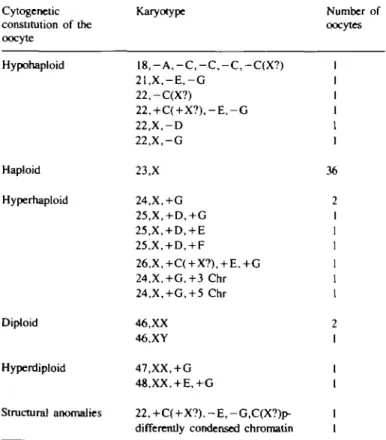
Ninety-one oocytes from 37 women were prepared for cyto-genetic investigation and chromosome analysis was possible on 55 oocytes. The number of oocytes per woman ranged from one to eight (one woman, eight oocytes; two women, four oocytes; two women, three oocytes; eight women, two oocytes and 17 women, one oocyte). The remaining 39.6% of the oocytes were unsuitable for analysis because chromosomes were either inadequately spread or lost due to mechanical damage of the cell. Table I shows the chromosome complements from 55 oocytes failing to fertilize after IVF. Thirty-six oocytes (65.5%) had a normal haploid karyotype. Eight complements were hyperhaploid
Table I. Chromosome complement of uncleaved human oocytes Cytogenetic constitution of the oocyte Karyotype Number of oocytes Hypohaploid Haploid Hyperhaploid Diploid Hyperdiploid Structural anomalies 1 8 , - A , - C , - C , - C , - C ( X ? ) 2 1 , X , - E , - G 22,-C(X?) 22,+C( + X ? ) , - E , - G 22.X.-D 22,X,-G 23,X 24,X,+G 25,X, + D,+G 25,X, + D, + E 25,X, + D, + F 26,X,+C( + X?), + E.+G 24,X,+G,+3 Chr 24,X,+G, + 5 Chr 46,XX 46.XY 47,XX, + G 48.XX. + E.+G 22, +C(+X?), - E , -G,C(X?)p-differemly condensed chromatin
36 : ) 2 1 1 1 1 1
(14.5%; Figure 1), six were hypohaploid (10.9%) and one had both numerical and structural abnormalities (Figure 2). One of two oocytes from the same patient showed decondensed and
Fig. 1. Karyotype of a hyperhaploid human oocyte (n = 25)
showing one extra chromosome in the D and G group, respectively. A \
X
J* f U
Hin
b
A
-H.
"FFig. 2. Karyotype of a hypohaploid human oocyte (n = 22) showing absence of the p arm in a chromosome of the C group (big arrow). The small arrow shows one chromosome fragment attached to one chromosome in the F group. One extra
chromosome is presented in the C group and one chromosome is absent in the E and G groups, respectively.
partially unstained regions in the long arms of three chromosomes from group C (Figure 3). The most frequent numerical aber-rations were observed in chromosomes of groups G, F, E, D and C of the human karyotype. Three diploid metaphases were detected and two of them displayed mitotic chromosomes (46.XX and 46, XY). These two oocytes exhibited no visible pronuclei. The chromosome complements of the remaining two oocytes were without either polar body or polar body chromatin and were hyperdiploid. The overall frequency of chromosomal abnor-malities was 32.7%.
Considering clinical parameters, no correlations were found between the type of infertility, the stimulation treatment, the dose of gonadotrophins administered and the frequency or type of chromosome anomaly. The mean age of patients with normal oocytes was 34.1 years whereas patients with aneuploid oocytes averaged 36.2 years old. The frequency of aneuploid oocytes was significantly higher (P < 0.05) in patients older than 35 (34.8 versus 18.4%).
Aside from second meiotic and mitotic chromosomes, two other types of morphologically variant chromosomes were observed. Typical polar body chromosomes were found in two cells, one of which apparently contained a normal haploid polar body complement with 23 chromosomes. The second had only
18 chromosomes and in addition to 18 chromosomes, as did the oocyte at second meiotic metaphase (Figure 4).
Prematurely condensed chromosomes (PCC) have also been observed in this study. Eighty-one oocytes were investigated for the presence of PCC. Of these, 13 (16%) showed sperm chromosomes in an arrested stage of PCC of the G, phase. In addition, maternal ME chromosomes were detected in the diploid (two oocytes) or haploid range (11 oocytes). Asynchronous condensation of pronuclear chromosomes was observed in four oocytes which were in G,/G2, G|/S, G,/S and Gi/S/G? phase of the cell cycle.
Discussion
The exact frequency of chromosomal aberrations in the human preimplantation embryo has so far been very difficult to deter-mine (Warburton, 1987). In-vitro fertilization offers a unique possibility to study chromosomal abnormality as early as the
Table Da. Summary of frequencies of hypohaploidy and hyperhaploidy presented in nine different cytogenetic studies
Authors n Haploidy Hypohaploidy Hyperhaploidy
Bongso et al. (1988) Ma et al. (1989) Martin et al. (1986) Papadopoulos et al. (1989) Pellestor et al. (1988) Plachot el al. (1986) Veiga et al. (1987) Wramsby et al. (1987) Macas et al. (Present study) 251 65 50 30 201 55 117 52 55 76.6 52.3 68.0 40.0 81.9 54.4 77.0 34.6 65.5 13.0 15 4 28.0 13.3 10.6 5.5 2.6 51.9 10.9 8.0 10.8 2.0 6.7 8.0 5.5 7.7 5.8 14.5
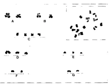
Fig. 3. The arrows point to differently condensed regions on the long arms of three chromosomes from group C.
9 K «[ c
"" " F " " '
E
Fig. 4. Karyotype of a hyperhaploid human oocyte (n = 18). 18 polar body chromosomes of the same oocytes are presented in the inset.
Table lib. Summary of frequencies of aneuploidy and structural aberrations presented in nine previous cytogenetic studies
Authors Frequency Frequency of Total frequency of structural of chromosomal aneuploidy aberrations aberrations
Bongso et al. (1988) 251 21.0 0.4 21.4 M a r t al. (1989) 65 47.7* - 47.7 Martin et al. (1986) 50 4.0* 4.0 8.0 Papadopoulos et al. (1989) 30 20.0 20.0 60.0° Pellestor et al. (1988) 201 18.6d - 20.0c Plachot et al. (1986) 55 2O.2f 1.8 22.0 Veiga etal. (1987) 117 15.4b 4.3 26.5« Wramsby et al. (1987) 52 59.6 1.9 61.5 Macas et al.
(Present study) 55 29.1 3.6 32.7
•Including eight mature oocytes with hyperdiploid, hypodiploid and diploid complements.
""Frequency of aneuploidy is derived by doubling the hyperhaploid frequency.
cIncluding diploid and polyploid zygotcs. ''Total aneuploidy metaphase II. ^Including five hypodiploid metaphases I. including three hyperdiploid metaphases I. •Including eight unreduced oocytes in metaphase II.
precleavage stage (Rudak et al., 1984). An examination of the chromosomes of gametes has also been undertaken as the zona-free hamster test allows analysis of human sperm chromosomes. These results have shown that some 9% of sperm nuclei carry a chromosomal anomaly (Martin, 1985). The data available on aneuploidy in human oocytes on the other hand gives an extremely variable range of chromosomal anomaly (Table Ila.b). Wramsby and Fredga (1987), Papadopoulos et al. (1989) and Ma et al. (1989) detected similar percentages of abnormal numbers of chromosomes, while Martin et al. (1986), using a conservative protocol for the evaluation of the frequency of aneuploidy, found only 4% numerical aberrations. In contrast, Veiga et al. (1987), applying the same criteria, found values about four times higher. The frequency in our study with 32.7% abnormalities was more similar to the data reported by Bongso et al. (1988), Pellestor and Sele (1988) and Plachot et al. (1986) (see Table lib).
The process of meiotic non-disjunction should produce an equal number of n + 1 and n — 1 gametes. The proportion of hypo-haploidy and hyperhypo-haploidy presented in the different cytogenetic studies is not equal (Table Ha). The current study shows that hyperhaploid complements are slightly more common than hypohaploid (14.5 versus 10.9%), although, as compared to other groups, we found fewer hypohaploid than hyperhaploid comple-ments (see Tables I and Da). It has been suggested that anaphase lag is a source for induction of the hypohaploid oocytes, by loss of chromosomes within the cytoplasm. An excess of hypohaploidy was found in studies of the one-cell embryo in the Chinese hamster (Mikamo and Kamiguchi, 1983), in Syrian hamster oocytes (Martin, 1984) and human oocytes (Wramsby and Fredga, 1987; Martin et al., 1986). One of the oocytes in our study showed 18 polar body and 18 second meiotic chromosomes (Figure 4). This finding undoubtedly suggests that a mechanism similar to anaphase lag is involved in the production of chromo-somaJly abnormal gametes. Most probably, chromosomal loss occurs more frequently in female meiosis than in male, because of the large quantity of cytoplasm. Under certain conditions, when using various superovulation methods during IVF treatment, anaphase lag occurs more frequently than does non-disjunction. This could be an explanation for the high incidence of hypo-haploidy reported by Wramsby and Fredga (1987) and Martin et al. (1986) (see Table Ha). At any rate, the incidence of aneuploidy in preimplanation embryos appears to be very high indeed, meaning that a large proportion of such embryos will never develop further (Angell et al., 1983, 1986; Plachot et al.,
1988).
A correlation between ageing and the frequency of oocyte aneuploidy has been demonstrated. Previous reports showed a significant correlation of oocyte chromosome anomalies with age ( > 3 5 years) (Plachot et al., 1988; Bongso et al., 1988). Our results are in agreement with these and show a significantly higher incidence of oocyte aneuploidy in patients > 35 years of age. In our observations, two oocytes with structural anomalies (3.6%) were detected, a percentage similar to that observed by others (Bongso et al., 1988; Martin et al., 1986; Plachot et al., 1986; Wramsby and Fredga, 1987). Fragmentation of chromo-somes is the most common structural anomaly, accounting for 20% of all anomalies (Papadopoulos etal., 1989). Structural
chromosome anomalies have also been found in human pre-embryos (Papadopoulos et al., 1989). Deletion and translocation are rather infrequent, representing < 2 % of the aberrations encountered in some cytogenetic studies (Bongso et al., 1988; Martin et al., 1986; Plachot et al., 1986; Wramsby and Fredga, 1987). The incidences of deletion and translocation in our study are thus consistent with those of previous reports (Bongso et al., 1988; Martin et al., 1986). The significance of what appears to be differently condensed sites on C-group chromosomes in one oocyte remains obscure. The fragile sites appear as unstained and often stretched regions within a chromosome. The chromo-somes have a tendency to break at these points, but at present there are only a few data to interpret the genetic significance of these fragile sites (Sutherland, 1982).
In this first series of chromosomal preparations, we observed only two oocytes with additional polar body chromosomes. These were localized far from the main group of second meiotic chromosomes. Most oocytes however, did not display polar body chromosomes. It is suggested that they disappeared quickly at an early stage. Schmiady et al. (1986) first observed prematurely condensed sperm chromosomes (PCC) in human oocytes. The mechanism is not completely understood; PCC may arise during sperm penetration into immature oocytes, inducing their arrest. It has been suggested that the presence of chromosome-condensing factors in the cytoplasm triggers the premature sperm chromosome condensation (Papadopoulos et al., 1989). In our study, the frequency of oocytes with PCC was 20.9%, a figure which agrees with the data of Ma et al. (1989). A prominent factor in the success of fertilization is the level of maturation of the oocyte. Thus, the presence of numerous oocytes with sperm PCC might suggest that they were not completely mature at insemination.
In conclusion, our observation has shown that the number of oocytes with hyperhaploid complements (14.5%) was slightly higher than hypohaploids (10.9%) though not significantly, and this differs from other cytogenetic studies on the human oocyte. The high proportion of chromosomal aberrations in human oocytes may contribute to the high frequency of abnormal embryonic development in vitro.
Acknowledgements
We thank Mrs M.Widmer and Mr M.Bossi for excellent technical help, Dr C.D.DeLozier-Blanchet, University Institute of Medical Genetics. Geneva, Switzerland, for editorial assistance and Dr P.Emmerich for helpful comments.
References
Angell.R.R., Aitken.R.J., Van Look,P.F.A., Lumsden.M.A. and Templeton,A.A. (1983) Chromosome abnormalities in human embryos after in vitro fertilization. Nature, 303, 336—338. Angell,R.R., Templeton,A.A. and Aitken.RJ. (1986) Chromosome
studies in human in vitro fertilization. Hum. Genet., 72, 333—339. Bongso,A., Chye,N.G., Raman,S., Sathananthan,H. and Kono,P.C. (1988) Chromosome anomalies in human oocytes failing to fertilize after insemination in vitro. Hum. Reprod., 3, 645 — 649. Boue\A., Boue\J. and Gropp.A. (1985) Cytogenetics of pregnancy
wastage. In Harris,H. and Hirschhorn,K. (eds), Advances in Human Genetics. Plenum Press, New York, pp. 1-57.
Chandley.A.C. (1982) Normal and abnormal meiosis in man and other mammals. In Crosignani,P.G. and Rubin,B.L. (eds). Genetic Control of Gamete Production and Function. Academic Press, London, pp. 229-237.
Edmonds,D.K., Lindsay,K.S., Miller.J.F., Williamson,E. and Wood,P.J. (1982) Early embryonic mortality in women. Fertil. Steril., 38, 447-453.
Hassold,T.J. and Jacobs,P.A. (1984) Trisomy in man. Armu. Rev. Genet., 18, 69-97.
Johnson,I.W.H., Lopata.A., Speirs,A., Hoult,I., Kellow,G. and du Plessis,Y. (1981) In vitro fertilization: the challenge of the eighties. Fertil. Steril., 36, 698-711.
Lopata,A., Johnson.I.W.H., Hoult,I. and Speirs,A. (1980) Pregnancy following intrauterine implantation of an embryo obtained by in vitro fertilization of a preovulatory egg. Fertil. Steril., 33, 117—120. Ma,S., Kalousek,D., Zouves,C, Yeen.B., Gomel,V. and Moon.Y.
(1989) Chromosome analysis of human oocytes failing to fertilize in vitro. Fertil. Steril., 51, 992-997.
Martin,R.H. (1984) Comparison of chromosomal abnormalities in hamster egg and human sperm pronuclei. Biol. Reprod., 31, 819-825. Martin,R.H. (1985) Chromosomal abnormalities in human sperm. In
Dellarco,V.L., Voytek,P.E. and Hollaender,A. (eds), Aneuploidy: Etiology and Mechanisms. Plenum Press, New York, pp. 91 — 102. Martin,R.H., Mahadevan.M.M., Taylor,P.J., Hildebrand,K.,
Long-Simpson,L., Peterson,D., Yamamoto.J. and Fleetham,J. (1986) Chromosomal analysis of unfertilized human oocytes. J. Reprod. Fertil., 78, 673-678.
Mikamo.K. and Kamiguchi.Y (1983) Primary incidences of spontaneous chromosomal anomalie and their origins and causal mechanisms in the Chinese hamster. Mutat. Res., 108, 265-278.
Papadopoulos,G., RandallJ. and Templeton,A.A. (1989) The frequency of chromosomal anomalies in human unfertilized oocytes and uncleaved zygotes after insemination in vitro. Hum. Reprod., 4, 568-573.
Pellestor,F. and Sele.B. (1988) Assessment of aneuploidy in the human female by using cytogenetics of IVF failures. Am. J. Hum. Genet.,
42, 274-283.
Plachot,M., Junca,A.M., MandelbaumJ., de Grouchy,J., Salat-Baroux,J. and Cohen.J. (1986) Chromosome investigation in early life. I. Human oocytes recovered in an IVF programme. Hum. Reprod., 1, 547-551.
Plachot.M., de Grouchy,J., Junca.A.M., Mandelbaum,J., Salat-Baroux,J. and Cohen,J. (1988) Chromosome analysis of human oocytes and embryos: does delayed fertilization increase chromosome imbalance? Hum. Reprod., 3, 125-127.
Rudak,E., Dor.J., Mashiach,S., Nebel.L. and Goldmann.B. (1984) Human embryo chromosomes: preliminary results of a study to karyotype multipronuclear human oocytes fertilized in vitro. In Feichtinger.W. and Kemeter.P. (eds), Recent Progress in Human in Vitro Fertilization. Cofese Edizioni, Palermo, pp. 231—239. Schmiady,H., Sperling,K., Kentemch,H. and Stauber.M. (1986)
Prematurely condensed human sperm chromosomes after in vitro fertilization (IVF). Hum. Genet., 74, 441-443.
Seppala,M. (1985) The world collaborative report on in vitro fertiliza-tion and embryo replacement: current state of the art in January 1984. Ann. N.Y. Acad. Sci., 442, 558-563.
Sutherland,G.R. (1982) Heritable fragile sites on human chromosomes. VIII. Preliminary population cytogenetic on the folic-acid-sensitive fragile sites. Am. J. Hum. Genet., 34, 452-458.
Tarkowsky.A.K. (1966) An air drying method for chromosome preparations from mouse eggs. Cytogenetics, 5, 394—400. Veiga,A., CaJderon.G., Santalo,J., Barri.P. and Egozcue,J. (1987)
Chromosome studies in oocytes and zygotes from an IVF programme. Hum. Reprod., 5, 425-430.
Warburton,D. (1987) Reproductive loss: how much is preventable? N. Engl. J. Med., 316, 158-160.
Wramsby,H. and Fredga,K. (1987) Chromosome analysis of human oocytes failing to cleave after insemination in vitro. Hum. Reprod., 2, 137-142.