Intrauterine light delivery for photodynamic therapy of
the human endometrium
M.K.Fehr1'3, S-J.Madsen1, L.O.Svaasand1-4, BJ.Tromberg1, J.Eusebio1, M.W.Berns1 and Y.Tadir1-2'5
'Beckman Laser Institute and Medical Clinic and department of Obstetrics and Gynecology, 1002 Health Sciences Road East University of California, Irvine, CA 92715, USA, department of Obstetrics and Gynecology, University of Zurich, CH 8091 Zurich, Switzerland and 4Department of Physics, University of Trondheim, 7000 Trondheim, Norway
5
To whom correspondence should be addressed at Beckman Laser Institute
Photodynamic therapy is currently being evaluated as a minimally invasive procedure for endometrial ablation not requiring anaesthesia. Light penetration depths at 630, 660 and 690 nm and the optimal configuration of intrauterine light-diffusing fibres were determined in 14 human uteri to assist in the design of a light intrauterine device. Post-menopausal ex-vivo uteri showed a significantly lower light penetration depth than pre-menopausal uteri. With a single central diffusing fibre inserted, the fluence rate measured in the uterine wall at the most remote point of the cavity decreased to 1.1 ± 0.4% of that measured at closest proximity, whereas it decreased to only 40.0 ± 9.0% with three fibres. Distension of the uterine cavity with 2 ml of an optically clear fluid increased the fluence rate at the fundus between the fibres at a depth of 2 mm by a factor of 4. We conclude that in normal-sized pre-menopausal uterine cavities, three diffusing fibres will deliver an optical dose above the photodynamic threshold level at a depth of 4 mm, even in the most remote areas, in <30 min without causing thermal damage. For distorted and elongated cavities, either slight distension of the cavity or the insertion of a fourth diffusing fibre is required.
Key words: endometrium/intrauterine light delivery/light dosimetry/photodynamic therapy
Introduction
Photodynamic therapy (PDT) is an experimental medical treatment which requires specific interactions between light and a photosensitizer to induce oxidation reactions and cause tissue necrosis. Several animal studies (Schneider et ai, 1988; Manyak et ai, 1989; Bhatta et ai, 1992; Yang et ai, 1993; Wyss et ai, 1994a,b; Steiner et ai, 1995) have shown that the selective destruction of the endometrium using PDT is feasible and may provide a viable alternative to routinely performed surgical treatments which require anaesthesia and imply specific risks and complications (Goldrath et ai, 1981; De
Cherney and Polan, 1983; Jedeikin et ai, 1990; Perry and Baughman, 1990; Brooks et ai, 1991; Serden and Brooks, 1991; Daniell et ai, 1992). PDT may, therefore, offer a simple, cost-effective and safe alternative to more radical surgical procedures for the treatment of dysfunctional uterine bleeding. Systemic and topical applications of various photosensitizers have been studied. Intrauterine administration offers potential advantages, including the lack of skin photosensitivity. Several studies have suggested that the endometrial surface regenerates from the residual epithelium of the gland stumps where stem cells are most likely to be present (Ferenczy, 1976). In humans, the glandular crypts of the basal endometrial layer, the regeneration site for new endometrial cells, lie within the innermost myometrial layer. Accordingly, a sufficient optical dose must be delivered to the entire endometrium, including the innermost myometrial layer, to induce irreversible photo-chemical destruction. The thickness of the normal endometrium varies between 1 and 5 mm, depending on the phase in the menstrual cycle (Santolaya-Forgas, 1992).
The aim of this study was to determine the light distribution inside the uterine wall using between one and three intrauterine light-diffusing fibres in intact uteri immediately following hysterectomy. Given this information, light exposure times necessary for the photodynamic destruction of the entire endometrium can be calculated for different configurations of intrauterine light delivery devices.
Materials and methods
A total of 10 pre-menopausal [age (mean ± SE) 40.3 ± 1.0 years] and four post-menopausal (age 57.8 ± 3.0 years) patients scheduled for hysterectomy for benign conditions, such as leiomyoma and uterine prolapse, participated in this study. The intact fresh uterine specimens were obtained immediately after hysterectomy and put on ice. The uterine vessels were clamped proximal and distal to the dissection site to keep the blood in the tissue. During the measurement period (2-4 h), wet gauze was applied to the specimen to prevent dehydration.
The clinical histories and pathology reports were reviewed to obtain the following information: patient age, menopausal status, day of menstrual cycle at surgery, hormonal treatment during the last 3 months and histopathological findings. The study was approved by the Human Subject Review Committee of the University of California, Irvine, CA, USA.
The light source was an argon-ion laser (Innova 90-5; Coherent, Palo Alto, CA, USA) used to pump a dye laser (Coherent Model 599) containing DCM dye (Exciton Inc., Dayton, OH, USA). The laser beam was coupled into flexible quartz fibres with cylindrical diffusing tips of 3 cm length and 1.2 mm diameter (Model 4420-A02; PDT Systems Inc., Santa Barbara, CA, USA) via a three-way fibre splitter (PDT Systems). One, two or three fibres were inserted
into the uterine cavity through the cervical canal under hysteroscopic guidance. Two fibres were placed at the lateral edges of the uterine cavity with the fibre tips located at the entrance to the corresponding Fallopian tube. The third fibre was positioned in the middle of the uterine cavity with the tip touching the fundus. The fibres were held in place by a single 6-0 monofilament polypropylene suture attached to the tip of the diffusing fibre and pulled through the uterine wall. To collect light with equal efficiency from all directions, a 200 \).m core diameter detection fibre with a spherical isotropic collection tip (diameter = 0.8 mm; PDT Systems) was used. The detection fibre was connected to a photomultiplier tube (Hamamatsu R 928; Bridgewater, NJ, USA) to measure the steady-state light intensity (ftuence rate). The detector probe was calibrated in water using a 7.2 mW (k = 633 nm) He-Ne laser. The optical powers emitted from each of the cylindrical fibres were evaluated by inserting them into an integrating sphere and comparing the signals with those obtained from the He-Ne laser.
The detection fibre was inserted into an 18 G needle which perforated the uterine wall in a direction normal to the uterine surface and the cylindrical diffusing fibres. The needle was removed and the detector fibre retracted under hysteroscopic visualization until the spherical probe entered the endometrium. The hysteroscope was then removed and the CO2 gas was evacuated to allow the cavity to collapse. The detection fibre was then retracted into the tissue using a micropositioner, and steady-state measurements of light intensity were recorded as a function of separation from the uterine cavity. The optical penetration depth is defined as the distance from the surface of the tissue at which the fluence rate drops to 37% of its initial value (Wyss et al., 1995). The calculation of the penetration depth from the measured fluence rates at different source detector separations has been described elsewhere (Svaasand, 1984; Tromberg
et al., 1995). If fibroids were visible macroscopically, the
measure-ments were made in tissue which appeared to be normal.
To determine light distribution, measurements were made at the corners of the uterine cavity when only one central fibre was inserted, between the source fibres when three fibres were inserted, and at the centre of the fundus and corpus when only two lateral source fibres were in place. Measurements were recorded at 630, 660 and 690 nm wavelengths. The optical power of each fibre was kept constant regardless of the number of fibres illuminated. To determine the effect of uterine distension on the fluence rate at distances of 2 and 6 mm from the endometrial surface, 9 ml of a high viscosity, optically clear liquid (Hyskon®; Pharmacia Inc., Piscataway, NJ, USA) were injected into the uterine cavity. To avoid spillage, the isthmic portion of the Fallopian tubes was clamped, and the Hyskon was injected through a Bard Cervical Cannula® (C.R.Bard Inc., Billerica, MA, USA), with an inflated balloon in place to block the cervical canal.
After making these measurements, the uteri were opened by a Y-shaped incision of the anterior wall, and the dimensions of the uterine cavity were measured. The unpaired, two-tailed /-test was used for a statistical comparison of light penetration depth between pre- and post-menopausal women. For the comparison of penetration depths at different wavelengths, the paired, two-tailed r-test was used. Statistical significance was taken as P < 0.05. Data are presented as mean ± SE.
Results
The pre-menopausal uteri weighed 498 ± 188 g, with a transverse diameter of the uterine cavity of between 30 and 95 mm (48 ± 12). The post-menopausal uteri weighed 71 ± 21 g, with a transverse diameter of the uterine cavity ranging from 28 to 56 mm (40 ± 6). Histology revealed that six of
800 700 600 500
-I
£ 400 2 0 " 300 200 100V
V
-»-calculated • patient 1 • patient 2 • patient 3 2 4 6 8 Distance from applicator axis (mm)10
Figure 1. Decay of fluence rate by retracting the detector probe from the endometrial surface into the uterine wall of three pre-menopausal women. The power of the cylindrical light-diffusing fibre is 100 mW per cm of fibre length. The curve represents the calculated values after an equation given by Svaasand (1984) and Tromberg et al. (1995).
penetration depth, premenopausal penetration depth, postmenopausal fluence rate Increase in %, premenopausal fluence rate increase In %, postmenopausal
.2
s
630 660
Wavelength (nm)
690
Figure 2. Penetration depth of light into the uterine tissue at different wavelengths and percentage increase of fluence rate at a depth of 4 mm compared with the fluence rate measured at 630 nm light. A total of 10 pre-menopausal and four post-menopausal patients were studied. The light source lies in the uterine cavity and the detector probe is retracted from the endometrial surface into the uterine wall. Histogram bars represent mean ± SE. At 630 nm, light penetration into post-menopausal uteri is significantly lower than into pre-menopausal (P = 0.004, unpaired r-test). In pre- and post-menopausal women, light penetration increases significantly with increasing wavelength from 630 to 690 nm (P = 0.02 and
P = 0.04, paired r-test).
the pre-menopausal patients had secretory phase endometrium and four had proliferative endometrium. Of the post-menopausal uteri, two displayed proliferative and two inactive endometrium.
Fluence rate drops to 1.1 +0.4 o/(
Above light sources - I Fluence rate 100%
Fluence rate
drops to 18 ± 3% Fluence rate
drops to 60 ± 5 %
Fluence drops to 40 +
Light diffusing fibres
Figure 3. Percentage of fluence rate measured at different sites in the uterine cavity compared with fluence rate in closest proximity to light-diffusing fibre at a depth of 2 mm (wavelength 630 nm). Longitudinal and transverse sections of the uteri are shown with the endometrium embedding the light source fibres. (A) With a single diffusing fibre as the light source. (B) With two and (C) with three inserted light-diffusing fibres. Numbers represent mean ± SE.
20 15 <D CO to 2 o Z 10 o •ac t 5 n - -. . - _ - e- - at 2mm depth —•— at 6mm depth ) / J Is ' r —< i""" / ) / / r f r A 1 ' r 1 1 •— — i I 1 1 1 1 1 1 1 1 • 1 0 1 2 3 4 5 6 7 8 9 Injected volume (ml)
Figure 4. Increase of fluence rate at depths of 2 and 6 mm from the endometrial surface during uterine distension by the instillation of Hyskon into the cavities of four uteri. The isthmic portions of the Fallopian tubes and cervical canals are blocked. Points represent mean ± SE.
mm to 1.08 ± 0.4% of the fluence rate measured at the same depth directly above the light source (Figure 3A). When two lateral light sources were inserted, the fluence rate at the middle of the fundus and the centre of the cavity decreased to 17.5 ± 3.0 and 59.5 ± 5.0% respectively (Figure 3B). In Figure 3C, measurements were made between three illuminated fibres at the fundus, and the fluence rate dropped to 40.0 ± 9.0%. In general, the distance between source fibres increased with cavity size, resulting in a more pronounced drop in fluence rate between the fibres (data not shown).
Distension of the uterine cavity produces an optically integrating cavity which results in a more uniform light distribution. Figure 4 shows that at depths of 2 and 6 mm, the fluence rate between two diffusing fibres at the fundus increased after distension with 2 ml of fluid by factors of 4.0 and 5.5 respectively. Following further dilation of the uterine cavity using an additional 3 ml of Hyskon, the fluence rate increased by factors of 8.0 and 13.5.
Figure 1 shows the approximately exponential decay of the fluence rate of light in uterine tissue with increasing source-detector separation in three examples. A comprehensive mathe-matical model describing the decay in fluence rate with increasing source-detector separation shown by the curve in Figure 1 has been described by Svaasand (1984).
The penetration depth of light into the uterine tissue increased with increasing wavelength (Figure 2). The increase from 630 to 690 nm was statistically significant for both pre- (P = 0.02) and post-menopausal uteri (P = 0.04). The higher penetration depth resulted in an increase in fluence rate at a depth of 4 mm of 58.0 ± 11.0% for pre-menopausal and 71.0 ± 23.0% for post-menopausal uteri. Post-menopausal uteri showed a statistically significant lower light penetration depth at 630 nm than pre-menopausal samples (2.5 ± 0.3 versus 3.5 ± 0.1 mm; P = 0.004).
With a central fibre as the sole light source, the fluence rate at the corners of the uterine cavity dropped at a depth of 2
Discussion
PDT is currently being evaluated as a minimally invasive procedure for endometrial ablation, which may not require anaesthesia and therefore could be performed in an office setting (Wyss et al., 1995). Once a photosensitizer has accumu-lated in the target tissue, the delivery of a sufficient optical dose throughout the entire endometrium is necessary to induce irreversible destruction. However, optical irradiation will initi-ate a temperature rise in the tissue. To minimize the thermal effects of irradiation, the power coupled into a cylindrical light applicator should not exceed 100 mW/cm fibre length (Svaasand, 1984; Tromberg et al., 1995). On the other hand, to be clinically acceptable, treatment times should be kept short, e.g. not longer than 30 min. Two strategies can be used to minimize the time required to achieve a sufficient optical dose at the endometrial-myometrial interface: (i) a photosensit-izer with an excitation peak at a long wavelength can be chosen, and (ii) the uterine cavity can be distended to achieve 3069
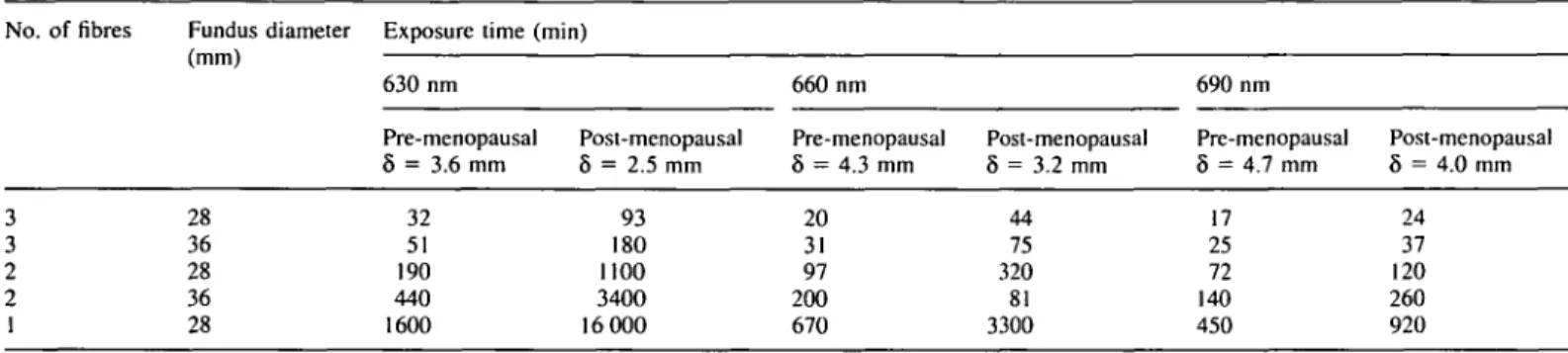
Table I. Calculated exposure times required to apply an optical dose of 70 J/cm2 at a depth of 4 mm at the most remote point from the source fibre for differently sized uterine cavities
No. of fibres Fundus diameter Exposure time (min) (mm) 630 nm 660 nm 690 nm 3 3 2 2 1 28 36 28 36 28 Pre-menopausal 8 = 3.6 mm 32 51 190 440 1600 Post-menopausal 8 = 2.5 mm 93 180 1100 3400 16 000 Pre-menopausal 8 = 4.3 mm 20 31 97 200 670 Post-menopausal 8 = 3.2 mm 44 75 320 81 3300 Pre-menopausal 8 = 4.7 mm 17 25 72 140 450 Post-menopausal 8 = 4.0 mm 24 37 120 260 920
Optical power = 100 mW per cm length of 0.8 mm diameter diffusing fibre. Times are given for pre-menopausal and post-menopausal samples using 630, 660 and 690 nm light. Penetration depths (8) vary with wavelength and physiological status, as shown. Reduced scattering coefficients are assumed to be 0.8 mm"1 for all cases. Exposure time calculations are based on a uterine light propagation model (Tromberg et al., 1995) and measured penetration depths.
Table II. Calculated exposure times required to apply an J/cm2 at a depth of 4 mm at the most remote point from differently sized uterine cavities
Diameter at fundus (mm)
28 36
Exposure time (min) Three fibres, no distension
11.3 12.5
optical dose of 70 the source fibre for
Three fibres, 2 ml distension 2.5 2.8
Exposure time calculations are based on absolute fluence rate measurements in collapsed and distended uteri in the region of interest. Optical power = 100 mW per cm length of 1.2 mm diameter diffusing fibre for 660 nm light.
a more uniform light distribution. Light of a longer wavelength will penetrate more deeply into tissue. At a given depth, a higher optical dose will therefore be delivered for equal light exposure times.
Here, we measured the penetration depths at 630, 660 and 690 nm because these are the excitation wavelengths used for the photosensitizers 5-aminolevulinic acid-induced proto-porphyrin IX (Goff et al, 1992), tin-etiopurpurin (Morgan and Tertel, 1986) and benzoporphyrin monoacid ring A (Richter et al., 1990). Our results show that by increasing the wavelength from 630 to 690 nm, the penetration depth increases signific-antly in both pre- and post-menopausal uteri. As shown in Figure 2, this corresponds to an average increase in fluence rate at 4 mm of 58% for pre-menopausal and 71 % for post-menopausal uteri. Provided that photobiological effects are comparable, exposure times can be reduced proportionally by selecting a photosensitizer that is activated by the 690 nm irradiation wavelength. The fact that optical penetration depths in post-menopausal uteri are lower at all three wavelengths can be explained by either a higher light absorption or an increased scattering of post-menopausal uterine tissue. Indeed, in post-menopausal uteri, both higher absorption and scattering coefficients have been measured and attributed to post-menopausal changes in water content, cell density and haemo-globin absorption (Madsen et al., 1994).
To improve the uniformity of light distribution, the distension of the organ by fluid instillation has been used in intracavitary PDT of the bladder (Marynissen et al, 1989). Because acute
distension of the uterine cavity induces contractions and pain, the insertion of multiple, thin, cylindrical diffusing fibres to improve light uniformity seems to be a viable alternative. As with the insertion of an intrauterine contraceptive device, this could be carried without excessive discomfort or pain and therefore without anaesthesia. Because the uterine cavity is normally collapsed, an intrauterine light-diffusing fibre will be surrounded by endometrial tissue and the light intensity will rapidly decrease with increasing distance in all directions. According to theory, the fluence rate will drop to -2% of the initial value at a distance of four times the tissue-specific penetration depth. This relationship was confirmed during measurement at the corners of the uterine cavity (one source fibre) when the fluence rate fell to <2% of initial values at a source-detector separation of just over 14 mm (about four times the penetration depth).
With multiple fibres inserted, the most critical region for obtaining an adequate fluence rate is where the distance from the source fibres is maximal, i.e. between the fibre tips in the fundus. Calculated light exposure times for the photodynamic destruction of the entire endometrium are shown for various wavelengths and uterine optical properties in Table I. Details of the uterine light dosimetry model are presented elsewhere (Tromberg et al., 1995). These results are based on the assumption that an optical dose of 70 J/cm2 is required at a depth of 4 mm to destroy the endometrial gland stumps at the endometrial-myometrial interface. Because measurements of protoporphyrin IX photodegradation indicate that the proto-porphyrin IX bleaching fluence is in the range 30-50 J/cm2 (Hoeydalsvik, 1994), an optical dose far above this value will only lead to the destruction of the photosensitizer without an additional photodynamic effect.
The uniformity of light distribution can be improved mark-edly by separating the uterine walls. Under these conditions, the uterine cavity acts as an 'integrating sphere' by reflecting light at the endometrial surface (Tromberg et al., 1995; Wyss et al, 1995). This can be achieved by filling the uterine cavity with fluid. However, limitations include spillage through the Fallopian tubes and pain/contractions which may occur with large-volume injections. In practical terms, disadvantages to fluid instillation can be minimized by using a small volume
of viscous material, because even moderate distension of the cavity can significantly reduce the required exposure time. These concepts are illustrated in Figure 4, which shows that the instillation of only 2 ml of a viscous, optically lossless liquid such as Hyskon can increase the fluence rate (at 6 mm) by a factor of - 5 . Thus, from Table I, the required exposure time for 630 nm radiation with three fibres in a distended uterus could be reduced to 10 and 30 min for pre- and post-menopausal patients respectively.
Despite this enhancement, the exposure time required using a single source fibre is clearly unacceptable, even for small, distended uterine cavities. Because choosing a longer wave-length (690 nm) can only reduce the single-fibre exposure time to, at best, 7.5 h, multiple source fibres are clearly required for complete tissue destruction. However, even two fibres are inadequate in a collapsed uterus, and distension may be necessary for delivering the required light dose in a reasonable time period, provided that optical penetration depths are relatively high (e.g. - 4 mm).
The delivery of an in-situ optical dose of 70 J/cm2 during 20 min of exposure at 100 mW/cm thus requires that the maximum distance from the nearest fibre is in the range of twice the optical penetration depth. An order-of-magnitude estimate of the required exposure time in the case of a distance between fibres equal to n times the penetration depth is: time (min) = 20Xe(" ~ 2). For example, when n is equal to twice the optical penetration depth, only 20 min are required; however, doubling the distance between fibres so they are separated by four penetration depths (n = 4) demands nearly an 8-fold increase in exposure time (-150 min) to maintain an equivalent optical dose.
Actual treatment times needed for irreversible endometrial destruction may be different from those shown in Table I. These estimates were determined by measuring ex-vivo penetration depths, assuming a fixed scattering parameter, and calculating the uterine light distribution using a dosimetry model for three cylindrical fibres (Tromberg et ai, 1995). Because the model assumes homogeneous optical properties, several 'real-world' variations could contribute to errors in our calculations. These may include non-uniform tissue structure and, despite our attempts to retain blood in the specimen, differences in optical properties between intact and surgically removed uteri. Consequently, model calculations should be confirmed with in-vivo measurements. To test our Table I predictions, we recorded absolute fluence rates at the most remote locations from three cylindrical fibres inserted into 24 and 36 mm diameter uterine cavities (at depths of ~4 mm; however, since the detector fibre was buried in tissue, its exact location could only be estimated). Each cylinder received 100 mW per cm of 660 nm light. Table II shows the exposure times required to apply an optical dose of 70 J/cm2 based on: exposure time = optical dose (70 J/cm2)/measured fluence rate (mW/cm2). Uteri were probed in both collapsed and distended (2 ml) states.
Without distension, measurements indicate that the desired light dose obtained from fluence rate measurements agrees with model predictions within a factor of -2.0. Given that the detector fibre position and uterine tissue optical properties (Penetration depth 8 is assumed to be 4.3 mm at 660 nm)
were not determined accurately, we believe that this is an encouraging result. The addition of 2 ml of Hyskon to distend the uterus increased the measured fluence rate by a factor of -4.5, resulting in concomitant reductions in required exposure times. Overall, these measurements provide further confirma-tion of the accuracy of our predictive light dosimetry model and suggest that Table I results may be conservative 'worst case' estimates. Deviations between measured (Table II) and predicted (Table I) data are probably a result of experimental uncertainties as well as tissue structural inhomogeneities.
In conclusion, our results suggest that a sufficient optical dose for photodynamic destruction of the entire endometrium without a risk of thermal effects by high optical powers can reasonably be achieved using multiple diffusing fibres. In normal-sized pre-menopausal uterine cavities (28 mm fundus), a trifurcated intrauterine light delivery device will deliver an optical dose of 70 J/cm2 (630 nm) at a depth of 4 mm, even to the most remote areas, in -30 min (Table I). However, when the uterine cavity is distorted and elongated by fibroids, slight distension of the cavity may be required to provide a sufficient optical dose throughout the endometrium. These results have encouraged us and others, and suggest that endometrial PDT may ultimately provide several benefits compared with conventional surgical techniques. Among these advantages is the possibility that endometrial PDT will not require anaesthesia and operating room facilities. Thus, although it is difficult to predict cost, this improvement alone would make endometrial PDT a more economical alternative to existing surgical procedures. In addition, with the increased availability of inexpensive optical components, such as com-pact, efficient diode lasers, flexible light diffusers and novel photosensitizers, we expect that a variety of technological developments will have a positive impact on its availability.
Acknowledgements
We thank M.Shell, PhD, for help in statistical analyses. M.K.F. wishes to acknowledge fellowship assistance from the Swiss National Science Foundation. This work was supported by the National Institutes of Health (NIH grant nos. R29GM50958 and R01CA32248) and the Whitaker Foundation (WF16493). In addition, Beckman Laser Insti-tute programmatic support was provided by the National InstiInsti-tutes of Health (NIH grant no. 5P41RR01192-15), the Department of Energy (DOE grant no. DE-FG03-91ER61227) and the Office of Naval Research (ONR grant no. N00014-91-C-0134).
References
Bhatta, N., Anderson, R.R., Flotte, T., Schiff, I., Hasan, T. and Nishioka, N.S. (1992) Endometrial ablation by means of photodynamic therapy with photofrin II. Am. J. Obslel. Gynecol., 167, 1856-1863.
Brooks, P.G., Serden, S.P. and Davos, I. (1991) Hormonal inhibition of the endometrium for resectoscopic endometrial ablation. Am. J. Obstel.
Gynecol., 164, 1601-1606.
Daniell, J.F., Kurtz, B.R. and Ke, R.W. (1992) Hysteroscopic endometrial ablation using the rollerball electrode. Obstel. Gynecol., 80, 329-332. De Cherney, A. and Polan, M.L. (1983) Hysteroscopic resection of intrauterine
lesions and intractable uterine bleeding. Obstet. Gynecol., 61, 392-397. Ferenczy, A. (1976) Studies on the cytodynamics of human endometrial
regeneration. I. Scanning electron microscopy. Am. J. Obstet. Gynecol., 124, 64-74.
Goff, B.A., Bachor, R., Kollias, N. and Hasan, T. (1992) Effects of photodynamic therapy with topical application of 5-aminolevulinic acid on 3071
normal skin of hairless guinea pigs. J. Pholochem. Pholobiol. B: BioL, 15, 239-251.
Goldrath, M.H., Fuller, T.A. and Segal, S. (1981) Laser photovaporization of endometrium for treatment of menorrhagia. Am. J. Obstet. Gynecoi, 140,
14-19.
Hoeydalsvik, E. (1994) Characterization of the distribution of porphyrins in malignant tumors by fluorescence. MSc Thesis, Division of Physical Electronics, Norwegian Institute of Technology, Trondheim, Norway. Jedeikin, R., Olsfanger, D. and Kessler, I. (1990) Disseminated intravascular
coagulopathy and adult respiratory distress syndrome: life-threatening complications of hysteroscopy. Am. J. Obstet. Gynecoi, 162, 44-45. Madsen, S.J., Wyss, P., Svaasand, L.O., Haskell, R.C., Tadir, Y. and Tromberg,
B.J. (1994) Determination of the optical properties of the human uterus using frequency-domain photon migration and steady-state techniques. Phys.
Med. Biol., 39, 1191-1202.
Manyak, M.J., Nelson, L.M. and Solomon, D. (1989) Photodynamic therapy of rabbit endometrium transplants: a model for treatment of endometriosis.
Fertil. Steril., 52, 140-145.
Marynissen, J.P., Jansen, H. and Star, W.M. (1989) Treatment system for whole bladder wall photodynamic therapy with in vivo monitoring and control of light dose rate and dose. J. Vrol, 142, 1351-1355.
Morgan, A.R. and Tertel, N.C. (1986) Observations on the synthesis and spectroscopic characteristics of purpurins. J. Org. Chem., 51, 1347. Perry, P.M. and Baughman, V.L. (1990) A complication of hysteroscopy: air
embolism. Anesthesiology, 73, 546-547.
Richter, A.M., Waterfield, E., Jain, A.K., Ethan, D., Sternberg, E.D., Dolphin, D. and Levy, J.G. (1990) In vitro evaluation of phototoxic properties of four structurally related Benzoporphyrin derivatives.
Photochem. Photobiol., 52, 495-500.
Santolaya-Forgas, J. (1992) Physiology of the menstrual cycle by ultrasonography. J. Ultrasound Med., 11, 139-142.
Schneider, D.F., Schellhas, H.F., Wesseler, T.A. and Moulton, B.C. (1988) Endometrial ablation by DHE photoradiation therapy in estrogen-treated ovariectomized rat. Colposc. Gynecoi. Laser Surg., 4, 73-77.
Serden, S.P. and Brooks, P.G. (1991) Treatment of abnormal uterine bleeding with the gynecologic resectoscope. J. Reprod. Med., 36, 697-699. Steiner, R.A., Tromberg, B.J., Wyss, P., Krasieva, T, Chandanani, N.,
McCullough, J., Berns, M.W. and Tadir, Y. (1995) Rat reproductive performance following photodynamic therapy with topically administered Photofrin. Hum. Reprod., 10, 227-233.
Svaasand, L.O. (1984) Optical dosimetry for direct and interstitial photoradiation therapy of malignant tumors. In Doiron, D. and Gomer, C.J. (eds), Porphyrin Localization and Treatment of Tumors. Alan R.Liss, New York, NY, USA, pp. 91-114.
Tromberg, B.J., Svaasand, L.O., Fehr, M.K., Madsen, S.J., Wyss, P., Sansone, B. and Tadir, Y. (1995) A mathematical model for light dosimetry in photodynamic destruction of human endometrium. Phys. Med. Biol, in press. Wyss, P., Tadir, Y, Tromberg, B.J., Liaw, L., Krasieva, T. and Berns, M.W. (1994a) Benzoporphyrin derivative: a potent photosensitizer for photodynamic destruction of rabbit endometrium. Obstet. Gynecoi., 84, 409-414.
Wyss, P., Tromberg, B.J., Wyss, M.T., Krasieva, T, Schell, M., Berns, M.W. and Tadir, Y. (1994b) Photodynamic destruction of endometrial tissue with topical 5-aminolevulinic acid in rats and rabbits. Am. J. Obstet. Gynecoi., 171, 1176-1183.
Wyss, P., Svaasand, L.O., Tadir, Y, Haller, U., Berns, M.W., Wyss, M.T. and Tromberg, B.J. (1995) Photomedicine of the endometrium: experimental concepts. Hum. Reprod., 10, 221-226.
Yang, J.Z., Van Vugt, D.A., Kennedy, J.C. and Reid, R.L. (1993) Evidence of lasting functional destruction of the rat endometrium after 5-aminolevulinic acid-induced photodynamic ablation: prevention of implantation. Am. J. Obstet. Gynecoi., 168, 995-1001.