Surgical left atrial appendage occlusion: evaluation of a novel
device with magnetic resonance imaging
§Sacha P. Salzberg
a,1,*
, Alan Marc Gillinov
b, Anelechi Anyanwu
a, Javier Castillo
a,
Farzan Filsoufi
a, David H. Adams
aa
Department of Cardiothoracic Surgery, Mount Sinai Medical Center, New York, NY, United States
bDepartment of Thoracic and Cardiovascular Surgery, The Cleveland Clinic Foundation, Cleveland, OH, United States
Received 27 February 2008; received in revised form 13 May 2008; accepted 15 May 2008; Available online 6 August 2008
Abstract
Objective: Management of the left atrial appendage (LAA) is considered an important adjunct to ablation in cardiac surgical patients with atrial fibrillation (AF). However, current surgical techniques, both cut-and-sew and stapling, have been associated with incomplete LAA occlusion and complications. Using cardiac magnetic resonance imaging (MRI), we studied the safety and effectiveness of a new device for LAA occlusion in a primate model. Methods: Seven adult baboons underwent off-pump placement of an LAA clip (AtriCure Inc., Westchester, Ohio). LAA occlusion was confirmed intraoperatively by direct incision. All animals had MRI before and after clip placement to assess LAA perfusion, architecture, and overall cardiac function. Pathologic and histological studies were performed at 7, 30 and 180 days. Results: Clip placement was successful in all (n = 7) without any clip related complications. Complete LAA occlusion was demonstrated intraoperatively in all subjects. LAA occlusion was confirmed on pre-sacrifice MRI, and left and right ventricular function were unchanged from preoperative studies; however, clip placement caused small reductions in left ventricular end-diastolic, end-systolic, and stroke volumes. At sacrifice, direct inspection confirmed stable location, persistent LAA exclusion, tissue in-growth and homogenous epithelialization without damage to adjacent structures. Histological analysis revealed a regular in-growth pattern in all studied specimens. Conclusion: We demonstrated a safe, straightforward, persistent and effective method for LAA occlusion with this new LAA clip. MRI effectively demonstrated LAA occlusion and only minor changes in left ventricular volumes.
#2008 European Association for Cardio-Thoracic Surgery. Published by Elsevier B.V. All rights reserved.
Keywords: Left atrial appendage; Atrial fibrillation; Surgery; Device
1. Introduction
Patients with atrial fibrillation (AF) have a five-fold increased risk of stroke, and it is hypothesized that most embolic strokes in such patients arise from thrombi forming in the left atrial appendage (LAA)[1]. In patients undergoing cardiac surgery for AF, the current guidelines recommend some form of LAA amputation for patients undergoing mitral valve surgery[2]. In an attempt to provide minimally invasive solutions for LAA management, novel percutaneous approaches have been developed which come with the risks of LAA injury, incomplete occlusion, and device dislocation with peripheral embolization [3—5,8,11]. Because the LAA
can be accessed from the pericardial space, it is likely that a properly constructed device placed from the epicardial surface of the LAA will be both safe and effective. Currently, there is considerable interest in the development of new technologies for management of the LAA. This study used magnetic resonance imaging (MRI) to assess the impact of LAA exclusion with a new epicardial clip in a healthy primate model.
2. Materials and methods
This study utilized eight adult primate animals (Papio anubis), and was composed of two substudies. The first trial was a pulmonary vein isolation with a new bipolar ablation device (manuscript in preparation) and the second actual LAA clip deployment. Throughout the trial animal welfare was closely supervised by a trained veterinarian team. Prior to the start of the study, time points for study termination were defined at 7, 30 and 180 days. The animals were randomly assigned to the different time points. All the animals
www.elsevier.com/locate/ejcts
§
This research was funded by an unrestricted grant by Atricure Inc., the research was performed independently and outcomes were not influenced by the origin of funding. Cleveland Clinic has an indirect equity interest in Atricure by an investment in a fund that has an investment in Atricure.
* Corresponding author. Address: Clinic for Cardiovascular Surgery, Univer-sity Hospital, Ra¨mistrasse 100, CH-8091 Zu¨rich, Switzerland.
Tel.: +41 44255 11 11; fax: +41 44255 50 47.
E-mail address:[email protected](S.P. Salzberg).
1Dr S. Salzberg is a consultant for AtriCure Inc.
1010-7940/$ — see front matter # 2008 European Association for Cardio-Thoracic Surgery. Published by Elsevier B.V. All rights reserved. doi:10.1016/j.ejcts.2008.05.058
underwent MRI both before clip placement and before sacrifice (Fig. 4a). The procedures were all performed aseptically in an American Association for Laboratory Animal Science (AALAS) approved facility. The protocol was approved by the Institutional Animal Care and Use Commit-tee (IACUC) at Mount Sinai School of Medicine, and all experimental procedures were consistent with the Guide for the Care and Use of Laboratory Animals.
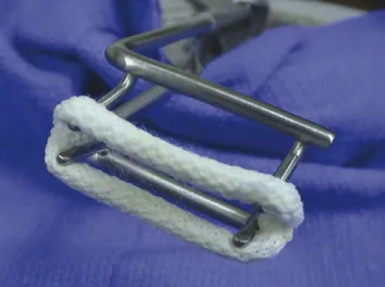
This second generation LAA clip was developed by AtriCure Inc. (West Chester, Ohio, USA). The first generation clip[6]had a curved design with the identical components and performance as the current straight version; however the titanium parallel elements had a 2 in radius of curvature. The curvature was originally intended to aid in accessing the anatomy of the LAA. However, it was determined in animal testing that the curvature of the parallel titanium (tubes) elements did not aid in accessibility and was therefore unnecessary. The current design is virtually identical to the initial device, with the exception of having straight parallel members instead of slightly curved parallel members (Fig. 1). All materials in the device are the same for both versions of the clip and are composed of a deflectable nitinol frame covered with a proprietary polyester mesh. The internal design of the clip consists of two polycarbonate elastomers, two nitinol springs, and two titanium elements. The nitinol springs provide the clamping force needed to approximate the LAA tissue thus providing immediate occlusion. The polyester mesh assists in the pressure distribution as well as serving as a matrix for epithelial tissue growth. The clip comes in different sizes, based on the length of the LAA base (from 25 mm to 45 mm in 10 mm steps). In this study a 25 mm clip was used in all animals. The applier is configured such that the clip can be removed and redeployed if initial deployment is judged to be unsatisfactory.
MRI was performed in a 1.5-T clinical magnet (Magneton Sonata#
, Siemens Medical Solutions, Erlangen, Germany) with a dedicated cardiac coil. Maximum and minimum end-systolic, end-diastolic and stroke volumes were quantified as previously described [7]. In addition, the long-axis views were employed to determine flow in the left atrial appendage.
3. Surgical implant
After endotracheal anesthesia, median sternotomy and pericardial incision were performed. In addition to LAA exclusion with the LAA clip, a complete off-pump epicardial modified Cox Maze IV lesion set was performed with a bipolar pen and clamp. While manually stabilizing the heart, the clip was lowered onto the LAA and deployed at its base. Care was taken to orient the clip parallel to the base of the appendage and not to impinge upon the circumflex coronary or pulmonary artery. Confirmation of circulatory exclusion and non-perfusion of the LAA was assessed by incising the LAA distal to the clip. The defect was then oversewn with a 7/0 Prolene suture.
On the day of the terminal study an MRI was performed to assess cardiac function, patency of the pulmonary veins, and circulatory exclusion of the LAA. After the MRI, the chest was re-entered through the original incision to expose the heart with the clip, after which full heparinization proceeded humane euthanasia and en-bloc cardiac resection.
4. Results
4.1. Surgical results
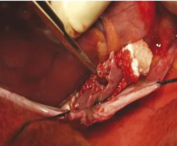
Clip placement was swift and straightforward. The clip was successfully deployed in a satisfactory manner on the first attempt (Fig. 2). No conflict with any surrounding structures (coronary artery, pulmonary artery, or left atrial body) was noted. Postdeployment, direct intraoperative LAA incision demonstrated total circulatory isolation from the left atrium, with absence of blood within the LAA (Fig. 3). Additionally, we performed a complete left atrial epicardial ablation using a bipolar ablation/pacing pen (AtriCure Inc., Westchester, OH). All of the animals survived the procedure and there were no intra- or postoperative complications due to clip placement or ablation.
Fig. 1. Clip on standard open chest deployment tool.
Fig. 2. Intraoperative view after clip (25 mm) deployment on 17 kg female baboon.
One death occurred on postoperative day 1 due to acute respiratory failure in an older female subject. The other animals survived in excellent health until their randomly pre-assigned time points of 7 days (n = 2), 30 days (n = 2) and 180 days (n = 2). No animal appeared sick or required medical attention during the entire study period. At sacrifice, all animals were in documented sinus rhythm.
Preoperative MRI demonstrated normal cardiac architec-ture and function in all animals (mean EF 54%) (Table 1). The LAA was readily identified on MRI in all animals. There was an interspecies variation of ejection fraction and cardiac volumes, as these were wild animals of different age, gender and physical constitution. On pre-sacrifice MRI, left ven-tricular function was seen to be preserved (mean EF 52%). The LAA was present at 7 days, totally retracted at 30 days and macroscopically unidentifiable with only a slight shadow on the MRI pointing to the presence of the clip at 6 months (n = 2) (Fig. 4b). MRI also demonstrated normal adjacent tissues without erosion or spatial displacement. Quantitative calculation from cardiac MRI images revealed a discrete decrease in stroke volumes, end-diastolic volumes and systolic volumes when compared to preoperative values (Table 1). The clip components did not cause any artifact to the MRI imaging. The MRI provided impeccable sliced for quantitative analysis and functional imagery for real time reproduction of cardiac function.
4.2. Pathology
At gross epicardial inspection, the LAA clip was easily identified in all animals. At 7 days the LAA was present but empty and there were no pericardial adhesions. At 30 days the LAA was still identifiable, but there were adhesions at the epicardial aspect. By 180 days, the LAA was no longer present
Fig. 3. Intraoperative view after incision of LAA. No LAA perfusion confirmed on the beating heart.
Table 1
Quantitative MRI data
Animals EF1 EF2 Diff ED1 ED2 Diff ES1 ES2 Diff SV1 SV2 Diff
7 d_1 53 42 11 38 30 8 18 18 0 20 13 7 7 d_2 46 45 1 22 19 3 12 11 1 10 9 1 30 d_1 62 60 2 27 16 11 11 6 5 10 8 2 30 d_2 53 50 3 24 21 3 11 9 2 13 13 0 180 d_1 56 56 1 33 20 13 14 9 5 19 11 8 180 d_2 57 56 1 21 21 0 9 9 0 12 12 0
Pre, before clip; post, after clip; EF, ejection fraction; ED, end-diastolic volume; ES, end-systolic volume; SV, stroke volume.
Fig. 4. (a) MRI depicting LAA before clip placement (white circle). (b) MRI 180 days after clip placement showing shadow of clip and no LAA (white circle).
and the clip was completely covered by tissue in-growth. There was no erosion into adjacent structures (e.g. the circumflex coronary artery, pulmonary artery, or left atrium). Examination of the endocardial surface of the left atrium revealed no thrombus in any animal. At 6 months, the LAA opening appeared to be covered by a smooth endocardial tissue layer (Fig. 5).
4.3. Histology
On microscopic examination, a regular growth pattern was seen on the polyester grain covering the clip. At 7 days, the cross sections through the clip showed healing mural necrosis. After 30 and 180 days main findings included a fibrous replacement scar between the two clip arms in addition to a smooth layer of endothelial cells on the atrial side.
5. Discussion
In this first primate trial, we have demonstrated safe, straightforward, dependable and effective LAA occlusion with a novel epicardial LAA clip. Moreover, with the use of cardiac MRI we have clearly shown that LAA occlusion is durable and that LAA exclusion with the LAA clip device does not substantially impact cardiac function.
During cardiac surgery, the most common technique for LAA occlusion is to perform suture ligation from within or epicardially, concomitant to a maze or left atrial ablative procedure. While LAA management is a common component of surgical ablation as an adjunct to heart valve surgery, there is limited data documenting safety and clinical impact of stand-alone LAA ligation or excision. In a recent series[9]
with 15 patients and 3 year follow-up, thoracoscopic stand-alone LAA ligation was shown to be feasible and potentially beneficial; however, in one patient intraoperative LAA injury occurred, requiring emergency thoraocotomy. This report highlights the interest but also emphasizes the challenge and difficulty in surgically addressing the LAA[9].
There has been recent interest in assessment of surgical techniques for LAA management during open-heart surgery. In a recent report from the left Atrial Appendage Occlusion Study (LAAOS) where patients without AF were randomized to LAA occlusion during routine coronary artery bypass grafting[10], the authors found that LAA occlusion is safe. However, they found that suture ligation was frequently incomplete, allowing continued blood flow into the LAA. In addition, stapled excision often left a large cul-de-sac, amounting to an incomplete left atrial appendage excision
[10]. Thus, advances in technique and technology are necessary.
The new LAA clip presented herein, addresses another limitation of LAA occlusion, which is the wide spectrum of LAA dimensions present in patients with AF. The hetero-geneity of LAA dimensions in patients with AF has recently been documented in an MRI series[12]. It is well established that complete and effective LAA occlusion are paramount to ensure optimal clinical outcomes[13]. The availability of the clip in several different sizes, accommodating the variety of LAA bases, will most certainly be beneficial in this patient population and help ensure complete LAA occlusion.
6. Summary
In this series, we have shown the LAA clip to enable effective and durable epicardial LAA occlusion in healthy primates. It is straightforward to implant and there were no device related complications. MRI demonstrates only minor impact on cardiac volumes without important changes in cardiac function. These animals were in sinus rhythm and had normal cardiac function; therefore, it is possible that clinical results in humans with AF and structural heart disease will differ from the findings in this study. However, human clinical trials in patients with AF undergoing open-heart surgery are currently underway. In addition, a thoracoscopic platform for clip application and AF ablation is under development.
References
[1] Wolf PA, Abbott RD, Kannel WB. Atrial fibrillation as an independent risk factor for stroke: the Framingham Study. Stroke 1991;22(Aug (8)):983—8. [2] Fuster V, Ryden LE, Cannom DS, Crijns HJ, Curtis AB, Ellenbogen KA, Halperin JL, Le Heuzey JY, Kay GN, Lowe JE, Olsson SB, Prystowsky EN, Tamargo JL, Wann S, Smith Jr SC, Jacobs AK, Adams CD, Anderson JL, Antman EM, Halperin JL, Hunt SA, Nishimura R, Ornato JP, Page RL, Riegel B, Priori SG, Blanc JJ, Budaj A, Camm AJ, Dean V, Deckers JW, Despres C, Dickstein K, Lekakis J, McGregor K, Metra M, Morais J, Osterspey A, Tamargo JL, Zamorano JL. ACC/AHA/ESC 2006 guidelines for the manage-ment of patients with atrial fibrillation: full text: a report of the American College of Cardiology/American Heart Association Task Force on practice guidelines and the European Society of Cardiology Committee for Practice Guidelines (Writing committee to revise the 2001 guidelines for the management of patients with atrial fibrillation) developed in collabora-tion with the European Heart Rhythm Associacollabora-tion and the Heart Rhythm Society. Europace 2006;8(Sep (9)):651—745.
[3] Ostermayer SH, Reisman M, Kramer PH, Matthews RV, Gray WA, Block PC, Omran H, Bartorelli AL, Della Bella P, Di Mario C, Pappone C, Casale PN, Moses JW, Poppas A, Williams DO, Meier B, Skanes A, Teirstein PS, Lesh MD, Nakai T, Bayard Y, Billinger K, Trepels T, Krumsdorf U, Sievert H. Percutaneous left atrial appendage transcatheter occlusion (PLAATO system) to prevent stroke in high-risk patients with non-rheumatic atrial Fig. 5. Intraoperative photograph showing atrial surface of LAA occluded by
the LAA clip. A smooth surface of endothelium covers the remaining few millimeter long ‘cul-de-sac’.
fibrillation: results from the international multi-center feasibility trials. J Am Coll Cardiol 2005;46(Jul (1)):9—14.
[4] Meier B, Palacios I, Windecker S, Rotter M, Cao QL, Keane D, Ruiz CE, Hijazi ZM. Transcatheter left atrial appendage occlusion with Amplatzer devices to obviate anticoagulation in patients with atrial fibrillation. Catheter Cardiovasc Interv 2003;60(Nov (3)):417—22.
[5] Fountain RB, Holmes DR, Chandrasekaran K, Packer D, Asirvatham S, Van Tassel R, Turi Z. The PROTECT AF (WATCHMAN Left Atrial Appendage System for Embolic PROTECTion in Patients with Atrial Fibrillation) trial. Am Heart J 2006;151(May (5)):956—61.
[6] Kamohara K, Fukamachi K, Ootaki Y, Akiyama M, Cingoz F, Ootaki C, Vince DG, Popovic ZB, Kopcak Jr MW, Dessoffy R, Liu J, Gillinov AM. Evaluation of a novel device for left atrial appendage exclusion: the second-gen-eration atrial exclusion device. J Thorac Cardiovasc Surg 2006;132(Aug (2)):340—6.
[7] Tseng WY, Liao TY, Wang JL. Normal systolic and diastolic functions of the left ventricle and left atrium by cine magnetic resonance imaging. J Cardiovasc Magn Reson 2002;4(4):443—57.
[8] Onalan O, Lashevsky I, Hamad A, Crystal E. Nonpharmacologic stroke prevention in atrial fibrillation. Expert Rev Cardiovasc Ther 2005;3(4): 619—33.
[9] Blackshear JL, Johnson WD, Odell JA, Baker VS, Howard M, Pearce L, Stone C, Packer DL, Schaff HV. Thoracoscopic extracardiac obliteration of the left atrial appendage for stroke risk reduction in atrial fibrillation. J Am Coll Cardiol 2003;42(7):1249.
[10] Healey JS, Crystal E, Lamy A, Teoh K, Semelhago L, Hohnloser SH, Cybulsky I, Abouzahr L, Sawchuck C, Carroll S, Morillo C, Kleine P, Chu V, Lonn E, Connolly SJ. Left Atrial Appendage Occlusion Study (LAAOS): results of a randomized controlled pilot study of left atrial appendage occlusion during coronary bypass surgery in patients at risk for stroke. Am Heart J 2005;150(2):288.
[11] Bayard YL, Ostermayer SH, Sievert H. Transcatheter occlusion of the left atrial appendage for stroke prevention. Expert Rev Cardiovasc Ther 2005;3(6):1003—8.
[12] Heist EK, Refaat M, Danik SB, Holmvang G, Ruskin JN, Mansour M. Analysis of the left atrial appendage by magnetic resonance angiography in patients with atrial fibrillation. Heart Rhythm 2006;3(11):1313—8. [13] Almahameed ST, Khan M, Zuzek RW, Juratli N, Belden WA, Asher CR,
Novaro GM, Martin DO, Natale A. Left atrial appendage exclusion and the risk of thromboembolic events following mitral valve surgery. J Cardio-vasc Electrophysiol 2007.