International Immunology, Vol. 6, No. 6, pp. 925-930
Exposure of monocytes to heat shock does
not increase class II expression but
modulates antigen-dependent T cell
responses
Ewa Mariethoz, Fabienne Tacchini-Cottler
1, Muriel Jacquier-Sarlln,
Fiona Sinclair
2and Barbara S. Polia
Allergy Unit, University Hospital and 'Department of Pathology, CMU, University of Geneva, 1211 Geneva 14, Switzerland
2Present address: University Department of Surgery, Western Infirmary, 44 Church Street, Glasgow G11 6NT, UK
Key words: antigen processing, chaperones, diphtheria toxin, heat shock proteins, hsp70, MHC class II
mdlecules, MLR, superantigens
Abstract
Expression of heat shock (HS) proteins (HSP) Increases after exposure to elevated temperatures or other types of Injury, such as oxldative Injury. Because of their function as 'molecular chaperones', HSP are suggested to participate In antigen processing and presentation. We have previously reported that HS modulates antigen presentation In a human EBV-transformed B cell line. Here we investigated the effects of HS on MHC class II expression and on antigen
processing and presentation by human monocytes. Monocytes were Isolated from peripheral blood of normal human volunteers, purified by adherence, then exposed to temperatures ranging from 37 to 45°C for 20 mln, allowed to recover for 2 h at 37°C and used for Immunofluorescence or as antigen presenting cells In autologous and heterologous lymphocyte proliferation assays. No Increase In class II expression was detected as assessed by flow cytometry. Monocytes (3 x 10⁴) and lymphocytes (1 x 10s) were co-cultured for 5 days In the presence of several antigens [diphtheria toxold, tetanus toxold or purified peptlde derivative (PPD)] and labeled with 1 /xCI [3H]thymldlne for 16 h. Pre-exposure to HS (44°C) significantly (P < 0.001) Increased T cell responses to diphtheria toxold, whereas the effect on the responses to other antigens (tetanus toxold or PPD) were not significant. HS did not increase heterologous T cell responses nor T cell proliferation Induced by the non-processed superantigens such as staphylococcal enterotoxln B. The effect of HS was Inhibited by actlnomycln B and thus appeared dependent upon HSP synthesis. HSP-mediated Increases in antigen processing may potentiate the ongoing Immune response at Inflammatory sites.
The heat shock (HS) response is an ubiquitous response to cellular stress characterized by the increased expression of HS or stress proteins (HSP). HSP are usually classified into families according to their apparent molecular weight. The hsp70 family contains several members of mol. wt ranging from 68 to 74 kDa, expressed constitutively (cognates) and/or increased or induced after exposure to HS or a number of other types of injury, including oxidative injury (1,2). Hsp70 molecules function as 'molecular chaperones' (3) and participate in the proper folding, assembly and sorting of nascent peptides, as well as in
transmembrane transport. Members of the hsp70 family may play a role in antigen processing and presentation (4 - 6). Indeed, the hsp70 family contains the clathrin uncoating ATPase (7,8); other members of the hsp70 family participate in targeting cytosolic proteins to the lysosomal compartment [prp73, (9)] or in the intracellular transport of selected antigens [peptide binding protein (PBP72-74; 5,6)]. Furthermore, endoplasmic resident stress proteins can associate with HLA-DR molecules in invariant chain-negative BALB/c 3T3 cells transfected with the HLA-DR gene (10).
Correspondence to B. S Polla
We previously found that HS increases class II expression and presentation of peptides in an HLA-DR1 human B cell line (11). The increase in class II expression was associated with an increased ability to stimulate autologous and alloreactive T cell
clones and to present peptide to an influenza-specific T cell clone. HS also increased the ability of this B cell line to present whole influenza. Other studies reported no significant increase in class II expression following HS in monocytes (12) or in the
if.
B
*'• *••J W 1BJ ^ ^ ^ J * ^
-Actin-37*C 39C 41 C 43C 44'C 45C
37C 44C 37C
+AD
44C
+AD
70 kD
37°C
A D : - +
39°C
+
41 °C
43°C
+
44°C
+
45°C
+
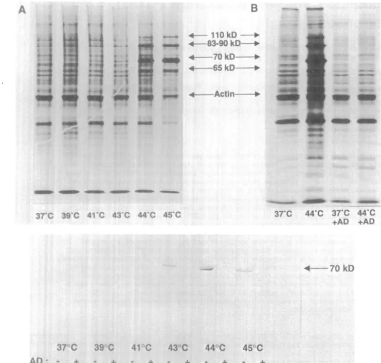
Fig. 1 . (A) Human monocytes were exposed to the indicated temperatures for 20 min, allowed to recover for 2 h, labeled with [^SJmethionine (9 /tCi/ml) for 90 min, washed and rysed in SDS buffer. Aliquots corresponding to equal cell numbers were etectrophoresed on slab gels with 10% polyacrytamkJe. Constitutive hsp70 was detectable at 37°C, whereas the inducible HSP, 65, 70, 90 kDa, were observed after exposure to 44 and
AS°C. The decrease in normal protein synthesis only occurred after exposure of monocytes to 45°C. (B) Actinomycin D (5 ^g/ml) was added 10
min before HS to parallel cultures and removed at the end of HS. The cells were then processed as described in (A). The 65, 70 and 90 kDa HSP induced after exposure to 44°C were not detectable anymore when actinomycin D had been added, whereas total protein synthesis was unaltered. (C) Actinomycin D (5 /ig/ml) was added 10 min before HS to parallel cultures and removed right at the end of HS. The cells were then processed as described in (A) and Western blot performed. Equal amounts of proteins for each sample were electrophoresed and transferred to nitrocellulose paper (26). The membrane was saturated with casein-containing buffer for 2 h and hybridized with a mouse monoclonal anti-human hsp70 antibody (Stress Gen). Bound hsp70 antibody was revealed with anti-mouse IgG peroxidase conjugated (Sigma) in the presence of H2O2 and
Heat shock modulation of APC function 927
Epstein-Barr virus-transformed B cell line (EVB-LCL) (13). In the present study we analysed the effects of HS on class II expression and antigen presentation and processing in normal human peripheral blood monocytes.
Cells were prepared and processed in RPMI 1640 (Gibco, Paisley, UK) with 10% FCS (Kibutz Beth, Heamek, Israel), 1 % glutamine (Gibco) and 25 mM HEPES (Gibco) [HEPES medium (HM)] and cultured in RPM11640 with 20% pooled human male AB serum (Centre de Transfusion sanguine, Annemasse, France) and 1 % glutamine (Gibco) [(complete medium (CM)]. Peripheral blood mononuclear cells were isolated by gradient centrifugatjon and monocytes purified as previously described (14), recovered, washed with PBS (Gibco), re-suspended in HM and then sampled. Selected samples were exposed for 20 min to temperatures ranging from 37 to 45°C as described previously (14), whereas control samples were maintained at 37°C. After HS, the cells were washed twice with PBS, allowed to recover
for 2 h at 37°C, then counted again and re-suspended in CM or in CM without methionine at 1 x 10⁸ cells/ml. Neither HS (up to 44°C) nor actmomycin D had any effect on cell viability as tested by trypan blue exclusion and protein synthesis.
In the monocytes, maximal hsp70 synthesis without inhibition of normal protein synthesis was obtained after exposure to 44°C as shown by metabolic labeling with [^SJmethionine (sp. act. 1145 Ci/mmol, Amersham International, Amersham, UK) (Fig. 1 A). We thus elected this temperature for most subsequent studies. As previously reported in human neutrophils (15), the transcriptional inhibitor actinomycin D (Sigma Chemicals Company, St Louis, MO); final concentration 5 uglmty, added 10 min before and during the HS, selectively prevented HSP induction by HS without altering normal protein synthesis (Fig. 1B), as shown for hsp70 in Fig. 1C (Western blot performed with mouse monoclonal anti-human inducible hsp70 from Stress Gen, San Francisco, CA and anti-mouse IgG peroxidase
3
pro
Fig. 2. Immunofluorescence analysis of cell surface class II HLA-DR expression in monocytes exposed or not to HS. Some 2 x 10s cells/well were
plated onto a 96-well plate in 100 ^ PBS with 5% human serum and 0.01% azide. After HS, the plate was centrifuged and the cells stained with 5 ^g mouse anti-human DR antibody (CR3/43, L243, XD5.A11) for 30 min at 4°C. The cells were washed three times in PBS, and stained with 2 ng rabbit anti-mouse FITC conjugated antibody (DAKO) for 30 mm at 4°C The negative control was obtained by staining the cells with rabbit anti-mouse FITC conjugated antibody (DAKO) and the positive control was obtained by staining the cells with mouse ant human CD14 antbody
(DAKO). The cells were analysed on a Epics •-Profile II Counter (Coulter, Miami, FL) provided with an argon laser (488 nm), calibrated by computer, and optimized for sensitvity and resolution with fluorospheres (Immune-Check, PN 6603487, Epics Division). The data were analysed using Epics •-Profile Version 2.0 Software. Dead cells and lymphocytes were excluded on the basis of forward and sideward scatter. 10,000 events were analysed. PBMC not heated (plain line) and heated (bold line) were stained with different mAbs: (A) rabbit anti-mouse FITC conjugated antibody; (B) anti-CD14 antibody; (C) the mouse anti-human class I monoclonal antibody WR6/32 (M736 DAKO, Denmark); (D) the mouse anti-human DR a/3 monoclonal L243 (kindly provided by E O. Long, Rockville, MD); (E) mouse ant-human class II p chain monoclonal antibody XD5:A11 (kindly provided by E O. Long, Rockville, MD); and (F) ant-class II 0, CR3/43, a mouse ant-human class II (3 chain monoclonal (M775, DAKO). Cells were washed and antibody binding revealed with FITC rabbit ant-mouse antbody No significant difference was observed after HS.
conjugated Fab from Sigma). This inhibition of HSP synthesis was thereafter used to distinguish possible physical effects of elevated temperature on cell metabolism from effects relating to HSP themselves (15).
As previously reported by others in these cells (12), no increase in class II expression was detectable 2 h after HS using FACS analysis (Fig. 2) (for detailed methods see Fig. 2 legend). Following HS, class II molecules (but not control molecules such
Table 1 . Effects of HS on lymphocyte proliferative response
to mitogen (PHA), to antigen (DT, TT, PPD) or to staphylococcal antigen (SEB) n [3H]Thymidine incorporation P (c.p.m. x 10"3) ± SEM Control PHA DT TT PPD MLR SEB 40 40 28 18 12 15 5 37°C 2 ± 35 ± 14 ± 27 ± 46 ± 23 ± 11 ± 0.3 6.0 2.0 5.0 10.0 5.0 2.0 44°C 2 ± 32 ± 23 ± 29 ± 49 ± 22 ± 13 ± 0.3 4.0 4.0 6.0 13.0 5.0 3.0 NS NS 0.0009 NS NS NS NS
as CD14) clustered in patches on the cell membrane, in a manner reminiscent of the capping of antibody molecules, suggesting redistribution of class II molecules secondary to membrane altera-tions (16). The results previously obtained in B cells therefore could not be extended to human monocytes and thus in these latter cells a positive effect of HS may relate to increased pro-cessing rather than presentation. Along these lines, we tested the effects of HS on processing-independent T cell activation by super- or allo-antigens.
We performed mixed lymphocyte reaction (MLR) experiments with cells from 15 distinct donors. Non-adherent mono-nuclear cells were applied to nylon wool columns (Biotest Diagnostics, Dreieich, Germany) previously wetted with 3 ml HM for 1 h. Cells were slowly eluted in HM, washed and re-suspended in CM at a final cell concentration of 1 x IC^/ml. Lymphocytes were not exposed to HS. After preliminary studies determined optimal monocytes: lymphocyte ratios in control experiments, 3 x 1 0 * monocytes and 1 x 10⁵ heterologous lymphocytes were co-cultured in 96-well flat-bottom tissue culture plates (Costar, Cambridge, UK) and antigens or mitogens were added. Each condition was tested in quintuplicate. On day 5, [3H]thymidine (1 /iCi/well) (sp. act. 2 Ci/mmol; Amersham) was added for 16 h. Cells were harvested and lysed (Skatron automatic cells harvester, Skatron Instruments, Lier, Norway), and labeled DNA counted in scintillation vials using a Beckman Liquid Scintillation
37-C
Arc
o
L06 FLUORESCENCE
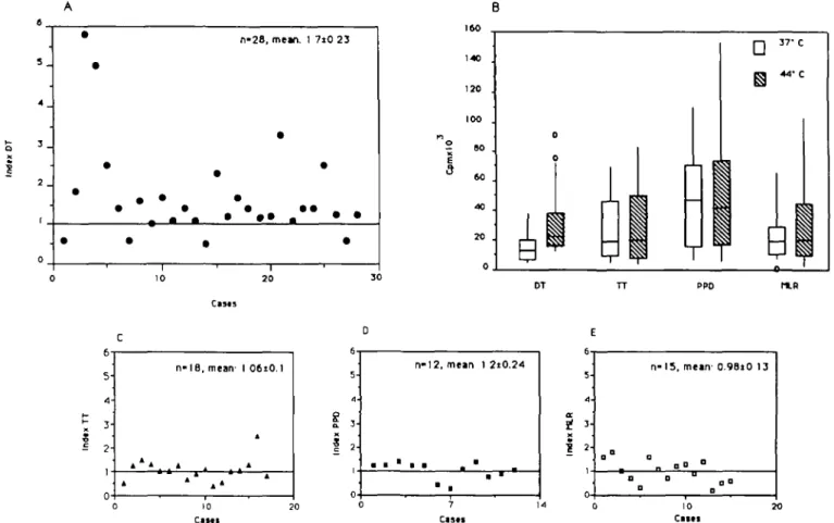
Fig. 3. Proliferation assays: 1 x 10⁵ T lymphocytes were co-cultured in the presence of 3 x 10⁴ autologous monocytes maintained at 37°C or
exposed to 44°C and [3H]thymidine incorporation determined at day 5. The cells co-cultured in the RPMI + 20% human serum + 1 % glutamine
and: (A) DT at 8900 U/ml; (C) TT at 525 U/ml; (D) PPD at 7.5 /ig/ml; or (E) MLR. On day 5, [3H]thymidine was added for 16 h, then the cells were
harvested. The results are expressed as the proliferative index:
c.p.m. antigen at 44°C - c.p.m. control at 44°C c.p.m. antigen at 37°C - c.p.m. control at 37°C
(A, C, D and E) Pre-exposure to HS (44°C) increased the stimulation index for DT (A) but had no significant effect on T cell responses to TT (C), or to PPD (D), or in MLR (E). (B) Pre-exposure to HS (44°C) significantly increased DT-dependent T cell proliferative responses but not TT, PPD or MLR dependent T cell proliferative responses (B). The results are expressed in c.p.m. x 103 and represented as a box plot: a rectangle parallel
to one axis, with its two ends at the upper and lower quartiles; the median or the middle value is the line cutting the box; lines extended from the ends show the smallest and the largest observation; most extreme values are shown as individual observations. Only the median DT 44°C is increased when compared with the control.
Heat shock modulation of APC function 929
Counter (Beckman LS 7500). The experiments in which the negative controls (monocytes heated or not with autologous lymphocytes, without antigen nor mitogen) were >50O0 c.p.m. were dismissed [3H]Thymidme incorporation into monocytes or lymphocytes alone was < 500 c.p.m In these and all subsequent experiments heated and control samples were compared by the Wilcoxon signed-rank test.
HS did not affect T cell responses in MLR (Table 1, Fig. 3E), except at the higher temperatures, at which there was a decline in T cell proliferation. Others have reported previously that exposure of human or murme antigen presenting cells (APC) to higher (45°C) or to lower temperatures (43°C) but for prolonged periods of time (3 h) abolishes presentation ability (12,13,17). We found similarly that when the allogeneic APC were heated to 45°C for 20 min lymphocyte proliferation was abolished or greatly reduced (data not shown). These findings could relate to (i) non-specific membrane alterations induced by HS; (ii) a decrease in expression and/or production of cytokines important for presentation such as membrane or secreted interleukin 1 (12); or (iii) an HS-induced inhibition of the synthesis of proteins involved in antigen processing, presentation or internalization via specific receptors (18).
Similarly, we compared the effect of HS on the autologous T cell response to superantigens such as staphylococcal entero-toxin B (SEB; final concentration 10~6 mg/ml, Sigma Chemicals Company), which do not appear to require processing although they need to associate with MHC class II molecules (19). Pre-exposure of APC to 44°C did not affect lymphocyte prolifera-tion in response to SEB (Table 1).
In contrast, pre-exposure of the monocytes to HS resulted in enhanced T cell responses to diphtheria toxoid (DT), (8900 U/ml; Institut Pasteur-Merieux, Lyon, France) (Table 1, Fig. 3A and B). The ability of the cells to induce autologous T cell response to nominal antigen was compared at 37 and 44°C (HS) in 40 distinct experiments. Whereas HS altered neither basal levels of [3H]thymidine incorporation into T cells (negative control) nor T cell proliferation in response to PHA (0.75 fig/ml; Wellcome Diagnostics, Dartford, UK) (positive control) (Table 1), T cell proliferation in response to DT was significantly increased when the APC were pre-exposed to 44°C (n = 28; Table 1, Fig. 3A and B). This increase could be blocked by actinomycin D. As shown in Fig. 4, the drug had no effect on lymphocyte prolifera-tion when the APC were maintained at 37°C, but prevented the HS-induced increase in T cell responses to DT. These experiments support the hypothesis that increased levels of HSP within the APC play a role in the observed increases in T cell responses to DT.
The intracellular pathways for processing and presentation of exogenous antigens is assumed to involve antigen proteolysis, binding to MHC class II molecules and translocation of the resulting complex across membranes. Increasing evidence suggests that the delivery of processed peptides to MHC class II molecules requires peptde transporters, as is the case for MHC class I (20). This hypothesis is supported by the fact that synthetic antigenic peptides and purified MHC class II molecules incor-porated into lipid bilayers are sufficient to activate T cells, whereas the peptide concentration required for presentation in vivo is considerably lower than required in vitro (21). Peptide transporters such as PBP72/74 or other members of the HSP families could participate in the association between peptide and class II (4)
as an equivalent of the invariant chain. Chaperones may also take part in the biosynthesis and exocytosis of MHC class II molecules themselves (20), scavenge peptides from degradative compartments, prevent complete proteolysis or concentrate peptides for binding to MHC class II molecules (4-6). Several genes coding for members of the hsp70 family have been mapped within the MHC, as is the case for transporter molecules such as the RING genes (22).
We extended these studies to other vaccination antigens, but induction of HSP within the APC did not significantly increase T cell responses to either tetanus toxoid (TT; Institut Pasteur-Merieux, Lyon, France at 525 U/ml) nor purified peptide derivative (PPD, 7.5 nQlm\\ Statens Serum Institut, Copenhagen, Denmark) (Table I, Rg. 3C and D). It thus appears that there may be some antigen specificity in the HS-induced modulation of accessory cell function.
Several aspects of DT may explain this selectivity. The toxin is synthesized as a single polypeptide chain (mol. wt =58 kDa), further cleaved in two disulfide-linked fragments. Fragment A ( - 2 1 kDa) is enzymatically active and by ADP-ribosylation inactivates elongation factor 2; fragment B ( - 3 7 kDa) binds to specific receptors at the cell surface (23). After binding to its receptor, the toxin is internalized by endocytosis from coated pits (24). Thus, a first hypothesis to explain the HS-induced selective increase in DT processing relates to the fact that the clathrin uncoating ATPase is a member of the hsp70 family. The clathrin uncoating ATPase, however, is a cognate protein with moderate responsiveness to HS Second, the small size of DT may facilitate its binding to chaperones. However, T cell responses to small (30 ammo acid) TT peptides (kindly provided by G. Coarradin, Lausanne, Switzerland) did not appear to be modulated by HS (unpublished data), indicating that peptide size may not be a major determinant for the effects of HS. Third, the selective increase in T cell responses to DT may relate to the fact that basal T cell responses to this antigen were usually lower than to TT
50 n
— o
Control PHA DT • AD
R g . 4. Actinomycin D inhibits the increase in the T cells proliferation induced by the HS in the DT stimulated APC T lymphocytes (1 x 10⁵)
were co-cultured in the presence of 3 x i O4 autologous monocytes
maintained at 37°C or exposed to 44°C and [3H]thymidine
incorpora-tion determined in six proliferaincorpora-tion assays performed with or without actinomycin D (5 /xg/ml) added 10 min before HS. The results are expressed as c.p.m. x i O3. Bars represent means ± SEM. The
HS-induced increase in lymphocyte proliferation was significantly decreased in the presence of actinomycin D (n - 6, P s 0.05).
or PPD. However, the HS-induced increase in DT-specific T cell proliferation was essentially observed in the cases with the highest levels of T cell proliferation in response to DT at 37°C, and there was no significant effect of HS in titration experiments with lower concentrations of TT (unpublished data).
Finally, HSP may selectively bind DT and thus efficiently chaperone this antigen through the cell or protect immunogenic DT fragments from complete proteolysis. The selectivity for a given antigen may relate to the biochemical properties of HSP, as is the case for TT chaperoned by C3b (25). The observation that HSP partially prevented the inhibition of DT processing by chloroquine (unpublished data) suggests that a member of the HSP family (prp73?) directly targets DT into the lysosomes or the processing vesicle, allowing its processing even in the presence of drugs preventing lysosome fusion and antigen processing in the acidification compartment. However, there is no KFERQ sequence (the prp73-binding amino acid sequence) (9) in DT fragment A.
The data presented here provide further support to the concept that HSP play a role in antigen processing and presentation. HSP-mediated amplification of T cell responses to certain antigens may be of relevance to the ongoing immune response in inflammatory sites, where HSP may be up-regulated in monocytes by cytokines or reactive oxygen species.
Acknowledgements
We are grateful to Laurie Glimcher and Jean-Pierre LJautard for helpful suggestions and critical review, to Amos Bairoch for sequence research, to G. Corradin for TT peptides, to E. O. Long and E. Roosnek for antibodies, to D. Wohlwend for assistance in FACS analysis and to Sibyl Baled! for technical help. This work was supported by the Swiss National Research Foundation grant no. 32-028645.90 to BSP. MJ was supported by a grant from ARC.
Abbreviations APC CM DT HM HS HSC HSP MLR PBP PPD SEB TT References
antigen presenting cells complete medium diphtheria toxoid HEPES medium heat shock heat shock cognate heat shock proteins mixed lymphocyte reaction peptide binding protein purified peptide derivative staphytococcal enterotoxin B tetanus toxoid
1 Hightower, L. E. 1991. Heat shock, stress proteins, chaperones and proteotoxicity. Cell 66:191.
2 Kantengwa, S., Donati, Y. R. A., Cterget, M., Maridonneau-Parini, I., Sinclair, F., Mariethoz, E., Perin, M., Rees, A. D. M., Slosman, D. O. and Polla, B. S. 1991 Heat shock proteins: an autoprotective mechanism for inflammatory cells? Sem. Immunol. 3.49.
3 Ellis, R. J. and van der Vies, S. M. 1991. Molecular chaperones. Annu.
Rev. Biochem. 60:321.
4 DeNagel, D. C. and Pierce, S. K. 1992. A case for chaperones in antigen-processing. Immunol. Today 13:86.
5 VanBuskirk, A., Crump, B. L , Margoliash, E. and Pierce, S. K. 1989. A peptide binding protein having a role in antigen presentation is a
member of the HSP70 heat shock family. J. Exp. Med. 170:1799. 6 Manara, G C , Sansoni, P., Badiah-De Giorgi, L., Galmella, G.,
Ferrari, C , Brianti, V., Fagnoni, F F., Reugg, C. L , de Panfilis, G. and Pasquinelli, G. 1993. New insights suggesting a possible role of a heat shock protein 70 kD family-related protein in antigen processing/ presentation phenomenon in humans. Blood 82:2865
7 Chappell, T G , Welch, W J., Schlossman, D. M., Palter, K. B., Schlesinger, M. J. and Rothman, J. E. 1986. Uncoating ATPase is a member of the 70 kilodalton family of stress proteins. Cell 45:3. 8 Ungewickell, E. 1985. The 70-kd mammalian heat shock proteins are structurally and functionally related to the uncoating protein that releases clathrin triskelia from coated vesicles. EMBO J. 4:3385. 9 Chiang, H.-L, Terlecky, S. R., Plant, C. P. and Dice, J. D. 1989. A
role for a 70 kilodalton heat shock protein in lysosomal degradation of mtracellular proteins. Science 246:382.
10 Schaiff, W. T., Hruska, K. A., Jr, McCourt, D W., Green, M. and Schwartz, B. D. 1992. HLA-DR associates with specific stress proteins and is retained in endoplasmic reticulum in invariant chain negative cells. J. Exp. Med. 176:657.
11 Rees, A. D. M., Donati, Y. R. A., Lombardi, G., Lamb, J., Polla, B. S. and Lechler, R. 1991. Stress induced modulation of antigen-presenting cells function. Immunology 74:386.
12 Goeken, N. E., Ballas, 2. K. and Staggs, T. S. 1986. Alteration of human accessory cells function by heat treatment: role of IL-1 and class II molecules. Human Immunol. 16:234.
13 Loertscher, R., Abbud-Filho, M., Leichtman, A. B., Ythier, A A., Williams, J. M., Carpenter, C. B. and Storm, T. B. 1987. Differential effect of gamma-irradiated and heat-treated lymphocytes on T cell activation and interleukin-2 and interleukin-3 release in human mixed lymphocyte reaction. Transplantation 44:673.
14 Polla, B. S., Healy, A. M., Wojno, W. C. and Krane, S. M. 1987. Hormone 1 a, 25-dihydroxyvitamin D3 modulates heat shock
response in monocytes. Am. J. Physiol. 252:C641.
15 Maridonneau-Parini, I., Malawista, S. E., Stubbe, H., Russo-Marie, F. and Polla, B. S. 1993. Heat-shock proteins in human neutrophils. superoxide generation is inhibited by a mechanism distinct from heat-denaturation of NADPH oxidase and is protected by heat shock proteins in thermotolerant cells J Cell. Physiol. 156:204. 16 Polla, B. S., Mariethoz, E. and Rees, A. D. M. 1992 More evidence
for a case for chaperones in antigen processing. Immunol. Today 13:421.
17 Mandell, R. B., Hank, J. A., Chen, B. P., Robins, H. I. and Sondel, P. M. 1987. Differential antigen presentation by heat-treated peripheral blood mononuclear cells and Epstein - Barr virus transformed lymphc-blastoid cell lines (EVB-LCL): heated EBV-LCL present alloantigen and soluble antigen but are deficient in the stimulation of autologous EBV-LCL primed T cells. Human Immunol. 19:163.
18 Kakiuchi, T., Watanabe, M., Hozumi, N. and Nariuchi, H. 1990. Differential sensitivity of a specific and non specific antigen-preserrtation by B cells to a protein synthesis inhibitor. J. Immunol. 145:1653. 19 Fraser, J. D. 1989. High-affinity binding of Staphykxoccalenterotoxins
A and B to HLA-DR. Nature 339221.
20 Parham, P. 1989. MHC molecules, a profitable lesson on heresy.
Nature 340:426.
21 Buus, S., Sette, A., Colon, S. M., Jams, D. M. and Gray, H. M. 1987. Isolation and characterization of antigen - la complexes involved in T cell recognition. Cell 47:1071.
22 Sargent, C. A., Dunham, I., Trowsdale, J. and Campell, R. D. 1989. Human major histocompatibility complex contains genes for the major heat shock protein HSP70. Proc. Natl Acad. Sd. USA 86:1968. 23 Olsen, S., Moskaug, J. O., Stenmark, H. and Sandvig, K. 1990
Transtocaton of diphtheria toxin to the cytosol and formation of cation selective channels. J. Physiol. (Pans) 84.191.
24 Morris, R E , Gerstein, A. S., Bonventre, P F. and Saelmger, C. B. 1985 Receptor mediated entry of diphtheria toxin into monkey kidney (Vero) cells: electron microscopic evaluation. Infect. Immunol. 50:721. 25 Jacquier, M. R., Gabert, F., Villiers, C. L and Colomb, M. G. 1993.
Disulfide linkage between C3b and tetanus toxin on tetanus toxin-specific EBV transformed B cells. J. Immunol. 150:4253. 26 Towbin, H., Staehlng, T. and Gordon, J. 1979. Electrophoretic transfer
of proteins from polyacrylamide gels to nitrocellulose sheets: procedure and some applications. Proc. Natl Acad. Sd. USA 76:4350.
![Table 1 . Effects of HS on lymphocyte proliferative response to mitogen (PHA), to antigen (DT, TT, PPD) or to staphylococcal antigen (SEB) n [ 3 H]Thymidine incorporation P (c.p.m](https://thumb-eu.123doks.com/thumbv2/123doknet/14910744.658380/4.931.204.760.556.909/effects-lymphocyte-proliferative-response-mitogen-staphylococcal-thymidine-incorporation.webp)
