Immune response of bovine milk somatic cells to endotoxin in
healthy quarters with normal and very low cell counts
Olga Wellnitz, Amandine Baumert, Machabbat Saudenowa and Rupert M Bruckmaier*
Veterinary Physiology, Vetsuisse Faculty, University of Bern, Bern, SwitzerlandReceived 12 January 2010; accepted for publication 30 June 2010; first published online 8 September 2010
Low somatic cell count (SCC) is a reliable indicator of high-quality milk free of pathogenic microorganisms. Thus, an important goal in dairy practice is to produce milk with low SCC. Selection for cows with low SCC can sometimes lead to extremely low SCC in single quarters. The cells in milk are, however, predominantly immune cells with important immune functions. To investigate the mammary immune competence of quarters with very low SCC, healthy
udder quarters of cows with normal SCC of (40–100)r103cells/ml and very low SCC
of < 20r103cells/ml were challenged with lipopolysaccharide (LPS) from Escherichia coli. In the first experiment, SCC and cell viability after a challenge with 50 ng of LPS/quarter was investigated. In the second experiment, tumour necrosis factor a (TNF-a) concentration and lactate dehydrogenase (LDH) activity in milk, and mRNA expression of various innate immune factors in milk cells were measured after a challenge with 100 mg LPS/quarter. LPS challenge induced an increase of SCC. SCC levels reached were higher in quarters with normal SCC and maximum SCC was reached 1 h earlier than in very low SCC quarters. The increase of TNF-a concentrations in milk in response to LPS challenge was lower in quarters with very low SCC than in quarters with normal SCC. The viability of cells and the LDH activity in milk increased in response to LPS challenge, however, without a difference between the groups. The mRNA expression of IL-1b and IL-8 was increased in milk cells at 12 h after LPS challenge, whereas that of TNF-a and lactoferrin was not increased at the measured time points (12, 24 and 36 h after LPS challenge). No differences of mRNA expression of measured immune factors between normal and very low SCC samples were detected. The study showed that udder quarters with very low SCC responded with a less marked increase of SCC compared with quarters with normal SCC. This difference corresponded with simultaneously lower TNF-a concentrations in milk. However, the immune competence of the cells themselves based on mRNA expression of TNF-a, IL-8, IL-1b, and lactoferrin, did not differ. The results may indicate that very low SCC can impair the immune competence of udder quarters, because the immune response in udder quarters with lower SCC is less efficient as fewer cells contribute to the production of immunoregulators.
Keywords : Bovine somatic cells, immune response, endotoxin, SCC.
Somatic cell count (SCC) is used worldwide as a hygienic parameter of milk. Low SCC is a reliable indicator of healthy mammary gland quarters because increasing SCC is mostly the result of pathogen invasion (Schukken et al. 2001). In dairy production exceeding a fixed SCC limit invokes penalties. Thus, it is economically advantageous to reduce SCC to very low levels, and a goal in dairy practice can be
the breeding of cows that produce milk with very low SCC (Shook, 1989).
The cells in milk are, however, predominantly immune cells. These somatic cells play, evidently, important roles in the defence of the mammary gland. They recognize in-vading pathogens and initiate the innate immune reaction through liberation of immunomodulators (Rainard & Riollet, 2006). Each cell type (macrophages, neutrophils, lymphocytes, and epithelial cells) has a vital role in the immunity of the mammary gland e.g. phagocytosis, antigen presentation, secretion of antibacterial factors, immu-nological memory, or regulation of the immune response
*For correspondence ; e-mail : Rupert.Bruckmaier@physio. unibe.ch
(Sordillo & Streicher, 2002). An excessive reduction of SCC might therefore have a negative effect on the mam-mary immune competence.
There are numerous reports concerning low SCC and clinical mastitis incidence. Green et al. (1996) and Suriyasathaporn et al. (2000) showed an association be-tween low herd SCC and increasing mastitis incidence. Beaudeau et al. (2002) found a greater risk of clinical mastitis in herds containing a high proportion of cows with low SCC ( < 50r103cells/ml). In addition, Deluyker et al. (1993) showed that in a low-SCC herd, cows with clinical mastitis had a higher SCC (> 245r103cells/ml) prior to mastitis than control cows ( < 90r103cells/ml). Further-more, Sarikaya et al. (2005) found that cells from milk with very low SCC ( < 12r103cells/ml) had lower mRNA ex-pression levels of inflammatory factors compared with cells from milk with higher SCC. No previous investigation had, to our knowledge, the specific focus of studying the immune competence of mammary gland quarters with very low SCC (VLS) and with normal SCC (NS) in response to a defined challenge of the mammary immune system.
With intramammary injections of the Escherichia coli lipopolysaccharide (LPS), it is possible to mimic bacterial invasion and to induce a mammary inflammation very similar to natural mastitis (Schmitz et al. 2004). In contrast to a bacterial infection, the intensity of the inflammatory stimulus is exactly defined by the LPS dose, and the im-mune response can be compared between animal groups. SCC augmentation following stimulation with pathogen or endotoxin is due to the release of immunomodulators from resident somatic cells and mammary epithelial cells after recognition of the antigen (Sordillo & Streicher, 2002). TNF-a is a rapidly responding mediator of inflammatory reactions that increases in milk after an immune stimu-lation with LPS (Paape et al. 2002). IL-1b is a cytokine that is rapidly up-regulated in milk cells during infection to initiate the immune response (Lee et al. 2006). IL-8 is an important chemokine that is involved in the further re-cruitment of immune cells into the milk during the im-mune response (Persson-Waller et al. 2003). Lactoferrin is known to have antibacterial effects and during acute mastitis neutrophil leucocytes are besides the epithelial cells an important source (Harmon & Newbould, 1980). Therefore, the production of the factors TNF-a, IL-1b, IL-8 and lactoferrin indicates the immune activity of the cells. Parallel to the release of these factors and the induced immune response, an increased recruitment of new leuco-cytes from blood into the milk is induced. Finally the enhanced population of young leucocytes increases the viability of the total milk cell population (Mehrzad et al. 2004).
The present study was performed to compare the re-sponse to LPS of udder quarters with NS and VLS milk in relation to the SCC increase, changes of TNF-a and LDH in milk and the mRNA expression of TNF-a, IL-1b, IL-8 and lactoferrin in milk cells. This could give an idea of whether the immune protection of udder quarters differs
where the SCC is below 20r103cells/ml compared with
the quarters with SCC of (40–100)r103cells/ml. Materials and Methods
Experiment 1
Thirty-three quarters of 19 lactating dairy cows (8 Holstein, 11 Red HolsteinrSimmental) from udders free of clinical signs of mastitis were used for this experiment. All cows were chosen randomly and were in months 2–15 of lac-tations 1–7. Cows were kept in a tethered barn and were fed twice daily with hay, corn and concentrate. Water was available ad libitum. Milking was performed routinely twice daily.
One or two quarters of each cow were randomly sel-ected based on foremilk SCC determined with the DeLaval cell counter (DCC, DeLaval, Tumba, Sweden). Quarters were divided into two groups: very low SCC (VLS; SCC < 20r103cells/ml; SCC = 16.2 ± 3.0r103cells/ml; n = 16)
and normal SCC [NS; (40–100)r103cells/ml; SCC =
56.3 ± 4.4r103cells/ml; n = 17). The other quarters of each used udder did not show SCC above 300 000 cells/ ml. The number of lactations of the cows was normally distributed between the two groups (5, 8, 2 and one quarter of cows in the 1st, 2nd, 3rdand 6thlactation in VL group, and 6, 7, 3, and one quarter of cows in the 1st, 2nd, 3rd and 7thlactation, respectively). In each group, 7 con-trol quarters (C quarters) were randomly chosen and all other quarters were treated with LPS (LPS quarters).
Cows were milked during routine milking procedures using a quarter milking device for separate quarter milk collection (Sarikaya et al. 2005). Immediately after morning milking LPS, isolated from a mastitis-inducing Esch. coli strain by the University of Constance group (Prof. Hartung and Dr von Aulock), dissolved in sterile saline solution (9 g/l), was intramammarily infused into LPS quarters (50 ng/10 ml saline solution). Into the C quarters 10 ml saline solution was infused. Before injection teat openings were carefully disinfected with 70% alcohol. To measure the change of SCC after treatment, strict foremilk samples (first 3 squirts) were collected hourly from all quarters until the evening milking and daily (except day 6) before morning milking until day 7 post challenge (p.c.). Samples were obtained by hand milking without udder preparation to prevent any milk ejection, and therefore alveolar milk, in the samples (Bruckmaier & Hilger, 2001). SCC was measured with the DCC. From 26 of the quarters (6 C and 6 LPS for VLS and 7 C and 7 LPS for NS group) 1 l of milk at the morning milking (0 h) and at the evening milking (10 h p.c.) was collected and filtered (Ø 100 mm). Milk samples were diluted 1 : 1 with cold (4 8C) sterile PBS and centrifuged at 1000 g at 4 8C for 15 min. Fat layer and supernatant were removed (aspiration) and cell pellets re-suspended and centrifuged twice in 50 ml PBS at (400 g and 300 g at 4 8C for 10 min). Pellets were resuspended in cold PBS (2–10 ml, depending on SCC). Cell suspension
(25 ml) of the cell suspension was used for Trypan Blue staining (0.25%) for determination of viability in Neubauer’s counting chamber (Brand, Wertheim, Germany) with direct light microscopy.
Experiment 2
Thirty-two udder quarters from 17 lactating dairy cows (6 Holstein and 11 Red HolsteinrSimmental) were grouped into a VLS group (n = 10) or into a NS group (n= 22). Udders from experimental cows did not show clinical signs of mastitis. One day before the experiment sterile milk samples were collected from all quarters and cultured over night at 37 8C on blood agar plates to assure that all quarters were free of bacterial infections.
Immediately after the morning milking 5 VLS quarters and 15 NS quarters were infused intramammarily with 100 mg Esch. coli LPS (SIGMA ; O26 : B6; # L8274) dis-solved in 10 ml saline solution, as described in Exper-iment 1.
Milk samples (y5 ml) were taken within 40 s before the start of milk ejection after 0 h, 3 h, 6 h, 9 h, 12 h, 24 h and 36 h p.c. and frozen at – 20 8C until analyses. The protein content of TNF-a was measured by radioimmunoassay according to Blum et al. (2000). In addition, LDH activity in milk was measured using a commercial kit (Roche, Rotkreuz, Switzerland).
Milking was performed immediately before LPS chal-lenge and at 12 h, 24 h and 36 h p.c. with a special milking device for collection of quarter milk. One litre of the re-moved milk of each quarter was centrifuged in 500-ml flasks at 2000 g at 4 8C for 20 min. The pellet was redis-solved by up-and-down pipetting with 200 ml ice cold PBS and centrifuged again at 2000 g at 4 8C for 10 min. The final pellet was redissolved in 1 ml of Trifast (Peqlab), trans-ferred into 1.5-ml microtubes and frozen at – 80 8C until the extraction of total RNA according to manufacturer’s protocol.
Final RNA concentration was quantified by spectro-photometry (Biophotometer, Vaudaux-Eppendorf, Basel,
Switzerland) by measuring optical density (OD) at 260 nm. cDNA was produced using 50 ng RNA with 200 U Moleney Murine Leukemia Virus Reverse Transcriptase RNAse H minus, Point Mutant (MMLV-RT, Promega, Madison WI, USA) and 100 pmol random hexamer primers (Invitrogen, Leek, The Netherlands).
Quantitative PCR analysis was carried out on Rotor Gene 6000 (Corbett Research, Sydney, Australia) using Sensimix DNA Kit (Quantace, Biolabo, Chaˆtel St Denis, Switzerland). Primers for housekeeping (GAPDH) and the target genes TNF-a, IL-8, IL-1b and lactoferrin were syn-thesized commercially (Microsynth, Balgach, Switzerland) (Table 1). The following 3-step programme was used: 10 min at 95 8C, 40 cycles of 95 8C for 15 s, primer specific annealing temperature for 30 s, and 20 s 72 8C, ending with a melting curve programme (60–99 8C, heating rate of 0.1 8C/s, continuous measurement). Take-off values (second derivative maximum; CP) were achieved by Rotor Gene software version 1.7.40. mRNA expression is given in DDCP values : to calculate the impact of treatment the gene ex-pression was normalized to the exex-pression of the house-keeping gene GAPDH. Then the gene expression values of non-treated control cells [DCP (control cells)] and treated cells [DCP (treated cells)] were set in relation, according to following equation:
DDCP = DCP(control cells)– DCP(treated cells) Statistical analyses
Data are presented as means ±SEM. SCC are presented and statistically evaluated at a logarithmic scale (log10) to ensure normal distribution. Differences of SCC, cell vi-ability and mRNA expression of immunomodulators between groups (VLS and NS group, C and LPS quarters) were tested by analysis of variance using the PROC MIXED procedure of SAS (1999–2001). The model in-cluded time, group, and their interaction as fixed effects. Differences between means were considered significant if P < 0.05.
Table 1. Experiment 2: Sequences of PCR Primers [forward (for) and reverse (rev)], PCR product length (bp), accession number, annealing temperature (8C)
Primer Sequence (5kp3k) Length Accession no. 4
Annealing temperature
TNF-a for CCA CGT TGT AGC CGA CAT C 155 NM_173966 60
rev CCC TGA AGA GGA CCT GTG AG
Lactoferrin for GGC CTT TGC CTT GGA ATG TAT C 338 DQ522305 60
rev ATT TAG CCA CAG CTC CCT GGA G
IL-8 for ATG ACT TCC AAG CTG GCT GTT G 150 EU276073 60
rev TTG ATA AAT TTG GGG TGG AAA G
IL-1b for AGT GCC TAC GCA CAT GTC TTC 114 M37211 62
rev TGC GTC ACA CAG AAA CTC GTC
GAPDH for GTC TTC ACT ACC ATG GAG AAG G 197 NM_00103403 60
Results Experiment 1
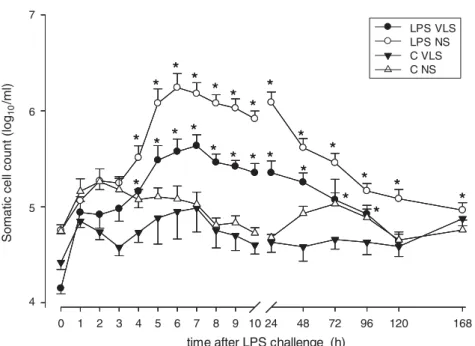
Milk SCC data from LPS and C quarters at each time point are shown in Fig. 1. Before LPS challenge (0 h) SCC of VLS (14.1 ± 1.5r103cells/ml) and NS group (56.7 ± 4.2r 103cells/ml) were significantly different. SCC of C quarters from both groups did not change significantly throughout the entire experiment. At 4 h p.c. SCC began to increase (P < 0.05) in both LPS-treated groups to reach a maximum at 6 h p.c. for NS group (1747 ± 696r103cells/ml) and at 7 h p.c. for VLS group (432± 130r103cells/ml).
Cell viability (Fig. 2) of all groups at time 0 h was 43.7 ± 3%. There was no difference between groups. At 10 h p.c. viability did not change in both C groups (48 ± 6.3%) but increased significantly in LPS-treated quar-ters with no differences between VLS and NS groups (88.9 ± 2.3%).
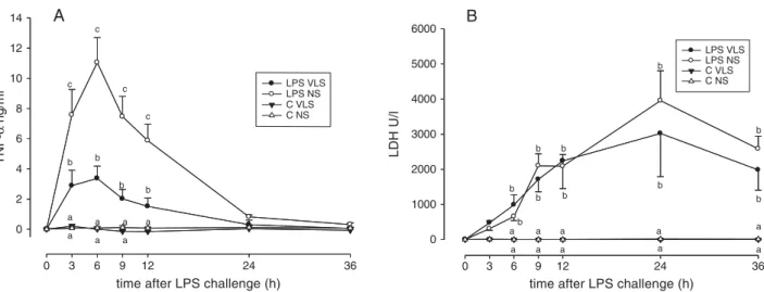
TNF-a concentrations in milk (Fig. 3A) increased within 3 h after LPS treatments in both groups and stayed elevated until 12 h p.c. Significant differences between VLS and NS groups was found from 3 h until 12 h p.c. with higher TNF-a concentrations in the NS group. In controls TNF-a concentrations did not increase.
LDH activity in milk (Fig. 3B) increased after 6 h p.c. in LPS quarters with no differences between groups and stayed elevated until the end of the experiment. In C quarters no increase of LDH activity was detect-able.
Experiment 2
At time 0 h no differences within relative mRNA ex-pression of milk cells of all measured genes were found between the controls and treatments and between groups.
0 1 2 3 4 5 6 7 8 9 10 24 48 72 96 120 168
Somatic cell count (log
10 /ml) 6
*
*
*
*
*
*
*
*
*
*
*
*
*
*
*
*
*
*
*
*
*
*
*
*
5 7 4time after LPS challenge (h)
LPS VLS LPS NS C VLS C NS
*
*
*
*
*
*
*
*
*
*
*
*
*
*
*
*
*
*
*
*
*
*
*
*
Fig. 1. Experiment 1: SCC (in log10/ml) after intramammary injection of 0 (C) and 50 ng LPS (LPS) dissolved in 10 ml saline solution
into quarters of very low SCC (VL; SCC < 20 000 ells/ml) and normal SCC (NS ; 40 000–100 000 cells/ml). $ LPS VL (n = 9), # LPS NS (n = 10), h C VL (n = 7), D C NS (n = 7). Data are represented as mean ±SEM. *: Significant differences between LPS challenge and
control within each group (P < 0.05). SCC between groups were significantly different (P < 0.05). SCC increased in response to LPS in both groups, whereas the maximum was reached 1 h earlier in the NS group.
viability of milk cells (%)
0 20 40 60 80 100 0 h 10 h * * C VLS LPS VLS C NS LPS NS
Fig. 2. Experiment 1: Proportion of viable cells (%) in the milk at 0 h (&) and 10 h (%) after LPS challenge in C (control without LPS challenge) and LPS quarters of very low SCC (VLS; SCC < 20 000 cells/ml) and normal SCC group (NS; SCC = 40 000–100 000 cells/ml). Data are represented as mean ±SEM
(n = 5–7). *: Significant differences between before and after LPS challenge within each group (P < 0.05). Cell viability increased after LPS challenge with no differences between the groups.
At 12 h p.c. an increased mRNA expression of IL-8 (Fig. 4 IL-8) and IL-1b (Fig. 4 IL-1b) in LPS quarters from both groups was observed. In C quarters IL-8 and IL-1b mRNA expression was decreased at 24 h p.c. in VLS and was not changed at other time points. Lactoferrin mRNA expression (Fig. 4 Lactoferrin) was increased at 36 h p.c. in VLS and C quarters. A decrease of lactoferrin mRNA expression could be detected in LPS-treated quarters at 24 h and 36 h p.c. of the NS group and in LPS-treated quarters at 12, 24 and 36 h p.c. of the VLS group. TNF-a relative mRNA ex-pression (Fig. 4 TNF-a) of milk cells was decreased at 12 h in C quarters of both groups and was not changed at all time points in all treatments.
Discussion
In the present study the immune competence of udder quarters with very low SCC ( < 20r103cells/ml; VLS) was compared with quarters with normal SCC (NS) of (40–100)r103cells/ml. Previous investigations already compared low SCC and high SCC but definitions of low
SCC varied: 200r103cells/ml (Vangroenweghe et al.
2004; Kauf et al. 2006 ; Olde Riekerink et al. 2007), 150r103cells/ml (Schukken et al. 1991; Barkema et al. 1998; Schmitz et al. 2004; Werner-Misof et al. 2007), 100r103cells/ml (Paape et al. 1977; Boutet et al. 2004;
Koess & Hamman, 2008), 50r103cells/ml (Beaudeau
et al. 2002), 18r103cells/ml (Cheng et al. 2008) or
12r103cells/ml (Sarikaya et al. 2006). However, a direct
comparison of very low SCC levels below 20r103cells/
ml with normal SCC levels of (40–100)r103cells/ml that are found very often in dairy practice, in regard to the
immune response of the cells, had not been done before as far as we were aware.
The quarters for the experiments were selected based on their SCC of < 20r103cells/ml for VLS group and (40–100)r103cells/ml for NS group. The selected quarters were of experimental cows with a broad range of lac-tational stage and number of lactation because other cows were not available. However, all quarters were from udders without any signs of mastitis and they were grouped with normal distribution of number of lactation and lactational stages. If two quarters from one cow were used the treat-ments of the two quarters within this cow were randomly chosen (control and control, control and LPS treatment, or LPS and LPS treatment). Therefore, results were not influ-enced by a potential crosstalk between quarters.
For the measurement of responses of SCC and cell viability during an immune response in VLS and NS groups, quarters were stimulated with 50 ng LPS in 10 ml saline solution to induce a clear but not too strong immune re-sponse so as to be able to detect possible differences in cell count and viability. To investigate the immune response of the milk cells by mRNA expression of immune factors, a strong immune response is necessary to be able to detect possible differences. Therefore, it was not possible to use the same treatment for the measurements of all factors and two experiments had to be performed with a lower LPS dose for the measurement of SCC, cell viability, and TNF-a concentration and LDH activity in milk, and a higher LPS dose for mRNA expression of immune factors in milk cells. According to Bannerman et al. (2004), Didier & Bruckmaier (2004) and Werner-Misof et al. (2007) SCC of C quarters were stable over the time of the experiment. Therefore, comparisons between C and LPS quarters were possible.
a c a a b c b a a a b b c c a a a a a a a a a a b b b b b b b b b b A
time after LPS challenge (h)
36 24 12 9 6 3 0 TN F-α ng /m l 0 2 4 6 8 10 12 14 LPS VLS LPS NS C VLS C NS B
time after LPS challenge (h)
36 24 12 9 6 3 0 LDH U/l 0 1000 2000 3000 4000 5000 6000 LPS VLS LPS NS C VLS C NS a
Fig. 3. Experiment 1: TNF-a concentration (A) and LDH activity (B) in milk after LPS challenge in C quarters (control without LPS challenge) and LPS quarters of very low SCC (VL; SCC < 20 000 cells/ml) (n = 5 for LPS- and C quarters, respectively) and normal SCC (NS; 40 000–100 000 cells/ml) (n = 15 and 7 for LPS- and C quarters, respectively). Data are represented as mean ±SEM.a,b,c: Different
letters indicate significant differences between groups and treatments within one time point (P < 0.05). If there were no differences between groups and treatments within that time point no letters are given. TNF-a concentration and LDH activity were not different between groups at 0 h and increased in both groups in response to LPS challenge. TNF-a concentration increase in response to LPS was higher in NS compared with VLS. No group differences in LDH activity increase in response to LPS were detected.
SCC started to increase simultaneously in both groups 4 h after the challenge with LPS. NS group reached higher SCC levels, although the highest level of SCC was reached 1 h earlier than in VLS group. The time required for SCC in-crease is the time needed for the recruitment of new cells from blood into milk (Harmon & Heald, 1982; Nickerson & Pankey, 1984; Kehrli & Schuster, 1994). This recruit-ment of cells was faster in NS quarters than in VLS quarters, which can indicate a more effective immune response.
Like SCC, milk cell viability is crucially involved in the outcome of mammary gland infection. In the present study cell viability was stable during the time of the exper-iment in quarters that were not stimulated. According to Mehrzad et al. (2001, 2004) the viability in LPS-treated quarters increased from 42.9 ± 4.3% to 88.9 ± 2.3%. This is due to the increased presence of young leucocytes that migrate from blood into milk and these cells undergo slower apoptosis than older cells (Van Merris et al. 2002). Moreover, LPS stimulation prolongs cell survival (Burvenich et al. 2003). Without stimulation and, therefore,
without this entry of new leucocytes, resident somatic cells have a long storage period (milking interval) in the cisternal cavities. During this time striking changes occur in the morphology of somatic cells owing to the ingestion of fat globules and casein (Paape et al. 1975; Paape & Guidry, 1977). Owing to these morphological and physio-logical changes, phagocytic and bactericidal activities, as well as cell viability are decreasing and, therefore, the mammary gland defence will be impaired (Burvenich et al. 2007; Rainard & Riollet, 2006). However, no difference in the cell viability between VLS and NS groups was observed after LPS challenge, because at 10 h p.c. the viability of the cells was already nearly 90%. Differentiation of cell populations was for technical reasons not possible in these experiments; however, it is known that the increase of SCC is due mainly to the increase of polymorpho-nuclear neutrophils (Sarikaya et al. 2006).
LDH activity increased after LPS challenge in NS quarters and in VLS quarters as expected in response to intra-mammary LPS administration (Bogin & Ziv, 1973). LDH is
TNF-α
rel. mRNA expression
-4 -2 0 2 4 LPS VLS LPS NS C VLS C NS
*
*
IL-1βtime after LPS challenge (h)
36 24
12 0
time after LPS challenge (h)
36 24
12 0
rel. mRNA expression
-4 -2 0 2 4 LPS VLS LPS NS C VLS C NS IL-8
time after LPS challenge (h)
36 24 12 0 re l. m R NA e x pr es sio n -2 0 2 4 6 LPS VLS LPS NS C VLS C NS
*
*
*
*
*
*
Lactoferrintime after LPS challenge (h)
36 24
12 0
rel. mRNA expression
-6 -4 -2 0 2 LPS VLS LPS NS C VLS C NS
*
*
*
*
*
*
Fig. 4. Experiment 2 : Relative mRNA expression (DDCP) of IL-8, lactoferrin, IL-1b, and TNF-a in milk cells in C (control without LPS challenge) and LPS quarters of very low SCC (VL ; SCC < 20 000 cells/ml) (n = 5 for LPS and C quarters, respectively) and normal SCC group (NS; 40 000–100 000 cells/ml) (n = 15 and 7 for LPS and C quarters, respectively). Data are represented as mean ±SEM.
*: Significant differences within groups between LPS treatment and controls (P < 0.05). There were no considerable differences in mRNA expression of measured immune factors in response to LPS in the cells between both groups.
a ubiquitous enzyme that is found in all cells and is an indicator of inflammation as it is released into the extra-cellular fluid during cell damage and cell death (Glick, 1969). The increase of milk cells after LPS challenge led to a greater number of cells contributing to the LDH activity besides cells from the tissue. The differences in SCC in-crease between the NS and VLS groups were most likely not great enough to induce differences in LDH increase.
Cytokines and other pro-inflammatory factors produced after pathogen invasion or intramammary stimulation in-itiate the immune reaction (Rainard & Riollet, 2006). Some of the most important factors were analysed in this study. TNF-a is a cytokine, which serves as a rapidly responding central mediator of inflammatory functions known to play an important role in mastitis and increases in milk after LPS challenge (Paape et al. 2002). It is also involved in the endotoxin shock during an acute phase of coliform mastitis (Sordillo & Streicher, 2002). However, TNF-a mRNA ex-pression in milk cells was not changed at 12 h p.c. TNF-a is a cytokine which is expressed in the early immune response and important for the initiation of the innate immune response. It is likely that the milk cells responded immediately within the first hours after LPS challenge with an increase of TNF-a transcription. Measurement of mRNA expression 12 h p.c., however, could not detect a TNF-a mRNA expression increase because it was already nor-malized to levels before stimulation. Unfortunately RNA of milk cells earlier than 12 h p.c. was not available because of an insufficient amount of milk. The protein accumulates in the milk and, therefore, an increase 12 h after LPS challenge was detectable although the increased synthesis had already stopped after 12 h. The increase of TNF-a concentration in milk after LPS challenge was significantly higher in NS than in VLS group. This is because more cells could contribute to the production of TNF-a as the SCC was higher in NS group. It is possible that the increased TNF-a concentration in milk could elicit the cure of a potential infection due to a faster and more effective immune response in udder quarters with slightly higher SCC.
IL-8 and IL-1b mRNA expression of milk cells was in-creased 12 h p.c. in response to LPS treatment with no differences between the groups. This increase was ex-pected as it is known that after intramammary injection of LPS the expression of IL-8 and IL-1b increases (Schmitz et al. 2004; Werner-Misof et al. 2007) in mammary tissue. In addition, Lee et al. (2006) found increasing expression of cytokines in milk cells after intramammary Esch. coli stimulation. After 12 h the milk cells that had direct con-tact with the LPS are removed by milking and, for this reason milk cells at 24 h p.c. did not show an increased cytokine mRNA expression.
Surprisingly, unlike the other factors, relative mRNA expression of lactoferrin in milk cells decreased. Pfaffl et al. (2003) found a lower lactoferrin expression in quar-ters with SCC > 150r103cells/ml than in quarters with SCC < 150r103cells/ml. The difference between high-SCC
quarters and low-SCC quarters in Pfaffl et al. (2003) might be compared to the difference between LPS and C quarters of both groups in the present study. Thus, quarters with elevated SCC (with or without stimulation) have similar or lower lactoferrin expression in milk cells than quarters with low SCC. Why lactoferrin mRNA expression in VLS decreased earlier (12 h) than in NS group remains unclear. The increasing lactoferrin expression after intramammary injection of 100 mg LPS in mammary tissue that was found by Schmitz et al. (2004) is due to the increased expression of lactoferrin by mammary epithelial cells, the main source of lactoferrin in milk. These cells represent only a very small population of the somatic cells.
In conclusion, the results of the present study showed differences in the immune response between quarters with normal and very low SCC. The time until SCC started to increase after LPS challenge was approximately equal in quarters with normal and with very low SCC. However, quarters with very low SCC did not reach SCC levels as high as quarters with normal SCC. Although increasing less, maximum SCC in response to LPS challenge was reached slightly later in quarters with very low SCC com-pared with those with normal SCC. The immune response of the milk cells themselves, based on the mRNA ex-pression of the immune factors TNF-a, IL-1b, IL-8 and lactoferrin, was not different between cells of normal and very low SCC groups. Although the selection of exper-imental quarters was limited, the results suggest that the immune response of udders with slightly higher SCC might be more efficient, as more cells can contribute to the pro-duction of immune factors like TNF-a. It is possible that this effect can impair the immune response of udder quarters with very low SCC.
We thank the H. Wilhelm Schaumann-Stiftung, Hamburg, Germany, for financial support of Mrs A Baumert and Mrs Machabbat Saudenowa.
References
Bannerman DD, Paape MJ, Lee JW, Zhao X, Hope JP & Rainard C 2004 Escherichia coli and Staphylococcus aureus elicit differential innate immune responses following intramammary infection. Clinical and Diagnostic Laboratory Immunology 11 463–472
Barkema HW, Schukken YH, Lam TJ, Beiboer ML, Wilmink H, Benedictus G & Brand A 1998 Incidence of clinical mastitis in dairy herds grouped in three categories by bulk milk somatic cell counts. Journal of Dairy Science 81 411–419
Beaudeau F, Fourichon C, Seegers H & Bareille N 2002 Risk of clinical mastitis in dairy herds with a high proportion of low indi-vidual milk somatic cell counts. Preventive Veterinary Medicine 53 43–54
Blum JW, Dosogne H, Hoeben D, Vangroenweghe F, Hammon HM, Bruckmaier RM & Burvenich C 2000 Tumor necrosis factor-a and nitrite/nitrate responses during acute mastitis induced by Escherichia coli infection and endotoxin in dairy cows. Domestic Animal Endocrinology 19 223–235
Bogin E & Ziv G 1973 Enzymes and minerals in normal and mastitic milk. Cornell Veterinarian 63 666–676
Boutet P, Boulanger D, Gillet L, Vanderplasschen A, Closset R, Bureau F & Lekeux P 2004 Delayed neutrophil apoptosis in bovine subclinical mastitis. Journal of Dairy Science 87 4104–4114
Bruckmaier RM & Hilger M 2001 Milk ejection in dairy cows at different degrees of udder filling. Journal of Dairy Research 68 369–376
Burvenich C, Van Merris V, Mehrzad J, Diez-Fraile A & Duchateau L 2003 Severity of E. coli mastitis is mainly determined by cow factors. Veterinary Research 34 521–564
Burvenich C, Bannerman DD, Lippolis JD, Peelman L, Nonnecke BJ, Kehrli ME Jr & Paape MJ 2007 Cumulative physiological events in-fluence the inflammatory response of the bovine udder to Escherichia coli infections during the transition period. Journal of Dairy Science 90 (Suppl 1) 39–54 Review
Cheng JB, Wang JQ, Bu DP, Liu GL, Zhang CG, Wei HY, Zhou LY & Wang JZ 2008 Factors affecting the lactoferrin concentration in bovine milk. Journal of Dairy Science 91 970–976
Deluyker HA, Gay JM & Weaver LD 1993 Interrelationships of somatic cell count, mastitis, and milk yield in a low somatic cell count herd. Journal of Dairy Science 76 3445–3452
Didier A & Bruckmaier RM 2004 mRNA expression of apoptosis-related genes in mammary tissue and milk cells in response to lipopolysac-charide challenge and during subclinical mastitis. Milchwissenschaft 59 119–123
Glick JH 1969 Serum lactate dehydrogenase isoenzyme and total lactate dehydrogenase values in health and disease, and clinical evaluation of these tests by means of discriminant analysis. American Journal of Clinical Pathology 53 320–328
Green MJ, Green LE & Cripps PJ 1996 Low bulk milk somatic cell counts and endotoxin-associated (toxic) mastitis. Veterinary Record 138 305–306
Harmon RJ & Heald CW 1982 Migration of polymorphonuclear leuko-cytes into the bovine mammary gland during experimentally induced Staphylococcus aureus mastitis. American Journal of Veterinary Research 43 992–998
Harmon RJ & Newbould FHS 1980 Neutrophil leukocyte as a source of lactoferrin in bovine milk. American Journal of Veterinary Research 41 1603–1606
Kauf AC, Vinyard BT & Bannerman DD 2006 Effect of intramammary infusion of bacterial lipopolysaccharide on experimentally induced Staphylococcus aureus intramammary infection. Research in Veterinary Science 82 39–46
Kehrli EM & Schuster DE 1994 Factors affecting milk somatic cells and their role in health of the bovine mammary gland. Journal of Dairy Science 77 619–627
Koess C & Hamann J 2008 Detection of mastitis in the bovine mammary gland by flow cytometry at early stages. Journal of Dairy Research 75 225–232
Lee JW, Bannerman DD, Paape MJ, Huang MK & Zhao X 2006 Characterization of cytokine expression in milk somatic cells during intramammary infections with Escherichia coli or Staphylococcus aureus by real-time PCR. Veterinary Research 37 219–229
Mehrzad J, Dosogne H, Meyer E & Burvenich C 2001 Local and systemic effects of endotoxin mastitis on the chemiluminescence of milk and blood neutrophils in dairy cows. Veterinary Research 32 131–144
Mehrzad J, Duchateau L & Burvenich C 2004 Viability of milk neutrophils and severity of bovine coliform mastitis. Journal of Dairy Science 87 4150–4162
Nickerson SC & Pankey JW 1984 Neutrophil migration through test end tissues of bovine mammary quarters experimentally chal-lenged with Staphylococcus aureus. Journal of Dairy Science 67 826–834
Olde Riekerink RG, Barkema HW, Veenstra W, Berg FE, Stryhn H & Zadoks RN 2007 Somatic cell count during and between milkings. Journal of Dairy Science 90 3733–3741
Paape MJ, Guidry AJ, Kirk ST & Bolt DJ 1975 Measurement of phago-cytosis of 32P-labeled Staphylococcus aureus by bovine leukocytes: lysostaphin digestion and inhibitory effect of cream. American Journal of Veterinary Research 36 1737–1743
Paape MJ & Guidry AJ 1977 Effect of fat and casein on intracellular killing of Staphylococcus aureus by milk leukocytes. Proceeding of the Society for Experimental Biology and Medicine 155 588–593 Paape MJ, Pearson RE, Wergin WP & Guidry AJ 1977 Enhancement of
chemotactic response of polymorphonuclear leukocytes into the mammary gland and isolation from milk. Journal of Dairy Science 60 53–62
Paape MJ, Rautiainen PM, Lilius EM, Malstrom CE & Elsasser TH 2002 Development of anti-bovine TNF-alpha mAb and ELISA for quantitat-ing TNF-alpha in milk after intramammary injection of endotoxin. Journal of Dairy Science 85 765–773
Persson-Waller K, Colditz IG, Lun S & Ostensson K 2003 Cytokines in mammary lymph and milk during endotoxin-induced bovine mastitis. Research in Veterinary Science 74 31–36
Pfaffl MW, Wittmann SL, Meyer HH & Bruckmaier RM 2003 Gene ex-pression of immunologically important factors in blood cells, milk cells and mammary tissue of cows. Journal of Dairy Science 86 538–545 Rainard P & Riollet C 2006 Innate immunity of the bovine mammary
gland. Veterinary Research 37 369–400 Review
Sarikaya H, Werner-Misof C, Atzkern M & Bruckmaier RM 2005 Distribution of leucocyte populations, and milk composition, in milk fractions of healthy quarters in dairy cows. Journal of Dairy Research 72 486–492
Sarikaya H, Schlamberger G, Meyer HH & Bruckmaier RM 2006 Leukocyte populations and mRNA expression of inflammatory factors in quarter milk fractions at different somatic cell score levels in dairy cows. Journal of Dairy Science 89 2479–2486
Schmitz S, Pfaffl MW, Meyer HH & Bruckmaier RM 2004 Short-term changes of mRNA expression of various inflammatory factors and milk proteins in mammary tissue during LPS-induced mastitis. Domestic Animal Endocrinology 26 111–126
Schukken YH, Grommers FJ, Van de Geer D, Erb HN & Brand A 1991 Risk factors for clinical mastitis in herds with a low bulk milk somatic cell count. 2. Risk factors for Escherichia coli and Staphylococcus aureus. Journal of Dairy Science 74 826–832
Schukken YH, Bennet G, Green L & Van Werven T 2001 Can somatic cell counts get too low? National Mastitis Council Annual Meeting Proceedings
Shook GE 1989 Selection for disease resistance. Journal of Dairy Science 72 1349–1362
Sordillo LM & Streicher KL 2002 Mammary gland immunity and mastitis susceptibility. Journal of Mammary Gland Biology and Neoplasia 7 135–146
Suriyasathaporn W, Schukken YH, Nielen M & Brand A 2000 Low so-matic cell count : a risk factor for subsequent clinical matitis in a dairy herd. Journal of Dairy Science 83 1248–1255
Vangroenweghe F, Rainard P, Paape M, Duchateau L & Burvenich C 2004 Increase of Escherichia coli inoculum doses induces faster innate immune response in primiparous cows. Journal of Dairy Science 87 4132–4144
Van Merris V, Meyer E & Burvenich C 2002 Functional maturation during bovine granulopoiesis. Journal of Dairy Science 85 2859–2868 Werner-Misof C, Pfaffl MW & Bruckmaier RM 2007 Dose-dependant
immune response in milk cells and mammary tissue after in-tramammary administration of lipopolysaccharide in dairy cows. Veterinarni Medicina 52 231–244