HAL Id: hal-02068079
https://hal-amu.archives-ouvertes.fr/hal-02068079
Submitted on 3 May 2019HAL is a multi-disciplinary open access archive for the deposit and dissemination of sci-entific research documents, whether they are pub-lished or not. The documents may come from teaching and research institutions in France or abroad, or from public or private research centers.
L’archive ouverte pluridisciplinaire HAL, est destinée au dépôt et à la diffusion de documents scientifiques de niveau recherche, publiés ou non, émanant des établissements d’enseignement et de recherche français ou étrangers, des laboratoires publics ou privés.
Cushing Syndrome Is Associated With Subclinical LV
Dysfunction and Increased Epicardial Adipose Tissue
Flavia Maurice, Benedicte Gaborit, Clara Vincentelli, Ines Abdesselam,
Monique Bernard, Thomas Graillon, Frank Kober, Thierry Brue, Frederic
Castinetti, Anne Dutour
To cite this version:
Flavia Maurice, Benedicte Gaborit, Clara Vincentelli, Ines Abdesselam, Monique Bernard, et al.. Cushing Syndrome Is Associated With Subclinical LV Dysfunction and Increased Epicardial Adi-pose Tissue. Journal of the American College of Cardiology, Elsevier, 2018, 72 (18), pp.2276-2277. �10.1016/j.jacc.2018.07.096�. �hal-02068079�
Cushing syndrome is associated with subclinical left ventricular dysfunction and increased epicardial adipose tissue
Flavia Maurice MDa,b, Bénédicte Gaborit MD,PhDa,b, Clara Vincentelli MDa,b , Ines Abdesselam PhDa,c, Monique Bernard PhDc, Thomas Graillon MD,PhDd, Frank Kober PhDc, Thierry Brue MD,PhDe, Frédéric Castinetti MD,PhDe,*, Anne Dutour MD,PhDa,b *
*authors contributed equally to the work
a
Aix Marseille Univ, INSERM, INRA, C2VN, Marseille, France
b
Department of endocrinology, pôle ENDO, APHM Conception 147 bd baille 13005 Marseille, France.
c
Aix Marseille Univ, CNRS, CRMBM, Marseille, France.
d
Department of neurosurgery, APHM, CHU Timone, Marseille, France.
e
Aix-Marseille Univ, INSERM U1251, Marseille Medical Genetics (MMG), Marseille, France and Assistance Publique - Hôpitaux de Marseille (AP-HM), Department of Endocrinology, Hôpital de la Conception, Centre de Référence des Maladies Rares Hypophysaires HYPO, 13005 Marseille, France
Funding: ARARD, Marseille, France
Disclosures: The authors report no conflicts of interest in this work.
Clinical Trial: NCT02335996 NCT02848703
Correspondence:
Dr Benedicte Gaborit
Pole ENDO, Hôpital De La Conception 147 Boulevard Baille 13005 Marseille France Telephone: + 33 4 91 38 36 50
Fax: +33-491254336
2 Cushing syndrome (CS) results from chronic glucocorticoids (GC) excess exposure. This disease is associated with increased mortality and adverse cardiovascular (CV) outcomes due to visceral adiposity, insulin resistance, hypertension, and hypercoagulability. Interestingly, CV risk persists even after remission of the disease (1,2) which remains partly unexplained.
The impact of cardiac adiposity on coronary artery disease (CAD) and left ventricular (LV) function has been suggested. Prospective studies have shown that epicardial adipose tissue (EAT) amount is an independent predictor of CAD (3). However, the impact of hypercortisolism on EAT is yet unknown, while it would be of interest as CS is classically associated with upper body adiposity. Our aim was to compare EAT volume and LV function in patients with active and in remission ACTH-dependent CS, and controls.
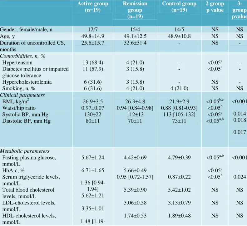
Thirty-eight patients with ACTH-dependent CS (19 active and 19 in remission for at least 2 years, mean 41.2± 4.2 months) and 19 controls of similar age and sex distribution were included. A magnetic resonance imaging was performed on a 3-Tesla wide-bore magnet to determine abdominal fat distribution, EAT volume, systolic and diastolic LV function and structure as previously described (4). The expected increase in hypertension, diabetes, dyslipidemia was evidenced in active CS (Table) as well as visceral adiposity (higher VAT/sSCAT). Patients from the active and remission groups did not differ in uncontrolled disease duration and had comparable BMI at diagnosis.
Cushing syndrome induces EAT accumulation and subclinical LV dysfunction: Increased
EAT volume was found in active patients compared to controls (93.5±6.1 versus 55.2±3.6 mL, p<0.01) and remission patients (68.2±5.1 mL p<0.01). However, patients in remission had a higher EAT volume than controls (p<0.05.). Active patients compared to controls had higher LV mass index (LVMi 55.3±2.6 vs 43.2±2.3 g/m2, p<0.01), concentric remodeling (ie indexed LV
mass to end-diastolic volume ratio (EDVi) 1.10±0.07 vs 0.68±0.03g/mL, p<0.01) and impaired early LV relaxation (ie E/A ratio 1.34±0.07 versus 1.66±0.09, p=0.01), while LV ejection function did not differ. In remission patients, LVMi and E/A ratio were not different from controls but LVMi/EDVi was higher (0.81±0.04, p=0.02). Multivariate analysis (including age, sex, BMI, blood pressure, fasting plasma glucose and EAT) revealed that EAT volume was independently associated with LVMi (β=0.40, p=0.008) and LVMi/EDVi (β=0.41, p=0.011).
EAT is a perivascular depot releasing inflammatory adipokines that pass through the coronary wall and interact with endothelial cells, enhancing monocytes adhesion. EAT correlates with the extent and severity of CAD, chest pain, unstable angina and coronary flow reserve and predicts coronary events independently of CV risk factors .We hypothesize that together with other CV risk factors, increased EAT in CS could contribute to its excess cardiovascular morbidity.
EAT volume was independently correlated with LV mass index and mass to end-diastolic volume. This suggests a major role for EAT in CS induced LV dysfunction. Indeed, the EAT anatomical proximity to the myocardium free of any fascia gives it the distinctive function to act locally and directly on the myocardium. A link between EAT secreted adipokines and both contractile function and insulin signaling in cardiomyocytes was demonstrated (3). Furthermore, EAT volume and secretome have been linked to cardiac fibrosis, a substratum widely recognized to impair LV function (3).
In conclusion, we demonstrated a unique pattern of ectopic fat deposition in patients with active ACTH-dependent CS, with a relatively high epicardial fat accumulation. This increase in EAT was associated with subtle LV dysfunction. Furthermore, in remission patients, EAT volume was higher than in controls, possibly contributing to the persistent cardiovascular risk.
4
References
1.Clayton RN, Jones PW, Reulen RC, et al. Mortality in patients with Cushing’s disease more than 10 years after remission: a multicentre, multinational, retrospective cohort study. Lancet Diabetes Endocrinol. 2016;4:569–576.
2. Colao A, Pivonello R, Spiezia S, et al. Persistence of Increased Cardiovascular Risk in Patients with Cushing’s Disease after Five Years of Successful Cure. J. Clin. Endocrinol. Metab. 1999;84:2664–2672.
3.Gaborit B, Sengenes C, Ancel P, Jacquier A, Dutour A. Role of Epicardial Adipose Tissue in Health and Disease: A Matter of Fat? Compr. Physiol. 2017;7:1051–1082.
4.Gaborit B, Jacquier A, Kober F, et al. Effects of bariatric surgery on cardiac ectopic fat: lesser decrease in epicardial fat compared to visceral fat loss and no change in myocardial triglyceride content. J. Am. Coll. Cardiol. 2012;60:1381–1389.
Table 1: Characteristics of study population. Data presented as n (%) or mean ± SD or median
[25th – 75th percentile]. BMI: Body mass index, BP: Blood pressure, EAT: Epicardial adipose
tissue, NS: Not Significant, sSCAT : superficial Subcutaneous adipose tissue, VAT: Visceral adipose tissue. Comparison between groups p<0.05 a active vs remission groups, b active group
vs controls, c remission group vs controls, 3-groups comparison with one way-ANOVA and
Holm-Sidak post-hoc test or Kruskal-Wallis and Dunn’s as appropriate
Active group (n=19) Remission group (n=19) Control group (n=19) 2 group p value 3-group pvalue Gender, female/male, n 12/7 15/4 14/5 NS NS Age, y 49.8±14.9 49.1±12.5 48.9±10.8 NS NS Duration of uncontrolled CS, months 25.6±15.7 32.6±31.4 - NS - Comorbidities, n, % Hypertension
Diabetes mellitus or impaired glucose tolerance Hypercholesterolemia Smoking, n, % 13 (68.4) 11 (57.9) 6 (31.6) 6 (31.6) 4 (21.0) 3 (15.8) 3 (15.8) 4 (21.0) - - - 4 (21.0) <0.05a <0.05a NS NS - - - NS Clinical parameters BMI, kg/m2 Waist/hip ratio Systolic BP, mm Hg Diastolic BP, mm Hg 26.9±3.5 0.97±0.07 130±22 80±11 26.3±4.8 0.94 [0.84-0.98] 112±13 70±11 21.9±2.9 0.88 [0.81-0.93] 113 [105-132] 73±11 <0.05b,c <0.05b <0.05a <0.05a,b <0.001 0.014 0.018 0.017 Metabolic parameters
Fasting plasma glucose, mmol/L
HbA1c, %
Serum triglyceride levels, mmol/L
Total blood cholesterol levels, mmol/L LDL-cholesterol levels, mmol/L HDL-cholesterol levels, 5.67±1.24 6.71±1.65 1.36 [0.94-1.94] 5.62±1.21 3.35±1.01 4.42±0.69 5.66±0.49 0.95 [0.72-1.57] 5.39±0.90 3.06±0.58 1.74±0.53 4.79±0.39 - 0.87±0.22 5.42±1.02 3.13±0.79 1.89±0.48 <0.05a,b <0.05a <0.05b NS NS NS <0.001 - 0.024 NS NS NS
6 2.04]
Inflammation biomarkers
Plasma fibrinogen levels, g/L High-sensitivity-C-reactive protein, mg/L
Serum ferritin levels, μg/L
3.43±0.73 1.4 [0.5-2.9] 135 [44-249] 3.52±0.87 1.6 [0.8-5.2] 78 [35-114] - - - NS NS NS - - -
MRI Body composition
VAT/sSCAT 0.98
[0.78-2.14] 0.63 [0.44-1.06] 0.84±0.36 <0.05a
0.017
MRI Epicardial adipose tissue (EAT) assessment
EAT volume, mL 93.5±26.6 68.2±22.1 55.2±15.9 <0.05a,b,c <0.001
LV morphology and function
E/A ratio
LV Mass index, g/m2 LV Mass index to end-diastolic volume, g/mL 1.34±0.30 55.3±11.3 1.10 ±0.30 1.59±0.37 40.9 [36.0-45.6] 0.75 [0.66-0.99] 1.66±0.33 43.2±10.2 0.68±0.11 <0.05b <0.05a,b <0.05a,b,c 0.031 <0.001 <0.001