Abstract Langerhans’ cell histiocytosis (LCH) is a rare
and enigmatic clonal disorder that affects mainly children.
It is characterized by single or multiple granulomatous
mass lesions composed of cells with the Langerhans’ cell
phenotype. Clinical presentation and behavior are
hetero-geneous and can range from a solitary lytic bone lesion
(i.e., eosinophilic granuloma) with a favorable course to a
fatal disseminated leukaemia-like form, with a wide
spec-trum of intermediate clinical presentations between these
two extremes. Although LCH typically involves the bone,
lesions can be found in almost all organs. We are reporting
the case of a multisystem LCH in a 47-year-old patient
who presented with a panhypopituitarism and diabetes
in-sipidus, and who, 5 years later, developed mandibular,
mastoid and femoral lesions. The final diagnosis of LCH
was made on mandibular biopsy.
Keywords Langherhans’ cell histiocytosis · Diabetes
insipidus · Panhypopituitarism · Mastoiditis · Mandibular
lytic lesions
Introduction
Langerhans’ cell histiocytosis (LCH) encompasses a
dis-parate group of diseases that has been referred to as
Hand-Schüller-Christian disease, Letterer-Siwe disease,
eosino-philic granuloma, histiocytosis X, Hashimoto Pritzker
syn-drome, self-healing histiocytosis, pure cutaneous
histiocy-tosis, Langerhans’ cell granulomahistiocy-tosis, type II
histiocyto-sis and non-lipid reticuloendotheliohistiocyto-sis [8]. Langerhans’
cell histiocytosis is characterized by the proliferation of
ab-normal dendritic antigen-presenting histiocytes, known as
Langherhans’ cells, with an accompanying infiltrate of
lym-phocytes, eosinophils and neutrophils resulting in the
de-struction of a variety of tissues. Lesions may occur as
lo-calized lesions or as widespread systemic disease.
Langher-hans’ cells have distinctive morphologic and
immunohis-tochemical features (such as S-100 protein and CD1a
anti-gen) and represent an immature stage in the development
of interdigitating cells [7, 13]. Although some authors
have recently demonstrated clonal proliferation in all forms
of LCH, suggesting a neoplastic disorder, the exact
etio-pathogenesis of this enigmatic disease still remains obscure,
and the clinical course is unpredictable [17, 19].
Langer-hans’ cell histiocytosis is usually considered to be a
dis-ease of childhood, with a peak incidence from 1 to 3 years.
However, it should be included in the differential
diagno-sis of adult patients, particularly those with multisystem
disease [18, 14, 4]. We are reporting a case of a
47-year-old patient with a multisystemic form of Langherhans’
cell histiocytosis.
Case report
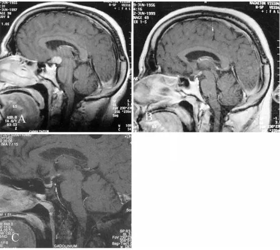
A 42-year-old patient was referred to the endocrinological depart-ment in May 1997 for investigation of hypothyroidism. For 2 years, he had complained of chronic fatigue, memory impairment, dimin-ished libido, impotence, a loss of hair, gynecomastia and poly-uria/polydipsia, which had started a few months previously. Pan-hypopituitarism and central diabetes insipidus were diagnosed. Mag-netic resonance imaging (MRI) showed evidence of an infiltrating hypothalamic lesion involving the mammillary bodies, a slightly thickened pituitary stalk (both homogeneously enhanced after in-travenous gadolinium administration), a loss of the normal hyper-intense signal of the posterior pituitary on T1-weighted images (“bright spot”) and a partially empty sella (Fig. 1a). Microbiologi-cal cultures of blood and CSF and serologiMicrobiologi-cal markers for
Tre-ponema pallidum (VDRL, TPHA), Borrelia burgdorferi, Brucella
P. Scolozzi · T. Lombardi · P. Monnier · B. Jaques
Multisystem Langerhans’ cell histiocytosis
(Hand-Schüller-Christian disease) in an adult:
a case report and review of the literature
DOI 10.1007/s00405-003-0690-z
Received: 28 May 2003 / Accepted: 3 September 2003 / Published online: 10 October 2003
O N C O L O G Y
P. Scolozzi (✉) · B. Jaques
Division of Oral and Maxillofacial Surgery, Centre Hospitalier Universitaire Vaudois (CHUV), 1011 Lausanne, Switzerland
Tel.: +41-213142731, Fax: +41-213141347, e-mail: scolozzi@hotmail.com
T. Lombardi
Laboratory of Oral Histopathology, Division of Stomatology, Faculty of Medicine,
19, Rue Barthélemy-Menn, 1205 Genève, Switzerland P. Monnier
Department of Otolaryngology and Head and Neck Surgery, Centre Hospitalier Universitaire Vaudois (CHUV),
Lausanne, Switzerland © Springer-Verlag 2003
melitensis, Bartonella henselae and HIV were negative. The
re-sults of cerebrospinal fluid immunoelectrophoresis were normal. The level of angiotensin-converting enzyme, tested in blood and CSF, was normal. Further work up with chest X-ray for sarcoïdosis was normal. Neuropsychological tests showed a fronto-subcortical syndrome characterized by severe short- and long-term memory impairment (verbal and spatial) and marked executive dysfunction. Replacement therapy was begun with oral levothyroxine 0.15 mg daily, oral hydrocortisone acetate 20 mg daily, intramuscularly testos-terone enantate 250 mg weekly and intranasal DDAVP 10µg twice daily. In July 1998, the patient presented with hyperosmolar dia-betic decompensation, and treatment with insulin and metformine 850 mg twice daily was begun. A stereotaxic biopsy was per-formed in January 1999 because the neuropsychological status of the patient had become worse. Histopathologic examination revealed gliosis together with non-specific perivascular chronic inflamma-tory infiltration. Oral prednisone 60 mg daily for 3 months was
be-gun in March 1999, which stabilized the situation temporarily. The follow-up MRI in June 1999 demonstrated a slight regression of the hypothalamic lesion and the intensity of the contrast enhance-ment following I.V. gadolinium administration (Fig. 1b). In July 2001, the patient complained of intermittent mandibular pain lo-cated in the right molar and symphyseal areas. Periapical X-ray showed a discrete periapical radiolucent lesion associated with a non-vital mandibular right second molar (Fig. 2a) and a large peri-apical radiolucent lesion associated with the non-vital mandibular incisors (Fig. 2b). The right second mandibular molar was ex-tracted and a devitalisation of the left central and lateral incisors and right central incisor was performed. The panoramic radiograph in December 2001 demonstrated a 3.5×3-cm well-defined non-cor-ticated radiolucency with a scalloped superior border of the right gadolinium administration.
B Sagittal T1-weighted MRI images after 3 months of pred-nisone therapy showing a slight regression of the hypothalamic lesion following gadolinium administration. C Sagittal T1-weighted MRI images after 5 years showing an important regression of the hypothalamic lesion
Fig. 2 Periapical radiographs showing well-defined radiolucency associated with the apex of the right mandibular second molar (A) and mandibular incisors (B)
Fig. 3 Panoramic radiograph showing two unilocular radiolucen-cies with well-defined margins in the right mandibular angle and symphyseal region
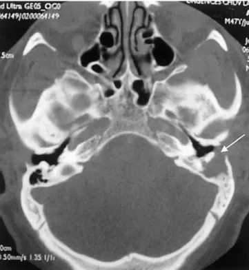
mandibular area and the persistence of a punched-out radiolucency in the symphyseal region (Fig. 3). Chronic osteomyelitis of the mandible was diagnosed, and antibiotic and antiphlogistic therapy was prescribed without any modification of the symptomatology. In February 2002, the patient underwent a surgical curettage and enucleation of both mandibular lesions, and the histologic diagno-sis was that of chronic osteomyelitis. Follow-up panoramic radio-graphs in April and August 2002 demonstrated no changes in the size or radiographic characteristics of the mandibular lesions, but the patient was asymptomatic. The patient presented in June 2002 with a 1-month history of transient otalgia, pain in the left retro-au-ricular area, disturbances of gait, dizziness and left hypoacusia, which had started 1 week previously. Otitis externa was diagnosed and antibiotic and antiphlogistic therapy was given with a slight modification of the symptomatology. One week later, he presented at the hospital for the persistence of the symptomatology, appear-ance of fever and significant red pulsatile swelling in the mastoid region. Audiological evaluation revealed mixed hearing loss. The CT-scan of the temporal bone revealed a massive destruction of the bone of the mastoid area, and the patient underwent a left mas-toidectomy (Fig. 4). The mastoid was destroyed by adherent gran-ulations to the peri-temporal tissues, which prevented complete surgical exeresis. Histopathological diagnosis was that of a chronic non-specific inflammatory infiltrate.
In November 2002, the patient presented with a worsening of mandibular pain associated with a discharge of whitish material in the mouth. The intraoral exam revealed the persistence of a sinus tract in the retromolar and symphyseal regions from which whitish secretions were exuding. The panoramic radiograph demonstrated no changes in the size or radiographic characteristics of the mandibu-lar lesions. The patient underwent a new surgical curettage and enucleation of both mandibular lesions. Microscopic examination showed a diffuse infiltration of large pale-staining mononuclear cells with indistinct cytoplasmic borders and indented vesicular nuclei with a histiocytic appearance (Fig. 5a). The infiltrate was also composed of a large amount of eosinophils, plasma cells and lym-phocytes. Immunohistochemical studies using CD1a and S-100 protein antibodies showed that the histiocytic cells were consistent with Langerhans’ cells (Fig. 5b). The final diagnosis was that of a Fig. 4 CT scan showing left mastoid infiltrated by soft tissue with the destruction of the bony cortex (arrow)
Fig. 5 A Photomicrographs of the mandibular biopsy specimen showing the cells of the infiltrate exhibiting the characteristic mor-phologic features of Langerhans’ cells with a convoluted shape, elongated nuclei exhibiting longitudinal grooves, and pale cyto-plasm. Numerous eosinophils are also visible (haematoxylin-eosin, original magnification ×25) B Immunostaining revealing numer-ous strongly positive Langerhans’ cells for the CD1a antibody (im-munoperoxidase, original magnification ×25)
Fig. 6 Technetium-99-m bone scan showing a lesion of the distal epiphysis of the left femur, the persistence of a left temporal and mandibular lesions and a frontal bone lesion
hypothalamic-pituitary axis lesion (Fig. 1c).
Given that the patient presented no symptoms at the 3-month follow-up and given the lack of an international consensus con-cerning treatment for adult LCH, the oncologists decided to pro-pose no further treatment.
Discussion
In 1893, Hand reported a case of “polyuria and
tuberculo-sis” in a 3-year-old boy who presented with diabetes
in-sipidus (DI), skull lesions and exophthalmos [6]. In
retro-spect, he gave the first detailed clinical description of a
new group of polymorphic disorders involving the
reticu-loendothelial system (RES) and characterized by a
histio-cytic infiltrate. In 1953, Lichtenstein proposed the general
term “histiocytosis X” to describe a group of disorders
in-cluding Hand-Schuller-Christian syndrome, Letterer-Siwe
disease and eosinophilic granuloma [12]. Twenty years later,
Nezelof identified the Langerhans’ cells as the original
cells of histiocytisis X [15]. The Langerhans’ cell (LC), first
described by the Berlin medical student Paul Langerhans
in 1868, is a bone-marrow-derived dendritic,
antigen-pre-senting cell network, related to the mononuclear
phago-cyte system [10]. These cells are found in the skin, lymph
nodes, bone marrow, spleen and probably also the brain, and
have characteristic surface proteins, which identify them
as immature or mature. Within the LC is a five-layered
structure, the Birbeck granule, first seen by electron
mi-croscopy in 1961 [13]. The gene coding for the endocytic
receptor that induces the formation of Birbeck granules
has been identified and the protein called “langerin” [13,
17, 19]. Pathological LC is positive for HLA-DR, CD1a,
S100 and Fc receptor, as with their normal counterparts
[8, 13, 17, 19, 18]. In 1987, the Histiocyte Society
estab-lished a new classification of these disorders based on the
lineage of lesional cells and biological behavior related to
the ontogeny of histiocytes, and distinguished three classes:
class I or Langerhans’ cell histiocytosis (LCH); class II or
non-Langerhans’ cell histiocytosis; class III or malignant
histiocytosis [18].
Two main forms of LCH have been described:
local-ized LCH (eosinophilic granuloma) and multisystem LCH
(formerly Hand-Schuller-Christian syndrome or chronic
disseminated disease and Letterer-Siwe disease or acute
disseminated disease).
Langerhans’ cell histiocytosis often presents as a
puz-zling syndrome, which renders the diagnosis problematic.
Four important considerations explain the reasons for a
delayed diagnosis of LCH in our patient. First is the age
of the patient (42 years) at the time of the first symptoms.
In fact, these disorders are typically found in children –
more than 50% of all cases are seen in patients under age
10 – even though recent studies have shown that
approxi-was found to be the site of presentation in only 11 patients
[14]. By contrast, CNS involvement is not uncommon and
is strongly associated with the systemic disease, but it is
rarely found alone. The hypothalamic-pituitary axis
re-mains the unexplained site of predilection of CNS LCH;
however, hypothalamic mass lesions, as found in our
pa-tient, are not frequently encountered [5, 9]. Grois et al.
re-viewed 38 patients with CNS LCH and found only 4
pa-tients (10%) with such lesions, whereas 22 papa-tients (63%)
presented with structural changes in the pituitary region
(infundibular thickening in 8 patients and empty sella in
14 patients) [5]. Diabetes insipidus (DI) is the most
com-mon endocrine abnormality, reported in 5–50% of patients
with LCH, and it occurs when more than 80% of the
par-aventricular-supraoptic neurons are destroyed [9].
Dia-betes insipidus occurs in only 5% of patients at the time of
original diagnosis, whereas it develops in one quarter to
one third of patients within 5 years of the diagnosis.
Al-though panhypopituitarism is reported in only 5–20% of
patients, it is almost always associated with DI [9].
The third consideration concerns the difficulty in
ob-taining a correct diagnosis of LCH from cerebral biopsy.
According to the literature, whether or not a biopsy should
be taken from such lesions still remains controversial.
Nevertheless, neurosurgeons are not inclined to approach
lesions in certain delicate regions, such as the
hypothala-mus, pons, basal ganglia and cerebellar peduncles, for only
the purpose of biopsy [5]. Normann et al. described four
stages of CNS LCH: hyperplastic-proliferative,
granulo-matous, xanthomatous and gliosis. The histological and
immunohistochemical LCH diagnostic features are more
likely to be found in the hyperplastic-proliferative stage,
whereas in the gliosis stage, as in our patient’s case, these
are more likely to be absent [5].
Finally, the fourth consideration concerns the clinical
presentation of the LCH, especially in a diabetic patient,
which can mimic an acute or chronic infection. Temporal
bone involvement may mimic otitis media and/or acute
mas-toiditis [11, 1, 16], and the mandibular involvement may
mimic a periapical infection and/or an osteomyelitis [3].
The skull is the most common site involved in both adults
and children, with calvarial bones as the most common
site for solitary lesions [8, 14, 4]. Ear disease has been
re-ported in 18–61% of cases, and when the temporal bone is
involved, 30% of patients present with bilateral disease
[11, 1]. A helpful feature that can distinguish LCH from
infectious otomastoiditis is the lack of middle ear or
pos-terior canal wall suppuration, because the LCH rarely
in-volves the middle ear cavity [16]. Our patient, on the
con-trary, presented with supporative otitis externa as well as
a significant involvement of the middle ear cavity, with
granulations adherent to the tympanic ossicles.
Jaw involvement (particularly the mandible) is usually
found in LCH patients. Howarth et al. [8] reviewed 314
patients of the Mayo Clinic and found osseous lesions in
60%, 7% of whom had mandibular lesions. Of 47 patients
in the study by Baumgartner [4], the primary sites were
jaw (30%), skull (21%), extremities (17%), vertebrae (13%),
pelvis (13%), and ribs (6%). This is in contrast to children,
where the skull is the major bone area involved (40%),
and the jaw is involved in only 8% of those patients.
Radiographically, LCH lacks pathognomonic
charac-teristics and may mimic a wide spectrum of lesions such
as radicular cysts, periodontal disease, osteomyelitis and
ma-lignancies. Most often, lesions appear as sharply
punched-out radiolucencies, and when extensive alveolar
involve-ment occurs, the teeth appear as if they are “floating in
air.” Ardekian et al. reviewed 25 patients with 41 eosinophilic
granulomas and found pain to be the most common
pre-senting symptom (92% of patients), often accompanied
by swelling [3].
Treatment for LCH remains empirical even though
sig-nificant progress has been made in the understanding of
the pathophysiological basis of this disorder [8, 2].
Usu-ally, the localized form of LCH as an isolated bone lesion
requires minimal treatment involving only biopsy or
curettage. If the patient is asymptomatic, then a “wait and
see” attitude is also possible, given the sometimes
sponta-neous resolution and healing of these lesions. Treatment
for multifocal/multisystem LCH normally benefits from
systemic therapy, which usually reduces morbidity and
mortality. As suggested by different international trials,
three important observations result: (1) the importance of
identifying the risk group that could benefit from
treat-ment with systemic cytotoxic agents; (2) that patients with
minimal involvement require minimal or no treatment; (3)
that in patients with extensive involvement, the initial
re-sponse to therapy predicts outcome [2].
Our patient benefited from enucleation and curettage
for temporal and mandibular lesions, as recommended by
the international literature [8, 14, 4, 2]. Although systemic
prednisone therapy considerably reduced the size of the
diencephalic lesion, substitutive hormonal treatment was
required. In fact, standard treatment for preventing or
de-laying hormonal deficit is yet to be determined; therefore,
the damage to the pituitary/hypothalamus axis results in
life-long hormonal replacement.
In conclusion, it should be kept in mind that
multisys-tem LCH can occur in adults and that particular attention
must be paid to osteolytic jaw lesions associated with
en-docrine or neurological symptoms.
References
1. Angeli S, Alcade J, Hoffman HT (1995) Langherhans’ cells histiocytosis of the head and neck in children. Ann Otol Rhinol Laryngol 104:173–180
2. Arceci RJ (1999) The histiocytosis: the fall of the tower of Ba-bel. Eur J Cancer 35:747–767
3. Ardekian L, Peled M, Rosen D, Rachimel A, Abu el-Naaj I, Laufer D (1999) Clinical and radiographic features of eosino-philic granuloma in the jaws. Review of 41 lesions treated by surgery and low-dose radiotherapy. Oral Surg Oral Med Oral Pathol Oral Radiol Endod 87:238–242
4. Baumgartner I, von Hochstetter A, Baumert B, Luetolf U, Fol-lath F (1997) Langerhans’ cell histiocytosis in adults. Med Ped Oncol 28:9–14
5. Grois N, Favara B, Mostbeck G, Prayer D (1998) Central ner-vous system disease in Langerhans’ cell histiocytosis. Hematol Oncol Clin North Am 12:287–305
6. Hand A (1893) Polyuria and tuberculosis. Arch Pediat 10:673– 675
7. Herzog KM, Tubbs RR (1998) Langerhans’ cell histiocytosis. Adv Anat Pathol 5:347–358
8. Howarth DM, Gilchrist GS, Mullan BP, Wisemann GA, Ed-monson JH, Schomberg PJ (1999) Langerhans’ cell histiocyto-sis: diagnosis, natural history, management and outcome. Can-cer 85:2278–2290
9. Kaltsas GA, Powles TB, Evanson J, Plowman PN, Drinkwater JE, Jenkins PJ, Monson JP, Besser GM, Grossman AB (2000) Hypothalamo-pituitary abnormalities in adult patients with Langerhans cell histiocytosis: clinical, endocrinological, and radiological features and response to treatment. J Clin Endo-crinol Metab 85:1370–1376
10. Langherhans P (1868) Uber die Nerven der menschlichen Haut. Virchows Arch Pathol Anat 44:325
11. Laurence J, Di Nardo MD, Ralph F, Wetmore MD (1999) Head and neck manifestations of histiocytosis-X in children. Laryn-goscope 99:721–724
12. Lichtenstein L (1953) Histiocytosis X: integration of eosino-philic granuloma of the bone, Letterer-Siwe disease and Schüller-Christian disease as related manifestations of a single noso-logic entity. Arch Pathol 56:84
13. Lombardi T, Hauser C, Budtz-Jorgensen E (1993) Langerhans cells: structure, function and role in oral pathological condi-tions. J Oral Pathol Med 22:193–202
14. Malpas JS, Norton AJ (1996) Langherhans’ cells histiocytosis in adults. Med Pediatr Oncol 27:540–546
15. Nezelof C, Basset F, Rousseau MF (1973) Histiocytosis X: his-togenetic arguments for a Langherhans’ cell origin. Biomedi-cine 18:365–371
16. Sellari-Franceschini S, Forli F, Pierini S, Favre C, Berrettini S, Macchia PA (1999) Langherhans’ cells histiocytosis: a case re-port. Int J Pediatr Otorhinolaryngol 48:83–87
17. Willman Cl, Busque L, Griffith BB, McClain KL, Duncan MH, Gilliland DG (1994) Langerhans’ cell histiocytosis (histiocyto-sis X) – a clonal proliferative disease. N Engl J Med 331:154– 160
18. Writing Group of the Histiocyte Society (1987) Histiocytosis syndromes in children. Lancet 1:208–209
19. Yu RC, Chu C, Buluwela L, Chu AC (1994) Clonal prolifera-tion of Langherhans’ cells in Langerhans’ cell histiocytosis. Lancet 343:767–768