ORIGINAL ARTICLE
Aplastic anemia and concomitant autoimmune diseases
Magnus P. Stalder&Alicia Rovó&Jörg Halter&
Dominik Heim&Tobias Silzle&Jakob Passweg&
Johannes Rischewski&Martin Stern&Caroline Arber&
Andreas Buser&Sandrine Meyer-Monard&
André Tichelli&Alois Gratwohl
Received: 5 September 2008 / Accepted: 11 December 2008 / Published online: 13 January 2009
# Springer-Verlag 2009
Abstract The association of aplastic anemia (AA) with other autoimmune diseases (AID) has been described but so far not systematically evaluated. We assessed the incidence and the outcome of concomitant AID in a retrospective, single-center study of 243 patients with severe AA treated between 1974 and 2006 with either immunosuppression (186) or hematopoietic stem cell transplantation (57) and a median follow-up time of 9.3 years (0–33). Clinically manifest AID were observed in 24 out of 243 (10 ±3.7%) patients. Age at diagnosis of AA was significantly younger in patients without AID compared to patients with AID (median, 20 versus
52 years; P<0.001). In 12 patients where the diagnosis
of AID was done before AA therapy, response to antithymocyte globulin was good for AA (ten out of 12) but not for AID (2 out of 12). In 13 patients in which AID occurred after first-line therapy, the median time to the AID was 7 years (range 3 months–27.5 years).
Keywords Aplastic anemia . Autoimmune diseases . Antithymocyte globulin
Introduction
Aplastic anemia (AA) is defined as a pancytopenia with unexplained bone marrow hypocellularity. Acquired AA can be considered in most cases as a T cell-mediated autoimmune disorder, targeted against the hematopoietic
progenitors, leading to the failure of the bone marrow [1].
Viral infections, drugs, chemical exposure, pregnancy, or unknown agents seem to trigger the autoimmune dysregu-lation in patients with predisposition. Associations of AA and other autoimmune diseases (AID) have been shown in
single case reports [2, 3]. However, so far, there are no
published data on a systematic review of concomitant AID in AA patients. Moreover, data on the impact of immuno-suppressive strategies to treat AA, e.g., antithymocyte globulin (ATG) and cyclosporine A (CSA) on the outcome
of AID, are scarce [4].
We sought to determine the incidence and characteristics of concomitant AID diagnosed before or during the course of AA and to compare AA patients with and without AID.
Materials and methods
This single-center, retrospective cohort study included all 243 patients with the diagnosis of AA, treated at the University Hospital Basel, between 1974 and 2006. The severity of AA was defined according to the widely
accepted criteria described by Camitta [5]. The general
treatment strategy was uniform with only minor changes throughout the observation period. Patients younger than 40 years with a matched sibling donor received hemato-poietic stem cell transplantation (HSCT) as first-line therapy. Older patients and without an eligible sibling donor were treated with intensive immunosuppression M. P. Stalder
:
A. Rovó:
J. Halter:
D. Heim:
T. Silzle:
J. Passweg
:
J. Rischewski:
M. Stern:
C. Arber:
A. Buser:
S. Meyer-Monard:
A. Tichelli:
A. GratwohlBasel Stem Cell Transplant Team, University Hospital Basel, Basel, Switzerland
A. Rovó (*)
Division of Hematology, University Hospital Basel, Petersgraben 4,
CH-4031 Basel, Switzerland e-mail: [email protected]
containing ATG [6] with or without CSA. Since 2001, patients allocated to immunosuppression were included in the prospective European study and randomized to receive ATG and CSA with or without granulocyte
colony-stimulating factor [7]. In order to circumvent serum
sickness of ATG, most patients received steroids during the early phase of the treatment. Patients with relapse or nonresponse (NR) at 3 months usually were splenectomized
[8] and, if necessary, retreated with a second course of ATG
[9]. Response of AA to therapy was defined according to
the Camitta criteria [10]: Splenectomy was performed in
nonresponder patients before second-line therapy.
Concomitant AID were defined according to
interna-tional diagnostic criteria [11–17]. We retained in this study
only clinically evident AID. Isolated positive antibody titers without further clinical signs were not included. Response criteria for the concomitant AID were those used for patients with autoimmune disorders treated with HSCT, e.g., absence of all clinical signs without additional
therapy and normalization of laboratory values [18].
Patients were controlled yearly; clinical outcome data were collected prospectively and stored in our local database. To identify AID, medical records were system-atically reviewed. This study was approved by local Institutional Review Boards.
Statistical analysis
Left-truncated Cox models were used to assess the impact of patients’ characteristics (gender, presence of HLA-DR2, severity of AA, type of AA treatment [ATG, HSCT],
splenectomy) on the probability of developing an AID. Severity of AA, AA treatment, and splenectomy were coded as time-dependent covariates. Similarly, the impact of development of AID on survival was assessed by coding
AID as a time-dependent risk factor in a Cox model. P
values <0.05 were considered significant.
Results
Of the 243 patients, there were 115 (47%) females. The median age at diagnosis was 20 years (range 1–80 years), the median follow-up time was 9.3 years (0–33). Very severe AA was diagnosed in 128 (53%) patients and severe AA in 115 (47%). Splenectomy was performed in 89 (37%) patients. First-line therapy was ATG in 186 (77%) patients
and HSCT in 57 (23%) (Table 1). In 24 out of 243 (10%)
AA patients, a concomitant AID was diagnosed. Thirteen of the patients (54%) had an AID before diagnosis of AA, and 11 (46%) after therapy for AA. Four out of these 24 patients had more than one AID. In two of them, the first AID was diagnosed before AA appearance and a second AID after first-line therapy. We identified 16 different types of AID
(Table 2). The most frequent AID were autoimmune
gastritis (six patients) and autoimmune thyroiditis (six patients). The median age at diagnosis of AA was significant lower for patients without AID than patients
with a concomitant AID (20 versus 52 years; P<0.001)
(Fig. 1a, b). In our cohort, the cumulative incidence
according to the age of the patients at diagnosis of AA increases mainly during the first three decades of age. In
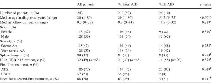
Table 1 Pretreatment patients’ characteristics comparing patients with and without concomitant AID
All patients Without AID With AID P value
Number of patients,n (%) 243 219 (90) 24 (10)
Median age at diagnosis, years (range) 20 (1–80) 20 (1–80) 51.5 (9–75) <0.001a
Median follow-up, years (range) 9.3 (0–33) 9.3 (0–33) 11.5 (0–32) 0.219a
Sex,n (%) Female 115 (47) 106 (46) 9 (38) 0.310b Male 128 (53) 113 (54) 15 (62) Severity,n (%) Severe AA 115(47) 101 (46) 14 (58) 0.255b Very severe AA 128 (53) 118 (54) 10 (42) Splenectomy,n (%) 89 (37) 81 (37) 8 (33) 0.723b HLA DRB1*15 present,n (%) 32 (49) (n=65) 21 (47) (n=45) 11 (55) (n=20) 0.598b First-line treatment,n (%) ATG 186 (77) 164 (75) 22 (92) 0.019b HSCT 57 (23) 55 (25) 2 (8)
Need for a second-line treatment,n (%) 68 (28) 63 (29) 5 (21) 0.441b
aMann–Whitney U test
T able 2 Characteristics of the 24 patients with concomitant AID and SAA UPN Sex Age at diagnosis of SAA (years) Autoimmune disorder Severity of AA HLA- DR15 T ime interval diagnosis AID –SAA Splenectomy Therapy for AID Remission of SAA Outcome AID after A T G First-line therapy: A TG, AID before AA 292 M 2 2 Diabetes type 1 vSAA No 232 months before Y es Insulin NR No change 352 F 7 1 Eosinophilic fasciitis SAA Y es 3 months before No No therapy CR Remission 353 M 9 Chron. juv . polyarthritis vSAA No 29 months before Y es Not known CR Remission 499 M 6 3 Autoimmune gastritis SAA Y es 5 1 months before No V it. B12 substitution PR No change 580 M 6 2 Derm. herp. Duhring vSAA Y es 2 6 months before No Danazol, steroids, dapsone CR No change Celiac disease 27 months after Diet NA Hashimoto thyroiditis 128 months after Not known NA 747 M 4 9 Autoimmune gastritis SAA Y es A t same time No V it B12 substitution PR No change 748 M 7 2 Psoriasis SAA No Before, not specified No Steroids PUV A N R N o change 778 M 5 7 Sjogren syndrome SAA Y es 259 months before No Prednisone PR No change 786 M 3 1 Autoimmune gastritis SAA No 1 months before No No periodic vit. B12 substitution CR No change 883 F 7 1 ITP vSAA Y es 181 months before Y es Danatrol, steroids CR No change 919 M 7 5 Microscopic polyangitis SAA No 5 months before No Cyclophosphamide, prednisone, mesna PR No change 1052 M 5 5 Colitis ulcerosa SAA Y es 139 months before No Not known PR No change 1248 F 6 5 Guillain –Barré syndrome SAA Unknown 61 months before No IvIg NR Remission before AT G Small vessel vasculitis 1 month after Cortisone injections NA First-line therapy: A TG, AID after AA 19 M 3 1 Systemic sclerosis SAA Y es 330 months after Y es Not known CR NA 39 M 1 0 Psoriasis vSAA Unknown Not known, after Y es Not known CR NA 126 M 2 6 Autoimmune thyroiditis vSAA No 276 months after Y es Hormone replacement CR NA 286 F 1 5 Graves ’ disease SAA Unknown 232 months after No Thyreostatic treatment CR NA 291 F 5 5 Autoimmune gastritis SAA No 238 months after Y es V it. B12 surveillance CR NA 501 F 2 2 Hashimoto thyroiditis vSAA Y es 123 months after No Hormone replacement CR NA Antiphospholipid syndrome 108 months after No NA 507 F 6 2 SLE with vasculitic neuropathy SAA No 87 months after No Cyclophosphamide, steroids, azathioprine CR NA Hashimoto thyroiditis 86 months after No NA Autoimmune gastritis 61 months after No NA 513 F 1 0 Guillain –Barré syndrome vSAA Y es 7 months after No IvIg, steroids CR NA 646 M 5 6 Autoimmune gastritis SAA Y es 1 2 months after No V it. B12 substitution CR NA
contrast, the cumulative incidence of AA patients with concomitant AID increases predominantly after the fifth
decade of life (Fig.1c).
In multivariate analysis comparing patients with and without AID, there were no differences with respect to
gender (hazard ratio [HR]=0.540; 95%CI=0.232–1.260;
P=0.154), severity of the disease (HR=1.343; 95%CI= 0.503–3.588; P=0.556), presence of HLA-DRB1*15 (HR= 0.731; 95%CI=0.297–1.796; P=0.494), type of treatment (HR=0.994; 95%CI =0.406–2.434; P=0.990), and splenec-tomy (HR =0.987; 95%CI=0.214–4.547; P=0.987).
We evaluated the influence of the AA treatment on the outcome of the concomitant AID in the 12 patients where the AID appeared before the diagnosis of AA (one patient was not included because Guillain–Barré syn-drome resolved before ATG therapy). Five patients obtained complete response (CR = 41.5%) of the AA, five obtained partial response (PR= 41.5%), and two were nonresponders (NR = 17%). Complete response of both diseases (AA and AID) was observed only in two out of 12 (17%) patients. One patient presented an eosinophilic fasciitis and the other a chronic juvenile polyarthritis. In the other ten patients, the course of the AID was not changed by the treatment of AA.
In 13 patients, the AID occurred after first-line therapy for AA (11 patients, ATG; two patients, HSCT); we evaluated the influence of this treatment on the develop-ment of the concomitant AID. Two out of 13 patients had had a first AID before diagnosis and therapy for AA. The median time from ATG treatment to diagnosis of the first
AID was 7 years (range 3 months–27.5 years). At onset of
the AID, the AA was in CR in seven patients and in PR in three patients. In two patients, there was NR and in one patient, the type of response was unknown. After allogeneic HSCT, two patients developed an AID. One patient with graft rejection and subsequent autologous reconstitution developed Graves’ disease 2 years later. He is now in CR of the AA since more than three decades. A second patient developed an immune thrombocytopenia controlled by splenectomy. None of their donors had had a documented AID.
Discussion
In this study, we show that about one in ten AA patients will develop a concomitant AID during their lifetime, which can appear at any time before and/or after the onset of the AA. The frequency of a concomitant AID is higher in older AA patients. Hence, more than 25% of AA patients diagnosed after 50 years of age presented a concomitant AID. The main type of concomitant AID appeared to be either gastritis or thyroiditis. AA response to ATG was
T able 2 (continued) UPN Sex Age at diagnosis of SAA (years) Autoimmune disorder Severity of AA HLA- DR15 T ime interval diagnosis AID –SAA Splenectomy Therapy for AID Remission of SAA Outcome AID after A T G First-line therapy: HSCT , AID after AA 2 F 30 Graves ’ disease SAA Unknown 21 months after No Radiojod-therapy CR, AR NA 521 M 4 5 ITP vSAA No 9 months after Y es Splenectomy CR NA The patients are grouped according to their first-line therapy (A TG versus HACS) and the time of appearance of the AID (before SAA, after first-line th erapy). Some of the patients presented more than one AID SAA severe aplastic anemia, NR no response, vSAA very severe aplastic anemia, CR complete remission, AR autologous reconstitution, PR partial remission, NA not applicable
similar in patients with or without AID, but AID response to ATG was poor.
There are some particular features of the AID occurring in AA patients. In contrast to the general population where
AID are more common in females [19], in AA patients,
AID were more frequently observed in males (15 out of 24, 62%). Interestingly, we did not observe cases of rheumatoid arthritis and only one case of systemic lupus erythematosus. HLA-DRB1*15 has been shown to be involved in the development and the outcome of AA and other
autoim-mune disorders [20–23]. It seems unlikely that
HLA-DRB1*15 plays a relevant role in the appearance of concomitant AID in AA patients.
Most cases of acquired AA can be considered as autoimmune disorders characterized by T cell-mediated, organ-specific destruction of bone marrow hematopoietic cells. However, the usual trigger of this autoimmune reaction remains unclear. In individual patients, the aberrant immune response can sometimes be linked to a viral infection or to drug or chemical exposure. There is much less evidence for other mechanisms including the associa-tion with other AID. In consideraassocia-tion of the high frequency of a concomitant AID in AA patients, it is unlikely that both diseases appear together just by chance. It, therefore, raises the question of related pathophysiologic mechanisms. In posthepatitis AA, which typically occurs in young, Fig. 1 Age repartition at
diagno-sis of AA without AID (a) and with AID (b). The data are presented in percentage of the whole group. Patients are divided into three age groups: <20, 20–50, and >50 years (P<0.001). AA patients without AID are significantly younger than AA patients with a concomitant AID. c Cumulative incidence of AA and the concomitant AID according to the age at diagnosis of each disease. The slope of the curve is different for both dis-eases. In AA, the cumulative incidence increases mainly dur-ing the first three decades of age, whereas in concomitant AID, the cumulative incidence increases predominantly after the fifth decade of life
healthy males with self-limited but severe liver inflamma-tion, a common inciting infectious cause could be involved
[24]. Indeed, in hepatitis-associated AA, similar skewed T
cell repertoires have been detected in the liver and in the peripheral blood lymphocytes, suggesting that a similar antigen-driven pathogenic mechanism is involved for both
diseases [25]. This might be different for the concomitant
AIDs. Here, there are arguments in favor of distinct mechanisms: the different age repartition of AA patients with and without concomitant AID; the nonresponse of the AID in patients responding to immunosuppressive treat-ment for the AA and the fact that AA is a T cell-mediated AID, while in many of the concomitant AID, autoanti-bodies are involved. Common genetic backgrounds, addi-tional immunogenetic, environmental, or hormonal factors may be responsible for the formation of subsets of AID
clustering [26]. The AIDs occurring after successful
allogeneic HSCT with full donor chimerism do not probably belong to the same category. Late secondary autoimmune-like phenomena have been described after allogeneic HSCT as a possible consequence of skewed
immune reconstitution [27].
The older age of our AA patients with concomitant AID suggests that immunosenescence could play a role. Recent studies in healthy octogenarian patients indicate that the immune system, instead of suffering a general-ized deterioration, undergoes a remodeling/readjustment of its major functions. Two divergent phenomena may coexist in immunosenescence: a decrease in the capacity of immune response and, simultaneously, autoantibody
production [28].
Our study has limitations arising mainly from its retrospective, single-center character and the lack of a control population. There is a relatively small number of patients at risk; however, considering that AA is a rare disease, this is the first and largest study reporting on the frequency of concomitant AID in AA patients followed up systematically over a long period of time. The advantage of a single-center study is the homogeneity of therapeutical approaches and the consistent follow-up. At last control, 80% of the long-term survivors had a follow-up of more than 7 years.
In conclusion, in this study, we show that the development of a concomitant AID is frequent, partic-ularly in older AA patients. The AID may appear at any time before or after the AA, and the outcome of the AA is not impaired by the concomitant AID, but the AID does not usually respond to the immunosuppression applied for the AA. The difference in response to ATG therapy between AA and AID suggests independent underlying immune mechanisms. Alternatively, one of these diseases could be the trigger for a second immune dysregulation.
Acknowledgements This work was supported by grants of the Horten Foundation and the Swiss National Research Foundation grant no. 3200 BO-118/76. We thank Katherine Perret and Mariana Gimpelewicz for the English corrections.
Conflicts of interest The authors declare no competing financial interests.
References
1. Young NS, Calado RT, Scheinberg P (2006) Current concepts in the pathophysiology and treatment of aplastic anemia. Blood 108:2509–2519
2. Antic M, Lautenschlager S, Itin PH (2006) Eosinophilic fasciitis 30 years after—what do we really know? Report of 11 patients and review of the literature. Dermatology 213:93– 101
3. Hinterberger-Fischer M, Kier P, Forstinger I, Lechner K, Kornek G, Breyer S et al (1994) Coincidence of severe aplastic anaemia with multiple sclerosis or thyroid disorders. Report of 5 cases. Acta Haematol 92:136–139
4. Lytton SD, Denton CP, Nutzenberger AM (2007) Treatment of autoimmune disease with rabbit anti-T lymphocyte globulin: clinical efficacy and potential mechanisms of action. Ann N Y Acad Sci 1110:285–296
5. Camitta BM, Thomas ED, Nathan DG, Santos G, Gordon-Smith EC, Gale RP et al (1976) Severe aplastic anemia: a prospective study of the effect of early marrow transplantation on acute mortality. Blood 48:63–70
6. Viollier R, Tichelli A (2000) Predictive factors for cure after immunosuppressive therapy of aplastic anemia. Acta Haematol 103:55–62
7. Tichelli A, Schrezenmeier H, Socié G, Marsh J, Chapion K, Passweg J A (2002) radomized controlled study in newly diagnosed severe aplastic anemia patients receiving antilympho-cyte globulin, cyclosporin A, with or without G-CSF: a study from the EBMT, Severe Aplastic Anemia Working Party. Available at http://www.ebmt.org/5WorkingParties/AAWP/wpar-ties-aa5.html
8. Speck B, Tichelli A, Widmer E, Harder F, Kissling M, Wursch A et al (1996) Splenectomy as an adjuvant measure in the treatment of severe aplastic anaemia. Br J Haematol 92:818–824
9. Tichelli A, Passweg J, Nissen C, Bargetzi M, Hoffmann T, Wodnar-Filipowicz A et al (1998) Repeated treatment with horse antilymphocyte globulin for severe aplastic anaemia. Br J Haematol 100:393–400
10. Camitta BM (2000) What is the definition of cure for aplastic anemia? Acta Haematol 103:16–18
11. Albers JW, Kelly JJ Jr (1989) Acquired inflammatory demyelin-ating polyneuropathies: clinical and electrodiagnostic features. Muscle Nerve 12:435–451
12. Boin F, Hummers LK (2008) Scleroderma-like fibrosing disor-ders. Rheum Dis Clin North Am 34:199–220
13. Dayan CM, Daniels GH (1996) Chronic autoimmune thyroiditis. N Engl J Med 335:99–107
14. Hochberg MC (1995) Classification criteria for childhood arthritic diseases. J Rheumatol 22:1445–1446
15. McMillan R (2007) The pathogenesis of chronic immune thrombocytopenic purpura. Semin Hematol 44:S3–S11
16. Nikolaus S, Schreiber S (2007) Diagnostics of inflammatory bowel disease. Gastroenterology 133:1670–1689
17. Strickland RG, Mackay IR (1973) A reappraisal of the nature and significance of chronic atrophic gastritis. Am J Dig Dis 18:426–440 18. Gratwohl A, Passweg J, Bocelli-Tyndall C, Fassas A, van Laar JM, Farge D et al (2005) Autologous hematopoietic stem cell transplantation for autoimmune diseases. Bone Marrow Transplant 35:869–879
19. Eaton WW, Rose NR, Kalaydjian A, Pedersen MG, Mortensen PB (2007) Epidemiology of autoimmune diseases in Denmark. J Autoimmun 29:1–9
20. Callander M, Haghighi S, Landtblom AM, Ahlgren CE, Nilsson SI, Rydberg L et al (2007) Multiple sclerosis immunopathic trait and HLA-DR(2)15 as independent risk factors in multiple sclerosis. Mult Scler 13:441–445
21. Graham RR, Ortmann W, Rodine P, Espe K, Langefeld C, Lange E et al (2007) Specific combinations of HLA-DR2 and DR3 class II haplotypes contribute graded risk for disease susceptibility and autoantibodies in human SLE. Eur J Hum Genet 15:823–830 22. Kapustin SI, Popova TI, Lyschov AA, Togo AV, Abdulkadyrov
KM, Blinov MN (1997) HLA-DR2 frequency increase in severe aplastic anemia patients is mainly attributed to the prevalence of DR15 subtype. Pathol Oncol Res 3:106–108
23. McCombe PA, Csurhes PA, Greer JM (2006) Studies of HLA associations in male and female patients with Guillain–Barre syndrome (GBS) and chronic inflammatory demyelinating poly-radiculoneuropathy (CIDP). J Neuroimmunol 180:172–177 24. Brown KE, Tisdale J, Barrett AJ, Dunbar CE, Young NS (1997)
Hepatitis-associated aplastic anemia. N Engl J Med 336:1059– 1064
25. Lu J, Basu A, Melenhorst JJ, Young NS, Brown KE (2004) Analysis of T-cell repertoire in hepatitis-associated aplastic anemia. Blood 103:4588–4593
26. Theander E, Jacobsson LT (2008) Relationship of Sjogren’s syndrome to other connective tissue and autoimmune disorders. Rheum Dis Clin North Am 34:935–947
27. Trendelenburg M, Gregor M, Passweg J, Tichelli A, Tyndall A, Gratwohl A (2001) Altered immunity syndrome, a distinct entity in long-term bone marrow transplantation survivors? Bone Marrow Transplant 28:1175–1176
28. Ramos-Casals M, Garcia-Carrasco M, Brito MP, Lopez-Soto A, Font J (2003) Autoimmunity and geriatrics: clinical significance of autoimmune manifestations in the elderly. Lupus 12:341–355