HAL Id: inserm-02966905
https://www.hal.inserm.fr/inserm-02966905
Submitted on 14 Oct 2020
HAL is a multi-disciplinary open access
archive for the deposit and dissemination of
sci-entific research documents, whether they are
pub-lished or not. The documents may come from
teaching and research institutions in France or
abroad, or from public or private research centers.
L’archive ouverte pluridisciplinaire HAL, est
destinée au dépôt et à la diffusion de documents
scientifiques de niveau recherche, publiés ou non,
émanant des établissements d’enseignement et de
recherche français ou étrangers, des laboratoires
publics ou privés.
Do RA associated HLA-DR molecules bind citrullinated
peptides or peptides from PAD4 to help the
development of RA specific antibodies to citrullinated
proteins?
Nathalie Balandraud, Isabelle Auger, Jean Roudier
To cite this version:
Nathalie Balandraud, Isabelle Auger, Jean Roudier. Do RA associated HLA-DR molecules bind
cit-rullinated peptides or peptides from PAD4 to help the development of RA specific antibodies to
citrul-linated proteins?. Journal of Autoimmunity, Elsevier, 2020, pp.102542. �10.1016/j.jaut.2020.102542�.
�inserm-02966905�
Journal of Autoimmunity xxx (xxxx) xxx
Available online 11 September 2020
0896-8411/© 2020 The Authors. Published by Elsevier Ltd. This is an open access article under the CC BY license (http://creativecommons.org/licenses/by/4.0/).
Do RA associated HLA-DR molecules bind citrullinated peptides or peptides
from PAD4 to help the development of RA specific antibodies to
citrullinated proteins?
Nathalie Balandraud
a,b,1, Isabelle Auger
a,1, Jean Roudier
a,b,* aAix Marseille University, INSERM, UMRs1097, Marseille Luminy, FrancebAPHM, Rheumatology, IML, Marseille, France
A R T I C L E I N F O
Keywords:
Rheumatoid arthritis HLA-DR association
Anticitrullinated protein antibodies Citrullinated peptide binding PAD4 peptide binding Prevention
A B S T R A C T
Purpose: Rheumatoid arthritis (RA) is associated with HLA-DRB1 genes encoding a five amino acid basic motive, the shared epitope SE). Each HLA-DRB1 genotype defines a genotype specific risk of developing RA. RA is preceded by the emergence of anti citrullinated protein antibodies (ACPAs). Citrullin is a neutral version of arginin, a basic amino acid, formed after post translational modification by Peptidyl Arginyl Deiminases (PADs). HLA-DRB1 genes associated with RA are also associated with ACPAs.
Two models might explain this association.
Here we tested both models for prediction of HLA-DRB1 genotypic risks of developing RA.
Methods: We calculated the likelihoods for the 2 HLA-DR molecules encoded by 12 common HLA-DRB1 geno-types to bind at least one randomly chosen peptide from PAD4 or fibrinogen(native or citrullinatd) and compared them with the 12 respective HLA-DRB1genotypic risks of developing RA.
Results: HLA-DRB1 Genotypic risks of developing RA correlate with likelihoods of binding PAD4 peptides, not citrullinated Fibrinogen peptides. Thus, the molecular basis for the association of HLA-DR and ACPA positive RA is most likely the capability for RA associated HLA-DR molecules to bind peptides(s) from PAD4.
- RA associated HLA-DR molecules might bind citrullinated peptides better than non RA associated HLA-DR molecules.
- RA associated HLA-DR molecules might bind PAD4 peptide(s) better than non RA associated HLA-DR molecules.
1. Introduction
Rheumatoid arthritis (RA) is a chronic destructive autoimmune joint disease of unknown origin.
HLA-DRB1 genes are the major genetical component of RA suscep-tibility. Indeed, HLA-DR molecules with the so called “shared epitope” (SE), a basic five amino acid motive in the third hypervariable region of their HLA-DRВ1 chain are associated with RA [1]. Both HLA-DR mole-cules expressed by an individual influence his/her risk to develop RA with Odds Ratios (ORs) ranging from 30 for HLA-DRB1 genotypes
encoding 2 RA associated HLA-DR molecules to 0.2 for HLA-DR geno-types encoding no shared epitope positive HLA-DR molecule [2].
The development of RA is preceded by the emergence of anti cit-rullinated protein antibodies (ACPAs). Citrullin is a neutral version of arginin, a basic amino acid formed after post translational modification of arginin by enzymes called Peptidyl Arginyl Deiminases (PADs).
HLA-DRB1 genes associated with RA are also associated with the presence of ACPAs in patients with RA [2]. This suggests that RA asso-ciated HLA-DR molecules predispose to RA because they contribute to the development of ACPAs.
Indeed, the function of HLA-DR molecules is to present peptides to Follicular B Helper T cells (TFH) to allow the development of IgG
anti-bodies, suggesting that RA associated HLA-DR molecules might present peptides to the TFH cells which help the development of ACPAs.
How-ever, the identity of the peptide(s) presented by RA associated HLA-DR molecules to the TFH cells which help the development of IgG ACPAs is
* Corresponding author. INSERM, UMRs1097, Parc Scientifique de Luminy, 163 avenue de Luminy, case 939, 13009, Marseille, France. E-mail address: jean.roudier@inserm.fr (J. Roudier).
1 These authors contributed equally.
Contents lists available at ScienceDirect
Journal of Autoimmunity
journal homepage: www.elsevier.com/locate/jautimm
https://doi.org/10.1016/j.jaut.2020.102542
Journal of Autoimmunity xxx (xxxx) xxx
2
unknown.
It was first proposed that RA associated HLA-DR molecules bound and presented citrullinated peptides to TFH cells allowing them to help
the development of IgG antibodies to citrullinated proteins. This was demonstrated by showing that one peptide from Vimentin: Vim 65-77, when citrullinated, bound RA associated HLA-DR molecules better than non RA associated HLA-DR molecules. This suggested that RA associated HLA-DR molecules were, in general, better than non RA associated HLA-DR molecules at binding citrullinated peptides, explaining why they were associated with ACPAs and RA [3].
However, thorough HLA-DR peptide binding studies did not confirm the preferential binding of citrullinated peptides to RA associated HLA- DR molecules [4,5].
We have proposed an alternative hypothesis, suggesting that TFH
cells recognize PAD4, the citrullinating enzyme and help the develop-ment of ACPAs of multiple specificities by a hapten carrier mechanism in which PAD4 is the carrier and proteins bound by PAD4 and being cit-rullinated are the haptens [6,7]. We found evidence for this hypothesis in mice and in humans.
In short, two models may explain the RA/ACPA/HLA-DR association. One states that RA associated HLA-DR molecules bind citrullinated peptides better, the second that RA associated HLA-DR molecules bind PAD4 peptide(s) better.
Here, we decided to evaluate how each of the two models is consistent with the risk to develop ACPA positive RA associated with 12 common HLA-DRB1 genotypes.
To do so, we used previously published binding data of 65 peptides from PAD4, 96 peptides from Fibrinogen (under their native or cit-rullinated form) to HLA-DR molecules encoded by HLA-DRB1*04:01, *04:04, *01:01, *04:02, *07:01 [4,7]. We evaluated the likelihoods of binding at least one randomly chosen peptide from PAD4 or native or citrullinated fibrinogen for the 2 HLA-DR molecules encoded by 12 different HLA-DRB1 genotypes and compared them with HLA-DRB1 genotypic risks of developing RA (2).
We found that the risk of developing RA associated with each HLA- DRB1 genotype matches the likelihood of binding at least one peptide from PAD4, but not the likelihood of binding a peptide from native or citrullinated Fibrinogen.
2. Methods
2.1. What are the relevant citrullinated antigens to test for HLA-DRB1 binding?
There is no evidence that any particular citrullinated antigen is at the origin of anti citrullinated protein immunization. We chose to test the binding of peptides from Fibrinogen because Fibrinogen is strongly expressed in the rheumatoid joint and because positivity of antibodies to citrullinated Fibrinogen matches closely that of the anti CCP2 test [8].
2.2. How to characterize the peptide binding capabilities conferred to each individual by the two HLA-DRB1 alleles he/she expresses?
To evaluate the capability for an individual with a given HLA-DRB1 genotype, to mount an efficient T cell response to a relevant, unknown, peptide from PAD4 or Fibrinogen, we calculated, for the two HLA-DR molecules expressed by each HLA-DRB1 genotype, the likelihood to bind at least one peptide from PAD4 or Fibrinogen or citrullinated Fibrinogen. We counted how many peptides tested from 65 overlapping peptides from PAD4 or 96 peptides from Fibrinogen or citrullinated Fibrinogen could be bound by HLA-DRB1*0401, *0404, *0101, *0402, *0101 molecules, with a signal as strong or higher than that obtained with HA, a peptide from the Haemophilus Influenzae hemagglutinin known to be a pan HLA-DR binder [5].
2.3. Binding of peptides from PAD4 and fibrinogen to different HLA-DR molecules
We previously published the results of a direct binding assay of peptides from PAD4 and native and citrullinated Fibrinogen to HLA-DR molecules [4,7]. Homozygous cell lines expressing SE positive HLA-DRB1*01:01 (JESTHOM), *04:01 (SAVC), and *04:04 (PEYSSON), or SE negative HLA-DRB1*04:02 (YAR) and DRB1*07:01 (MOU) were cultured in RPMI 1640 supplemented with 10% fetal calf serum. After cell lysis (in 10 mM Tris pH8, 10 mM NaCl, 10 mM MgCl2, 1% Triton X100, 0.05 mg/ml Dnase and protease inhibitors), total protein extracts were immunoprecipitated by anti-HLA-DR LB3.1 antibody covalently coupled on CNBr–activated Sepharose 4B (Sigma Aldrich, St. Quentin-Fallavier, France). After washing, HLA–DR molecules were eluted in phosphate buffered saline (pH 2) with 0.5% octyl glucoside, neutralized in 1 M Tris, and quantified.
The binding of each purified HLA–DR allele was tested on ELISA plates coated with 10 μg of peptide from PAD4 or native and
citrulli-nated fibrinogen. Peptides were synthesized using the solid-phase sys-tem and purified (>60%) (Neosyssys-tem, Strasbourg, France). Each peptide was tested in duplicate wells and, as controls, 2 empty wells (not coated with any peptide) and 2 wells coated with a positive binder, influenza hemagglutinin (HA) peptide (amino acid sequence: PKYVKQNTLKLAT) [5]. After peptide coating, plates were blocked with 1% bovine serum albumin. One microgram of each purified HLA–DR molecule was added to each well. After washing, bound HLA–DR was detected by bio-tinylated anti-HLA-DR antibody (Immunotech, Marseille, France) fol-lowed by peroxidase-conjugated avidin (Sigma Aldrich, St. Quentin-Fallavier, France). After tetramethyl benzidine incubation, optical density (OD) was read at 405 nm. Positive binding was defined as an OD value equal or higher than to the OD for the HA peptide.
2.4. Peptides from PAD4 tested for HLA-DR binding
65 overlapping peptides covering the entire PAD4, a 663 aa protein (locus NM_012387) were tested for HLA-DR binding as described above. Relevant peptides may be 10-20 amino acid long. For each peptide length, there are about 660 possible peptides on PAD4. Thus, a maximum number of 6600 PAD4 peptides may exist, and 65 peptides represent about 1% of this maximum number of possible peptides. Se-quences and HLA-DR binding properties of the 65 tested peptides from PAD4 have been published recently and are shown in Fig. 1 [7].
2.5. Peptides from fibrinogen and citrullinated fibrinogen tested for HLA- DR binding
96 overlapping peptides covering the entire alpha (631 aa) (locus NP_000499) and beta (447 aa) (locus NP_005132) chains of Fibrinogen were synthesized. A native set included 40 peptides from the alpha chain, 31 peptides from the beta chain, 13 arginin free peptides from the alpha chain and 12 arginin free peptides from the beta chain. A cit-rullinated set included the same peptides, with all arginine residues replaced by citrullins. Sequences and HLA-DR binding properties of these 96 citrullinated or native peptides from Fibrinogen have been published previously and are shown in Fig. 2 [4].
The alpha chain and beta chains of Fibrinogen encompass a total 1078 amino acids, thus may theoretically yield up to 10.000 possible peptides in the 10-20 amino acid range. Thus, 96 peptides from the alpha and beta chains of Fibrinogen represent about 1% of the maximum number of possible peptides, a coverage very similar to that of PAD4.
2.6. Likelihood for an HLA-DR molecule of binding a random peptide from a given protein
To evaluate the capability of HLA-DR molecules encoded by different HLA-DRB1 alleles of binding an unknown relevant peptide from a given
protein, we used a surrogate marker: we evaluated the likelihood of binding an unknown, randomly chosen peptide from this protein.
This can be done by studying the binding of a limited number of overlapping peptides, covering the entire protein by the HLA-DR molecule. The likelihood of binding is the ratio of the number of bound peptides divided by the total number of tested peptides.
2.7. Likelihood for one (at least) of the 2 HLA-DR molecules encoded by an HLA-DRB1 genotype of binding a random peptide from a given protein
The likelihood for none of the two HLA-DR molecules encoded by an HLA-DRB1 genotype to bind a peptide is the product of the likelihood for each HLA-DR molecule of not binding it.
The likelihood for one (at least) of the 2 HLA-DR molecules encoded by an HLA-DRB1 genotype of binding a random peptide from a protein is 1 minus the likelihood for none of the two HLA-DR molecules to bind it.
2.8. HLA-DRB1 genotypic risk of developing RA
We previously genotyped for HLA-DRB1 857 patients with ACPA positive RA and 2178 controls from South Eastern and Eastern France and calculated Odds Ratios (OR) for developing RA for 106 of 132 possible genotypes accounting for 97% of subjects. We found that HLA- DRB1 genotypic ORs for developing ACPA positive RA range from 30 to 0.2 [2].
2.9. Statistics
Correlation between the HLA-DRB1 genotypic Odds Ratios to develop RA and the likelihood to bind at least one peptide from PAD4 or Fibrinogen for this genotype was evaluated by Pearson’s correlation test. Comparison between numbers of peptides bound by each HLA-DR molecule was done by chi-square test. GraphPad Prism 5.02 (Graph-Pad Software) was used for all statistical analyses.
3. Results
3.1. Likelihood of binding one randomly chosen peptide from PAD4, native or citrullinated fibrinogen for individual HLA-DR molecules encoded by HLA-DRB1*01:01, *04:01, *04:02, *04:04, *07:01
The results of the direct binding assay of 65 peptides from PAD4, 96 citrullinated peptides from Fibrinogen and their 96 native counterparts to 5 purified HLA-DR molecules, HLA-DRB1*01:01, *04:01, *04:04, *04:02, *07:01, previously published [4,7], are presented in Figs. 1 and 2.
Each purified HLA-DR molecule bound 2 to 9 of 65 PAD4 peptides, 12 to 23 of 96 fibrinogen peptides, 11 to 23 of 96 citrullinated fibrinogen peptides.
For each HLA-DR molecule, the number of bound citrullinated Fibrinogen peptides was not different from the number of bound native Fibrinogen peptides (Chi2, N.S.).
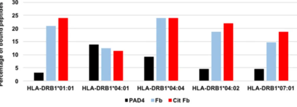
For each HLA-DR molecule, we calculated a likelihood of binding at least a peptide from PAD4, native Fibrinogen and citrullinated Fibrin-ogen. This likelihood was defined as the ratio of the number of bound peptides divided by the number of tested peptides. Likelihoods of binding ranged from 3% to 24% (Fig. 3).
Our binding data did not show differential HLA-DR binding between citrullinated or native Fibrinogen peptides, and allowed us calculate for each of 5 HLA-DR molecules, a likelihood of binding a peptide from PAD4 or native and citrullinated Fibrinogen.
Fig. 1. Binding of PAD4 peptides to HLA-DRB1*01:01, *04:01, *04:04, *04:02,
*07:01 molecules. Ten micrograms of peptide were coated to each well. One microgram of purified HLA–DR molecule was added. After washing, bound HLA–DR was detected by biotinylated anti–HLA–DR antibody followed by peroxidase-conjugated avidin and tetramethyl benzidine incubation. OD was read at 405 nm. Positive binding was defined by an OD value equal or higher than the OD for HA peptide. Numbers indicate the ratios tested peptide OD divided by HA peptide OD. Thus, a number higher than 1 indicates posi-tive binding.
Journal of Autoimmunity xxx (xxxx) xxx
4
Fig. 2. Binding of native and citrullinated fibrinogen peptides to HLA-DRB1*01:01,*04:01, *04:04, *04:02, *07:01 molecules. Ten micrograms of peptide were
coated to each well. One microgram of purified HLA–DR molecule was added. After washing, bound HLA–DR was detected by biotinylated anti–HLA–DR antibody followed by peroxidase-conjugated avidin and tetramethyl benzidine incubation. OD was read at 405 nm. Positive binding was defined by an OD value equal or higher than the OD for the HA peptide. Numbers indicate the ratios peptide signal OD divided by HA peptide OD. Thus, a number higher than 1 indicates posi-tive binding.
3.2. Correlation between Odds ratios to develop ACPA positive RA and peptide binding likelihoods for one (at least) of the two HLA-DR molecules encoded by 12 different HLA-DRB1 genotypes
We calculated the likelihood for one (at least) of the 2 HLA-DR molecules encoded by each of 12 HLA-DRB1 genotypes containing HLA-DRB1*04:01, *04:04, *01:01, *04:02, *07:01 to bind at least one peptide from PAD4 or native or citrullinated Fibrinogen, as described in the Methods section (Table 1).
We then compared the likelihood for each of the 12 sets of HLA-DR molecules of binding at least one peptide from PAD4 or native or cit-rullinated Fibrinogen with each of the 12 respective genotypic risks of developing RA, using Pearson’s rank correlation.
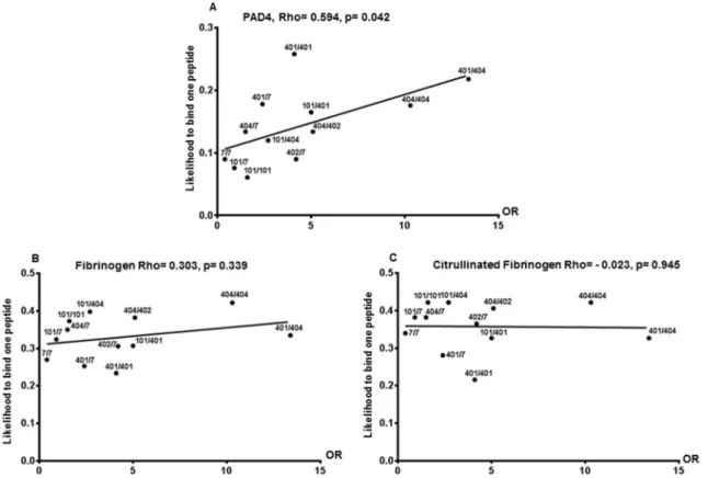
We found that genotypic risks of developing RA correlate with likelihoods of binding PAD4 peptide, not native or citrullinated Fibrin-ogen peptide (Pearson’s, p = 0.042) (Fig. 4).
4. Discussion
HLA-DRB1 genes predispose to both rheumatoid arthritis and the development of antibodies which precede and are specific of RA: anti citrullinated protein antibodies (ACPAs). ACPAs recognize citrullin residues on many different proteins. Citrullins are arginin residues which have been modified after translation by enzymes called Peptidyl Arginyl Deiminases (PAD). Citrullins are neutral while Arginins are charged and basic.
How RA associated HLA-DRB1 alleles help the development of RA is
unknown. However, since RA is preceded by ACPAs, antibodies believed to cause RA, it is assumed that HLA-DRB1 genes contribute to the development of RA by allowing the development of ACPAs. This is ex-pected, because the function of HLA-DR molecules is to present peptides to CD4 T cells, among which TFH cells which help the development of
IgG antibody responses. Still, the identity of the peptides presented by HLA-DR molecules to the TFH cells that will help the development of
ACPAs is unknown.
An early, classical hypothesis, proposed that HLA-DR molecules associated with RA were capable of binding citrullinated peptides with higher affinity than non RA associated HLA-DR molecules. The demonstration of this hypothesis relied on a classical study of the binding of one peptide from Vimentin: Vim 65-77 under its native (arginine) or citrullinated form, called Vim R70Cit, to 8 HLA-DR mole-cules, of which 3 (HLA-DRB1*04:01, *04:04, *01:01) contained the shared epitope. VimR70Cit was found to bind shared epitope positive HLA-DR molecules with higher affinity than Vim 65-77 [3]. This study was confirmed in 2018 by binding and structural studies performed with a few selected peptides including Vim 65-77 and its citrullinated variant [9].
However these findings have been challenged by many thorough peptide binding studies. The binding of 167 peptides from the alpha and beta chains of human Fibrinogen to 5 HLA-DR molecules did not give any indication of preferential binding of citrullinated fibrinogen pep-tides to SE positive HLA-DR molecules [4]. More recently, Sette et al. studied the binding of 200 peptides from Collagen, Fibrinogen, Aggre-can and Vimentin (including Vim 65-77) to 28 different HLA-DR mole-cules and failed to demonstrate a higher binding affinity of citrullinated peptides to SE positive HLA-DR molecules [5].
We have developed an alternative hypothesis. Indeed, the emergence of ACPAs is often preceded by the development of anti PAD4 IgG anti-bodies [10]. In normal mice, immunization with PADs can trigger the development of ACPAs by a hapten carrier mechanism [6]. Similarly, RA patients have both antibodies and T cell responses to PAD4, suggesting that the target for the helper T cells involved in the development of ACPAs may be not citrullinated antigens but PADs [7].
To sort between the two hypotheses, we evaluated which one matches HLA-DRB1 genotypic risks for the development of ACPA posi-tive RA better. Indeed, the risk to develop ACPA posiposi-tive RA can be calculated based on an individual’s HLA-DRB1 genotype. In 2013, we calculated these risks for 106 of the 136 most common HLA-DRB1 ge-notypes in France [2]. Here, we compared the risks (Odds Ratios ORs) of developing ACPA positive RA for 12 common HLA-DRB1 genotypes with the likelihood for one at least of the two HLA-DR molecules encoded by each genotype to bind one randomly chosen peptide from PAD4, native and citrullinated Fibrinogen. We found that HLA-DRB1 genotypic ORs to develop RA correlate with the likelihood for at least one of the encoded HLA-DR molecules to bind one randomly chosen peptide from PAD4 but not from citrullinated or native Fibrinogen.
This result suggests that HLA-DRB1 genes act on the triggering of ACPA immunization and the development of RA by allowing presenta-tion of peptide(s) from PAD4, according to a classical hapten carrier
Fig. 3. Percentages of peptides bound by purified
HLA-DR molecules encoded by HLA- DRB1*01:01,*04:01, *04:04, *04:02, *07:01. The in-dividual binding of each of 65 peptides from PAD4, each of 96 native or citrullinated peptides from Fibrinogen to purified HLA-DR molecules encoded by HLA-DRB1*01:01, *04:01, *04:04, *04:02, *07:01 was tested by direct binding to purified HLA-DR. The ratios number of bound peptides/number of tested peptides define a likelihood of binding specific for a protein and an HLA-DR molecule. PAD4: Peptidyl Arginyl Deiminase 4, Fb: fibrinogen, Cit Fb: citrulli-nated fibrinogen.
Table 1
HLA-DRB1 genotypic risk for RA and likelihood to bind PAD4 or Fibrinogen peptides for each of 12 different HLA-DRB1 genotypes.
HLA-DRB1*
Genotypes Risk to develop RA (Odds Ratio) Likelihood to bind at least one peptide from Native Citrullinated PAD4 Fibrinogen Fibrinogen
DRB1*07/07 0.4 0.090 0.270 0.340 DRB1*01:01/07 0.9 0.076 0.324 0.382 DRB1*04:04/07 1.5 0.134 0.350 0.382 DRB1*01:01/ 01:01 1.6 0.061 0.373 0.422 DRB1*04:01/07 2.4 0.178 0.253 0.281 DRB1*01:01/ 04:04 2.7 0.120 0.398 0.422 DRB1*04:01/ 04:01 4.1 0.258 0.234 0.216 DRB1*04:02/07 4.2 0.090 0.306 0.365 DRB1*01:01/ 04:01 5 0.165 0.307 0.327 DRB1*04:04/ 04:02 5.1 0.134 0.382 0.406 DRB1*04:04/ 04:04 10.3 0.176 0.422 0.422 DRB1*04:01/ 04:04 13.4 0.218 0.335 0.327
Journal of Autoimmunity xxx (xxxx) xxx
6
model in which PAD4, the citrullinating enzyme, is the carrier. Thus, B lymphocytes recognizing citrullinated epitope(s) on any protein being citrullinated by PAD4 may process the PAD4/citrullinated protein complex, present PAD4 peptide on HLA-DR and benefit from the help of PAD4 peptide specific TFH cells (7).
We compared the capabilities of the HLA-DR molecules encoded by 12 different genotypes to bind peptides from PAD4, native or citrulli-nated Fibrinogen. We found that the risk to develop RA, associated with each pair of HLA-DR molecules, matches the pair’s capability to bind a peptide from PAD4, not a peptide from citrullinated or native Fibrin-ogen. This finding is consistent with the T cell proliferation and acti-vation data in patients with RA. Indeed, We found that T cell response to the PAD4 protein, but not to citrullinated or native Fibrinogen is com-mon and associated with antibodies to PAD4 and the shared epitope of HLA-DR, in RA patients, not controls. Furthermore, p8, the peptide from PAD4 which binds HLA-DR best according to our binding data is recognized by T cells in almost half of RA patients, not controls and this is associated with shared epitope positive HLA-DR alleles (7).
Thus, far from solving a purely theoretical issue, our findings identify PAD4 as an important triggering antigen in the development of RA. Their practical implication is the prevention of RA by PAD4 tolerization in high risk individuals identified by their high risk HLA-DRB1 geno-types (2).
Funding
INSERM and Fondation Arthritis. Declaration of competing interest
None.
References
[1] P.K. Gregersen, J. Silver, R.J. Winchester, The shared epitope hypothesis: an approach to understanding the molecular genetics of susceptibility to rheumatoid arthritis, Arthritis Rheum. 30 (1987) 1205–1213.
[2] N. Balandraud, C. Picard, D. Reviron, C. Landais, E. Toussirot, J. Roudier, HLA- DRB1 genotypes and the risk of developing anti citrullinated protein antibody (ACPA) positive rheumatoid arthritis, PloS One 8 (2013), e64108, https://doi.org/ 10.1371/journal.pone.0064108.
[3] J.A. Hill, S. Southwood, A. Sette, A.M. Jevnikar, D.A. Bell, E. Cairns, Cutting edge: the conversion of arginine to citrulline allows for a high-affinity peptide interaction with the rheumatoid arthritis associated HLA-DRB1*0401 MHC class II molecule, J. Immunol. 171 (2003) 538–541.
[4] I. Auger, M. Sebbag, C. Vincent, N. Balandraud, S. Guis, L. Nogueira, B. Svensson, A. Cantagrel, G. Serre, J. Roudier, Influence of HLA-DR genes on the production of rheumatoid arthritis-specific autoantibodies to citrullinated fibrinogen, Arthritis Rheum. 52 (2005) 3424–3432.
[5] J. Sidney, S. Becart, M. Zhou, K. Duffy, M. Lindvall, E.C. Moore, E.L. Moore, T. Rao, N. Rao, M. Nielsen, B. Peters, A. Sette, Citrullination only infrequently impacts peptide binding to HLA class II MHC, PloS One 12 (2017), e0177140. [6] F. Arnoux, C. Mariot, E. Peen, N. Lambert, N. Balandraud, J. Roudier, I. Auger,
Peptidyl arginyl deiminase immunization induces anti citrullinated protein antibodies in mice with particular MHC types, Proc. Natl. Acad. Sci. U.S.A. 114 (2017) E10169–E10177.
[7] I. Auger, N. Balandraud, E. Massy, M. Hemon, E. Peen, F. Arnoux, C. Mariot, M. Martin, P. Lafforgue, J.M. Busnel, J. Roudier, Peptidyl arginine deiminase autoimmunity and the development of ACPA in rheumatoid arthritis. The "hapten carrier" model, Arthritis Rheum. 72 (2020) 903–911, https://doi.org/10.1002/ art.41189.
[8] C. Masson-Bessiere, M. Sebbag, E. Girbal-Neuhauser, L. Nogueira, C. Vincent, T. Senshu, G. Serre, The major synovial target of the rheumatoid arthritis-specific antifilaggrin autoantibodies are deiminated forms of the a and b chains of fibrin, J. Immunol. 166 (2001) 4177–4184.
[9] Y.T. Ting, J. Petersen, S.H. Ramarathinam, S.W. Scally, K.L. Loh, R. Thomas, A. Suri, D.G. Baker, A.W. Purcell, H.H. Reid, J. Rossjohn, The interplay between citrullination and HLA-DRB1 polymorphism in shaping binding hierarchies in rheumatoid arthritis, J. Biol. Chem. 293 (2018) 3236–3251.
[10] J.R. Kolfenbach, K.D. Deane, L.A. Derber, C.I. O’Donnell, W.R. Gilliland, J. D. Edison, A. Rosen, E. Darrah, J.M. Norris, V.M. Holers, Autoimmunity to peptidyl arginine deiminase type 4 precedes clinical onset of rheumatoid arthritis, Arthritis Rheum. 62 (2010) 2633–2639.
Fig. 4. Correlation between OR to develop RA and likelihood to bind at least one peptide from PAD4 or native or citrullinated Fibrinogen. Correlation between HLA-
DRB1 genotypic risks (OR) for RA and likelihood of binding PAD4 (A), Fibrinogen (B) or citrullinated Fibrinogen (C) peptides for a given genotype was evaluated by Pearson’s rank correlation. Each HLA-DRB1 genotype is indicated by a black dot.