HAL Id: hal-03060060
https://hal.archives-ouvertes.fr/hal-03060060
Submitted on 13 Dec 2020HAL is a multi-disciplinary open access archive for the deposit and dissemination of sci-entific research documents, whether they are pub-lished or not. The documents may come from teaching and research institutions in France or
L’archive ouverte pluridisciplinaire HAL, est destinée au dépôt et à la diffusion de documents scientifiques de niveau recherche, publiés ou non, émanant des établissements d’enseignement et de recherche français ou étrangers, des laboratoires
Controlled crystallization of hierarchical monoliths
composed of nano-zeolites
Kassem Moukahhal, Nghia Le, Magali Bonne, Joumana Toufaily, Tayssir
Hamieh, T Daou, Bénédicte Lebeau
To cite this version:
Kassem Moukahhal, Nghia Le, Magali Bonne, Joumana Toufaily, Tayssir Hamieh, et al.. Controlled crystallization of hierarchical monoliths composed of nano-zeolites. Crystal Growth & Design, Amer-ican Chemical Society, 2020. �hal-03060060�
Controlled crystallization of hierarchical monoliths
composed of nano-zeolites
Kassem Moukahhal1,2,3, Nghia Huu Le1,2, Magali Bonne1,2, Joumana Toufaily3, Tayssir Hamieh3,
T. Jean Daou1,2*, Bénédicte Lebeau1,2*
1) Université de Haute Alsace (UHA), CNRS, IS2M UMR 7361, F-68100 Mulhouse, France
2) Université de Strasbourg, F-67000 Strasbourg, France
3) Laboratory of Materials, Catalysis, Environment and Analytical Methods Faculty of Sciences,
Section I, Lebanese University Campus Rafic Hariri, Hadath, Lebanon
KEYWORDS. Zeolite monolith, hierarchical zeolite, pseudomorphic transformation, ZSM-5,
shaping, nanosheets.
ABSTRACT. Micro/macroporous and meso/macroporous amorphous silica monoliths have been
prepared and transformed into ZSM-5 nanosheets zeolitic macroporous monoliths. The
hierarchized zeolite monoliths have been optimized in term of crystallinity rate by varying
durations and temperatures of hydrothermal treatments. Fully crystallized ZSM-5 nanosheets
monoliths were obtained after a combination of two consecutive hydrothermal treatments at 150
monolith similar in shape and dimensions to the original amorphous micro/macroporous silica
monolith was obtained, but a partial reduction in size has been observed for the ZSM-5 nanosheets
monolith obtained from the amorphous meso/macroporous silica monolith due to a partial
dissolution. Results showed that the macropores of the parent silica monoliths were preserved after
the transformation with similar diameter sizes but with a wider pores size distributions and
additional mesopores systems thanks to the nanosheets morphology.
INTRODUCTION
Zeolites are crystalline aluminosilicate materials commonly used in applications related to
adsorption, separation and catalysis 1–4. Zeolites are widely used as solid catalysts in the
(petro)chemical industry thanks to their high intrinsic acidity and thermal stability 5–11. The
micropores network in zeolites is very well controlled in terms of size and topology. However, the
micropore dimensions in major lower than 1 nm is a limit for molecules with kinetic diameter
larger than pore entrance. Moreover, the size of zeolite crystals that is in the few micron range is
limiting for diffusion within the microporous network and thus for the accessibility to active sites.
Consequently, to enlarge the application field of zeolites in catalysis to voluminous molecules it is
necessary to favor both their accessibility to the active sites and their diffusion within the solid
porosity 12,13. To reach these goals several investigations have been oriented not only on the
preparation of zeolites with larger pores and nanosized zeolites 14–21, but also on the synthesis of
zeolites with hierarchized porosity. Such hierarchized zeolites possess at least two grades of pore
systems including mesopores and/or macropores with the intrinsic zeolite micropores 8,22–28.
desilication or dealumination 31, hard-templating method 32–34, soft templating method 35–40 and
dual templating route with bifunctional surfactants 41–44. These latter are composed of long-chain
alkyl groups and polar quaternary ammonium groups separated by a short alkyl spacer: the
quaternary ammonium groups can generate the zeolite structure, while hydrophobic alkyl chains
block the zeolitic framework growth along one direction and can assemble into a mesoscale
micellar structure to create mesoporosity. This approach is quite interesting because the nature of
the bifunctional agent and the synthesis conditions allow controlling the morphology (e.g.
nanosheet, nanosponge) and the texture of the produced zeolitic materials 41–45.
The micron-size of zeolites crystals (0.5–25 µm) is also a limit for their use in some processes such
as continuous fluid flow ones that require shaping into larger particles. Zeolites must be therefore
shaped at the macroscopic scale with high mechanical resistance. Several methods have been
developed for shaping zeolite particles. In most processes, the zeolite particles are shaped as
extruded. Extrusion methods involve several steps usually including/following grinding, sieving,
binder additions and extrusion. Although these extruded particles exhibit high mechanical
resistance, they usually present an uncontrolled secondary porosity and require several stages of
development. In addition, the use of inorganic binders (e.g. alumina, clays) and organic binders
(e.g. carboxymethylcellulose) can strongly affect the catalytic and adsorption properties of the
material if they are used in a large amount. As a result, many efforts have been brought on the
implementation of methods for shaping zeolite particles without the addition of binders or by using
small amounts of binders (5 wt.%). These methods are mainly based on the compaction of zeolitic
powders, or the use of a mixer, introduction of sacrificial macrotemplates (e.g. resin), the
hydrothermal transformation of dense aluminosilicate gels and pseudomorphic transformation of
Pseudomorphism that consists in transforming one material to another one without modifying the
initial morphology is particularly interesting for materials shaping since it is a one-step and
binderless method 51. It involves the dissolution of the parent material with the simultaneous
precipitation or crystallization of the targeted material without any change of the external shape.
This concept has been used by Galarneau’s group for the synthesis of beads of 41 and
MCM-48 types mesoporous silica-based materials with micron-size (5–800 µm) from amorphous silica
beads of the same size 52,53. More recently, this method was extended to the synthesis of zeolite
beads (from 10 µm to 1 mm diameter) of type SOD, LTA, FAU-X, and cylindrical zeolite
monoliths (of 3-5 cm in length) of type LTA, FAU-X, from amorphous mesoporous silica beads
and amorphous meso/macroporous silica monoliths, respectively 54,55. The resulting LTA
monoliths showed high adsorption capacities for decontamination radioactive aqueous effluents in
continuous flow thanks to their fast ion-exchange kinetics 56. Our group has recently described the
one-step synthesis of ZSM-5 nanosheets beads with different sizes by using the pseudomorphism
concept 57. Here, we describe the pseudomorphic transformation of two types of amorphous silica
monoliths having bimodal micro/macro and meso/macro porosity, into ZSM-5 nanosheets
monoliths.
The amorphous silica monoliths with sponge-like continuous macroporous network have been
prepared by the Nakanishi’s method 58,59. The resulting macroporous monoliths present an
interparticular microporosity between silica nanoparticles constituting the silica skeleton. To
release the porosity a thermal treatment is usually applied but results in the partial destruction of
the macroscopic shape 56. Therefore, a post-synthesis treatment with a base is realized to create
mesoporosity that confers to the monoliths the mechanical stability necessary to stand the thermal
immersing monoliths in a basic solution to generate disordered mesopores in the silica skeleton of
monolith. This treatment involves the Ostwald ripening within the silica network to create
interparticular mesoporosity and improve the silica condensation. Temperature of the treatment
and basicity of the solution can be varied to control the mesopores size 61,63. In the present work,
meso/macroporous amorphous silica monoliths have been synthesized by a post-treatment with a
base but the synthesis of silica monoliths was also optimized to obtain for the first time
mechanically stable micro/macroporous monoliths. The transformation of porous amorphous silica
monoliths into hierarchized ZSM-5 zeolite monoliths was realized by following the protocol
developed for the ZSM-5 nanosheets beads 57.
EXPERIMENTAL SECTION
Synthesis of macroporous silica monoliths with microporosity. The macroporous silica
monoliths with microporosity were prepared by optimizing the protocol described by Galarneau’s
group60,62, which was inspired by the original work of Nakanishi 58,59. In a first step, a very accurate
amount of tetraethoxysilane (TEOS, Sigma-Aldrich) (12.62 g) was weighted with the aid of a
plastic syringe and kept in a refrigerator at 0 °C for 1 h. A precise amount (1.690 g) of 35 kDa
poly(ethylene oxide) (PEO, Sigma-Aldrich)) was weighted and introduced in a 50 mL beaker
containing a mixture of precisely measured 16 mL of distilled water and 1 mL of nitric acid. The
beaker was placed in a laboratory glass crystallizer well surrounded by ice (constant bath
temperature of 0 °C) and the mixture was stirred for 30 min under magnetic stirring at 400 – 500
rpm until total polymer dissolution. In a second step, the cold TEOS (0 °C) was directly added
from the syringe to the polymer solution in the beaker under stirring. The stirring was maintained
to obtain a homogeneous gel. This stirring step is important for optimizing the mechanical stability
to two different stirring ways were applied: (regime A) the solution was let for only 30 min at 400
rpm or (regime B) the solution was let for 30 min at 250 rpm and then for 30 min at 400 rpm. The
final molar composition of the gel is: 1SiO2: 4EtOH: 0.63EO: 0.25HNO3: 14.8H2O (EtOH stands
for ethanol obtained from the hydrolysis of TEOS; EO stands for ethylene oxide unit). In a third
step, 12 poly(vinyl chloride) (PVC) tubes (8 mm diameter and 40 mm length) stored in the
refrigerator at 0 °C were filled with the cold TEOS/PEO/HNO3/H2O mixture. The tubes were then
closed, sealed with Parafilm M® and put in an oven at 40 °C for 72 h. In a fourth step, monoliths
were removed from the PVC tubes and washed in 1 L of distilled water at room temperature.
Distilled water was renewed every 2 h (about 10 times) until a neutral pH was reached. In a last
step, monoliths were dried at room temperature. The monoliths were weighted every 12 h to follow
the loss of moisture until reaching a constant weight. This drying step (lasting about 6 days)
combined with sample weighting is necessary to obtain mechanically stable monoliths. It is
noteworthy that drying was also tested at 40 and 60°C in an oven but it resulted in cracks in the
monoliths.
Monoliths were calcined under air at 550 °C for 6 h in order to liberate the macropores occluded
by PEO. The heating rate of 0.7 °C/min from 20 to 550 °C is fast enough to warrant the mechanical
strength of the resulting silica monoliths. The obtained monoliths of 4 mm diameter and 40 mm
length were stored in a vacuum desiccator for preservation. The stable parent micro/macroporous
silica monoliths will be noted “M”.
Synthesis of macroporous silica monoliths with disordered mesoporosity. The macroporous
silica monoliths with disordered mesoporosity were prepared by applying a post-synthesis
treatment in basic medium to the wet silica monoliths obtained after the washing step (fourth step)
aqueous ammonia (NH4OH, 1 M) in a glass flask and left in an oven at 80 °C for 24 h. The resulting
monoliths were then washed in 1 L distilled water bath at room temperature. Distilled water was
renewed every 2 h (about 8 times) until a neutral pH was reached. The monoliths were dried,
calcined and conserved by following the last steps described above for micro/macroporous silica
monoliths. The parent meso/macroporous silica monolith will be noted “N”.
Synthesis of ZSM-5 nanosheets monoliths. For the pseudomorphic transformation of the micro/macroporous silica monolith M into ZSM-5 nanosheets monolith, the protocol for ZSM-5 nanosheets beads synthesis recently developed was followed 57. The bifunctional organic surfactant: [C
22H45–N+(CH3)2–
C6H12–N+(CH3)2–C6H13]Br2 named C22-6-6, was prepared according to the two steps procedure described in
the literature 41. The synthesis protocol is described for a calcined silica monolith with length of 26 mm and
diameter of 4 mm (256 mg of SiO2). In a beaker, 0.10 g sodium hydroxide (Carlo Erba, 99%) and 0.03 g
Al2(SO4)3.18H2O (Rectaptur, 99%) were dissolved in 3 g of distilled water, then 0.35 g C22-6-6 and 0.08 g
sulfuric acid (Sigma-Aldrich, 96%) were added under stirring. The resulting solution was poured onto the silica monolith previously placed into a 45 mL Teflon®-lined stainless steel autoclave to set a molar composition of: 1SiO2: 0.3Na2O: 0.01Al2O3: 0.18H2SO4: 0.1C22-6-6: 40H2O. The mixture was left at 60 °C
during 2 h without stirring and then put in an oven with rotating shaking (30 rpm) at 150 °C for 5 days and next in static mode at 120 °C for 1, 2 or 5 days. After cooling down, the monoliths were recovered and immersed in a distilled water bath: water was renewed every 15 min until a neutral pH was reached. The monoliths were then dried overnight at 80 °C and calcined in a furnace at 550 °C for 8 h to remove the C 22-6-6 surfactant. The resulting monoliths are named M-x-y, with x the number of hydrothermal treatment days
at 150 °C and y the number of hydrothermal treatment days at 120 °C.
The optimal conditions obtained for the pseudomorphic transformation of the monolith M into a
ZSM-5 nanosheet monolith have been applied to the meso/macroporous silica monolith N with 27
monolith after transformation is named N-x-y (x = number of hydrothermal treatment days at 150
°C; y = number of hydrothermal treatment days at 120 °C).
Characterizations. X-ray diffraction patterns were recorded in the range 3< 2θ <50° on a STOE STADI-P diffractometer with Cu-Kα1 radiation (λ = 0.15406 nm). The morphology of the synthesized materials was
investigated using a transmission electron microscope (TEM) JEOL model ARM-200F under an acceleration voltage of 200 kV and a scanning electron microscope (SEM) Philips model XL30 FEG under an acceleration voltage of 7 kV. Nitrogen sorption isotherms were recorded at -196 °C using a Micromeritics model ASAP 2420 analyzer after out-gassing samples under vacuum for 1 h at 90 °C then overnight at 300 °C. Specific surface areas (SBET) were calculated by using the BET method in the selected
relative pressure ranges (0.02 < p/p° < 0.1 for almost the analyzed samples and 0.05 < p/p° < 0.4 for the parent silica monolith N). The given total pore volumes (Vtot) were derived from the N2 adsorbed volumes
at p/p° = 0.99. The pore size distributions were deduced from the adsorption branches of the isotherms by applying the Density Functional Theory (DFT), which is reliable over the total range of micro- and mesopores 64,65. The microporous volumes (V
micro) of parent monoliths and ZSM-5 nanosheets monoliths
were determined from the cumulative pore volume. The amount of bifunctional surfactant present in ZSM-5 monoliths was evaluated by thermogravimetric analyses performed on a METTLER TOLEDO TG/DSC STARe system from 20 to 800 °C under air (heating rate of 5 °C/min). In order to determine the Si/Al molar ratio, the Si and Al contents of the different materials were determined by ICP-OES (Inductively Coupled Plasma Optical Emission Spectroscopy) on a Thermo-Model 6300DUO spectrometer. The samples were dissolved for at least 12 h under stirring in HF (0.025 gram of test sample + 2 mL of 48.9% HF) at room-temperature, diluted to 50 mL and filtered at 0.45 µm before analysis. Mercury intrusion experiments were performed on monoliths using a Micromeritics model AutoPore IV. Monoliths were preliminary outgassed one night at 150 °C under vacuum.
RESULTS AND DISCUSSION
Parent amorphous silica monoliths. In an effort to synthesize stable macroporous silica
monoliths with only microporosity inside the silica skeleton, the hydrodynamic regime of ripening
phase with stirring has been optimized. Two regimes corresponding to two different stirring ways
have been applied to the synthesis of monolith M. It is noteworthy that for each protocol at least 3
monoliths have been synthesized to check the reproducibility. After thermal treatment at 550 °C to
remove the PEO polymer from monoliths, some cracks were observed for those synthesized by
stirring the synthesis gel for only 30 min at a vigorously magnetic stirring of 400 rpm (regime A)
(Fig. 1a). On the other hand, monoliths synthesized by stirring the synthesis gel for 30 min at a
magnetic stirring of 250 rpm and then for 30 min at a magnetic stirring of 400 rpm (regime B) are
intact (Fig. 1b). There is a gain of stability of the silica monoliths when the regime B is used.
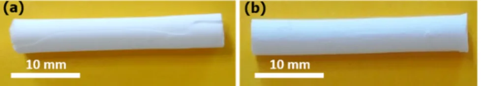
Figure 1. Macroscopic images of the calcined monoliths prepared by stirring the synthesis gel (a)
for only 30 min at a vigorously magnetic stirring of 400 rpm, and (b) for 30 min at a magnetic
Figure 2. SEM images of the calcined monoliths prepared by stirring the synthesis gel (a) and (b)
for only 30 min at a vigorously magnetic stirring of 400 rpm, (c) for 30 min at a magnetic stirring
of 250 rpm and then for 30 min more at a magnetic stirring of 400 rpm (Parent silica monolith M),
and (d) prepared by applying the treatment in basic medium (Parent silica monolith N)
The SEM images (Fig. 2a, 2b, 2c) show the morphology of the two synthesized monoliths
mentioned above. For the monolith prepared under only 30 min of vigorous stirring (400 rpm) a
macroporous silica skeleton with sponge-like morphology coexisting with several spherical
particles and holes inside the silica walls, was observed (Fig. 2a). The morphology of this monolith
is not homogeneous as confirmed by the micrograph displayed in Fig. 2b that shows another
longitudinal area of the monolith: absence of the sponge-like morphology, presence of larger
spherical silica particles and several holes in the silica walls. In the case of the monolith synthesized
by using the stirring regime B only a macroporous silica skeleton with sponge-like morphology
interconnected macropores network with average pores size of 3 µm and a silica skeleton of
average thickness of 2–3 µm. Thanks to these differences of the internal structure, this monolith
(M) exhibited a better mechanical stability after PEO removal upon thermal treatment at 550 °C.
After the basic treatment a sponge-like morphology with well-defined continuous macroporous
network similar to the one of monolith M was observed for monolith N (Fig. 2d).
Figure 3. (a) N2 adsorption-desorption isotherms at 77 K and (b) pore size distributions of the
calcined monoliths prepared by the stirring regime A (only 30 min at a vigorously magnetic
stirring of 400 rpm), the stirring regime B (for 30 min at a magnetic stirring of 250 rpm and then
for 30 min more at a magnetic stirring of 400 rpm – Parent silica monolith M)
The nitrogen adsorption/desorption isotherms of the two micro/macroporous monoliths obtained
by two stirring regimes indicate the difference in their textural properties (Fig. 3). The one of the
monolith synthesized by stirring regime B is type I (according to the IUPAC classifications of N2
synthesized by stirring regime A is a mixture of type I (microporosity) and type IV with a H2b
hysteresis (mesoporosity with pores partially blocking due to the pore topology). This means that
this monolith has both micropores and mesopores in silica skeleton. The presence of mesoporosity
is due to the formation of silica nanoparticles with brad particle size distribution and/or a
heterogeneous agglomeration of silica nanoparticles during gelification process. A more
homogeneous agglomeration of silica nanoparticles has occurred in the case of the monoliths
synthesized by stirring regime B. The pore size distributions confirm the existence of mesoporosity
in the monoliths synthesized with stirring regime A.
These results confirm that the hydrodynamic regime of the ripening phase induced by the stirring
during the silica monolith synthesis is an important factor to be controlled in order to obtain
mechanically stable micro-macroporous monoliths. After the simultaneous sol-gel
transition/spinodal decomposition the obtained monolith is macro and microporous.58 Walls
delimiting the continuous macroporous network are constituted of aggregated silica nanoparticles,
which create interparticular microporosity. The magnetic stirring applied during the ripening phase
can create a vortex at high speed, which generates heterogeneities in the liquid synthesis medium.
It is assumed that these heterogeneities generate nanoparticles more polydisperse in size and thus
result in a broad pore size distribution going from micro- to mesopores as observed in Fig 3b.
It should be noted that the drying and calcination steps have to be carefully respected to avoid any
perturbation from environment conditions (controlled temperature and relative humidity, no air
flow). Mechanically stable micro/macroporous silica monoliths M (Regime B) were used for the
As expected, the N2 adsorption/desorption isotherm of monolith N is type IV characteristics of
mesoporous solid (Fig. S1). Textural characteristics determined from the N2 adsorption/desorption
isotherms of monoliths and M and N are presented in Table 1. The pore size distributions
determined on nitrogen adsorption branches confirm that for the monolith M the average micropore
size is <1 nm, and that for the monolith N the average micropore size is of 1.5 nm and the average
mesopore size is of 14 nm. The microporous volume is very low for monolith N confirming its
predominant mesoporosity. The post-treatment with NH4OH realized in a second step has created
large mesopores in the monoliths walls by dissolution/re-precipitation of silica (Ostwald ripening):
the silica network of monolith M rearranges to form silica nanoparticles that aggregate and form
mesopores in the interparticles space 60,62. It is noteworthy that this step is controlled by the
temperature, the amount of base and the treatment duration, resulting in mesopores regular in size,
which can be controlled from 5 to over 50 nm 61. As a consequence, micropores almost disappear
and mesoporosity become predominant.A decrease of specific surface is thus observed from 609
m2/g for the monolith M down to 249 m2/g for the monolith N. Both monoliths M and N were
characterized by mercury intrusion. The mercury intrusion-extrusion curves and pore size
distributions are displayed in Fig. 4. Both monoliths M and N are characterized by narrow
distributed macropores centered at 4.5 and 8 µm, respectively. The appearance of a second step at
the position of around 14 nm on the mercury intrusion-extrusion curve of monolith N in comparison
with the curve of monolith M (only one step at 4.5 µm) is noted and consistent with N2
adsorption/desorption results. The monoliths M and N have a macroporous volume of 0.87 and
Figure 4. Mercury intrusion-extrusion curves (left) and pore size distributions (right) of the parent
silica monoliths M and N
Table 1. Textural properties of the two parent silica monoliths M and N
SBET (m2/g) Vtota (cm3/g) Vmicro (cm3/g) Vmesoa (cm3/g) Vmacrob (mL/g) Dmicroc (nm) Dmesod (nm) Dmacroe (µm) Monolith M 609 0.25 0.25 0.00 0.87 0.5 – 0.9 - 3 – 6 Monolith N 249 1.21 0.03 1.18 1.40 1.2 – 1.5 12 – 15 6 – 10 a Mesoporous volume: V
meso = Vtot – Vmircro total
b Macroporous volume determined by using mercury intrusion branch
e Macropores diameter determined from the pore size distribution deduced from the mercury intrusion branch
ZSM-5 nanosheets monoliths synthesized from macro/microporous silica monoliths. The
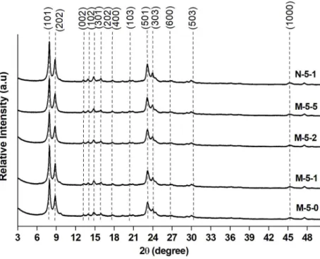
crystallinity of ZSM-5 nanosheets monoliths synthesized by pseudomorphism from the
micro/macroporous silica monolith M was analyzed by X-ray diffraction (Fig. 5). The XRD peaks
were indexed and are consistent with pure MFI-type zeolite. The presence of only h0l reflections
indicates the growth inhibition along the b-axis as expected with the use of C22-6-6 bifunctional
agent, and confirms the formation of ZSM-5 nanosheets. No halo between 20 and 30° 2θ
characteristic of amorphous silica was detected indicating the total dissolution of silica walls and
thus the well crystallization of ZSM-5 nanosheets.
Figure 5. XRD patterns of the ZSM-5 nanosheets monoliths obtained after different durations of
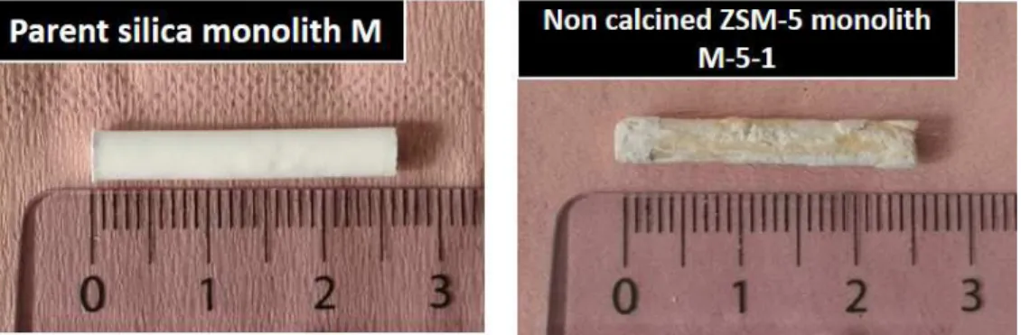
Fig. 6 displays the photographs of the parent silica monolith M and of the ZSM-5 nanosheets
monolith M-5-1. The macroscopic shape of the parent amorphous silica monolith was conserved
after pseudomorphic transformation. The resulting ZSM-5 nanosheets monolith have the same
length of the parent monolith M but the external surface of the monolith was slightly eroded during
the synthesis of the ZSM-5 nanosheet monolith M-5-1. It is assumed that a part of the silica
monolith was dissolved. Although the method was slightly different, Ocampo et al have noticed a
dissolution of the glass support when zeolitization was performed 67. The yellowish color of
ZSM-5 nanosheets monoliths was attributed to the C22-6-6 bifunctional agent since after calcination at ZSM-5ZSM-50
°C the monolith was recovered white with its initial shape (Fig. S2a).
Figure 6. Photographs showing the shape conservation of the obtained monolith after
pseudomorphic transformation
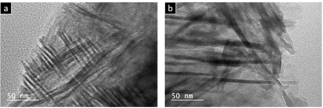
SEM images show the change in morphology of all samples after hydrothermal treatment: the
amorphous silica skeleton has crystallized into ZSM-5 zeolite with nanosheet morphology in all
a lamellar stacking of ZSM-5 nanosheets assembled together in the shape of a house of cards. The
size of the ZSM-5 nanosheets spheres increases slightly with the duration of the hydrothermal
treatment at 120 °C to reach 6-8 µm after 5 days. Moreover, a decrease of the space between the
stacked sheets that almost disappears after 5 days at 120 °C was also observed. The TEM images
Figure 7. SEM images of (a) the parent silica monolith M, and (f) monolith N and the obtained
ZSM-5 nanosheets monoliths (b) M-5-0, (c) M-5-1, (d) M-5-2, (e) M-5-5, and (g) N-5-1
Figure 8. TEM images of monoliths (a) M-5-1 and (b) N-5-1
The N2 sorption isotherms of the calcined monoliths obtained after different durations of the
hydrothermal treatment at 120 °C are a mixture type I at low p/p° and type II-b at high p/p° (Fig.
9a) 64. The hysteresis in the relative pressure range 0.4<p/p°<1 is characteristic of slit pores
resulting from the stacking of the nanosheets. The inflexions observed on the isotherms of M-5-0,
M-5-1 and M-5-2 monoliths at p/p° of about 0.3-0.4 indicate a capillary condensation in small
mesopores created during the formation of ZSM-5 nanosheets. Indeed, there is no capillary
condensation observed on the isotherm of the parent monolith. The textural properties determined
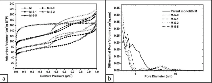
Figure 9. (a) N2 adsorption/desorption isotherms at 77 K and (b) pore size distributions of the
parent M monolith and all obtained monoliths after calcination
Micropores with sizes <1 nm that are assigned to the zeolite framework and secondary micropores
of 1.5 nm size are observed on the pore size distributions of synthesized monoliths (Fig. 9b). The
microporous volume of the secondary micropores remains constant (0.01 cm3/g) whatever the
duration of the hydrothermal treatment at 120 ° C. It is assumed that these secondary micropores
are formed during the crystallization of ZSM-5 nanosheets because of the presence of the
bifunctional organic agent. Indeed, secondary micropores of 1.59 nm diameter were also observed
for ZSM-5 nanosheets powder and conventional MFI microcrystals have only zeolitic micropores
with size <1 nm with a microporous volume of 0.12 cm3/g 57. An increase of the zeolitic
microporous volume from 0.07 cm3/g (M-5-0) to 0.12 cm3/g (M-5-1) is observed with an additional
1-day hydrothermal treatment at 120 °C in static mode (Table 2). But, the microporous volume
decreases slightly to 0.11 cm3/g after 2 days at 120 °C (M-5-2) and in a more pronounced way to
generates bigger particles not completely crystallized. The crystallization rates of obtained ZSM-5
nanosheets monoliths were calculated by comparing the zeolitic micropores volume to the
microporous volumes of ZSM-5 nanosheets powder (0.12 cm3/g) and revealed a total
crystallization for M-5-1 (Table 2).
Table 2. Textural characteristics and crystallization rates of the calcined ZSM-5 nanosheets
monoliths SBET (m2/g) Vtota (cm3/g) Vmicro (cm3/g) Vmesob (cm3/g) Dmeso (nm) Crystallization ratec (%) ze ol it ic se co nd ar y ZSM-5 nanosheets 57 486 0.37 0.12 0.02 0.19 4.4 100 M-5-0 308 0.23 0.07 0.01 0.15 3-6 58.3 M-5-1 428 0.33 0.12 - 0.20 2.5-6 100 M-5-2 426 0.32 0.11 0.01 0.20 3-6 91.6 M-5-5 247 0.26 0.06 - 0.20 2-15 50 N-5-1 440 0.30 0.12 0.005 0.18 2-8 100
a It is noteworthy that for all samples the presence of large mesopores does not allow determining accurately the total
pore volume
b Mesoporous volume: V
meso = Vtot – Vmircro total
c Crystallisation rate= V
micro zeolitic/Vmicro zeolitic of reference ZSM-5 nanosheets; Vmicro zeolitic of reference ZSM-5 nanosheets = 0.12 cm3/g
Mercury intrusion experiment was run for the sample M-5-1 with the highest crystallization rate
(Fig. 10). One step is observed corresponding to a macroporous volume of 0.30 mL/g. It is
noteworthy that the macropores size around 4.5 µm was conserved after pseudomorphic
transformation with a highly polydisperse distribution compared to the parent amorphous silica
monolith that have a narrow distribution of macropores size. Indeed, pore size distribution reveals
also the presence of meso-/macropores with broad distribution ranging from 20 to 200 nm and
mesopores of 8 nm with narrow distribution. These additional meso/macropores are the result of
Figure 10. Mercury intrusion/extrusion curves (left) and pore size distributions (right) of the parent
silica monolith M and the obtained monolith M-5-1 after calcination
ZSM-5 nanosheets monolith synthesized from meso/macroporous silica monolith. The optimal
conditions (i.e. 5 days at 150 °C under stirring and 1 day at 120 °C in static mode) observed for the
synthesis of ZSM-5 nanosheets monoliths from micro/macroporous silica monolith M have been
applied to transform the meso/macroporous silica monolith N into a monolith composed by
ZSM-5 nanosheets.
The photographs of the parent monolith N and the obtained monolith after pseudomorphic
transformation are displayed in Fig. S3 and show that the shape was conserved but the diameter of
the monolith was reduced to 70% compared to the diameter of the parent silica monolith N (0.50
cm for monolith N vs 0.35 cm for monolith N-5-1). Erosion also observed for monolith M appears
more important for monolith N. It is assumed that it is the result of the additional mesoporosity that
has increased the dissolution rates while re-precipitation rates were maintained.
XRD patterns of the obtained monolith N-5-1 show peaks corresponding to the reflections (h0l)
characteristic of a pure MFI-type zeolite phase, which is consistent with the inhibition of growth
along the b-axis characteristic of ZSM-5 nanosheets (Fig. 5).
SEM images show that the amorphous silica skeleton is crystallized into ZSM-5 zeolite with
nanosheets morphology (Fig. 7). Compared to the M-5-1 the morphology of N-5-1 is slightly
different with ZSM-5 nanosheets forming a more continuous network and not spherical aggregates.
The TEM image displayed in Fig. 8b confirms the nanosheet morphology with an average thickness
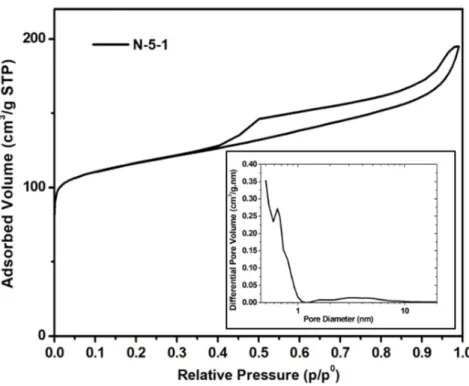
The N2 adsorption/desorption isotherm of monolith N-5-1 displayed in Fig. 11 is very similar to
the one of M-5-1 monolith and the textural properties are similar (Table 2). The zeolitic
microporous volume of 0.12 cm3/g corresponds to a crystallization rate of 100% and the mesopores
size is ranging from 2 to 8 nm.
Figure 11. N2 adsorption-desorption isotherms and pore size distributions (inset) of the obtained
N-5-1 monolith
Monoliths N and N-5-1 were characterized by mercury intrusion-extrusion. Fig. 12 displays their
mercury intrusion-extrusion curves and pore size distributions. For monolith N-5-1, 2 distinct steps
of 0.97 mL/g, smaller than the macroporous volume of the parent amorphous monolith (1.40 mL/g)
and one for smaller macropores pores ranging between 30 and 400 nm and centered at 80 nm,
which contribute to an additional pore volume of 0.15 mL/g. It is noteworthy the conservation of
the macropores with an average diameter around 8 µm after pseudomorphic transformation but
with a wider distribution between 3.5 and 14 µm compared to the narrow macropores size
distribution (between 6 and 10 µm) observed for the parent silica monolith. Mesopores observed
by mercury intrusion in the parent silica monolith are no more observed after pseudomorphic
transformation. However, mesopores were observed by N2 adsorption/desorption experiments as
well as in the M-5-1 monoliths and attributed to the mesopores created after the elimination of the
organic agent between the nanosheets packages by calcination (nanosheets do not collapse
perfectly after calcination).
Figure 12. Mercury intrusion-extrusion curves (left) and pore size distributions (right) of the parent
The ZSM-5 nanosheet monolith “M-5-1” and “N-5-1” were analyzed by TGA and two main weight
losses were observed from 20 and 800 °C (Table S1). The one (3-4 wt.%) in the temperature range
30 - 110 °C corresponds to physisorbed water and the second one (27-28% wt.%) in the temperature
range 110 - 700 °C corresponds to the decomposition of the bifunctional structuring agent. There
are 2 organic cations per unit cell in ZSM-5 zeolite and thus a weight loss of 16.5 wt.% is expected
43,66. However, the weight loss attributing to the decomposition of the organic cations is higher
indicating an excess of C22-6-6 surfactant as usually observed for zeolite nanosheets 68,69 and/or some
dehydroxylation due the presence of defects.
The molar Si/Al ratio from ICP-OES data for “M-5-1” and “N-5-1” are reported in Table S1. The
two monoliths show a high Si/Al ratio of 350 and 182 for M-5-1 and N-5-1, respectively. This
Si/Al is much higher than those measured for ZSM-5 nanosheets beads synthesized under the same
conditions (about 40) 57.
It is noteworthy that the ZSM-5 nanosheets monoliths remained intact after the mercury
intrusion-extrusion experiments under a pressure of 400 MPa reflecting a significant mechanical stability
Figure 13. Photographs of the synthetized monoliths M-5-1 and N-5-1 after mercury
intrusion-extrusion experiments
CONCLUSION
Mechanically stable cylindrical macroporous silica monoliths with only microporosity inside the
silica skeleton were obtained by optimizing the hydrodynamic regime during ripening phase of the
synthesis. The good stirring rate with a suitable duration contributed efficiently to the concurrence
of phase separation and silica condensation processes and further to the compact agglomeration of
silica particles creating a densified silica wall. In addition, macroporous silica monoliths with
disordered mesoporosity were generated by applying a treatment step in basic medium to wet silica
monoliths with only microporosity.
These two types of silica monoliths having different types of pores, micro/macropores and
meso/macropores, have been successfully transformed by following a pseudomorphism process
into ZSM-5 monoliths with nanosheets morphology featuring hierarchical porosities. The
formation of ZSM-5 nanosheets from the amorphous silica skeleton of the monoliths was
evidenced by XRD that showed the only presence of h0l reflections characteristic of MFI zeolite
with inhibition growth along the b-axis. The first attempt was carried out on the micro/macroporous
silica type monolith. A first 5-day treatment at 150 °C with stirring was found suitable to maintain
the macroscopic shape of the monolith but not sufficient to obtain a fully crystallized ZSM-5
monolith. Further crystallization was achieved without losing the monolith shape after an
additional treatment at 120 °C under static conditions. A fully crystallized ZSM-5 monolith with
120 °C. This latter features 3 levels of hierarchical porosity: macropores with an average diameter
centered at 4.5 µm similar to the parent silica monolith and a volume of 0.30 mL/g, secondary
meso-/macropores with broad distribution size ranging from 20 to 200 nm and mesopores of 8 nm
with narrow size distribution resulting of the stacking of the nanosheets and of the spherical
aggregates of nanosheets. The optimal conditions found for the previously discussed monolith (5
days at 150 °C under stirring and 1 day at 120 °C in static mode) have been applied to transform a
meso/macroporous silica monolith. The obtained monolith was fully crystallized into ZSM-5
monolith nanosheets and present macropores with a main diameter centered at 8 µm similar to the
macropores size of the parent silica monolith with a volume of 0.97 mL/g, and secondary
macropores centered at 80 nm contributing to an additional pore volume of 0.15 mL/g.
Both ZSM-5 nanosheets monoliths obtained from micro/macroporous and meso/macroporous
silica monoliths by following the same experimental protocol have similar textural characteristics
although mercury intrusion experiments showed some differences. Such hierarchically porous
ZSM-5 monoliths with nanosheets morphology are expected to be very efficient for use in
continuous flow process and industrial applications.
ASSOCIATED CONTENT
Supporting Information.
Figure S1. N2 adsorption-desorption isotherms and pore size distribution (inset) of the parent
silica monolith N
Figure S3. Photographs of parent silica monolith N and ZSM-5 nanosheets monolith N-5-1
Table S1. Experimental weight losses measured by thermogravimetric analysis and Si/Al ratio
measured by ICP-OES of the obtained monoliths
AUTHOR INFORMATION
Corresponding Authors
*E-mail: benedicte.lebeau@uha.fr ; jean.daou@uha.fr. Phone: +33389336882; +33389336739.
Fax: +33389896885
ORCID
Bénédicte Lebeau : 0000-0001-7447-6042 ; T. Jean Daou : 0000-0002-9973-3372
Author Contributions
T. Jean Daou and Bénédicte Lebeau: Conceptualization, Magali Bonne, Joumana Toufaily, Tayssir Hamieh, T. Jean Daou and Bénédicte Lebeau: Supervision, Kassem Moukahhal, Huu Nghia Le, T. Jean Daou and Bénédicte Lebeau: Writing - Original Draft, Kassem Moukahhal, Huu Nghia Le, Magali Bonne, T. Jean Daou and Bénédicte Lebeau: Review & Editing. Kassem Moukahhal and Huu Nghia Le: Performing the experiments.
All authors have given approval to the final version of the manuscript.
The XRD, adsorption, AMTR and ME platforms of IS2M are acknowledged. Laure Michelin,
Habiba Nouali, Ludovic Josien and Loïc Vidal are warmly thanked for their help in XRD, N2
adsorption/desorption, SEM and TEM data, respectively.
REFERENCES
(1) Kabalan, I.; Khay, I.; Nouali, H.; Ryzhikov, A.; Lebeau, B.; Albrecht, S.; Rigolet, S.;
Fadlallah, M.-B.; Toufaily, J.; Hamieh, T.; Patarin, J.; Daou, T. J. Influence of the Particle
Sizes on the Energetic Performances of MFI-Type Zeolites. J. Phys. Chem. C 2015, 119,
18074–18083. https://doi.org/10.1021/acs.jpcc.5b04484.
(2) Huve, J.; Ryzhikov, A.; Nouali, H.; Lalia, V.; Augé, G.; Daou, T. J. Porous Sorbents for the
Capture of Radioactive Iodine Compounds: A Review. RSC Adv. 2018, 8, 29248–29273.
https://doi.org/10.1039/C8RA04775H.
(3) Said, A.; Nouali, H.; Limousy, L.; Dutournié, P.; Josien, L.; Toufaily, J.; Hamieh, T.; Daou,
T. J. Synthesis of Mono- and Bi-Layer Zeolite Films on Alumina Substrates. C. R. Chim
2016, 19, 486–495. https://doi.org/10.1016/j.crci.2015.09.018.
(4) Rioland, G.; Nouali, H.; Daou, T. J.; Faye, D.; Patarin, J. Adsorption of Volatile Organic
Compounds in Composite Zeolites Pellets for Space Decontamination. Adsorption 2017, 23,
395–403. https://doi.org/10.1007/s10450-017-9870-9.
(5) Li, Y.; Yu, J. New Stories of Zeolite Structures: Their Descriptions, Determinations,
Predictions, and Evaluations. Chem. Rev. 2014, 114, 7268–7316.
(6) Martínez, C.; Corma, A. Inorganic Molecular Sieves: Preparation, Modification and
Industrial Application in Catalytic Processes. Coordin. Chem. Rev. 2011, 255, 1558–1580.
https://doi.org/10.1016/j.ccr.2011.03.014.
(7) Li, C.; Moliner, M.; Corma, A. Building Zeolites from Precrystallized Units: Nanoscale
Architecture. Angew. Chem. Int. Ed. 2018, 57, 15330–15353.
https://doi.org/10.1002/anie.201711422.
(8) Sachse, A.; García-Martínez, J. Surfactant-Templating of Zeolites: From Design to
Application. Chem. Mater. 2017, 29, 3827–3853.
https://doi.org/10.1021/acs.chemmater.7b00599.
(9) Dusselier, M.; Davis, M. E. Small-Pore Zeolites: Synthesis and Catalysis. Chem. Rev. 2018,
118, 5265–5329. https://doi.org/10.1021/acs.chemrev.7b00738.
(10) Li, J.; Corma, A.; Yu, J. Synthesis of New Zeolite Structures. Chem. Soc. Rev. 2015, 44,
7112–7127. https://doi.org/10.1039/C5CS00023H.
(11) Přech, J.; Pizarro, P.; Serrano, D. P.; Čejka, J. From 3D to 2D Zeolite Catalytic Materials.
Chem. Soc. Rev. 2018, 47, 8263–8306. https://doi.org/10.1039/C8CS00370J.
(12) Schneider, D.; Mehlhorn, D.; Zeigermann, P.; Kärger, J.; Valiullin, R. Transport Properties
of Hierarchical Micro–Mesoporous Materials. Chem. Soc. Rev. 2016, 45, 3439–3467.
https://doi.org/10.1039/C5CS00715A.
(13) Bingre, R.; Louis, B.; Nguyen, P. An Overview on Zeolite Shaping Technology and
Solutions to Overcome Diffusion Limitations. Catalysts 2018, 8, 163.
https://doi.org/10.3390/catal8040163.
(14) Awala, H.; Gilson, J.-P.; Retoux, R.; Boullay, P.; Goupil, J.-M.; Valtchev, V.; Mintova, S.
Template-Free Nanosized Faujasite-Type Zeolites. Nature Mater 2015, 14, 447–451.
(15) Mintova, S.; Jaber, M.; Valtchev, V. Nanosized Microporous Crystals: Emerging
Applications. Chem. Soc. Rev. 2015, 44, 7207–7233. https://doi.org/10.1039/C5CS00210A.
(16) Zaarour, M.; Dong, B.; Naydenova, I.; Retoux, R.; Mintova, S. Progress in Zeolite Synthesis
Promotes Advanced Applications. Micropor. Mesopor. Mat. 2014, 189, 11–21.
https://doi.org/10.1016/j.micromeso.2013.08.014.
(17) Bai, R.; Sun, Q.; Wang, N.; Zou, Y.; Guo, G.; Iborra, S.; Corma, A.; Yu, J. Simple
Quaternary Ammonium Cations-Templated Syntheses of Extra-Large Pore Germanosilicate
Zeolites. Chem. Mater. 2016, 28, 6455–6458.
https://doi.org/10.1021/acs.chemmater.6b03179.
(18) Jiang, J.; Yun, Y.; Zou, X.; Jorda, J. L.; Corma, A. ITQ-54: A Multi-Dimensional
Extra-Large Pore Zeolite with 20 × 14 × 12-Ring Channels. Chem. Sci. 2015, 6, 480–485.
https://doi.org/10.1039/C4SC02577F.
(19) Yang, J.; Zhang, Y.-B.; Liu, Q.; Trickett, C. A.; Gutiérrez-Puebla, E.; Monge, M. Á.; Cong,
H.; Aldossary, A.; Deng, H.; Yaghi, O. M. Principles of Designing Extra-Large Pore
Openings and Cages in Zeolitic Imidazolate Frameworks. J. Am. Chem. Soc. 2017, 139,
6448–6455. https://doi.org/10.1021/jacs.7b02272.
(20) Smeets, S.; Xie, D.; Baerlocher, C.; McCusker, L. B.; Wan, W.; Zou, X.; Zones, S. I.
High-Silica Zeolite SSZ-61 with Dumbbell-Shaped Extra-Large-Pore Channels. Angew. Chem.
Int. Ed. 2014, 53, 10398–10402. https://doi.org/10.1002/anie.201405658.
(21) Kang, J. H.; Xie, D.; Zones, S. I.; Smeets, S.; McCusker, L. B.; Davis, M. E. Synthesis and
Characterization of CIT-13, a Germanosilicate Molecular Sieve with Extra-Large Pore
Openings. Chem. Mater. 2016, 28, 6250–6259.
(22) Schwieger, W.; Machoke, A. G.; Weissenberger, T.; Inayat, A.; Selvam, T.; Klumpp, M.;
Inayat, A. Hierarchy Concepts: Classification and Preparation Strategies for Zeolite
Containing Materials with Hierarchical Porosity. Chem. Soc. Rev. 2016, 45, 3353–3376.
https://doi.org/10.1039/C5CS00599J.
(23) Chen, L.-H.; Li, X.-Y.; Rooke, J. C.; Zhang, Y.-H.; Yang, X.-Y.; Tang, Y.; Xiao, F.-S.; Su,
B.-L. Hierarchically Structured Zeolites: Synthesis, Mass Transport Properties and
Applications. J. Mater. Chem. 2012, 22, 17381. https://doi.org/10.1039/c2jm31957h.
(24) Sun, M.-H.; Huang, S.-Z.; Chen, L.-H.; Li, Y.; Yang, X.-Y.; Yuan, Z.-Y.; Su, B.-L.
Applications of Hierarchically Structured Porous Materials from Energy Storage and
Conversion, Catalysis, Photocatalysis, Adsorption, Separation, and Sensing to Biomedicine.
Chem. Soc. Rev. 2016, 45, 3479–3563. https://doi.org/10.1039/C6CS00135A.
(25) Pérez-Ramírez, J.; Christensen, C. H.; Egeblad, K.; Christensen, C. H.; Groen, J. C.
Hierarchical Zeolites: Enhanced Utilisation of Microporous Crystals in Catalysis by
Advances in Materials Design. Chem. Soc. Rev. 2008, 37, 2530.
https://doi.org/10.1039/b809030k.
(26) El Hanache, L.; Lebeau, B.; Nouali, H.; Toufaily, J.; Hamieh, T.; Daou, T. J. Performance
of Surfactant-Modified *BEA-Type Zeolite Nanosponges for the Removal of Nitrate in
Contaminated Water: Effect of the External Surface. J. Hazard. Mater. 2019, 364, 206–217.
https://doi.org/10.1016/j.jhazmat.2018.10.015.
(27) Astafan, A.; Benghalem, M. A.; Pouilloux, Y.; Patarin, J.; Bats, N.; Bouchy, C.; Daou, T. J.;
Pinard, L. Particular Properties of the Coke Formed on Nano-Sponge *BEA Zeolite during
Ethanol-to-Hydrocarbons Transformation. J. Catal. 2016, 336, 1–10.
(28) Kabalan, I.; Lebeau, B.; Fadlallah, M.-B.; Toufaily, J.; Hamieh, T.; Bellat, J. P.; Daou, T. J.
Hierarchical Faujasite-Type Zeolite for Molecular Decontamination. J. Nanosci.
Nanotechnol. 2016, 16, 9318–9322. https://doi.org/10.1166/jnn.2016.12884.
(29) Hua, Z. L.; Zhou, J.; Shi, J. L. Recent Advances in Hierarchically Structured Zeolites:
Synthesis and Material Performances. Chem. Commun. 2011, 47, 10536.
https://doi.org/10.1039/c1cc10261c.
(30) Na, K.; Choi, M.; Ryoo, R. Recent Advances in the Synthesis of Hierarchically Nanoporous
Zeolites. Micropor. Mesopor. Mat. 2013, 166, 3–19.
https://doi.org/10.1016/j.micromeso.2012.03.054.
(31) Groen, J. C.; Bach, T.; Ziese, U.; Paulaime-van Donk, A. M.; de Jong, K. P.; Moulijn, J. A.;
Pérez-Ramírez, J. Creation of Hollow Zeolite Architectures by Controlled Desilication of
Al-Zoned ZSM-5 Crystals. J. Am. Chem. Soc. 2005, 127, 10792–10793.
https://doi.org/10.1021/ja052592x.
(32) Ryoo, R.; Joo, S. H.; Kruk, M.; Jaroniec, M. Ordered Mesoporous Carbons. Adv. Mater.
2001, 13, 677–681.
https://doi.org/10.1002/1521-4095(200105)13:9<677::AID-ADMA677>3.0.CO;2-C.
(33) Fang, Y.; Hu, H. An Ordered Mesoporous Aluminosilicate with Completely Crystalline
Zeolite Wall Structure. J. Am. Chem. Soc. 2006, 128, 10636–10637.
https://doi.org/10.1021/ja061182l.
(34) Fan, W.; Snyder, M. A.; Kumar, S.; Lee, P.-S.; Yoo, W. C.; McCormick, A. V.; Lee Penn,
R.; Stein, A.; Tsapatsis, M. Hierarchical Nanofabrication of Microporous Crystals with
Ordered Mesoporosity. Nat. Mater. 2008, 7, 984–991. https://doi.org/10.1038/nmat2302.
(35) Ciesla, U.; Schüth, F. Ordered Mesoporous Materials. Micropor. Mesopor. Mat. 1999, 27,
(36) Monnier, A.; Schüth, F.; Huo, Q.; Kumar, D.; Margolese, D.; Maxwell, R. S.; Stucky, G. D.;
Krishnamurty, M.; Petroff, P.; Firouzi, A.; Janicke, M.; Chmelka, B. F. Cooperative
Formation of Inorganic-Organic Interfaces in the Synthesis of Silicate Mesostructures.
Science 1993, 261, 1299–1303. https://doi.org/10.1126/science.261.5126.1299.
(37) Hartmann, M. Ordered Mesoporous Materials for Bioadsorption and Biocatalysis. Chem.
Mater. 2005, 17, 4577–4593. https://doi.org/10.1021/cm0485658.
(38) Xia, Y.; Mokaya, R. On the Synthesis and Characterization of ZSM-5/MCM-48
Aluminosilicate Composite Materials. J. Mater. Chem. 2004, 14, 863–870.
https://doi.org/10.1039/B313389C.
(39) Prokešová, P.; Mintova, S.; Čejka, J.; Bein, T. Preparation of Nanosized Micro/Mesoporous
Composites via Simultaneous Synthesis of Beta/MCM-48 Phases. Micropor. Mesopor. Mat.
2003, 64, 165–174. https://doi.org/10.1016/S1387-1811(03)00464-5.
(40) Galarneau, A.; Cambon, H.; Renzo, F. D.; Ryoo, R.; Choi, M.; Fajula, F. Microporosity and
Connections between Pores in SBA-15 Mesostructured Silicas as a Function of the
Temperature of Synthesis. New J. Chem. 2003, 27, 73–79.
https://doi.org/10.1039/B207378C.
(41) Choi, M.; Na, K.; Kim, J.; Sakamoto, Y.; Terasaki, O.; Ryoo, R. Stable Single-Unit-Cell
Nanosheets of Zeolite MFI as Active and Long-Lived Catalysts. Nature 2009, 461, 246–
249. https://doi.org/10.1038/nature08288.
(42) Na, K.; Jo, C.; Kim, J.; Cho, K.; Jung, J.; Seo, Y.; Messinger, R. J.; Chmelka, B. F.; Ryoo,
R. Directing Zeolite Structures into Hierarchically Nanoporous Architectures. Science 2011,
333, 328–332. https://doi.org/10.1126/science.1204452.
(43) Dhainaut, J.; Daou, T. J.; Bidal, Y.; Bats, N.; Harbuzaru, B.; Lapisardi, G.; Chaumeil, H.;
Zeolite Nanosheets Using Mononitrogen Surfactants as Structure and Shape-Directing
Agents. CrystEngComm 2013, 15, 3009. https://doi.org/10.1039/c3ce40118a.
(44) Kabalan, I.; Rioland, G.; Nouali, H.; Lebeau, B.; Rigolet, S.; Fadlallah, M.-B.; Toufaily, J.;
Hamiyeh, T.; Daou, T. J. Synthesis of Purely Silica MFI-Type Nanosheets for Molecular
Decontamination. RSC Adv. 2014, 4, 37353–37358. https://doi.org/10.1039/C4RA05567E.
(45) Rioland, G.; Albrecht, S.; Josien, L.; Vidal, L.; Daou, T. J. The Influence of the Nature of
Organosilane Surfactants and Their Concentration on the Formation of Hierarchical
FAU-Type Zeolite Nanosheets. New J. Chem. 2015, 39, 2675–2681.
https://doi.org/10.1039/C4NJ02137A.
(46) Itani, L.; Valtchev, V.; Patarin, J.; Rigolet, S.; Gao, F.; Baudin, G. Centimeter-Sized Zeolite
Bodies of Intergrown Crystals: Preparation, Characterization and Study of Binder Evolution.
Micropor. Mesopor. Mat. 2011, 138, 157–166.
https://doi.org/10.1016/j.micromeso.2010.09.011.
(47) Fawaz, E. G.; Salam, D. A.; Nouali, H.; Deroche, I.; Rigolet, S.; Lebeau, B.; Daou, T. J.
Synthesis of Binderless ZK-4 Zeolite Microspheres at High Temperature. Molecules 2018,
23, 2647. https://doi.org/10.3390/molecules23102647.
(48) Tosheva, L.; Valtchev, V.; Sterte, J. Silicalite-1 Containing Microspheres Prepared Using
Shape-Directing Macro-Templates. Micropor. Mesopor. Mat. 2000, 35–36, 621–629.
https://doi.org/10.1016/S1387-1811(99)00256-5.
(49) Rioland, G.; Bullot, L.; Daou, T. J.; Simon-Masseron, A.; Chaplais, G.; Faye, D.; Fiani, E.;
Patarin, J. Elaboration of FAU-Type Zeolite Beads with Good Mechanical Performances for
Molecular Decontamination. RSC Adv. 2015, 6, 2470–2478.
(50) Rioland, G.; Daou, T. J.; Faye, D.; Patarin, J. A New Generation of MFI-Type Zeolite Pellets
with Very High Mechanical Performance for Space Decontamination. Micropor. Mesopor.
Mat. 2016, 221, 167–174. https://doi.org/10.1016/j.micromeso.2015.09.040.
(51) Martin, T.; Galarneau, A.; Renzo, F. D.; Fajula, F.; Plee, D. Morphological Control of
MCM-41 by Pseudomorphic Synthesis. Angew. Chem. Int. Ed. 2002, 41, 2590–2592.
https://doi.org/10.1002/1521-3773(20020715)41:14<2590::AID-ANIE2590>3.0.CO;2-3.
(52) Martin, T.; Galarneau, A.; Di Renzo, F.; Brunel, D.; Fajula, F.; Heinisch, S.; Crétier, G.;
Rocca, J.-L. Great Improvement of Chromatographic Performance Using MCM-41 Spheres
as Stationary Phase in HPLC. Chem. Mater 2004, 16, 1725–1731.
https://doi.org/10.1021/cm030443c.
(53) Petitto, C.; Galarneau, A.; Driole, M.-F.; Chiche, B.; Alonso, B.; Di Renzo, F.; Fajula, F.
Synthesis of Discrete Micrometer-Sized Spherical Particles of MCM-48. Chem. Mater 2005,
17, 2120–2130. https://doi.org/10.1021/cm050068j.
(54) Galarneau, A.; Sachse, A.; Said, B.; Pelisson, C.-H.; Boscaro, P.; Brun, N.; Courtheoux, L.;
Olivi-Tran, N.; Coasne, B.; Fajula, F. Hierarchical Porous Silica Monoliths: A Novel Class
of Microreactors for Process Intensification in Catalysis and Adsorption. C. R. Chim 2016,
19, 231–247. https://doi.org/10.1016/j.crci.2015.05.017.
(55) Didi, Y.; Said, B.; Micolle, M.; Cacciaguerra, T.; Cot, D.; Geneste, A.; Fajula, F.; Galarneau,
A. Nanocrystals FAU-X Monoliths as Highly Efficient Microreactors for Cesium Capture
in Continuous Flow. Micropor. Mesopor. Mat. 2019, 285, 185–194.
https://doi.org/10.1016/j.micromeso.2019.05.012.
(56) Said, B.; Grandjean, A.; Barre, Y.; Tancret, F.; Fajula, F.; Galarneau, A. LTA Zeolite
Strontium Capture in Continuous Flow. Micropor. Mesopor. Mat. 2016, 232, 39–52.
https://doi.org/10.1016/j.micromeso.2016.05.036.
(57) Moukahhal, K.; Daou, T. J.; Josien, L.; Nouali, H.; Toufaily, J.; Hamieh, T.; Galarneau, A.;
Lebeau, B. Hierarchical ZSM-5 Beads Composed of Zeolite Nanosheets Obtained by
Pseudomorphic Transformation. Micropor. Mesopor. Mat. 2019, 288, 109565.
https://doi.org/10.1016/j.micromeso.2019.109565.
(58) Nakanishi, K. Pore Structure Control of Silica Gels Based on Phase Separation. J. Porous
Mater. 1997, 4, 67–112. https://doi.org/10.1023/A:1009627216939.
(59) Nakanishi, K.; Soga, N. Phase Separation in Silica Sol-Gel System Containing Polyacrylic
Acid I. Gel Formation Behavior and Effect of Solvent Composition. J. Non-Cryst. Solids
1992, 139, 1–13. https://doi.org/10.1016/S0022-3093(05)80800-2.
(60) Babin, J.; Iapichella, J.; Lefèvre, B.; Biolley, C.; Bellat, J.-P.; Fajula, F.; Galarneau, A.
MCM-41 Silica Monoliths with Independent Control of Meso- and Macroporosity. New J.
Chem. 2007, 31, 1907. https://doi.org/10.1039/b711544j.
(61) Nakanishi, K.; Shikata, H.; Ishizuka, N.; Koheiya, N.; Soga, N. Tailoring Mesopores in
Monolithic Macroporous Silica for HPLC. J. High Resolut. Chromatogr. 2000, 23, 106–110.
https://doi.org/10.1002/(SICI)1521-4168(20000101)23:1<106::AID-JHRC106>3.0.CO;2-1.
(62) Galarneau, A.; Abid, Z.; Said, B.; Didi, Y.; Szymanska, K.; Jarzębski, A.; Tancret, F.;
Hamaizi, H.; Bengueddach, A.; Di Renzo, F.; Fajula, F. Synthesis and Textural
Characterization of Mesoporous and Meso-/Macroporous Silica Monoliths Obtained by
Spinodal Decomposition. Inorganics 2016, 4, 9. https://doi.org/10.3390/inorganics4020009.
(63) Galarneau, A.; Iapichella, J.; Brunel, D.; Fajula, F.; Bayram-Hahn, Z.; Unger, K.; Puy, G.;
Stationary Phases for Liquid Chromatography. J. Sep. Sci. 2006, 29, 844–855.
https://doi.org/10.1002/jssc.200500511.
(64) Landers, J.; Gor, G. Yu.; Neimark, A. V. Density Functional Theory Methods for
Characterization of Porous Materials. Colloids Surf. A 2013, 437, 3–32.
https://doi.org/10.1016/j.colsurfa.2013.01.007.
(65) Thommes, M.; Cychosz, K. A. Physical Adsorption Characterization of Nanoporous
Materials: Progress and Challenges. Adsorption 2014, 20, 233–250.
https://doi.org/10.1007/s10450-014-9606-z.
(66) Kabalan, I.; Lebeau, B.; Nouali, H.; Toufaily, J.; Hamieh, T.; Koubaissy, B.; Bellat, J.-P.;
Daou, T. J. New Generation of Zeolite Materials for Environmental Applications. J. Phys.
Chem. C 2016, 120, 2688–2697. https://doi.org/10.1021/acs.jpcc.5b10052.
(67) Ocampo, F.; Yun, H. S.; Pereira, M. M.; Tessonnier, J. P.; Louis, B. Design of MFI
Zeolite-Based Composites with Hierarchical Pore Structure: A New Generation of Structured
Catalysts. Cryst. Growth Des 2009, 9, 3721–3729. https://doi.org/10.1021/cg900425r.
(68) Schick, J.; Daou, T. J.; Caullet, P.; Paillaud, J.-L.; Patarin, J.; Mangold-Callarec, C.
Surfactant-Modified MFI Nanosheets: A High Capacity Anion-Exchanger. Chem. Commun.
2011, 47, 902–904. https://doi.org/10.1039/C0CC03604H.
(69) El Hanache, L.; Sundermann, L.; Lebeau, B.; Toufaily, J.; Hamieh, T.; Daou, T. J.
Surfactant-Modified MFI-Type Nanozeolites: Super-Adsorbents for Nitrate Removal from
Contaminated Water. Micropor. Mesopor. Mat. 2019, 283, 1–13.
Table of Contents Use Only
Controlled crystallization of hierarchical monoliths
composed of nano-zeolites
Kassem Moukahhal1,2,3, Nghia Huu Le1,2, Magali Bonne1,2, Joumana Toufaily3, Tayssir Hamieh3,
T. Jean Daou1,2*, Bénédicte Lebeau1,2*