HAL Id: tel-01148558
https://tel.archives-ouvertes.fr/tel-01148558
Submitted on 4 May 2015HAL is a multi-disciplinary open access archive for the deposit and dissemination of sci-entific research documents, whether they are pub-lished or not. The documents may come from teaching and research institutions in France or abroad, or from public or private research centers.
L’archive ouverte pluridisciplinaire HAL, est destinée au dépôt et à la diffusion de documents scientifiques de niveau recherche, publiés ou non, émanant des établissements d’enseignement et de recherche français ou étrangers, des laboratoires publics ou privés.
Effects of Interleukine-17A (Il-17A) and tumor necrosis
factor alpha (TNF-α) on osteoblastic differentiation
Bilal Osta
To cite this version:
Bilal Osta. Effects of Interleukine-17A (Il-17A) and tumor necrosis factor alpha (TNF-α) on os-teoblastic differentiation. Immunology. Université Claude Bernard - Lyon I, 2014. English. �NNT : 2014LYO10278�. �tel-01148558�
Acknowledgement
Firstly, I would like to thanks Region Rhône-Alpes for their finical support during my PhD program. As I wish to express my sincere gratitude to a lot of people, who help me to finish this PhD project. First I would like to express my deep gratitude to Pr. Pierre MIOSSEC my research supervisors, for his patient guidance, enthusiastic encouragement and useful critiques of this research work. Also, I would like to express my very great appreciation to Dr. Assia ELJAAFARI for her advice, assistance and constructive suggestions during the planning and development of this research work. Her willingness to give her time so generously has been very much appreciated.
My grateful thanks are also extended to Dr. Gulia for her advice, and helping in finalizing the manuscript. And of course special thanks for Dr. Ndieme and Fabien for their help in offering me the resources in running the program, splitting cells, data analysis and last but not least for the funny time that we spend it together. And of course I can’t forget Thomas, Paola, Melissa and Pr.Arnaud for their kindness and motivating me.
I would like to further acknowledge all BioMerieux equip for sharing the platform, equipments, and advices. Special thanks for Babeth and Marie, who taught me a lot about words, excel and were always patient to hear me. I would also like to thanks the staff of UMR INSERM 1033 - Physiopathology, Diagnostic and Treatments of bone diseases for enabling me to visit their platform and taught me how to perform histomorphometric studies, really I would like to thanks from my heart all the equip for their hospitality, advices and training. Especially Jean PAUL-ROUX (PAP-II), Marlene, Dr. Pascale CHAVASSIEUX and of course Dr. George BOVIN.
Finally, an honorable mention with grateful thanks goes to my parents and my wife Hejar for their Doaa, motivation and for pushing me through the tough moments of my PhD. My brothers and my friends especially Mohammad ZAATARI really I don’t know what to say, so god help and benefit you.
Table of Contents
Lists of Abbreviation ... 15
Lists of Figures and Tables ... 19
Part.I ... 17
Introduction ... 17
1. Human Bone tissue ... 18
1.1. Embryonic Development of Bone ... 18
1.2. Bone tissue and function ... 18
1.3. Macroscopic anatomy and organization of bone tissue ... 19
1.3.1. Bone architecture unit ... 19
1.3.2. Different bone tissue compartments ... 24
1.3.3. Bone classification ... 26
1.4. Bone tissue composition ... 26
1.5. Conclusion ... 29
2. Bone cells ... 30
2.1. Mesenchymal stem cells ... 30
2.1.1. MSCs and Heterogeneity ... 27
2.1.2. Circulating MSCs ... 28
2.1.3. Role of MSCs ... 29
2.1.3.1. Effects of MSCs on Immune cells ... 29
2.1.3.1.1. Dendritic cells (DCs) ... 34
2.1.3.1.2. Natural Killer cells (NK) ... 35
2.1.3.1.3. Neutrophils ... 36 2.1.3.1.4. T- Cells ... 36 2.1.3.1.5. B-Cells ... 36 2.1.3.1.6. Regulatory T-Cells ... 37 2.1.3.2. Tissue repair by MSCs ... 37 2.1.3.2.1. Bone ... 37
2.1.3.2.2. Central Nervous System ... 37
2.1.3.2.3. Heart ... 37
2.1.3.2.4. Other Tissues ... 38
2.3. Conclusion………..38
3. Osteoblastic lineages ... 37
3.1. Osteoblasts origin and differentiation ... 37
3.1.1. Runx2 ... 39 3.1.2. Osterix ... 41 3.1.3. Wnt and β-Catenin ... 45 3.1.4. BMPs and SMADs ... 46 3.1.5. AP-1 ... 47 3.1.6. ATF4 ... 48 3.1.7. Helix-loop-Helix protein ... 48
3.1.8. Zinc Finger Protein / Schnurri-3 ... 48
3.1.9. Homeobox proteins ... 49
3.2. Osteocytes ... 50
3.3. Lining Cells ... 48
3.4. Osteoblastogenesis and mechanical signaling ... 49
3.5. Conclusion ... 54
4. Epigenetic regulation of gene expression ... 55
4.1. Epigenetic regulation during osteoblastic differentiation ... 56
4.2. Conculsion ... 57
5. Osteoclastic lineages ... 58
5.1. Osteoclast Origin ... 58
5.2. Osteoclast Differentiation ... 59
5.2.1. Role of M-CSF in Osteoclast Differentiation ... 64
5.2.2. Role of RANKL in Osteoclast Differentiation ... 65
5.2.3 Costimulatory Signals for RANK ... 67
5.2.4. The OPG/RANKL/RANK Regulatory Axis ... 68
5.2.5 Other Regulatory Mechanisms ... 70
5.2.6. Osteoclast Fusion ... 67
5.3. Conclusion ... 67
6. Interleukin 17 ... 69
6.1. IL-17 receptor signaling ... 78
6.2. Effects of IL-17 on MSCs ... 79
6.4. IL-17 and inflammatory bone diseases………..84
6.4.1. Role of IL-17 in rheumatoid arthritis ... 84
6.4.2. Role of IL-17 in ankylosing spondylitis ... 85
6.5. Conclusion ... 86
7. Classical and paradoxical effects of TNF-α on bone homeostasis ... 86
Part.II ... 96
Study of the effects of interleukin-17A on osteogenic differentiation of
isolated human mesenchymal stem cells
Part.III ... 105
Bone marrow-derived and synovium-derived mesenchymal cells promote
Th17 cell expansion and activation through caspase
1 activation
Part.IV ... 118
TNF-α and IL-17A induce osteoblastic differentiation, but increase
synergistically Schnurri-3 in isolated synoviocytes
Part.V ... 144
Differential effects of IL-17A and TNF-α on osteoblastic function in isolated
mesenchymal cells and whole bone from arthritis patients
Part.VI. Conclusion and prespectives ... 164
Résumé de la thèse en Français
L’interleukine-17A (IL-17A) et le facteur de nécrose tumorale alpha (TNF-α) sont des cytokines pro-inflammatoires impliquées dans la pathogénèse de plusieurs maladies articulaires. Au cours de la polyarthrite rhumatoïde (PR), une augmentation de la destruction osseuse ainsi qu’un defaut de réparation sont responsables des dommages articulaires. Cependant au cours de la spondylarthrite ankylosante (AS), une importante ossification ectopique est observée, conduisant à la formation de syndesmophytes, associé à une perte de la masse osseuse systémique. Récemment, l’étude de ces cytokines a conduit à la publication de résultats contradictoires. Notre objectif a donc été d’étudier l’effet de ces deux cytokines sur la différenciation ostéogénique de cellules souches mésenchymateuses humaines isolées (hMSCs) et de fibroblastes de la membrane synoviale (FLS).
Nos résultats ont montré que l’IL-17A et le TNF-α agissent de façon synergique pour induire la différenciation ostéogénique des hMSCs via une augmentation de l’expression du récepteur II au TNF. Nous avons observé (i) une augmentation de l’expression de BMP2 dans les étapes précoces de la différenciation ostéogénique, (ii) une modulation plus tardive de l’expression de DKK-1 et (iii) une stimulation des dépôts de matrice minéralisée. Pour les FLS, lorsque nous avons comparé les FLS non inflammatoires provenant de donneurs sains et de patients souffrant d'arthrose avec les FLS inflammatoires de patients atteints de PR, nous avons observé que la combinaison de ces deux cytokines a entraîné une augmentation de la minéralisation et de l’expression de gènes impliqués dans la différenciation ostéogénique. Cependant, les FLS de PR produisent des taux tres élevés d'IL-6 et d’IL-8 associés à une expression stable de RANKL dans ces cellules. Ceci conduit probablement à l'activation d’ostéoclastogénèse. Enfin, une étude ex-vivo sur des biopsies osseuses, où ostéoblastes et ostéoclastes ainsi que d’autres cellules coexistent, n’a pas mis en évidence de différence sur les taux des marqueurs de la destruction osseuse (CTX ou dans l’activité de la phosphatase alcaline). Par contre, après stimulation avec l’IL-17A et le TNF-α, les niveaux d’IL-6 augmentent tandis que le rapport volume osseux/volume du tissu tend à diminuer. Ces résultats suggèrent que les ostéoblastes et ostéoclastes sont activés à différents niveaux, tandis qu’in vivo, les interactions sont probablement plus complexes.
Pour conclure, tous les modèles de cellules utilisés, ont démontré que l’IL-17A et le TNF-α augmentent de manière synergique l’ostéogénèse. Ceci semble se rapprocher du modèle de l’AS où une formation d’os ectopique est observée dans laquelle l’IL-17A et le TNF-α jouent un role majeur. En parallèle, ces deux cytokines stimulent localement les ostéoclastes, entraînant une perte de masse osseuse observée à la fois dans la PR et dans l’ostéoporose. Cibler simultanément l’IL-17A et le TNF-α pourrait conduire à une diminution de l’infiltration de cellules et de la destruction articulaire observée dans la PR et pourrait ainsi réduire les effets des FLS PR sur l’activation de l’ostéoclastogénèse.
Abstract of the thesis in English
Interleukin-17A (IL-17A) and tumor necrosis factor alpha (TNF-α) are pro-inflammatory cytokines involved in the pathogenesis of several arthritic diseases. In rheumatoid arthritis (RA), joint damage is a result of an increase in bone destruction and a decrease in bone repair. In contrast, in ankylosing spondylitis (AS), a bone mass loss accompanied by a significant ectopic ossification is observed leading to the formation of syndesmophytes. Recent studies led to contradictory findings regarding the role of IL-17A and TNF-α in arthritic disease. Therefore, our objective was to study the effect of these two cytokines on the osteogenic differentiation of isolated human mesenchymal stem cells (hMSCs) and fibroblasts of the synovial membrane (FLS).
In this study, IL-17A and TNF-α acted synergistically to induce the osteogenic differentiation of hMSCs via increased expression of the TNF receptor II (TNF-RII). We observed (i) an increase in the expression of BMP2 in the early stages of the osteogenic differentiation, (ii) a late modulation in the expression of DKK-1 and (iii) the stimulation of mineralized matrix deposition. In FLS, when we compared non-inflammatory FLS from healthy and osteoarthritis patients and inflammatory FLS from RA patients, we observed that the combination of the two cytokines resulted in increased mineralization and increased expression of genes involved in osteogenic differentiation. However, RA FLS produced highly levels of IL-6 and IL-8 and that the expression of RANKL was stabilized in those cells. This RANKL stabilization most likely led to activation of osteoclastogenesis. Finally, a study on ex-vivo bone biopsies, where osteoblasts, osteoclasts and other cells coexist, did not reveal any difference in the rate of bone destruction marker CTx or in the activity of alkaline phosphatase. However, after stimulation with IL-17A and TNF-α, IL-6 levels increased while the bone/ tissue volume ratio decreased. These results led us to postulate that osteoblasts and osteoclasts are activated at different levels and that in vivo interactions are probably more complex.
In conclusion, in all the cell models used, we demonstrated that Il-17A and TNF-α synergistically increase osteogenesis. This seems to approach the model of AS where ectopic bone formation is observed and in which IL-17A and TNF-α both are involved. These cytokines stimulate osteoclasts locally resulting in loss of bone mass observed in both RA and osteoporosis. Thus, targeting IL-17A and TNF-α could lead to a decrease in cell infiltration and joint destruction which is observed in RA and may reduce the effects of RA FLS on the activation of osteoclastogenesis.
Lists of Abbreviation
ADAS Adipose-derived adult stem cell
ALP Alkaline Phosphatase
APC Antigen presenting cell
AP-1 Jun proto-oncogene
ASCT Autologous stem cell transplantation
ATF4 Activating transcription factor 4
Atp6vd2 D2 isoform of vacuolar H+-ATPase V0 domain
BLCs Bone lining cells
BMUs Bone multicellular units
BM Bone marrow
BMPs Bone morphogenetic proteins
BSP Bone sialoprotein
BLCs Bone lining cells
CA II Carbonic anhydrase
CBP Sarcoplasmic calcium-binding protein
C/EBP CAAT/enhancer binding protein
CFU-F Colony-forming units of fibroblasts
CFU-GM CFU- granulocyte- macrophage
CINC Cytokine-induced neutrophil chemoattractant
CNS Central Nervous System
CSF Colony-stimulating factor
CTR Calcitonin receptor
CTK Cathepsin K
CXCL4-CXCL12 Alpha-chemokine receptor specific for stromal-derived-factor-1 (SDF-1 also known as CXCL12)
CX3CR1-CX3CL1 Chemokine (C-X3-C motif) : R1: receptor 1; L1: ligand 1
DAP12 DNAX activation protein of 12 kDa
DCs Dendritic cells
DC-STAMP Dendretic cell-specific transmembrane protein
EGF Epidermal growth factor
eNOS Endothelial nitric oxide synthase
FGF Fibroblast growth Factor
GROα Growth –regulated α protein
GSK-3 Serine/threonine protein kinase Gsk3
GVHD Graft-versus-host disease
HDACs Histone deacetylases
HLH Helix-loop-helix
HLA-DR Histocompatibility locus surface molecules
HLA-G Human leucocyte antigen- G
HGF Hepatocyte growth factor
HMGB1 High mobility group box1
HO-1 Heme oxygenase-1
HSCs Hematopoietic stem cells
IFN-γ Interferon gamma
IDO Indoleamine 2,3-dioxygenase
IL-R Interleukin receptor
IL-10 Interleukin-10
IL-1 Interleukin-1
IL-6 Interleukin-6
IGF Insulin-like growth factor
ITAM Immunoreceptor tyrosine-based activation motif
IKKi INB Kinase I
KLFs Kruppel-like factors
MAPK Mitogen-activated protein kinase
M-CSF Macrophage colony-stimulating factor
MLCs Mantle cells Lymphoma
MMPs Metalloproteinase
MOZ K(lysine) acetyltransferase 6A
MORF K(lysine) acetyltransferase 6B
MSCs Mesenchymal stem cells
Msx msh homeobox
NF-kB Nuclear factor-kappa B
NFATc1 Nuclear factor of activated T-cells, cytoplasmic, calcineurin-dependent 1
NK Natural killer cell
NO Nitric Oxide
OBs Osteoblasts
OCs Osteoclasts
OCN Osteocalcin
OCIF Osteoclastogenesis inhibitory factor
OCIL Osteoclast inhibitory lectin
OPG Osteoprotegerin
Osx Osterix
OSE2 Osteoblast specific elements
OSCAR Osteoclast-associated receptor
PCAF K(lysine) acetyltransferase 2B
PGE2 Prostaglandin E2
PI3K Phosphatidylinositol-4,5-bisphosphate 3-kinase
PIRA Paired immunoglobulin-like receptor A
PLCγ Phospholopase Cγ
PPARγ2 Proliferation activated receptor γ2
PTH Parathyroid hormone
P53 Tumor protein p53
p300 E1A binding protein p300
RANK Receptor activator of NF-kB
RANKL Receptor activator of NF-kB ligand
ROS Reactive oxygen species
Runx2 Runt-related transcription factor 2 sFRP1 Secreted frizzled-related protein1
Shn3 Schnurri-3
Sox9 SRY (sex determining region Y)-box 9
Sps Specificity proteins
TCFs Transcription factors
TIMP1 Tissue inhibitor of metallopeptidase 1
TNF Tumor Necrosis Factor
TRAP Tartrate-resistant acid phosphatase
TRAF6 TNF receptor associated factor 6
TREM-2 Triggering receptor expressed on myeloid cell-2
TRAIL Tumor necrosis factor (ligand) superfamily, member 10
Wnt Wingless
Lists of Figures and Tables
x Figures
Figure 1: Structure of compact bone.
Figure 2:
Bone tissue compartments.
Figure 3:
Collagen formation.
Figure 4: The multipotentiality of MSCs.
Figure 5: Mechanisms and cellular interaction of MSCs with immune cells.
Figure 6: Mechanical characteristic and differentiation potential of MSCs.
Figure 7: Model proposed for osteoblast differentiation from MSCs.
Figure 8: Regulation of osteoblast differentiation by transcription factors.
Figure.9:
Epigenetic regulation of osteoblastic lineage differentiation.
Figure 10: Osteoclastogenesis.
Figure 11: M-CSF/RANKL signalling pathways.
Figure 12: Cooperation betweenRANKL and ITAM induced signals in
osteoclastogenesis.
Figure 13: Regulation of osteoclast formation by the OPG/RANK/RANKL
axis.
x Tables
Table 1: Case studies and clinical application of MSCs
Table 2: miRNAs involved in the regulation of osteoblast differentiation
Table 3: Distribution and function of IL-17 family
Table 4: Distribution and function of IL-17 receptor family
Table 5: Inflammatory mediators and gene induced by IL-17
Table 6:
Biological activities of the IL-17 cytokine in related musculoskeletal
Part.I
Introduction
In healthy individuals, bone mass and structure are maintained by a balance between bone resorption mediated by osteoclasts that derives from hematopoietic stem cells (HSCs) and bone formation mediated by osteoblasts that arise from mesenchymal stem cells (MSCs). MSCs have a high capacity for self renewal while maintaining multipotency. Thus, they have enormous therapeutic potential for tissue repair. MSCs are capable of differentiating into multiple cell types including adipocytes, chondrocytes, osteocytes, and cardiomyocytes. Moreover, they possess immunomodulatory characteristics. This was confirmed with several in vitro and in vivo studies. One of the main influencing actors in the differentiation of MSC is the cytokines. Interleukin-17A (IL-17A) is an important cytokine involved in the bone inflammatory disease. IL-17A is a pro-inflammatory cytokine produced by different cell types mainly CD4+ T cells called Th17. The combination of IL-17A with other cytokines, specifically tumor necrosis factor alpha (TNF-α), results in a synergistic effect which contribute to the chronicity of the bone inflammatory diseases such as rheumatoid arthritis (RA) and ankylosis spondylitis (AS). RA causes massive destruction of juxta-articular and systemic bone while AS leads to concomitant bone destruction and ectopic bone formation because of inflammation of tendon/ligament insertions.
In this research, we studied the effects of these two cytokines, IL-17A and TNF-α, on (i) osteogenic differentiation of isolated human mesenchymal stem cells, .and (ii) fibroblasts like synoviocytes (FLS) which are abundantly found in inflammation site and mesenchymal in origin. We also developed an ex-vivo model (explant) to check the effects of the two cytokines on the bone compartment in the presence of both cell type osteoclasts and osteoblasts.
1. Human Bone tissue
1.1. Embryonic Development of Bone
During fetal development the skeletal system is developed from three distinct origins: 1) the paraxial mesoderm gives rise to the axial skeleton (vertebrae and ribs); 2) the lateral plate mesoderm gives rise to the appendicular skeleton (limb skeleton); and 3) the cranial neural crest, which is derived from the ectoderm, gives rise to the craniofacial skeleton [1].
Development of bone tissue occurs via two different processes: intramembranous and endochondral ossification. During the phase of intramembranous ossification, mesenchymal cells condense and directly differentiate to become osteoblasts that lay down bone matrix [2]. This process takes place in several craniofacial bones and in the lateral part of clavicles [3]. During the phase of endochondral ossification, mesenchymal cells condense in the center of the developing limp node and directly differentiate to become chondroblasts that synthesize a cartilage model. This cartilage model, in turn, is then replaced by bone and bone marrow [4]. The endochondrol ossification process takes place in the long bones of the limbs, basal part of the skull, vertebrae, ribs, and medial part of the clavicles [3, 5]. The main difference between the two types of ossification mentioned above is the presence of a cartilaginous precursor template in the endochondral ossification phase that is absent in intramembranous ossification phase.
1.2. Bone tissue and function
Bone tissue is classified as a connective tissue. It is made up of scattered cells that are abundant in extracellular matrix. This matrix is mainly composed of collagen type 1 mineralization that, in combination with cartilage, makes the skeletal system. The skeletal system serves four main functions: 1) A mechanical function as support and site of muscle
attachment for locomotion, 2) a protective function for vital organs and bone marrow, 3) provides a source to maintain mineral homeostasis particularly for calcium and phosphate; acid–base balance, and finally 4) serves as a source of growth factors, cytokines, and provides, within the marrow spaces, the environment for hematopoiesis [5-7].
1.3. Macroscopic anatomy and organization of bone tissue 1.3.1. Bone architecture unit
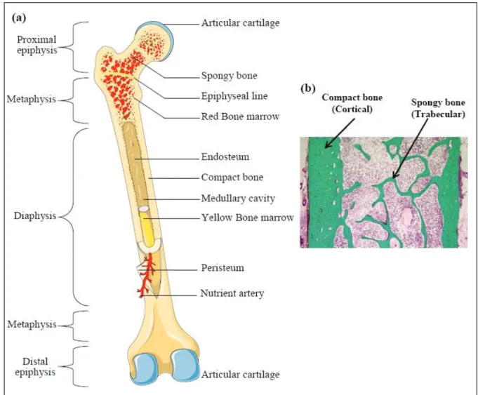
Bone architecture unit, also known as Bone Structural Unit (BSU), is labeled an Osteon. It is the primary building block of bone tissue. Osteon could be clearly visible under the microscope using a polarized light. Each Osteon makes up a concentric layer or lamellae. It is 3μm in thickness with a bright and dark appearance. This appearance is due to the 60o – 90o axis orientation of two consecutive lamellae. The Osteon diameter varies from 150 to 400 μm and can measure up to 2mm long [8, 9] (Figure.1).
Figure 1: Structure of compact bone. (a) Cross-sectional view of compact bone shows the basic structural unit, the
These lamella consist of type I collagen fibers and wherein calcium and calcium phosphate are deposited to form hydroxyapatite crystals. These collagen fibers are composed of fibrils that are morphologically characterized by periodic shells visible under electron microscopy. This mineralized matrix represents 90% of the bone tissue weight. The remaining 10% comes from other fundamental substances that are composed of diverse non-collagen proteins (osteocalcin, osteonectine, sialoprotein…etc), cytokines [10], lipids and proteoglycan. The noncollagenous proteins are at present under investigation, because of their importance in bone physiology. While playing a crucial role in binding of hydroxyapatite crystal to collagen fiber, osteocalcin seems to be involved in regulating mineralization phenomena.
1.3.2. Different bone tissue compartments
In adults, a bone tissue has two bone compartments: the first one is the cortical bone compartment that has a dense morphology. The other one is the trabecular bone compartment. It has a spongy morphology and is labeled the sponge bone. It also has a hexagonal three-dimensional cellular structure embracing the bone marrow.
The cortical bone makes up the epiphysis, metaphysis, and diaphysis of the long bones, and the envelope of plates and short bone. Spongy bone however makes up the interior part of epiphysis and metaphysis of long, flat, and short bones as illustrated in figure 2.
A-Cortical bone
Bone tissues are not made up of uniformly solid materials but rather constitute spaces between hard elements. Most bones have a thick and well organized outer shell known as the cortex (also known as compact bone) and they comprise 80% of human skeleton. The cortex has a slow turnover rate and a high resistance to bending and torsion. The cortical bone is made up of a dense and thick layer of mineralized tissue constituting the outer part of a bone
piece (Figure.2). It surrounds the medullary cavity and trabecular bone. The major part of the cortical bone is calcified which provides mechanical strength and protection, as well as regulates metabolic responses particularly under conditions of severe or prolonged mineral deficiency [11].
B-Trabecular bone
Trabecular bone also known as cancellous or spongy bone, makes up 20% of the skeletal mass. It has a higher surface area to mass ratio. Compared to cortical bone, it is less dense, more elastic, and has a higher turnover. Trabecular bone exhibits a major metabolic function of calcium-ions exchange [12]. Cancellous bone is typically found on the ends parts of long
Figure 2: Bone tissue compartments. (a) Scheme of a typical long bone shows the gross anatomical
bones, proximal to joints and within the interior of vertebrae (Figure.2). Cancellous bone is highly vascularized and frequently contains red bone marrow where hematopoiesis occurs. The primary anatomical and functional unit of cancellous bone is the trabecula.
Ratio of cortical to trabecular bone varies according to age and skeletal site. In adults, approximately 80% of the skeleton is composed of cortical bone and 20% of trabecular bone. However, the relative proportion between these two types, cortical and trabecular varies within skeletal parts. For example, the ratio of cortical bone to trabecular bone is 20 to 75 in human vertebrae, 50 to 50 in femoral head and 95 to 5 in radius in the diaphysis [13].
1.3.3. Bone classification
Anatomically, there are several types of skeletal bones. These are long (femur, humerus, radius, tibia ...), flat (skull, scapula, coast, mandible, calvarias ...), and short bones (ilium, vertebra ...) [5]. In addition to morphology, these bone types also differ in their mechanism of ossification during development. Flat bones are formed via membranous ossification while the development of bone tissue is directly from the surrounding mesenchymal cells without cartilaginous precursors. Short and long bones show a combination of membranous and endochondral ossification. The long bones are formed via endochondral ossification as an earlier step. More specifically, the long bones grow on a cartilaginous model that calcifies and is then gradually replaced by bone tissue. Long bones are predominantly distributed in the axial skeleton while short bones are distributed in the appendicular skeleton [14].
1.4. Bone tissue composition
Bones make up the largest proportion of the body’s connective tissue mass. Unlike most other connective tissue matrices, bone matrix is not only physiologically mineralized, but also unique in being constantly regenerated throughout life as a consequence of bone turnover.
Unlike most of the tissue, the extracellular matrix of the bone is mineralized by calcium salts in form of hydroxyapatite that gives the bone tissue a stiffness characteristic [15].
A- Organic bone matrix
The organic matrix of bone tissue is the protein matrix that interacts with the mineral phase. Ninety per cent of the organic matrix is composed of type I collagen while the remaining ten per cent is composed of other types of collagen-like material (Collagen III and V), proteoglycans and numerous non-collagenous proteins [16].
1. Collagen
Bone matrix fiber network is mainly composed of type I collagen also known as fibrillar collagen. Type I collagen is a ubiquitous protein heterotrimer composed of triple-helical molecules that contains two identical α1 chains and, a structurally similar but genetically different α2 chain [17]. Collagen α chains are characterized by a Gly-X-Y repeating triplet, where X is usually proline and Y is often hydroxyproline. There are also several post-translational modifications including: hydroxylation, glycosylation, and intra- and intermolecular covalent cross-links that differ from those found in soft connective tissue.
Collagen is released as procollagen, which is a triple helix form of non-active collagen that is assembled within the granules of endoplasmic reticulum of specialized bone cells. These cells are labeled osteoblasts. After the assembly process, procollagen is excreted in the intracellular matrix as illustrated in (Figure.3). Normally, bone matrix is predominantly made of type-I collagen and it gives the bone its ductility and the ability to absorb shocks without breaking. However, trace amounts of type III, type V, and FACIT collagens could be present during certain stages of bone formation. They also contribute to the regulation of collagen fibril diameter [18].
2. Noncollagenous proteins
Osteocalcin and osteonectin [19, 20] are two major non-collagenous proteins found in bone tissue, and play a role in bone mineralization. They belong to the family of γ-carboxylated glutamate proteins and regulate the activity of osteoclasts and their precursors. Osteocalcin and osteonectin are glycoproteins involved in regulating the genesis of collagen fibers and is a positive regulator of bone formation. Osteocalcin plays a role in the transitioning from resorption to formation phase during bone remodeling. Osteopontin, bone sailoprotein [21], phosphoproteins, phospholipids, proteoglycans [22] are other types of proteins incorporated into the bone matrix. Also, the bone extracellular matrix contains cytokines and growth factors [23] that play a primordial role in regulating bone remodeling
Figure 3: Collagen formation. Scheme representing different post-traductional modifications
and matrix mineralization. These proteins form the dynamic structure and function of the bone tissue organic matrix [24, 25]
B- Inorganic bone matrix or mineral phase
The bone is a reservoir for various minerals. It has the human body reserves for 99% of calcium, 85% of phosphors, 80% of carbonate and between 40 to 60% of sodium and magnesium of the organism [26]. The apatite crystal is generally flat, 2 to 5 nm thick and 20 to 80 nm long. The mineral phase is composed of phosphate and calcium crystal, under the form of hydroxyapatite [Ca10(PO4)6(OH)2]. The inorganic matrix of bone tissue is greatly
involved in phosphocalcic homeostasis in addition to protection and stiffness of the bones [27].
1.5. Conclusion
To recapitulate, bone tissue is a highly specialized connective tissue that serves four main functions: mechanical, protective, maintenance of mineral homeostasis and acid–base balance, and reservoir of growth factors and cytokines, and finally it provides the environment for hematopoiesis within the marrow spaces. Bone tissue development occurs in two distinct developmental processes: intramembranous and endochondral ossification. Unlike most of the tissues, bone tissue extracellular matrix is mineralized with calcium salts in the form of hydroxyapatite that gives the bone tissue its particular characteristic of stiffness.
2. Bone cells
Bone is a dynamic connective tissue, comprising an exquisite assembly of functionally distinct cell populations required to support its structural, biochemical, and mechanical integrity. Bone homeostasis is regulated by several cells. Bone resorption is mediated by osteoclasts derived from myeloid progenitor cells which originate from hematopoietic stem cells (HSCs). On the other hand, bone formation is mediated by osteoblasts derived from mesenchymal stem cells (MSCs). This progenitor cells MSCs produce bone mass, through osteoblasts, osteocytes and the protective bone surface lining cells.
2.1. Mesenchymal stem cells
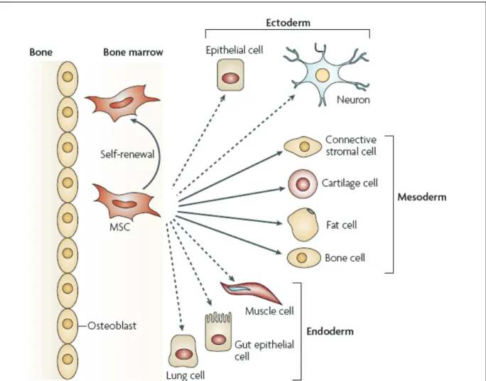
Mesenchymal stem cells (MSCs) were first described in the 1970s by Friedenstein et al. They reported the presence of fibroblastoid cells in the adult bone marrow (BM) that can differentiate into bone cells and reconstitute a hematopoietic microenvironment when transplanted subcutaneously [28]. MSCs represent less than 0.01% of the bone marrow cells. The ratio of MSCs to bone marrow-mononuclear cells decreases with age starting with 1:104 at birth decreasing to 1:105 in teenagers, and reaching as low as 1:2x10⁶ at the age of 80 [29]. Fortunately, MSCs can be recovered from several different locations such as adipose tissue [30], derma [31], dental pulp [32] and umbilical cord [33]. MSCs are multipotent precursors to many mesodermal cell lineages in vertebrate animals. They are present from early gestation through adulthood. Although they can be isolated from different adult tissues, MSCs display a stable phenotype in long-term culture, retaining the potential for adipogenic, chondrogenic, and osteogenic lineage differentiation in vitro [34], and are typically involved in the healing of damaged tissues such as bone, cartilage, muscle, ligament, tendon, adipose, and stroma in vivo [35]. Moreover, they are able to differentiate into a broad spectrum of cells that crosses
the oligolineage boundaries between mesodermal, ectodermal, and endodermal lineages (Figure.4) [36, 37].
2.1.1. MSCs and Heterogeneity
Studies of human bone marrow have revealed that about one-third of the MSC clones are able to acquire phenotypes of pre-adipocytes, osteocytes and chondrocytes [35]. This is in concordance with data showing that 30% of the clones from bone marrow have been found to exhibit a trilineage differentiation potential whereas the remainder display a bi-lineage (osteo-chondro) or uni-lineage (osteo) potential [38]. Moreover, MSC populations derived from
Figure 4: The multipotentiality of MSCs. This figure shows the ability of mesenchymal stem cells
(MSCs) in the bone-marrow cavity to self-renew (curved arrow) and to differentiate (straight, solid arrows) towards the mesodermal lineage. The reported ability to transdifferentiate into cells of other lineages (ectoderm and endoderm) is shown by dashed arrows, as transdifferentiation is controversial in vivo. (Figure adapted from Uccelli ,A. et al.2008)
adipose tissue and derma present a heterogeneous differentiation potential. In fact, only 1.4% of single cells obtained from adipose-derived adult stem cell (ADAS) populations were tri-potent while the others were bi-potent or unipotent [30]. Despite the similarities, differences appear to exist between MSC populations from different tissues. This presents an additional challenge to devise a universal definition determining their fate and functional characteristic [39, 40].
Pittenger et al. was the first to thoroughly study the surface antigens in MSCs [35]. Other groups subsequently attempted to characterize them until 2006, when the International Society for Cellular Therapy proposed that cells with the following characteristics should be considered as MSCs: (1) adherent to plastic in culture; (2) expression of CD105, CD73, and CD90 yet lacking CD45, CD34, CD14, CD11a, CD79a, or CD19 and histocompatibility locus antigen (HLA)-DR surface molecules; (3) capacity to differentiate into osteocytes, chondrocytes and adipocytes [41]. These criteria presented properties to purify MSCs and to enable their expansion by several-fold in-vitro, without losing their differentiation capacity. When plated at low density, MSCs form small colonies, called colony-forming units of fibroblasts (CFU-F), which are progenitors that can differentiate into one of the mesenchymal cell lineages [42, 43].
2.1.2. Circulating MSCs
Circulating MSCs in the peripheral blood stream are extremely rare in humans, even under normal conditions [44]. Although the role of these circulating MSCs in the normal state is unknown, this suggests that MSCs originally possess the ability to mobilize into the peripheral blood stream and migrate to organs. In cases of serious injury or stress, MSCs receive signals from the injured site and move to the bloodstream and migrate there [44, 45]. Following integration into the damaged site, they differentiate into cells to replenish the lost ones. Several factors and receptors related to these events have been suggested. Chemokines
and their receptors comprise a common system to recruit immunologic cells. The CXCR4-CXCL12 and CX3CR1-CX3CL1 systems were found to be involved in such migration. [46-48]. In addition, the urokinase receptor, the necrosis factor-related apoptosis-inducing ligand receptors (TRAIL) 2 and 4, and the endothelial nitric oxide synthase (eNOS) play a role in this process. High mobility group box 1(HMGB1), a chromatin protein released from damage cells and regulating gene expression specifically acts on MSCs to inhibit their proliferation and promote their migration and transdifferentiation [49]. Recently, in a corneal injury model, substance P, part of the atachykinin neuropeptide family and transmitter for specific sensory neurons, clearly triggered the recruitment of MSCs from bone marrow to the injured tissue and participated in the tissue repair process [50].
2.1.3. Role of MSCs
MSCs are pluripotent mesenchymal stromal cells with a number of common characteristics. The widely accepted niche of MSCs is at the abluminal surface of sinusoidal blood vessels in adult BM and at the interface between the BM and peripheral tissues. Their location is of importance given that their range of functions include regulation of local cellular homeostasis and immune regulation in case of inflammation or tissue injury [51, 52].
2.1.3.1. Effects of MSCs on Immune cells
MSCs have been shown to possess immunomodulatory characteristics through the inhibition of T-cell proliferation in vitro [53-55]. These observations have triggered a huge interest in the immunomodulatory effects of MSCs which was confirmed with several in vitro and in vivo studies [56]. The first in vivo study demonstrating this effect was performed in a baboon model in which infusion of ex vivo–expanded matched donor or third-party MSCs delayed the time to rejection of histo-incompatible skin grafts [54]. The delay indicated a potential role for MSCs in the prevention and treatment of graft-versus-host disease (GVHD)
in autologous stem cell transplantation (ASCT), prevention of organ transplantation rejection, and management of autoimmune disorders. Recently, MSCs were used to successfully treat a 9-year-old boy with severe treatment-resistant acute GVHD thus confirming the potent immunosuppressive effect in humans [57]. This characteristic was irrespective of the MSCs being derived from a third part or autologous with the stimulatory or the responder lymphocytes. The degree of MSCs suppression was found to be dose dependent. At high doses, MSCs were inhibitory whereas at low doses they enhanced lymphocyte proliferation in mantle cells lymphoma (MLCs) [58]. Broadly, MSC modulate cytokine production by the dendritic and T cell subsets DC/Th1 and DC/Th2 [59], block the antigen presenting cells (APC) maturation and activation [60], and increase the proportion of CD4+CD25+ regulatory cells in a mixed lymphocyte reaction [61].
The exact mechanisms by which MSCs are able to regulate immune functions are still not fully understood. However, the requirement of cell-to-cell contact is not clear. MSCs exert these effects via soluble inflammatory mediators and enzymatic action such as the expression of nitric oxide (NO) synthase, indoleamine 2,3-dioxygenase (IDO), transforming growth factor-beta (TGF-β), hepatocyte growth factor (HGF), and interleukin-10 (IL-10). A third mechanism is through secretion of human leukocyte antigen- G (HLA-G) and prostaglandin E2 (PGE2) [52, 62, 63] (Figure.5).
2.1.3.1.1. Dendritic cells (DCs)
DCs maturation and function are inhibited by MSCs [64, 65]. The effect on cytokine production results in an increased level of IL-10 production with a simultaneous decrease in the production of both pro-inflammatory cytokines TNF-α and IFN-γ [66]. The normal ability of DCs to upregulate T-cell activity is therefore suppressed in the presence of MSCs. It appears as well that MSCs are responsible for the induction of a more anti-inflammatory DC phenotype [59].
2.1.3.1.2. Natural Killer cells (NK)
The relationship between MSCs and NK cells is ambiguous and the mechanisms by which MSCs regulate inflammatory functions of NK cells are not well understood. While freshly isolated NK cells failed to attack MSCs, in vitro pre-activated NK cells acquired this ability [67]. On the other hand, MSCs can inhibit proliferation, cytokine secretion, and cytotoxicity of NK cells. IDO, TGF-β and PGE2, secretion by MSCs has been linked to anti-proliferative effects and reduction in cytokine production. Data suggest that cell-to-cell contact is also involved in this type of immunomodulatory effect. Several groups demonstrated the ability of
Figure 5: Mechanisms and cellular interaction of MSCs with immune cells. MSCs can inhibits both the
proliferation and function of natural killer (NK) cells via HLA-G, PGE2 and IDO. MSCs inhibit the differentiation of monocytes to immature myeloid dendritic cells via PGE2. Moreover, MSCs can suppress the proliferation and cellular activity of both of CD4+ and CD8+ T-cells via the release of several soluble molecules, including PGE2, IDO, TGFβ, HGF, iNOS, HO1 and HLA-G. The differentiation of regulatory T cells is mediated directly by MSCs through releasing of HLA-G. In addition, MSC-driven inhibition of B-cell function seems to depend on soluble factors and B-cell–B-cell contact. Finally, MSCs dampen the respiratory burst and delay the spontaneous apoptosis of neutrophils by constitutively releasing IL-6. (prostaglandin E2 (PGE2); indoleamine 2,3-dioxygenase (IDO); transforming growth factor-β1 (TGFβ1); hepatocyte growth factor (HGF); inducible nitric-oxide synthase (iNOS); haem-oxygenase-1 (HO1)). (Figure adapted and modified from Najib, H.2011 & Macfarlane. et al.2013).
MSCs to impair the proliferation of NK cells activated by IL-2 and IL-15. Secretion of Interferon gamma (IFN-γ), tumour necrosis factor-alpha (TNF-α), and IL-10 by activated NK cells is also reduced by MSCs in vitro [59, 67, 68].
2.1.3.1.3. Neutrophils
The two main effects of MSCs on neutrophils are an increase in their lifespan and inflammatory activity. Consistent results show a reduction of the spontaneous apoptosis rate in both resting and activated neutrophils mediated by MSCs secreting IL-6. Apoptosis in neutrophils is positively linked to production of reactive oxygen species (ROS). This leads to decreased inflammatory activity and onset of respiratory burst of neutrophils [69-71].
2.1.3.1.4. T- Cells
MSC-mediated inhibition of T cell proliferation has been largely described. In vitro, MSCs inhibited T cell proliferation regardless of the signaling pathway stimulated in the lymphocytes [53, 66]. The mechanisms by which MSCs are able to mediate immunosupression of T cells are diverse and complex. Several secreted effectors molecules have been linked to this process including IDO, PGE2, TGF-β, HGF, and HLA-G. The expression of HLA-G on adult MSCs is linked to their immunosuppressive effects on activated T cells via a mechanism including CD4+CD25+Foxp3+ regulatory T cells and IL-10 [63, 72].
2.1.3.1.5. B-Cells
The effect of human MSCs on B cells mainly depends on several factors linked to B cell biology, state of differentiation, strength of stimulus, and B to MSCs cells ratio. Evidence on the effect of MSCs on B cell proliferation is contradictory between inhibitory and stimulatory [73-75].
2.1.3.1.6. Regulatory T-Cells
MSCs are able to promote the proliferation of regulatory T cells and induce differentiated T-helper cells to display a regulatory phenotype through the production of heme oxygenase-1 (HO-1) [76, 77].
2.1.3.2. Tissue repair by MSCs
2.1.3.2.1. Bone
MSCs are able to migrate and engraft into multiple musculoskeletal tissues, especially at sites of injury, and undergo site-specific differentiation. These characteristics made MSCs a most promising candidate for cell therapy in many refractory diseases including bone joint diseases which can cause a great deal of pain, discomfort, and even disability. The initial clinical trials with MSCs were performed by Horwitz and colleagues in 1999 who demonstrated that bone marrow-derived mesenchymal cells improves the total-body bone mineral content and subsequent osteogenesis in children with osteogenesis imperfect [78]. Since then, MSCs have been used in patients with osteoarthritis and bone defects (Table.1).
2.1.3.2.2. Central Nervous System
Several studies using MSCs to recover from stroke (rat model) or repair spinal cord injury, and traumatic brain injury have been reported. In all of these models, MSC transplantation by either direct injection, intravenous infusion, or injection into the cerebrospinal fluid showed promising results with functional recovery [79, 80]. Clinical trial for CNS repair is most advanced in spinal cord injury (Table.1).
2.1.3.2.3. Heart
In myocardial infarction, transplanted MSCs or bone marrow mononucleated cells integrate into damaged tissue and differentiate into cardiac muscle cells. Autologous bone
marrow mononucleated cells transplantation in acute myocardial infarction patients resulted in efficient cardiac function recovery for up to one year[81] (Table.1).
2.1.3.2.4. Other Tissues
MSCs are involved in the repair of skeletal muscle degeneration [82], ischemic colonic anastomoses [83], chronic severe wounds such as skin ulcers [84], and airway (trachea) obstruction [85] (Table.1).
Despite the very optimistic results obtained in animal models, MSC-based preliminary clinical trials have not fully met the expectations. The lack of obvious outcomes in clinical trials may be the result of specific human MSCs features and MSC-niche interactions and must be further addressed by analyzing these factors in human contexts.
2.2. Hematopoietic Stem Cells
The history of stem cell research began in the early 1960s when James Till and Ernest McCulloch and their colleagues at the University of Toronto came across reservoirs of cells in mice with the properties of stem cells: the abilities to self-renew and differentiate into specialized cell. In the late 1960s, researchers began to infuse hematopoietic stem cells (HSCs) (this section will be discussed in more details below)
2.3. Conclusion
The principal cells that mediate bone homeostasis process of the mammalian skeleton are: myeloid progenitor cells which derived from HSCs (known as osteoclasts), and osteoprogenitor cells which are derived from MSCs (known as osteoblasts, chondrocytes, and osteocytes). Both types of cells play an important role in bone homeostasis. MSCs can be recovered from several different locations including bone marrow. MSCs are characterized by being adherent to plastic in culture, expressing CD105, CD73, CD90 and histocompatibility locus antigen (HLA)-DR surface molecules, and having the capacity to differentiate into
osteocytes, chondreocytes and adipocytes. Moreover, they have a considerable immunomodulatory potential on both innate and adaptive immune cells. Recently, MSC-based investigation for clinical applications has obtained intensive attention and shows very optimistic results in animal models. Further investigations are needed to explore the clinical application especially in human models.
40
Table.1|
Case studies and clinical ap
plication o f MSCs P athology Spe cies Target or gan Mechanism of action Re f. Rheumatoid Arthritis Mouse Jo in ts Inhibition of sec re tion of proinflammator y c yto kines and inhibitions of T cells [61] L upus e ry thematosus Mouse B one ma rr ow Reg en er ation of hematop oietic niche [86] Rejec tion of HSC transplantation She ep He matopoietic compar tment Im prov e transplant ation eff icien cy , increase h ema topoiesis [87] Osteog nesis imperf ecta Human B one ma rr ow In crease total -bod y bon e minera liz
ation and osteog
en esis [78] Ic tu s Ra t
Central nervous system
Secre tion of neu rotrophic factors [88] Ex perimental autoimmune enceph alom yeli ti s Ra t
Central nervous system
Inhibition of m yelin -spe ci fic T cells [89] M yoc ardial infa rc tion Ra t He art Secre tion of trophic fa cto rs SFRP2 [90] M yoc ardial infa rc tion Human He art Im prov ed gl
obal left vent
ricle fuctio n [91] Rejec tion of transplanted islets Mouse Kidne y capsule Inhibition of β cell -sp ecif ic T c ells [92] Ac ute r en al fa ilur e Ra t Kidne ys Secre tion of trophic factors, Inhibition of se cr et ion of proinflammator y c ytoki nes [93] Diabetes Mouse Pancre as and re nal g lomeruli Inhibition of macrophag e infiltra tion [94] Diabetes Mouse Pancre as Inhibition of β cell -sp ecif ic T c ells [95] He patic f ailure Mouse Li ve r Inhibition of inflammator y infiltra te [96] Ulcera tive colitis Mouse Gut Suppression of inflammator y infiltra tes and cy tokines. In crease of reg ulator y T cell activit y [97] Rejec tion of cutan eous grafts Monke y Skin Inhibitions of T c ells [54] Ac ute lun g injur y Mouse Lu ng s Inhibition of secre tion of proinflammator y c ytoki nes [98] Ac ute lun g injur y Mouse Lu ng s Inhibition of secre tion of proinflammator y cy tokines. Secre tion of IL -1 0 [99] Retinal dege ner ation Ra t Ey es Indu
ction and sec
re
tion o
f trophic fa
ctors
3. Osteoblastic lineages
Osteoblasts are the major cellular component of bone. They are mononuclear cells of 20 to 30μm in diameter, arising from mesenchymal stem cells that undergo a well defined program of gene expression as they progress through osteoblastic commitment, proliferation, and terminal differentiation [101]. At the end of the bone formation phase, osteoblasts can either become embedded in bone as osteocytes [102], become inactive osteoblasts or bone lining cells [103], or undergo programmed cell death (apoptosis)[104].
3.1. Osteoblasts origin and differentiation
Bone formation is a tightly regulated process which is characterized by a sequence of events starting by the commitment of osteoprogenitor cells, their differentiation into pre-osteoblasts and to mature pre-osteoblasts whose function is to synthesize the bone matrix that progressively mineralize.
The lineage commitment of multipotent mesencyhmal cells is driven by the selective expression of so-called master transcription regulators. Thus, MyoD (known as myogenic regulatory factors (MRFs)) directs these cells into the myogenic pathway [105]; proliferation-activated receptor γ2 (PPARγ2) promotes adipogenesis [106]; Sox9 drives chondrocyte development [107], and Runx2 is necessary for the osteoblast lineage [108-110] (Figure.6). Following lineage commitment, osteoprogenitors undergo a proliferative stage, characterized by the production of proteins such as histones, fibronectin, type I collagen c-Fos, c-Jun and p21 [111]. Subsequently, they exit mitosis, transition to expressing genes such as alkaline phosphatase (ALP), bone sialoprotein (BSP), and type I collagen, as they start to produce and mature an osteogenic extracellular matrix. Finally they express genes involved in mineralization of the extracellular matrix such as osteocalcin (OC), osteopontin and collagenase [112]. This highly regulated program of gene expression and cellular
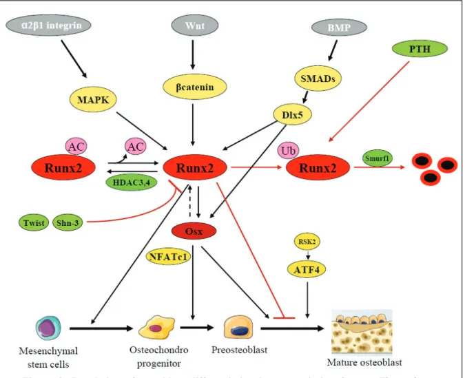
differentiation is governed by the expression and activity of transcription factors including Runx2, Osterix, SMADs, Wnt/β-Catenin, NFATc1, Twist, AP-1 and ATF4. These factors do not act alone, but interact to integrate diverse signals and to results in a fine-tune gene expression (Figure.7). The role of each of these factors as regulators of gene expression is presented and the key recent findings are discussed below.
Figure 6: Mechanical characteristic and differentiation potential of MSCs. Significant correlations
exist between stem cell mechanical biomarkers and the production of lineage-specific molecules that are characteristic of differentiated cell types. Stiff cells have greater osteogenic potential with specific gene markers such as Runx2 and Osx, highly viscous cells have greater chondrogenic potential (positive correlation with apparent viscosity) with high expression of the gene markers like Sox 9/5/6, and large, “soft” cells have greater adipogenic potential with gene marker PPARγ2 and C/EBP family (Figure adapted and modified from González-Cruz RD, et al 2012).
Figure 7: Model proposed for osteoblast differentiation from MSCs. Ihh is the initiator of
endochondral ossification. The Runx2 expressing biopontential progenitors can differentiate into either osteoblast or chondrocyte. Then cells differentiate into preostoblast, in which Runx2 plays an essential role. In the next step, preosteoblasts differentiate into mature osteoblast, a process in which Osx plays a critical role. In the last step osteoblasts differentiate into osteocytes, and during this process, Osx is required for the expression of the two gene markers DKK1 and Sost. It is speculated that Osx controls osteocyte differentiation from osteoblasts (Figure adapted from Zhang Chi 2012).
3.1.1. Runx2
A decade ago, Runx2 (Cbfa1, AML3) has been identified as the master regulator of osteoblastogenesis [113]. Runx2 is a member of the Runt family of transcription factors that is expressed by mesenchymal cells at the onset of skeletal development and it acts throughout the induction, proliferation, and maturation of osteoblasts and regulates expression of many osteoblastic genes [114]. Although Runx2 is the most abundant factor in mature osteoblasts, Runx1 and 3 are also present in osteoblast lineage cells [115]. The Runx regulatory element can be found in the promoter of all major osteoblastic genes controlling their expression, BSP and OCN, resulting in the establishment of an osteoblast phenotype. Additionally, Runx2 can be phosphorylated and activated by the mitogen-activated protein kinase (MAPK) pathway by binding of type I collagen to α2β1 integrins on the osteoblast surface [116]. In addition to control osteoblast differentiation, Runx2 was found to negatively control osteoblast proliferation by acting on the cell cycle [117]. It is intriguing that Runx2 can modulate the expression of kinases such as p85 PI3K that controls osteoblast differentiation and survival [118]. Recent studies indicate that Runx2 interacts with several regulatory proteins within the nuclear architecture, resulting in activation or repression of genes which control the program of osteoblast proliferation and differentiation. This indicates that Runx2 can control osteoblastogenesis through multiple mechanisms [113]. Despite its important role in osteoblast commitment, Runx2 is not essential for the maintenance of the expression of the major bone matrix protein genes in mature osteoblast [119]. Mice overexpressing Runx2 exhibit osteopenia, as a result of reduced number of mature osteoblast terminal differentiation and maintenance of osteoblastic cells in an immature stage [120]. Thus, Runx2 can act differently at multiple levels to control osteoblast differentiation and bone formation. As expected from the important role of Runx2 in osteoblastogenesis, both expression and activity of Runx2 are tightly controlled transcriptionally and post-translationally.
Several recent papers shed light into the factors and mechanisms that control Runx2 function. Histone acetyltransfereases, such as p300, CBP, PCAF, MOZ and MORF, are coactivators of Runx2 [121, 122]. They add acetyl groups to lysine residues of histone and nonhistone target proteins, which modifies protein function by a variety of mechanisms including altered protein-protein interaction and altered protein stability.
Histone deacetylation, catalyzed by histone deacetylases (HDACs) is correlated with chromatin condensation and transcriptional repression. The interaction between Runx2 and HDACs was first suggested by the discovery that histone deacetylases inhibitors reduce the activity of some Runx2 repression domains [123]. Subsequent studies have shown that Runx2 binds and is functionally inhibited by HDAC3, 4, 5 and 7 [124-129]. It remains poorly understood how these complexes participate in regulation of Runx2 activity, although it has been shown that Runx2 target gene expression is repressed by HDACs through multiple distinct mechanisms and in response to various osteogenic signals such as BMP2 and PTH. (Figure.8).
Runx2 and PTH
Parathyroid hormone (PTH) is another important regulator of skeletal physiology that stimulates Runx2 interaction with acetyltransferases. PTH is a strong inducer of MMP-13 transcription in osteoblasts [130]. Stimulation of these cells with PTH leads to a protein kinase A-dependent binding of p300 to Runx2 on the MMP-13 promoter, resulting in increased histone acetylation and gene transcription [131, 132]. PTH also regulates Runx2 activity through other mechanisms such as phosphorylation [133] and promoting interactions with AP-1 transcription factors [134, 135]. Finally, PTH decreases Runx2 protein stability by ubiquitin-mediated proteolysis, limiting PTH stimulation of osteoblastic genes [136] (Figure 8).
3.1.2. Osterix
Osterix (Osx, also known as Sp7) is a zinc finger transcription factors expressed in osteoblasts and, like Runx2, is required for bone formation [137]. Osx-null mice die at birth due to their lack in mineralized skeletons. Bones formed by intramembranous ossification are entirely non-mineralized, while endochondral bone exhibit regions of mineralized cartilage, indicating that Osx functions specifically in osteoblasts. However, Runx2 is expressed in Osx-deficient mice, indicating that Osx acts downstream of Runx2 [138] and acts by directing pre-osteoblasts to immature pre-osteoblasts. This was confirmed through characterization of a Runx2-binding element in the Osx gene promoter [139]. Osx activation of collagen IA1 promoter is enhance by binding of NFATc1 to Osx, an interaction that is disrupted by calcineurin [140] (Figure.8). Another function of Osx is as inhibitor of the canonical Wnt signaling by inhibiting DNA binding of (TCFs) [141]. One zinc finger protein, Schnurri-2, was found to negatively control Osx and thereby bone formation [142]. Additionally, p53 was found to repress Osx transcription [143]. Despite these interesting findings, the details concerning the regulation and function of Osx are incompletely understood.
3.1.3. Wnt and β-Catenin
Genetic studies have revealed that canonical Wnt signaling is an important pathway controlling bone formation and bone mass [144, 145]. Interaction of some Wnt proteins with frizzled and LRP5/6 co-receptors leads to inhibition of GSK-3-mediated β-catenin phosphorylation, resulting in β-Catenin accumulation and translocation into the nucleus, binding to LEF/TCF transcription factors and activation of down stream genes. Inactivation of catenin blunts osteoblast differentiation from mesenchymal progenitors, indicating that β-catenin plays an essential role in osteoblast differentiation and calcification in in vivo via inducing Runx2 expression [146, 147] (Figure.8).
3.1.4. BMPs and SMADs
Bone morphogenetic proteins (BMPs) were discovered in 1965 as potent inducers of ectopic bone formation when implanted subcutaneously. BMP2, 4, 6 and 7 are osteoinductive. BMP signaling leads to phosphorylation and nuclear translocation of receptor-activated SMADs (rSMADs), which interact directly with DNA and associate with other transcription factors (such as Dlx) to regulate gene transcription. rSMADs direct MSCs into the osteoblast lineage through induction of Runx2 expression [148]. They also interact with Runx2 protein to synergistically regulate transcription [149-151]. The SMAD-interaction domain in Runx2
Figure 8: Regulation of osteoblast differentiation by transcription factors. The major
transcription factors Runx2, Osx, and ATF4, which are activated and regulated by other important transcription factors, control the differentiation and maturation into the osteoblastic lineage starting from mesenchymal stem cells. In osteoblast differentiation, Runx2 is essential for the osteogenic potential of uncommitted osteochondroprogenitors and Osx is required for the commitment of osteochondroprogenitors to preosteoblasts and mature osteoblasts (Figure adapted and modified from Baek WY, et al.2011).
has been identified and is continuous with the nuclear matrix targeting sequence, which is necessary for Runx2 function [150, 152]. SMADs are inactivated by Smurf-directed ubiquitination, resulting in their proteolytic degradation. An interesting feedback loop between BMP/SMAD/Runx2 signaling is indicated by recent studies showing that BMPs act through Runx2 to induce the expression of BMP6, an inhibitory SMAD protein that represses BMP signalling [153]. SMAD6 stimulates Runx2 ubiquitination and degradation by Smurf [154]. Although, the role of BMPs in bone formation is well known, the current clinical data supporting their effectiveness are not robust, possibly in part because BMPs affect bone resorption as well. BMPs can reduce bone mass by inducing osteoclastogenesis via the RANKL-OPG pathway, which is a critical regulator of osteoclasts by osteoblasts. BMPs have both bone anabolic and catabolic effects by affecting multiple cell types in bone such as mesenchymal cells, chondrocytes, osteoblasts, osteoclasts, and endothelial cells [155] (Figure.8).
3.1.5. AP-1
AP-1, a transcription factor composed of heterodimers of Fos-related factors (c-Fos, Fra1, Fra2 and FosB) and Jun proteins (c-Jun, JunB and JunD). Multiple Fos and Jun proteins are highly expressed in proliferating osteoprogenitors. Their expression decrease during differentiation such that Fra2 and JunD are the primary AP-1 components present in mature osteoblasts [156]. Recent work by Chang et al. demonstrate that inhibition of the signaling cascade activated by the transcription factor nuclear factor κ B (NF-KB) specifically in
differentiated osteoblasts promotes bone formation through increased Fra1 expression [157]. These observations indicate that AP-1 proteins promote bone formation. In contrast, deletion of JunD increased bone mass, apparently by increasing expression of Fra1, Fra2and c-Jun, suggesting that JunD represses expression of other AP-1 proteins in osteoblasts [158]. A
number of direct targets of AP-1 in osteoblasts have been identified, and include the osteocalcin, collagenase-3 (MMP13), BSP, and ALP promoters.
3.1.6. ATF4
The positive role of ATF4 on osteoblast formation was recognized with the findings that it is a substrate for the RSK2 kinase. Deficiency in ATF4decreased bone formation [159], while forced accumulation of ATF4 induced osteoblastic gene expression in non-osseous cells [160]. ATF4 forms a complex with Runx2 [161] at the OCN promoter to increase OCN transcription (Figure.8).
3.1.7. Helix-loop-Helix protein
The helix-loop-helix (HLH) protein, including Id and Twist, are expressed during the proliferating stage in osteoblasts. One mechanism through which Twist acts to impair osteoblastogenesis is by binding to the Runx2 DNA binding domain and inhibiting its ability to bind DNA [162] (Figure.8). Twist also inhibits BMP/SMAD responsive transcription by forming a complex with Smad4 and HDAC1 [163].
3.1.8. Zinc Finger Protein / Schnurri-3
Two major families of Zinc-finger transcription factors are the kruppel-like factors (KLFs) and Specificity Proteins (Sps). Members of both groups participate in the regulation of gene expression in osteoblasts through interactions with other transcription factors at target gene promoters. ZFP521, a KLF protein, is expressed in osteoblast precursors, osteoblasts and osteocytes, as well as in chondrocytes [164]. Its expression increases during osteoblast differentiation and in response to PTHrP, while BMP2 decreases ZFP521 levels. ZEP521 binds Runx2 and antagonizes Runx2 gene transactivation, and overexpression of ZFP521 in in vitro osteoblast cultures impairs their differentiation. Recently, it was discovered that mice with germ line deletion of Schnurri-3 (Shn3 also known as ZAS3, a large zinc finger protein
[165]) displayed a massive increase in bone mass, revealing an unexpected role for this protein in the skeletal system [166, 167]. Shn3-deficient animals showed markedly augmented osteoblastic bone formation in vivo. Consistently, cultured primary osteoblasts lacking Shn3 express increased levels of classic osteoanabolic genes and produce increased amounts of mineralized ECM. In osteoblasts, Shn3 functions at least in part by regulating Runx2 protein levels via promoting its degradation through recruitment of the E3 ubiquitin ligase WWP1 [166] (Figure.8). Wein et al. showed that selective deletion of Shn3 in the mesenchymal lineage recapitulates the high bone mass phenotype of global Shn3 knock-out mice, including reduced osteoclastic bone catabolism in vivo and indicating that Shn3 expression directly controls osteoblastic bone formation and indirectly regulates osteoclastic bone resorption [168, 169].
3.1.9. Homeobox proteins
Cellular and genetic analyses showed that several transcription factors that belong to the homeobox proteins (Msx1, msx2, Dlx5, Dlx6) may play a role in osteoblast differentiation. These proteins act as a transcriptional repressors or activators and are essential for normal ossification [170-172]. Msx2 inactivation delays skull ossification in mice which is associated with decreased Runx2 expression [173]. Cellular analysis showed that Msx2 is expressed mainly in osteoprogenitor cells and is downregulated during differentiation [174]. Consistently, Msx2 promotes osteoblast proliferation and differentiation into mesenchymal cells [175, 176] but inhibits Runx2 activity and osteoblast gene expression in more mature osteoblasts in vitro [177, 178]. Additionally, Msx2 controls osteoblast apoptosis during in vitro osteogenesis [179], suggesting a stage specific action of Msx2. In vivo, however, Msx2 is a positive regulator of bone formation [180]. In contrast to Msx2, Dlx3 and Dlx5 are expressed at all stages of osteoblast differentiation and their expression increases in more mature osteoblasts [174, 181]. Dlx3 has a complex role as it has both positive and negative
effects on OCN gene transcription [174]. One function of Dlx5 is to activate the expression of Runx2 and of the bone markers BSP and OCN [182] (Figure.8). The potential role of other Dlx proteins is unknown, except for Dlx2 which is recently shows a potential osteogenic differentiation of stem cells [183].
3.2. Osteocytes
The osteocyte, defined as a cell located within the bone matrix, is descended from mesenchymal stem cells through osteoblast differentiation. Osteocytes are the most abundant cellular components of mammalian bones, making up to 90% of bone tissue cells and are derived from 10 to 20% of osteoblasts that are walled in the matrix during formation. As it can be derived from matured osteoblasts [184, 185]. Osteocytes possess morphologic and phenotypic characteristics that can live long [103]. The transformation of an osteoblast into an osteocyte has long been considered as a passive “burial” in the newly formed matrix [102]. Recently, it is recognized that this differentiation is an active mechanism in which the cell acquired certain characteristics [186]. Osteocytes are regularly spaced throughout the mineralized matrix and communicate with each other and with cells on the bone surface via multiple extensions of their plasma membrane that run along the canaliculi [187] (Figure.7). Osteocytes have alternatively resorbing activity (via OCs activation) then formation (OBs activation). The synthesis activity is strongly reduced and the osteocytes express specific genes which are different from those expressed by osteoblasts [188]. This kind of role by osteocytes is done in the presence of mechanical stimuli. As yet, it is not fully comprehended how osteocytes sense mechanical stimuli, and only a fraction of the whole range of molecules that osteocytes subsequently produce to regulate bone formation and degradation in response to mechanical stimuli is known. Recently, it was mentioned that mechanosensing is enabled by force-induced conformational changes in cellular structures, such as stretch-activated ions channels, integrin complexes, and cell-cell adhesions. The conformational changes enable the
influx and efflux of ions or the activation of signaling cascades, resulting in altered cell shape and altered activity and production of proteins [189]. With respect to osteocyte mechanosensing, the focal adhesion kinase inhibitor-14 has been shown to abolish fluid flow-induced stabilization of β-catenin and consequent activation of the Wnt/ β-catenin pathway in osteocytes, suggesting that focal adhesions and integrins play an important role in osteocytes mechanosensing [190]. It has been hypothesized that osteocytes in the skull and long bone have different sensitivity to mechanical stimulation, and this is based on their difference in mechanical environment, which could not be confirmed in vitro [191, 192]. Recently, a new study reported that mechanical loading in osteocyte can induce formation of Src/Pyk2/ MBD2 (methyl-CpG binding domain protein 2) complex that resulted in suppression of anabolic gene expression. This process occurred through translocation and accumulation of Src/Pyk2 in the nucleus and associates with protein involved in DNA methylation [193]. The osteocytes cytoskeleton might be altered during osteoporosis, since enhanced circulating levels of cytokines are present, which are known to modulate the cytoskeleton in several cell types. Cytokines are also highly expressed during inflammatory diseases such as Crohn’s disease and rheumatoid arthritis, and are associated with loss of bone mass. The cytokine TNF-α and IL-1β inhibit the increase of nitric oxide (NO) production and intracellular calcium that is normally observed in cultured osteocytes after application of a mechanical stimulus in the form of fluid flow [194]. TNF-α and IL-1β strongly reduce F-actin content, which results in a reduction of osteocyte stiffness as indicated by elastic moduli determined by twisting magnetic beads attached to the cell, providing a possible mechanism through which inflammation contributes to loss of bone mass [194].