HAL Id: hal-02192635
https://hal.archives-ouvertes.fr/hal-02192635
Submitted on 24 Jul 2019HAL is a multi-disciplinary open access
archive for the deposit and dissemination of sci-entific research documents, whether they are pub-lished or not. The documents may come from teaching and research institutions in France or abroad, or from public or private research centers.
L’archive ouverte pluridisciplinaire HAL, est destinée au dépôt et à la diffusion de documents scientifiques de niveau recherche, publiés ou non, émanant des établissements d’enseignement et de recherche français ou étrangers, des laboratoires publics ou privés.
Gut microbiome in Chronic Rheumatic and
Inflammatory Bowel Diseases: similarities and
differences
Fatouma Salem, Nadège Kindt, Julian Marchesi, Patrick Netter, Anthony
Lopez, Tunay Kokten, Silvio Danese, Jean-Yves Jouzeau, Laurent
Peyrin-Biroulet, David Moulin
To cite this version:
Fatouma Salem, Nadège Kindt, Julian Marchesi, Patrick Netter, Anthony Lopez, et al.. Gut mi-crobiome in Chronic Rheumatic and Inflammatory Bowel Diseases: similarities and differences. United European Gastroenterology Journal, SAGE Publications, In press, 7 (8), pp.1008-1032. �10.1177/2050640619867555�. �hal-02192635�
Gut microbiome in Chronic Rheumatic and Inflammatory Bowel
Diseases: similarities and differences
Fatouma SALEM1, Nadège KINDT1, Julian R MARCHESI2,3, Patrick NETTER1, Anthony LOPEZ4,5, Tunay KOKTEN4, Silvio DANESE6, Jean-Yves JOUZEAU1, Laurent PEYRIN-BIROULET4,5*and David MOULIN1,7*
1
IMoPA, UMR7365 CNRS-Université de Lorraine, Vandœuvre Les Nancy, France 2
Division of Integrative Systems Medicine and Digestive Disease, Imperial College London, England
3
School of Biosciences, Museum Avenue, Cardiff University, Cardiff, UK
4
NGERE, UMR_ U1256 INSERM-Université de Lorraine, Vandœuvre Les Nancy, France 5 Service d’hépato-gastroentérologie, CHRU de Nancy, Vandœuvre Les Nancy, France 6
Department of Biomedical Sciences, Humanitas University, Rozzano, Milan , Italy. 7
CHRU de Nancy, Contrat d’interface, Vandœuvre Les Nancy, France
*Authors contributed equally to this work and are corresponding:
Laurent PEYRIN-BIROULET: [email protected], Service d’hépato-gastroentérologie, CHRU de Nancy, Vandœuvre Les Nancy, France
David MOULIN: [email protected]; IMoPA, UMR7365 CNRS-Université de Lorraine, Vandœuvre Les Nancy, France
Contributors
FS and NK: study concept and design;acquisition, analysis, review and interpretation of data;manuscript preparation and critical revisions. NK: study concept and design; review and interpretation of data;manuscript preparation and critical revision. JRM, PN, AL, TK, SD: review and interpretation of data;manuscript preparation and critical revisions. JYJ, LPB DM: study concept and design; review and interpretation of data; manuscript preparation and critical revisions; study supervision.
Competing interests
1
ABSTRACT
Introduction: Inflammatory bowel diseases (IBD), and Chronic Rheumatic Diseases (CRD)
are systemic chronic disorders sharing common genetic, immune and environmental factors. About half of patients with IBD develop rheumatism affections and microscopic intestinal inflammation is present in up to half of CRD patients. IBD and CRD patients also share a common therapeutic armamentarium. Disequilibrium in the complex realm of microbes (known as dysbiosis) that closely interact with the gut mucosal immune system, has been associated with both IBD and CRD (Spondyloarthritis and Rheumatoid Arthritis). Whether dysbiosis represents an epiphenomenon or a prodromal feature remains to be determined.
Methods: In an attempt to further interrogate whether specific gut dysbiosis may be the
missing link between IBD and CRD in patients developing both diseases, we performed here a systematic literature review focusing on studies looking at bacterial microbiota in CRD and/or IBD patients.
Results: We included 80 studies, with a total of 3799 IBD patients without arthritis, 1084
CRD patients without IBD, 132 IBD patients with arthropathy manifestations and 12 SpA patients with IBD history. Overall, this systematic review indicates that an increase in
Bifidobacterium, Staphylococcus, Enterococcus, Lactobacillus, Pseudomonas, Klebsiella and Proteus genera, as well as a decrease in Faecalibacterium, Roseburia genera and species
belonging to Verrucomicrobia, Fusobacteria phyla are common features in IBD and CRD patients, whereas dozens of bacterial species are specific features of CRD and IBD.
Conclusion: Further work is needed to understand the functions of bacteria and of their
metabolites but also to characterize fungi and viruses that are commonly found in these patients.
Introduction
Inflammatory bowel diseases (IBD), are mainly represented by Crohn's disease (CD) and ulcerative colitis (UC), whereas Chronic Rheumatic Diseases (CRD), encompass Rheumatoid Arthritis and Spondyloarthritis (SpA). These systemic chronic disorders have relapsing and remitting clinical course arising from an interaction between genetic, immune and environmental factors.
CRD and IBD are intercurrent since articular manifestations are observed in up to 40% of IBD patients and intestinal inflammation is often present in CRD subjects 1. Co-occurring CRD and IBD can be very disabling and are associated with a more severe disease course in IBD patients2.
Interestingly, IBD and CRD share common pathophysiology, including common molecular and cellular actors and, consequently, common therapeutic armamentarium. Genetic studies have reinforced the importance of genes and pathways contributing to IBD pathogenesis, such as barrier function, the role of T cell subsets, and cytokine-cytokine receptor signalling 3. In addition, recent studies pointed out new genes and pathways, including autophagy or regulation of interleukin 23 (IL23) signalling, highlighting the importance of host defence pathways, specifically those involved in the management of mycobacteria 4. Heredity is also an important feature of CRD and notably in SpA, and several genetic polymorphisms have been shown to influence the disease risk. The most important one is the major histocompatibility complex (MHC) class I allele HLA-B27 5. Remarkably, a large subset of the IBD and CRD susceptibility identified genes are encoding for proteins involved in immune response, and particularly in the IL-23/Th17 pathway of T cell differentiation, which is primarily implicated in response against extracellular pathogens, including bacteria and yeasts, and/or in microbial sensing.
However, the link between pathological gut and joint inflammation in patients with both IBD and CRD is not fully understood. Taken together, these data suggest that the perturbation of the gut microbiome, also called dysbiosis represent an attractive target in this context.
In an attempt to further interrogate whether specific gut dysbiosis may be associated with IBD and CRD and promote pathological inflammation within joint-gut axis, we performed a systematic literature review investigating similarities and differences regarding faecal microbiota in these patients.
Methods
Search strategy and study selection
A systematic literature search was performed according to PRISMA guidelines 6. The literature review conducted using PubMed/MEDLINE (from 1950 to December 2018), Web of science (from 1958 to December 2018). Abstracts from annual meetings of national and international gastroenterology and rheumatology conferences (United European Gastroenterology Week [UegW], Digestive Diseases Week [DDW], European Crohn's and Colitis Organization [ECCO], European League Against Rheumatism [EULAR], American College of Rheumatology [ACR]) were searched manually from 2013 to 2018.
The following keywords were searched in various combinations using the boolean terms “AND” and “OR” ("Microbiota", "Microbiome", "Gut", "Gastrointestinal Microbiome","Microbiology", "Colitis", "Ileitis", "Intestinal", Enteritis", "Inflammatory Bowel Diseases", "Crohn Disease", "Ulcerative Colitis" , “Rheumatoid Arthritis”, "Spondyloarthritis" ,"Arthritis", "Reactive Arthritis", "Psoriatic Arthritis", "Rheumatoid Arthritis", "Infectious Arthritis", "Ankylosing Spondylitis", " Mycobiome", "Fungal Microbiota", "Intestinal Virome" ). This strategy was used both as Medical Subject Headings (MeSH) terms if available and as free text. Searching was limited to publications with human subjects. We only selected English language full text papers and abstracts.
Two authors independently reviewed all articles. Inclusion criteria included the presence of IBD and CRD patient samples and 16S rRNA gene sequencing or metagenomic methods to characterize the gut microbiota. Literature reviews did not include meta-analyses, as well as experimental studies based on in vitro findings and animal models.
Study characteristics and outcomes were reported in a Microsoft Excel Office 2016 Professional spread sheets.
5
Results
6519 were identified (Fig 1) based on defined criteria. After review of the titles and abstracts 5564 papers were excluded. Amongst the remaining studies, another 881 were excluded because they included reviews, data retrieved from studies using animal models and in vitro findings. Therefore, 80 studies were included: 56 from IBD patients, with 1 Case-reports 39 (Table 1.a, 1.b and 1.c), 21 from CRD patients (RA and SpA) including 5 congress abstracts 78,80,81,84,85
(Table 2.a and 2.b). Finally, three publications addressed gut microbiota study in IBD patients developing arthropathy 94,96,97 (Table 3). As microbiota from one individual is different from one sample location to another, table were generated by sample type and are detailled with studied populations characteristics.
1. Literature search results
A.
Distinct dysbiosis in IBD and CRD
In order to identify bacterial variations specific of IBD, (i.e. not found in CRD), and vice versa, we adopted two complementary methodologies: we first reviewed bacterial changes reported in studies enrolling IBD patients without information on possible concomitant arthritis, then all studies involving CRD patients without information on possible concomitant IBD. We looked finally at studies comparing gut microbiome in patients with or without IBD-associated CRD.
A.1. Gut bacterial changes reported in IBD patients
Fifty-six studies enrolling 3270 IBD patients from which gut microbiota was mainly analyzed by 16S rRNA gene sequencing or qRNA of DNA extracted from faeces and/or biopsies. A quantitative and qualitative (biodiversity) reduction of the gut microbiome in IBD patients 7,8 is generally observed.
Firmicutes phyla
A reduction of Clostridiales order species from the Firmicutes phylum is observed in the faecal microbiota of IBD and CD patients 9–11.Whereas an enrichment of R.gnavus is observed in the IBD patients faecal microbiota 12–14. This phylogenetic group includes several butyrate-producing bacteria, notably Faecalibacterium and Ruminococcus, which are among the main members of the Ruminococcaceae genera 15. Other bacteria that are considered as ‘beneficial’ for the host have been shown to be quantitatively reduced in the faecal microbiota of these patients. A few studies found a lower number of sequences of the bacterial phylum Firmicutes in the mucosal-associated microbiota (MAM) of CD and UC patients, especially species from the Lachnospiraceae genera (Roseburia and coprococcus) 12,13,16,17, ,18,19,20,21.
Within this phylum, an increase amount of Streptococcus genera was observed, in contrast to
Ruminococcaceae genera (Faecalibacterium) that seems to be particularly deficient in CD
15,20,22–24
. Furthermore, Rehman et al demonstrated a population-specific disease-related patterns of Firmicutes phyla, by observing a lower abundance in healthy German samples compared with patients samples, while Lithuanian and Indian patients with CD show the lowest Firmicutes abundances 25.
In a recent study using molecular methods of bacterial identification 19, it has been shown that
F. prausnitzii was one of the most underrepresented species of the Faecalibacterium genera
in the MAM of patients with IBD (compared with healthy subjects) 12,13,15,19,22,23,26–29. Therefore, similar to the results from faecal microbiota studies, a significant decrease of bacteria from the Firmicutes phylum was demonstrated in the MAM of CD patients 15,22,23. A reduction of Ruminococcaceae, Lactobacillaceae, Veillonellaceae and Erysipelotrichiaceae genera (Faecalibacterium, Streptococcus, Veillonella and Catenibacterium respectively) 10,13,30–32
7
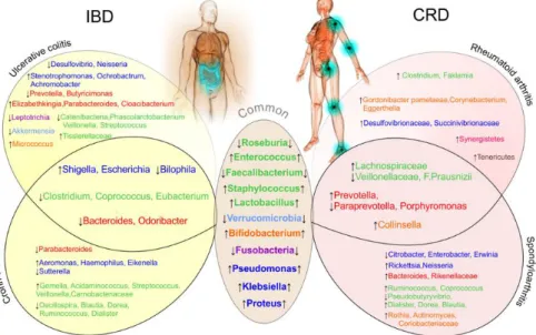
Butyricimonas genus is observed in IBD patients particularly those with UC 15,21,31,32,34 . A few studies, showed an increased amount of the Tissierellaceae family, and a decreased number of Eubacterium genera in inflamed colonic mucosa biopsy samples when compared to the non-inflamed sites in UC patients 35–37. (Fig 2)
Bacteroidetes phyla
Data concerning the Bacteroidetes phylum are more conflicting. Some studies reported a reduction of the Bacteroides group in IBD patients especially in CD patients 9,12,13,20. In contrast, Andoh and colleagues demonstrated an increase amount of this phyla in the context of IBD 38. To note, one study showed an increase of Bacteroidetes phylum in salivary microbiota in UC patients 39. Hirano and co-workers showed an enrichment of the
Cloacibacterium genus, and decreased abundance of Prevotella (at both inflamed and
non-inflamed mucosal site) and Butyricimonas genera at the non-non-inflamed mucosal site of UC patients compared to the corresponding site in non-IBD controls and in the faecal microbiota of UC patients12,34,35,40. A greater abundance in these two genera was found in the submucosal tissues of patients with CD 12,34,35,40,41. As with CD, this strongly suggest a restricted biodiversity in UC and an increased proportion of unusual bacteria 42,43. Bacteroidetes show also interesting age-related patterns and population-independent increase in abundance in the standing and active bacteria among healthy subjects and UC patients25.A decrease abundance of Parabacteroides genera and Odoribacteracae family in IBD and CD patients respectively is reported 10,13,15. Similar to the results from faecal microbiota studies, a significant decrease of bacteria from the phylum Firmicutes was demonstrated in the MAM of patients with CD 44,45. A recent study of Walujkar and collagues revealed significant differences in the MAM of patients manifesting acute exacerbations of UC with increased amount of Parabacteroides and Elizabethkingia genera in the MAM of UC patients as compared to the same patients during remission stage 46 (Fig 2).
Actinobacteria phyla
Concerning the Actinobacteria phylum, studies using both culture and recent molecular methods, demonstrated an increase of Bifidobacterium genera in the faecal microbiota as well as in the biopsy samples of IBD patients, notably in patients with CD 12,13,15,19,47. However, other authors reported an age-related reduction of bacteria of the Bifidobacterium genera was
shown in inflamed sites when compared to non-inflamed ones and salivary microbiota of UC patients 12,13,15,47–49,31,35,39,50. Walujkar and co-workers showed an increase amount of
Micrococcus genera in MAM of UC patients when compared to non-IBD subjects 46 (Fig 2).
Proteobacteria phyla
Published studies display a quantitative alteration of Proteobacteria phylum in IBD especially
Escherichia and Shigella from the Enterobacteriaceae family, 10,12,13,15,24,29,51. Thus, their increased abundance was reported in the MAM and faecal samples of patients with CD, whether using culture24,44 or molecular 52,17,53 methods. As with CD patients, the MAM of patients with UC contained an abnormally elevated concentration of bacteria, especially anaerobes 44,45. A restriction of the MAM biodiversity similar to that observed in patients with CD has been found such as reduction of Firmicutes and an overrepresentation of
Enterobacteriaceae 19,25 44 45 54 55 56. A decreased abundance of the genera Bilophila and
Desulfovibrio was evident at the inflamed site of UC patients compared to the corresponding
site of non-IBD controls, whereas a decreased amount of Bilophila genera and it’s species (B.wadsworthia) was detected in the faecal microbiota of CD patients 35,57,58. Moreover, an age-related reduction of the Neisseria genera bacteria was reported in inflamed sites when compared to non-inflamed ones and salivary microbiota of UC patients 12,13,15,47– 49,31,35,39,50
.Walujkar et al. suggested an increased abundance of Stenotrophomonas,
Ochrobactrum and Achromobacter genera in UC patients as compared to the same patients
during remission stage 46. Finally, Proteobacteria phyla displayed also an age-related patterns 25
. (Fig 2)
Other phyla
Finally, a decreased in abundance of Verrucomicrobia (Akkermansia) and Fusobacteria (Leptotrichia), was reported at the inflamed colonic mucosal sites of CD and UC patients compared to the corresponding site of non-IBD controls. However, further investigation concerning an eventual association between Leptotrichia and UC is necessary 12,31,32,35,59–61.
In summary, among the 56 available studies on IBD, differential abundance of 40 bacterial species has been reported, 15 were specifically found in CD studies while only 16 species reported in UC studies. These variations mainly concerned Firmicutes, Proteobacteria and Bacteroidetes.
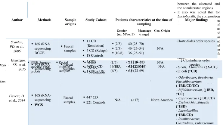
9
Table 1.a: Bacteria associated with inflammatory bowel disease analysed from biopsy samples.
Author Methods Sample origins
Study Cohort
Study Cohort characteristics at the time of sampling
Gender Mean age Geo. Origin (no. M/no. F) (range)
Major findings Seksik, P. et al 2005 TTGE of 16S rRNAs Biopsy samples 15 CD (6/9) 37.6 (21–63) France
No bacterial species was found to be specifically associated with CD ulceration, and ulceration did not qualitatively modify the dominant associated microbiota Ott, SJ. et al., 2004 16S rDNA based SSCP fingerprint Biopsy samples 26 CD 31 UC 15 Inflammatory controls 31 Non-inflammatory controls (9/17) (18/13) (6/9) (10/21) 35 (16–56) 44 (23–74) 50 (20–82) 52 (26–74)
N/A - Bacteroides, Prevotella (↓IBD)
Morgan, XC. et al., 2012 16S rRNA-sequencing WGS Biopsy samples 121 CD 75 UC 8 Indetermin ate 27 Controls (49/72) (38/37) (3/5) (12/15) 38 (35-41) 42 (38-45) 27(14-41) 36 (30-42) USA - Prevotella, Streptococcus, Catenibacteria (↓UC) - Roseburia, Ruminococcus (↓CD) - Lactobacillus, Acidaminococcus, Veillonella, Shigella, Aeromonas, Fusobacterium, Shigella (↑CD) - Asteroleplasma, Porphyromonas, Bifidobacterium, Faecalibacterium, Coprococcus (↓IBD) Ananthakrishnan, AN. et al. 2017 Metagenomic sequencing Biopsy samples 42 CD
43 UC N/A N/A N/A
- Roseburia inulinivorans,
Burkholderiales species (↑CD at 14 weeks remission)
Frank, D. N. et al
2007 16S rRNA sequencing Biopsy samples
68 CD 61 UC 61 Non-IBD Controls N/A 35 (21-49) 38 (22-54) 36 (23-49) N/A - Bacteroides (B. thetaiotaomicron),Lachnos piraceae (↓IBD) - Actinobacteria, Proteobacteria (↑IBD) Willing, BP. et al., 2010 T-RFLP Cloning and 16S rRNA Sequencing Biopsy from 5 locations between the ileum and rectum 6 L1-CD 8 L2-CD 6 Controls (3/3) (6/2) (3/3) Born between (1936-1986) N/A - F. prausnitzii (↓L1-CD) - E.coli (↑L1-CD) Png, C. W. et al. 2010 16S rRNA qPCR In vitro mucus degradation test Biopsy samples 26 CD 20 UC 20 Controls (6 / 20) (13 / 7) (9 / 11) 38 (19 – 74) 48 (24 – 71) 53 (22 – 84) N/A - R. gnavus R. torques (↑CD/UC) - Akkermansia muciniphila, (↓CD/UC) Hansen, R. et al 2012 16S rRNA RT-PCR and pyrosequencing Colonic mucosa biopsy samples 13 CD 12 UC 12 Controls (10/3) (9/3) (8/4) 13 (8- 17) 13 (9 - 16) 12 (7- 16) Scotland, UK - Faecalibacterium (↑CD) Wang, M. et al 2007 16S rRNA sequencing Colonic biopsy samples 1 UC (colonic
microbiota) (0/1) 12-year-old N/A
- Enterobacteriaceae,
Bacteroides fragilis, F. prausnitzii-like, Pseudomonas aeruginosa (↑UC)
Rehman, A. et al 2016 16S rRNA pyrosequencing Mucosal biopsy samples 27 CD (10 Ger.; 8 Lith.; 9 Ind.) 30 UC (10 Ger.; 10 Lith.; 10 Ind.) 30 Controls (10 Ger.; 9 Lith.; 11 Ind.) Ger. (14/16) Lith. (10/17) Ind. (21/19) Ger.(16-63) Lith.(19-81) Ind. (17-67) Germany Lithuania India - Firmicutes (↓Ger. Controls /CD Lith. Ind. ) - Bacteroidetes (↑UC) - Proteobacteria(↑CD Lith./Ind.) Hirano, A. et al. 2018 16S rRNA sequencing Mucosal biopsies samples 14 UC 14 Non-IBD (Controls) (6/8 ) (8/6) 45 (17-67) 59 (41-73) N/A - Cloacibacterium, Neisseria genus, Tissierellaceae family, (↑inflammed site UC compared to non-inflamed site UC)
- Prevotella, Eubacterium,
Neisseria, Leptotrichia, Bilophila, Desulfovibrio, Butyricimonas
(↓UC corresponding site of non-IBD controls).
- Prevotella, Butyricimonas
(↓UC patients compared with the corresponding site in non-IBD controls)
Chiodini, R. J. et al. 2015 Deep 16S rRNA sequencing Ilea mucosal and submucosal biopsy samples 20 CD 15 Non-IBD (Controls) (9/11) (4/11) 41 (24-66) 59 (32-88) USA - Desulfovibrionales (↑CD
in the subjacent submucosa as compared to the parallel mucosal tissue including)
- Ruminococcus spp., Oscillospira spp., Pseudobutyrivibrio spp., Tumebacillus spp., Propionibacterium spp., Cloacibacterium spp., Proteobacteria (Parasutterella spp. , Methylobacterium spp) (↑CD) Swidsinski, A. et al. 2002 16S rRNA sequences FISH 3 group-specific FISH probes Colonic biopsy samples 54 CD 119 UC 104 In.C 28 S.l.C 40 Controls (25/29) (52/67) (46/58) (16/12) (23/17) 35 (17-86) 45 (1786) 46 (19-81) 37(17-70) 50 (26-77) Berlin , Germany
No principal difference in the composition of the mucosal flora in IBD patients and controls. Species isolated from the washed mucosa were of faecal origin in all groups.
Proportion of
Enterococci/Streptococci, Clostridia, Peptostreptococci , Eubacteria were lower
- Proportion of Collinsella
aerofaciens or Propionibacte ria higher than usually found in faecal specimens
Swidsinski, A. et al. 2005 FISH 14 group-specific FISH probes Mucosal Biopsy samples 20 CD 20 UC 20 IBS 10 IBD + antibiotics 20 Controls (11/9) (9/11) (6/14) (4/6) (7/13) 33 45 48 40 47 N/A
An adherent mucosal biofilm mainly composed of
Bacteroides fragilis is a
prominent feature in patients with IBD, while biofilm composed of
Eubacterium rectale group in
IBS. Walujkar, SA. et al., 2018 16S rRNA gene-based sequencing Colon biopsy samples 12 UC 7 Non-IBD (Controls) N/A (30- 41) (37-54) Maharashtra, India Stenotrophomonas, Ochrobactrum, Achromobacter(↑UC) Kotlowski, R. et al. 2007 RISA DNA sequencing Biopsy samples 13 CD 19 UC 15 Controls
N/A N/A Canada Enterobacteriaceae (↑IBD)
Sokol, H. et al.
2007 TTGE Biopsies samples
3 Proctitis 7 Left-sided
colitis
N/A N/A N/A
E.coli subdominant bacteria
Zhang, M. et al. 2007 DGGE analysis Mucosal biopsy samples 24 UC (9/15) 40 (16–72) China - Lactobacilli, Clostridium
leptum subgroup were
11
Table 1.b:Bacteria associated with inflammatory bowel disease analysed from faecal samples.
between the ulcerated and the nonulcerated regions - It also was noted that for
Lactobacilli, the composition
varied significantly between biopsy sites irrespective of the location of UC in the gut but that the composition of the Clostridium leptum
subgroup showed significant differences between paired samples from UC in the rectum and not in the left colon Mylonaki, M., et al. 2005 FISH 5group-specific FISH probes Rectal biopsies samples 6 CD 33 UC 14 Controls (1/5) (19/14) (6/8) 51 (19–59) 53 (22–76) 33 (22-69) N/A
- E.coli, Clostridia (↑A-UC) - E. coli (↑CD) Earley, H. et al. 2015 16S rRNA PCR Mucosal biopsies 5 UC 7 Colonic cancer
N/A N/A Ireland A. muciniphila, Desulfovibrio spp. (↑UC)
Author Methods Sample
origins
Study Cohort Patients characteristics at the time of sampling
Gender Mean age Geo. Origin (no. M/no. F) (range)
Major findings Scanlan, PD. et al., 2006 16S rRNA sequencing DGGE Faecal samples 11 CD (Remission) 5 CD (Relaps) 18 Controls (7/3) (2/3) (10/8) 40 (25–70) 46 (25–54) 36 (25–51) N/A
↓ Clostridiales order species
Hourigan, SK. et al. 2015 16S rRNA sequencing Faecal samples 3 CDI 4CDI+ CD 1 CDI + UC N/A 13 (6–16) 14 (10–16) 17 N/A ↓ Clostridiales order species Gevers, D. et al., 2014 16S rRNA-sequencing WGS Faecal samples 447 CD
221 Controls N/A (<17) North America
- Odoribacter, Roseburia, Faecalibacterium (↓IBD/CD/UC) - Bifidobacterium, (↓IBD, ↑UC) - Coprococcus (↓IBD/CD) - Escherichia, Shigella (↑IBD) - Lactobacillus (↑IBD/CD) - Ruminococcus, Clostridium, Eubacterium
(↓CD) - Enterococci (↑CD) Hall, AB. et al., 2017 Metagenomic sequencing Faecal samples 9 CD 10 UC 1 Indeterminate Colitis 12 Controls (3 with Gastrointestinal symptoms)
N/A N/A N/A
- R.gnavus(↑IBD) Kaakoush, NO . et al. 2012 High-throughput sequencing of 16S rRNA Faecal samples 19 L1/L4 CD 21 Controls (12/7) (13/8) 12 (11-15) 10 (9-14) Sydney, Australia - Oscillospira (↓CD) Aomatsu, T. et al 2012 16S rRNA Sequencing T-RFLP analysis Faecal samples 10 CD 14 UC 27 Controls (4/6) (5/9) (12/15) (8-18) (8-15) (1-5) N/A - Parabacteroides, Bacteroides, Roseburia, Coprococcus , Blautia, Dorea, Ruminococcus, Oscillospira, Eubacteria, Dialister, Sutterella, Bilophila (↓CD) - Lactobacillus,Streptoc occus, Enterococcus, Gemella, Haemophilus (spp.) , Eikenlla (↑CD) - Bacteroides (↓IBD) Machiels, K. et al 2014 DGGE of 16S rRNA Metabolites quantification by gas chromatography– mass spectrometry Faecal samples 127 UC 87 Controls (74/53) (39/48) 43 (32-55) 42 (30-53) Belgium Roseburia (R. hominis), Clostridium, Butyricimonas, F. prausnitzii (↓IBD : UC) Duboc, H. et al 2013 16S rRNA qPCR Faecal samples 7 A-CD 5 R-CD 16 A-UC 14 R-UC 29 Controls (3/4) (2/3) (7/9) (9/5) (11/18 ) 38 (19-57 42 (23-61) 36 (22-50) 38 (26-50) 35 (21-49)
N/A - Clostridium (C. leptum) , Blautia (B. coccoides ) (↓IBD ) - F. prausnitzii (↓CD) - Escherichia(E.coli) (↑IBD) Fujimoto, T. et al 2012 16S rRNA qPCR T-RFLP Faecal samples 47 CD 20 Controls (31/16) (14/6) 36 (26-45) 45 (28/62) Japon F. prausnitzii (↓CD) Pascal, V et al. 2017 16S rDNA sequencing Faecal samples Spanish cohort (34 CD ,33 UC, 111 Controls) Belgian cohort (53 CD) (21/13) (25/28) 34 (18–58) 41 (27–53) Spain Belgium - Faecalibacterium, Peptostreptococcaceae, Anaerostipes, Methanobrevibacter, Christensenellaceae, Collinsella (↓CD) - Fusobacterium, Escherichia (↑CD) Swidsinski, A. et al 2008 Fluorescence in situ hybridization (FISH) Faecal samples 82 CD 105 UC 32 Controls N/A 34.8 (17-78) 41.2(18-84) 40 (18-60) Germany - F.prausnitzii, (↓CD/↑UC) - Enterobacteriaceae (↑ CD/UC) - Enterobacteriaceae (↓CD/↑UC) - Eubacterium hallii, E. cylindroides bacteria (↓CD) - Bifidobacteria, Atopobium (↑UC) Sokol, H. et al. 2009 16S rRNA Faecal samples 22 A-CD 10 R-CD 13 A-UC (7/15) (4/6) (8/5) 37 (34 - 41) 40 (35 – 44) 40 (37 – 44) N/A - F.prausnitzii
(↓R-IBD/ IC/ A-CD/A-UC)
13 4 R-UC 8 IC 27 Controls (1/3) (5/3) (11/16) 35 (31 – 40) 34 ( 29 - 39) 36 (35- 37) - Bifidobacterium (↓IC) Sabino, J. et al 2016 16S rDNA sequencing Faecal samples 18 PSC only 27 PSC-UC 21 PSC-CD 30 CD 13 UC 52 Controls (10/8) (20/7) (18/3) (15/15) (4/9) (49/3) Median age 49 (15.25) Median age 43 (14) Median age 49 (17) Median age 52 (14.25) Median age 50 (28) Median age 51.5 (17) Belgium Enterococcus, Fusobacterium, Lactobacillus (↑PSC only/ PSC-UC/PSC-CD) Bajer, L. et
al. 2017 16S rRNA Sequencing Faecal samples 32 PSC-IBD 31 Controls (17/15) (13/18) 40 (20-71) 44 (22-72) Prague
- Rothia, R. mucilaginosa, Fusobacteriaceae (↑PSC-IBD) - Adlercreutzia, Ruminococcus (↓PSC-IBD) - Butyricicoccus pullicaecorum sp (↓UC) Eeckhaut, V. et al. 2013 16S rRNA sequencing Genus-specific qPCR Faecal samples 51 CD 91 UC 88 Controls (23/21) (54/37) (39/49) Median age 39 Median age 44 Median age 41 N/A Butyricicoccus (↓CD/UC) Knoll, R. L. et al. 2016 Metagenomic analysis Faecal samples 6 CD 6 UC 12 Controls (3/3) (2/4) (6/6) (11-17) (11-16) (8-20) N/A - F. prausnitzii, E. rectale (↓CD/UC) - E.coli, F.nucleatum, E. coli, F. nucleatum (↑IBD) Andoh, A. et al. 2011 16S rRNA sequencing T-RFLP PCR T-RFL Faecal samples 31 CD 31 UC 30 Controls (16/15) (15/16) (12/18) 30 33 35 N/A - Clostridium (↓IBD) Sokol, H. et al. 2006 16S rDNA and rRNA PCR TTG Faecal samples 9 UC 9 Controls (5/4) (6/3) 39 (25-69) 43 (23-69) N/A - Clostridium coccoides (↓UC) Sokol, H. et al. 2006 FISH 6 group-specific FISH probes Flow cytometry Faecal samples 13 CD 13 UC 5 IC 13 Controls (2/11) (7/6) (2/3) (7/6) 37(24–50) 41(28–54) 29(25–33) 40(25–56) N/A - C. coccoides (↓UC) - C. leptum (↓CD) - Bacteroides (↑IC) Giaffer, MH. et al. 1991 16S rRNA quantitative and semi-quantitative bacterial culture techniques Faecal samples 22 A-CD 20 Quiescent CD 18 A-UC 19 Quiescent UC 21 Controls (6/16) (5/15) (8/10) (7/12) (11/10) 38 50 37 50 35 N/A Lactobacillus, Bifidobacteria (↓CD) Seksik, P. et al 2003 16S rDNA quantitative dot blot hybridization TTGE of 16S rDNA Faecal samples 8 A-CD 13 R-CD 16 Controls (1/7) (3/6) (7/9) 35 (16–68) 47 (32–62) N/A - Enterobacteria (↑CD) Schwiertz, A. et al 2010 16S rRNA
sequencing Faecal samples
21 A-CD 19 R-CD 13 A-UC 16 R-UC 25 Controls N/A 14 (5-19) N/A - Bifidobacteria (↓IBD) - Faecalibacterium (↓CD)
Thorkildsen , L. T. et al. 2013 16S rRNA sequencing MCR Faecal samples 30 CD 33 UC 3 IBDU 33 Non-IBD (10/20) (17/16) (1/2) (14/19) 33 (21-53) 34 (17-62) 42(35-53) 33 (20-56) Norway - Escherichia (↑CD) - Shigella (↑IBD/CD) Martinez-Medina, M. et al. 2006 16S rRNA gene sequencing PCR-DGGE BLAST database Faecal samples 19 CD 2 UC 1 Ischemic colitis 15 Controls (9/10) (1/1) (0/1) (5/11) (33-41) (29-34) 27 (43-50) N/A - Clostridium spp Ruminococcus Escherichia coli (↑CD) - γ-proteobacteria occasionally, in CD mucosal microbiota Jia, W. et al. 2012 DNA 454 sequencing DGGE In-depth sequencing NGS Faecal samples 20 CD 14 UC 21 IBS 18 Controls
N/A N/A England
- B. wadsworthia, Desulfovibrio piger (↑CD/UC/IBS) Vigsnæs, L. k. et al. 2012 DGGE Faecal samples 6 R-UC 6 UC 6 Controls
N/A N/A Danemark
- Lactobacillus spp. and Akkermansia (A. muciniphila ) (↓UC) Michail S. et al., 2012 PCR of bacterial 16S rRNA Microarray hybridization Faecal samples 27 UC 26 Controls (17/10) (14/12) (10-17) (10-16) N/A - Clostridia (↓UC) - γ-proteobacteria (↑UC) Papa, E. et al., 2012 DNA 454 pyrosequencing Sanger sequencing Faecal samples 23 CD 43 UC 1 IBDU 24 Controls (13/10) (21/22) (1/0) (10/14) 15 (3–20) 14 (4–24) 10 (3–17) 14 N/A Varela, E. et al. 2013 qPCR Faecal samples 116 R-UC 29 First degree relatives 31 Controls (55/61) (13/16) (17/14) 40 (32–46) 37 (27–54) 32 (23–41) Spain - F.prausnitzii (↓UC/relatives/ ↑R-UC)
15
Table 1.c : Bacteria associated with inflammatory bowel disease analysed from both faecal and biopsy samples.
A/R-CD / UC = Active / Remis sion Crohn Diseas e / Ulcera tive Colitis ; CD= Crohn diseas e, DGG E= Denat uring gradie nt gel Electr ophor esis; FISH = Fluore scence in situ hybrid izatio n; IBD= Inflam mator y bowel diseas e; IBDU = Inflammatory bowel disease unclassified; IBS = Irritable bowel syndrome; IC = Infectious Colitis; L1-CD= Ileum localized CD(Montreal classification); L1/L4 CD = Ileum localized CD with upper-gut involvement (Montreal classification); L2-CD= CD with primarily Colonic involvement (Montreal classification); L3-CD= Ileocolonic Crohn’s Disease (Montreal classification); MCR= Multivariate curve resolution; N/A = Not available; NGS = Next generation sequencing; PCR= Polymerase Chain Reaction; PSC = Primary sclerosing cholangitis; qPCR = quantitative Polymerase Chain Reaction; RISA = Ribosomal intergenic spacer analysis; RT-PCR = Reverse Transcription - Polymerase Chain Reaction; SSCP = Single Strand Conformation Polymorphism; S.l.c = Self-limiting colitis (s.l.c); T-RFLP = Terminal restriction fragment length polymorphism; TTGE= Temporal temperature gradient gel electrophoresis; UC = Ulcerative Colitis; In.C = Indeterminate colitis; WGS = Whole Genome Shotgun
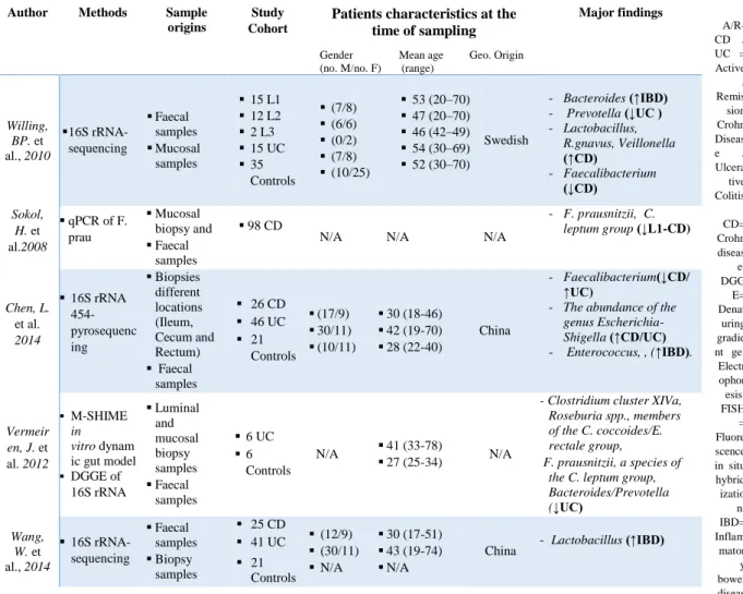
Author Methods Sample
origins
Study Cohort
Patients characteristics at the time of sampling
Gender Mean age Geo. Origin
(no. M/no. F) (range)
Major findings Willing, BP. et al., 2010 16S rRNA-sequencing Faecal samples Mucosal samples 15 L1 12 L2 2 L3 15 UC 35 Controls (7/8) (6/6) (0/2) (7/8) (10/25) 53 (20–70) 47 (20–70) 46 (42–49) 54 (30–69) 52 (30–70) Swedish - Bacteroides (↑IBD) - Prevotella (↓UC ) - Lactobacillus, R.gnavus, Veillonella (↑CD) - Faecalibacterium (↓CD) Sokol, H. et al.2008 qPCR of F. prau Mucosal biopsy and Faecal samples 98 CD
N/A N/A N/A
- F. prausnitzii, C. leptum group (↓L1-CD) Chen, L. et al. 2014 16S rRNA 454-pyrosequenc ing Biopsies different locations (Ileum, Cecum and Rectum) Faecal samples 26 CD 46 UC 21 Controls (17/9) 30/11) (10/11) 30 (18-46) 42 (19-70) 28 (22-40) China - Faecalibacterium(↓CD/ ↑UC)
- The abundance of the
genus Escherichia-Shigella (↑CD/UC) - Enterococcus, , (↑IBD). Vermeir en, J. et al. 2012 M-SHIME in vitro dynam ic gut model DGGE of 16S rRNA Luminal and mucosal biopsy samples Faecal samples 6 UC 6 Controls N/A 41 (33-78) 27 (25-34) N/A
- Clostridium cluster XIVa, Roseburia spp., members of the C. coccoides/E. rectale group,
F. prausnitzii, a species of the C. leptum group, Bacteroides/Prevotella (↓UC) Wang, W. et al., 2014 16S rRNA-sequencing Faecal samples Biopsy samples 25 CD 41 UC 21 Controls (12/9) (30/11) N/A 30 (17-51) 43 (19-74) N/A
A.2. Gut bacterial changes reported in chronic rheumatic diseases patients
A total of twenty-one studies, enrolling 993 CRD patients analyzing the gut microbiota by 16S rRNA gene sequencing from faeces. Breban et al. have demonstrated that -diversity analysis, which evaluates the shared diversity between different microbiomes in terms of various ecological distances, showed a microbiota composition significantly different between the RA, SpA and healthy subjects (HS) groups. Both SpA and RA patients differed from HSs as well as SpA from RA patients . This study showed also that α-diversity, which evaluates the species’ richness and evenness within the microbiota, assessed by the number of observed species was significantly decreased in both SpA and RA patients, as compared with HSs62,63. In ankylosing spondylitis (AS) patients, the diversity of the gut microbiome was similar to HSs at the genus level but was significantly higher in the controls at the species level 64.
Firmicutes phyla
Concerning the Firmicutes phylum, several bacteria from the Lachnospiraceae family, including Ruminococcus (R. gnavus sp.), Dorea, Coprococcus and Blautia genera are
overabundant in SpA 62. Increased amount of several Blautia and Ruminococcus could characterize HLA-B27+ siblings 62.Likewise, inflamed ileal biopsies of SpA patients revealed an increase in the Dialister genus which could be a microbial marker of disease activity 65,66. In contrast, SpA patients seemed to present a decreased amount of Roseburia species 62.
Concerning RA patients, a fewer Firmicutes of the Ruminococcaceae family but an increase in Lactobacillus species and Faklamia have been observed 62,67. A study by Picchianti-Diamanti et al. characterized the gut microbiota of RA patients on different immunosuppressants treatment strategies (ETN, MTX, or ETN plus MTX) and compared it with that of treatment-naïve patients. The drop in Proteobacteria caused by ETN which in general are abundant in both intestinal and extra-intestinal inflammatory diseases 68. Moreover, a decrease in Clostridiaceae was observed upon ETN treatment which were previously found enriched in patients with RA and IBD-associated arthropathy 69. In patients treated with MTX, analysis revealed a significant decrease in Enterobacteriales 67.
Liu et al. reported that RA patients, compared to HSs, exhibited an increased bacterial diversity within Lactobacillus community with increase in L.salivarius and L.iners 62,70,71 for instance. The analysis of faeces from RA patients have demonstrated the presence of a large cluster including Firmicutes bacteria belonging to Lachnospiraceae and Clostridiaceae (Clostridium) family, as well as small clusters containing strains from the Lactobacillus and
17
Ruminococcus genera70–73 . In the RA patients’ gut, a decrease of bacteria from the
Veillonellaceae family was observed 72,74. In contrast to SpA patients, PsA patients showed depletion in Coprococcus, Ruminococcus, Clostridium and Pseudobutyrivibrio compared to
HSs 62,74–76. Finally, SpA patients exhibited a decreased fecal abundance of F.prausnitzii compared to HSs. This bacterium may be, at least in part, responsible for the pathogenesis of SpA66,77,78.
Bacteriodetes phyla
There is a significant enrichment of the Prevotellaceae species, and more particularly of
Prevotella copri, within the Bacteriodetes phylum, in intestinal microbiota of patients with
new-onset RA, compared to chronic RA patients and HSs 79–81. This bacterium is relatively scarce in the general population. In addition, Bacteroides genera counts were lower in the same group, while being higher in SpA patients 79,66,72. However, P. copri decreased in the gut of RA patients along with disease chronicity 80. Breban et al. also demonstrated that SpA and RA patients have decreased populations of Prevotellaceae and Paraprevotellaceae genera compared to HSs 62. However, in AS patients,Prevotellaceae are more abundant in terminal
ileal biopsy samples 77.Furthermore, a quantitative metagenomics study has shown that the microbial communities in the AS cases were characterized by a higher abundance of
Prevotellaceae genera (Prevotella copri) compared to HSs 64. Other bacteria from the Bacteroidetes phylum, such as Porphyromonas, were shown to be decreased in RA patients while being increased in terminal biopsies of AS patients 77,82.
Actinobacteria phyla
Regarding the Actinobacteria phylum, which is a low-abundant one, patients with RA or SpA had a higher amount of bacteria from the Coriobacteriaceae family and especially of the
Bifidobacterium genus, including B. bifidum species than HSs 62,66. However, RA patients are also characterized by an increase of Corynebacterium species 62. The metagenomic analysis
and 16S sequencing have additionally brought into light the presence of the bacteria
Gordonibacter pamelaeae, Eggerthella lenta and Collinsella in RA patients63,64, 72. The latter could contribute to the increased permeability of the gut and enhanced production of pro-inflammatory cytokines 66. In SpA patients, an overabundance of Collinsella, Rothia and
Actinomyces genera was reported 63,64,76.
The Proteobacteria phylum is more abundant in RA patients than in HSs, concerning more
specifically the Klebsiella and Bilophila genera from Enterobacteriaceae,
Desulfovibrionaceae and Succinivibrionaceae families 62. In SpA patients there is a decrease of Citrobacter, Enterobacter and Erwinia genera 71,73,76. The latter was particularly reduced in the HLA-A24 positive group of patients. In contrast, an overabundance of Neisseria genera was reported SpA patients 64.
Other phyla
Finally, other phyla as Synergistetes, Tenericutes, Fusobacteria and Verrucomicrobia were also retrieved to be increased or decreased in RA and SpA patients 12,62,64,75,83. (Fig 2)
In summary, among the available studies on CRD (n= 21), 33 bacterial species were reported in CRD, among those 17 were specifically reported in SpA studies while only 9 species reported in RA studies. Variations mainly concerned Firmicutes, Bacteroidetes and
Actinobacteria phyla.
Table 2.a: Bacteria associated with chronic rheumatoid diseases analysed from faecal samples. Author Methods Samples
origins
Study Cohort Patients characteristics at the time of sampling
Gender Mean age Geo. Origin (no. M/no. F) (range)
19 Breban, M. et al. 2017 16S rRNA gene sequencing Faecal samples 86 SpA patients (74 SpA, 12 SpA+ IBD history) 28 RA 69 Controls (41/46) (6/22) (26/43) (35-63) (54-76) (27-63) France - Klebsiella, Desulfovibrionacae (bilophila), Succinivibrionaceae, Synergistetes, Tenericutes (↑RA)
- Bifidobacterium
(↓ RA /↑SpA)
- Paraprevotella (↓SpA)
- Coriobactericeae, Ruminococcus,
coprococcus, Dorea , Blautia (↑SpA) Picchianti -Diamanti, A. et al. 2018 NGS 16S Rrna Faecal samples 11 RA treatment naïve patients 11 RA received MTX 10 RA received ETN 10 RA received ETN+MTX 10 Controls (1/10) (2/9) (1/9) (2/8) N/A 56 63 60 65 N/A Finland - Lactobacillaceae, Lactobacillus (↑RA) - Faecalibacterium (↓RA) - Cyanobacteria phylum, Nostocophycideae ,Nostocales group (↑RA-ETN) - Deltaproteobacteria (↑RA-ETN/UC)
- Clostridiaceae upon (↓RA-ETN) - Enterobacteriales (↓RA- MTX ) Chen, J. et al. 2016 16S rRNA sequencing Faecal samples 40 RA patients, 32 Controls (12/28) (6/26) 56 53 USA - Eggerthella (↑RA) - Collinsella (↑RA/SpA) Wen, C. et al. 2017 Deep shotgun sequencing Faecal samples 97 AS 114 Controls (57/40) (72/42) (14-71) (23-70) China
- Collinsella, Prevotella copri
(↑RA/SpA)
- Actinobacteria, Neisseria, rothia,
Actinomyces (↑SpA) - Fusobacteria, Citrobacter, Verrucomicrobia (↓SpA) Stoll, ML. et al. 2018 16S rRNA sequencing Shotgun sequencing Faecal samples 30 ERA 19 Controls 11 SpA 10 Controls (19/11) (13/6) (4/7) (3/7) 14 (11-17) 14 (11-17) 52 (45-60) 47 (39-56) USA - Bifidobacterium, Actinobacteria, Lachnospiracea (↑RA/SpA) - F. Prausnitzii (↓RA/SpA) Liu, X. et al. 2013 16S sequencing Faecal samples 15 RA 15 Controls (3/12) (5/10) 48 41 China
Lactobacillus genera (Lactobacillus salivrius, L. Iners, L. ruminis) (↑RA) Maeda, Y. et al. EULAR 2012 RT-qPCR bacterial rRNA-targeted Faecal samples 37 RA patients 59 Controls (12/25) (6/53) 60 (49-71) 35 (25-45) Japan
L. ruminis, L.fermentum, L. reuteri, Enteroccocus (↑RA) Scher, JU. et al. 2015 16S rRNA sequencing Faecal samples 16 SpA 17 Controls (7/9) (7/10) 47 43 USA Verrucomicrobia, Pseudobutyrivibrio (↓SpA) Manasson . et al., 2018 16S rRNA sequencing Faecal samples 32 ReA 32 Controls N/A (18-55) USA - Rikenellaceae (↑SpA) - Pseudomonas (↑RA/SpA) Stoll, ML. et al. 2015 16S rRNA sequencing Faecal samples
12 recent onset ERA
21 Controls ACR meeting Abstract
F. Prausnitzii (↓RA/SpA) Scher, JU. et al. 2013 16S rRNA sequencing Shotgun sequencing Faecal samples 44 NORA 26 CRA 16 PsA 28 Controls (11/33) (3/23) (7/9) (7/21) 43 50 47 43 USA
Prevotella copri (↑RA/SpA)
Maeda, Y. et al. 2016 16S rRNA sequencing Shotgun sequencing Faecal samples 17 RA 14 Controls (3/14) (0/14) 64 (51-69) 53 (44-70) Japan
Prevotella copri (↑RA/SpA)
Vaahtovuo , J. et al. 2008 Flow cytometry 16S rRNA hybridization DNA-staining Faecal
samples 51 RA (9/42) 57 (44- 70) Finland Porphyromonas (↓RA/SpA)
Stoll, M. L. et al.
2014
16S rRNA
sequencing Faecal samples
25 ERA 13 Controls (14/11) (6/7) 13 (7-19) 13 (6-18) USA - F. prausnitzii (↓ERA) - Clostridium
leptum group (↓AS) Stebbings,
S. et al.
DGGE Faecal samples
15 AS
15 Controls N/A N/A N/A
Klebsiella pneumoniae, Bacteroides vulgatus
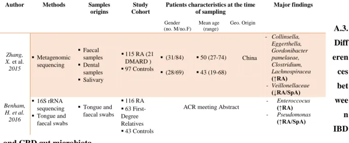
Table 2.b: Bacteria associated with chronic rheumatoid diseases analysed from biopsy samples.
Table 2.c: Bacteria associated with chronic rheumatoid diseases analysed from faecal and other origin samples.
ACR = American College of Rheumatology ; AS = Ankylosing spondylitis; CRA = Chronic, treated rheumatoid arthritis; DMARD = Disease-modifying antirheumatic drug; ETN = Etanercept; EULAR = European League Against Rheumatism; ERA = Enthesitis-related arthritis; IBD = Inflammatory bowel disease; NORA = new onset untreated rheumatoid arthritis; MTX = Methotrexate; PsA = Psoriasis arthritis; RA = Rhumatoide arthritis; ReA = Reactive arthritis; SpA = Spondylo-arthritis; ; UA = Undifferentiated Arthritis;UC = Ulcerative Colitis.
A.3. Diff eren ces bet wee n IBD and CRD gut microbiota
Three studies enrolling a total of 554 patients, directly compared 356 IBD patients without known arthropathy, and a total of 132 IBD with joint extra-intestinal-manifestation (EIM) patients were analysed (Table 3). One study indirectly compared three cohorts of patients,
2002 (↓AS)
Author Methods Samples origins
Study Cohort
Patients characteristics at the time of sampling
Gender Mean age Geo. Origin (no. M/no. F) (range)
Major findings Tito, RY. et al. 2017 16S rRNA sequencing Biopsy samples ileal and colonic 27 SpA 15 Controls (13/14) N/A (10-50) N/A
Belgium Dialister (↑SpA)
Costello, ME. et al. 2016 16S rRNA sequencing Intestinal biopsy 10 HLA-B27+ 85 HLA-B27
-ACR meeting Abstract Veillonellaceae (↓RA/SpA)
Costello, ME. et al. 2013
16S sequencing Terminal ileal Biopsy
N/A AS N/A CD N/A Controls
ACR meeting Abstract - Porphyromonas , F.
Prausnitzii (↓RA/SpA)
- Ruminococc (↑SpA)
Author Methods Samples
origins
Study Cohort
Patients characteristics at the time of sampling
Gender Mean age Geo. Origin (no. M/no.F) (range)
Major findings Zhang, X. et al. 2015 Metagenomic sequencing Faecal samples Dental samples Salivary 115 RA (21 DMARD ) 97 Controls (31/84) (28/69) 50 (27-74) 43 (19-68) China - Collinsella, Eggerthella, Gordonibacter pamelaeae, Clostridium, Lachnospiracea (↑RA) - Veillonellaceae (↓RA/SpA) Benham, H. et al. 2016 16S rRNA sequencing Tongue and faecal swabs Tongue and faecal swabs 116 RA 63 First-Degree Relatives 43 Controls
ACR meeting Abstract
- Enteroccocus (↑RA)
- Pseudomonas (↑RA/SpA)
21
SpA patients without IBD history (n=74) as well as SpA patients with an IBD history (n= 12),and RA patients (n=28) compared with HCs (n= 69) (Table 3)62.
Firmicutes phyla
Amongst included studies, some pointed out important differences, including variable amount of several Firmicutes genera. For instance, the overabundance of Veillonella observed in CD patients contrasted with its paucity in CRD (RA, SpA) patients. Conversely, the Eubacterium,
Clostridium, Ruminococcus and Coprococcus genera, that were increased in CRD (RA, SpA)
patients, were decreased in patients with CD 15,12,13, 19,72,77,79,80,82,83,84. Variation of the
Ruminococcus genus is the most surprising since a paradoxical overabundance, especially of R. gnavus, has been reported in SpA patients. This increased abundance correlated positively
with SpA activity whatever patients IBD history, even though IBD was inactive at the time of sampling in most of them12,62. In IBD, R. gnavus was mostly associated with the gut mucosa, which conferred to this mucolytic bacteria a possible role in the triggering or maintenance of inflammation 12,32. Whether its lonely increase could be linked to specific genetic predispositions to SpA warrants more investigation. As for the Dialister genera, belonging to the same bacterial family, an increased number of sequences was observed in SpA groups whereas a decrease was found in CD patients 62. In UC patients with a joint EIM, the
Staphylococcus genus was found more frequently in stool cultures 85.
Bacteroidetes phyla
Variations in Bacteroidetes phylum concerned mainly two genera: Bacteroides, which was in increased amount in SpA patients and in reduced amount in RA and IBD groups and
Prevotella which showed a high abundance in CRD (RA and SpA) patients and was lowered
in UC patients15,20, 63,64,66,79,80 84, 86.
Proteobacteria phyla
In the Proteobacteria phylum, the genus Bilophila was overabundant in RA and SpA patients while being found in reduced amounts in CD patients 12,51,62,87,88. Dorofeyev et al. showed a significant abundance of Enterobacter, Klebsiella and Proteus genera in stools cultures from UC patients with a joint EIM, compared to HSs and UC patients without EIM 85. In contrast, in UC a decreased amount of Neisseria was observed12,13,15,47–49,31,35,39,50. However, metagenomics studies of gut microbiome in patients with enteropathic arthritis are still lacking. Using qPCR, a relative overabundance of the Enterobacteriaceae family, concomitant to a reduction of the Clostridia group XIVa cluster, was reported in the gut microbiota in IBD patients with joint manifestations. As a whole, the Enterobacteriaceae
family seemed to be increased in the gut of IBD patients and this tendency is even more pronounced in those with arthropathy 89.
Actinobacteria phyla
Concerning the Actinobacteria phylum, an overabundance of Gordonibacter pamelaeae,
Eggerthella lenta and Collinsella was observed in RA patients 63,64, 72,90. However, an increase of Micrococcus genera was also characterized in MAM UC patients 46. In SpA patients, an overabundance of Collinsella, Rothia and Actinomyces genera was reported 63,64,76.
Other phyla
Finally, the Fusobacterium phylum is more abundant in CD patients and less abundant in SpA patients62. In contrast, amounts of the Tenericutes phylum are increased in SpA patients 10,62,64
.
Taken together, when considering all available studies (n=80), 40 bacterial species were reported only in IBD patients and 33 bacterial species were reported only in CRD subjects (Fig 2). Main variations were mostly observed in the Firmicutes phylum.
B. Similarities regarding bacterial microbiome in IBD and CRD
When comparing studies on IBD patients without known CRD versus studies on CRD patients without known IBD, we first observed that some dysbiotic changes share similarities between chronic IBD and chronic joint diseases, among which a lower microbial diversity and a diminished abundance of the Firmicutes phylum.
Firmicutes phyla
Amongst the Firmicutes genera, a common decreased amount was described for
Faecalibacterium and Roseburia species in both IBD subtypes (CD and UC), as well as in
SpA and RA patients 12,13,15,20,50,62,84. A few studies using bacterial culture, in addition to recent molecular methods, have demonstrated an increase amount of Lactobacillus and
Enterococcus in the faecal microbiota of IBD patients especially those with CD and RA
patients, although others demonstrated a reduction of Lactobacillus in CD patients7,12,15, 13, 47 , 30,48,49,52,70,72,84
. An overabundance of staphylococcus was observed in UC patients with arthritis when compared with patients without EIM and healthy population.
23
In the Proteobacteria phylum, an overabundance of several genera was observed, such as
Klebsiela and Proteus in all UC patients with arthritis. These facultative microbiota were
significantly higher in these patients than in the HSs and UC patients without EIM 46,62,85,91,92. An increase of Pseudomonas was recently showed by Walujkar et al in the MAM of UC patients as compared to the same patients during remission stage 46, as well as showed by Manasson et al. and Benham et al in patients with SpA or RA. 73,76.
Actinobacteria phyla
Concerning the Actinobacteria phylum, an overabundance of Bifidobacterium was reported in SpA patients, especially those with enthesitis-related arthritis (ERA), and in IBD patients notably in patients with CD 12,13,15,19,4750,62,64,66,72, 82, 83.
Other phyla
Finally, a common decrease of Verrucomicrobia and Fusobacteria belonging species was reported in both CD and UC patients compared to non-IBD controls 10,12,31,32,35,59–61, and in RA and SpA patients 12,62,64,75,83.
In summary, variations of species belonging to Firmicutes, Proteobacteria, Actinobacteria, Verrucomicrobia and Fusobacteria phyla represent the main common trait between IBD and CRD gut microbiota. A figure depicting similarities and differences observed in bacterial species amounts in biopsy and faeces from IBD and CRD patients is proposed (Fig 2).
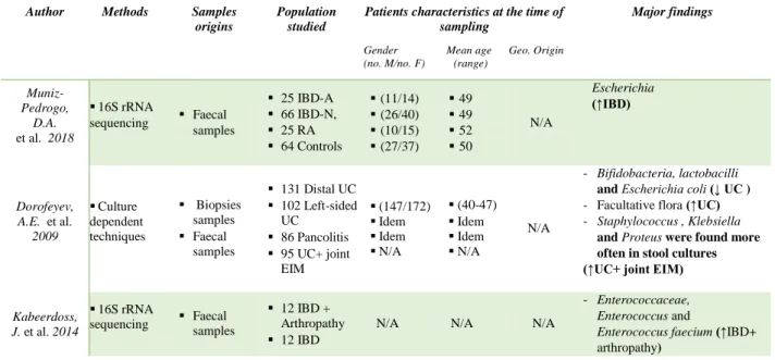
Table 3: Bacteria associated with inflammatory bowel disease and chronic rheumatic diseases.
EIM: Extra-intestinal manifestation; IBD= Inflammatory bowel disease; IBD-A/N = IBD-associated/ without arthropathy; N/A = Not available; RA = Rheumatoid arthritis.
Author Methods Samples origins
Population studied
Patients characteristics at the time of sampling
Gender Mean age Geo. Origin (no. M/no. F) (range)
Major findings Muniz-Pedrogo, D.A. et al. 2018 16S rRNA sequencing Faecal samples 25 IBD-A 66 IBD-N, 25 RA 64 Controls (11/14) (26/40) (10/15) (27/37) 49 49 52 50 N/A Escherichia (↑IBD) Dorofeyev, A.E. et al. 2009 Culture dependent techniques Biopsies samples Faecal samples 131 Distal UC 102 Left-sided UC 86 Pancolitis 95 UC+ joint EIM (147/172) Idem Idem N/A (40-47) Idem Idem N/A N/A - Bifidobacteria, lactobacilli
and Еscherichia coli (↓ UC )
- Facultative flora (↑UC) - Staphylococcus , Klebsiella
and Proteus were found more often in stool cultures (↑UC+ joint EIM)
Kabeerdoss, J. et al. 2014 16S rRNA sequencing Faecal samples 12 IBD + Arthropathy 12 IBD
N/A N/A N/A
- Enterococcaceae,
Enterococcus and
Enterococcus faecium (↑IBD+
25
Conclusion and perspectives
To our knowledge, this is the first systematic review concerning evidence regarding the gut microbiota in IBD and CRD patients. Our analysis highlights the general finding that microbiota favouring proteolytic-fuelled fermentation and lactic acid-producing bacteria, are increased in both CRD and IBD inflammatory conditions while those producing butyrate are generally decrease in both diseases. Secondly, variations of gut microbiota composition in IBD patients mainly concerned Firmicutes, Proteobacteria and Bacteroidetes. Within the Firmicute phylum variations of species as Roseburia, coprococcus, F. prausnitzii and
Streptococcus genera, was observed either in the mucosal-associated microbiota (MAM) of
CD patients or UC patients. In terms of Proteobacteria phylum published data display a quantitative alteration in IBD CD and UC patients compared to control groups especially of
Escherichia, Shigella, Bilophila, Desulfovibrio, Neisseria, Stenotrophomonas, Ochrobactrum and Achromobacter genera. Concerning the Bacteroidetes, variations of Cloacibacterium,
Prevotella, Butyricimonas, Parabacteroides, Elizabethkingia genera and Odoribacteracae family in IBD, CD and UC patients are observed.
However, in CRD patients, variations are mainly observed in Firmicutes, Bacteroidetes and Actinobacteria phyla. Alterations of gut microbiota observed in the Firmicutes phyla included
Ruminococcus (R. gnavus sp.), Dorea, Coprococcus, Blautia , and Dialister genus in RA and
SpA patients. In addition alterations of Roseburia, Lactobacillus, Faklamia, Staphylococcus,
Clostridium, Pseudobutyrivibrio, F.prausnitzii species and Veillonellaceae family was
observed in patients compared to healthy subjects. There is a significant variations of species within the Bacteriodetes phylum, particularly of, Bacteroides, Prevotellaceae (P.copri)
Paraprevotellaceae and Porphyromonas genera in RA and SpA patients compared to HSs.
Regarding the Actinobacteria phylum, which is a low-abundant one, in patients with RA or SpA variations of the Bifidobacterium genus, including among others B. bifidum species,
Gordonibacter pamelaeae, Eggerthella lenta, Collinsella, Rothia and Actinomyces genera was
reported compared to control groups.
Another major finding of this study, is the reduction of bacterial diversity, observed in both CRD and IBD and the presence of common bacterial phyla changes. We can mention an increased abundance in Lactobacillus, Enteroccocus, Staphylococcus, Bifidobacterium,
Klebsiella, Pseudomonas and Proteus genera in both CRD and IBD, whereas Faecalibacterium, Roseburia genera and Verrucomicrobia, Fusobacteria phyla are decreased
in both diseases.
Interestingly, experimental studies have confirmed the role of Faecalibacterium in immune controlled in both type of affections. First, Hablot and colleagues suggested that experimental Dextran Sulfate Sodium (DSS) induced colitis could altered the gut microbiota of mice with arthritis compared to mice with colitis alone and thus could delayed the appearance of “pro-arthritogenic” bacteria93
. This delay is associated with a difference of microbiota composition between mice with arthritis and colitis and mice with colitis only. Members of the Firmicutes phylum are mainly affected; Lactobacillus genus and Clostridiales order are more present in mice with arthritis and colitis compared to mice with only colitis. Several studies showed that species from Lactobacillus are beneficial in DSS-induced colitis 94,95. Thereby, Lactobacillus
sp increase in arthritis + colitis group might play a role in the subclinical improvement as
observed by the decrease in fecal lipocalin-2 level. A difference of the fecal microbiota composition is also observed between arthritis and arthritis + colitis groups. At arthritis and colitis onset, Lactobacillaceae, and notably Lactobacillus R.gnavus and S24_7 species belonging to Bacteroidales are more present in mice with arthritis and colitis compared to arthritis group. Interestingly, these groups of bacteria had been shown to be more present in mice with higher susceptibility to arthritis development 93,96.
Viladomiu and colleagues recently identified an enrichment of IgA-coated E. coli in CD-SpA with an adherent-invasive E. coli (AIEC) pathotype. Experimental models highlight two features of the host-pathogen interaction that must be considered to understand the specificity of pathogenetic mechanisms, namely, host susceptibility and strain variability97. CD SpA– derived AIEC protect against acute injury and death from DSS induced colitis in WT mice. Resident microbiota, including AIEC, induce colonic RORγt/Foxp3+ CD4+ T cells, which play an important role in restraining inflammatory colitis98. Consistent with a higher Enterobacteriaceae in 6-month-old infants correlated with better nutritional status 99. Thus, in situations of nutritional sufficiency or immunocompetence, the response to Enterobacteriaceae may have coevolved to protect the host; however, persistent nutritional deficiency 99 or genetic susceptibility (modeled in IL-10–deficient and K/BxN mice) evokes maladaptive responses, which, in turn, promote more severe inflammatory Th17 disease. Likewise, this data link the shared genetic susceptibility in the IL23R locus in both CD and SpA 100 with increased systemic E. coli sero-reactivity and Th17 inflammatory cytokines.