HAL Id: tel-02177132
https://tel.archives-ouvertes.fr/tel-02177132
Submitted on 8 Jul 2019HAL is a multi-disciplinary open access
archive for the deposit and dissemination of sci-entific research documents, whether they are pub-lished or not. The documents may come from teaching and research institutions in France or abroad, or from public or private research centers.
L’archive ouverte pluridisciplinaire HAL, est destinée au dépôt et à la diffusion de documents scientifiques de niveau recherche, publiés ou non, émanant des établissements d’enseignement et de recherche français ou étrangers, des laboratoires publics ou privés.
Combinatorial diversity of two-pore-domain k+ channels
and its involvement in migraine
Perrine Royal
To cite this version:
Perrine Royal. Combinatorial diversity of two-pore-domain k+ channels and its involvement in mi-graine. Molecular biology. Université Côte d’Azur, 2018. English. �NNT : 2018AZUR4114�. �tel-02177132�
2
LA DIVERSITÉ COMBINATOIRE DES CANAUX
POTASSIQUES À DEUX DOMAINES PORE ET
SON IMPLICATION DANS LA MIGRAINE
Jury
Président du jury
Pr. Michel LAZDUNSKI, Professeur, IPMC Rapporteurs
Pr. Stephan TUCKER, Professeur in Biophysics, University of Oxford Dr Michel VIVAUDOU, DR, IBS
Examinateurs
Dr Michel FINK, Scientific Content Manager, Cisbio Bioassays Dr Florian LESAGE, DR & Directeur adjoint, IPMC
3
LA DIVERSITÉ COMBINATOIRE DES CANAUX POTASSIQUES À DEUX DOMAINES PORE ET SON IMPLICATION DANS LA MIGRAINE
Résumé
Le maintien d'un potentiel de membrane de repos négatif est à la base de l'excitabilité neuronale. Ce potentiel négatif est généré par un courant de fuite de potassium induit par les canaux potassiques à deux domaines pore (K2P). Ils se sont révélés impliqués dans de nombreux mécanismes physiologiques et physiopathologiques tels que la dépression, la neuroprotection contre les ischémies, l'anesthésie, la migraine et la perception de la douleur. L'hétéromultimérisation est un mécanisme couramment utilisé dans la nature pour augmenter la diversité fonctionnelle des complexes protéiques. Par exemple, avec 15 gènes classés en 6 sous-familles, les canaux K2P pourraient générer 120 combinaisons et, en théorie, chacune d’elles possèderait des caractéristiques bien distinctes. Ici, nous avons d’abord étudié la capacité des membres de la même sous-famille K2P (sous-famille TREK) à s’assembler pour former des hétéromères fonctionnels dotés de nouvelles propriétés. En alliant l’optopharmacologie, une technique de précipitation de molécules uniques (SiMPull) et une technique de co-localisation à l’échelle de la molécule unique à la membrane plasmique, nous avons déterminé l’existence ainsi que la stœchiométrie des complexes créés entre TREK1, TREK2 et TRAAK. Nous avons caractérisé fonctionnellement les hétérodimères et avons constaté qu'ils formaient tous des canaux sélectifs au potassium rectifiant vers l'extérieur avec une sensibilité à la tension et aux pH variables. Ayant constaté que l’hétéromérisation est possible dans la même sous-famille, nous nous demandons si cela peut être fait entre membres de familles différentes et quelles pourraient en être les conséquences pathophysiologiques. Nous avons trouvé que TREK1 et TREK2 sont capable d’hétéromériser avec le canal plus distant TRESK, un canal K2P impliqué dans la migraine. Chez l'homme, la mutation TRESK-MT, une délétion de 2 paires de base (F139WfsX24) qui induit la formation de TRESK-MT1, un dominant négatif de TRESK, a été corrélé à la migraine. De manière surprenante, nous avons découvert que cette délétion induit un site alternatif de traduction (fsATI), menant à la formation d’un second fragment de TRESK, TRESK-MT2 qui s’assemble spécifiquement avec TREK1 et TREK2. Cet assemblage induit l’extinction des courants TREK, ce qui va augmenter l’excitabilité des neurones trijumeaux, une composante clé dans l’induction
4
de la migraine, à l’origine du phénotype migraineux observé. Ensemble, ces résultats démontrent que l’hétéromérisation des canaux K2P n’est pas rare et doit être considérée pour comprendre leurs fonctions pathophysiologiques. Enfin, les analyses génétiques des mutations liées à des pathologies devraient désormais prendre en compte les fsATI.
Mots-clefs : canaux potassiques, hétéromérisation, SiMPull, migraine, fsATI
COMBINATORIAL DIVERSITY OF TWO-PORE-DOMAIN K+ CHANNELS AND ITS INVOLVEMENT IN MIGRAINE
Abstract
Maintenance of a negative resting membrane potential underlies the basis of neuronal excitability. This negative potential is generated by a potassium leak current mediated by two-pore-domain potassium channels (K2P). Over the years, they have been shown to be involved in many physiological and pathophysiological mechanisms such as depression, neuroprotection, anesthesia, migraine and pain perception. Heteromultimerization is a mechanism commonly used to increase the functional diversity of protein complexes. For example, with 15 genes classified in 6 subfamilies, the K2P channel family can potentially generates 120 combinations and, in theory, each of them would show different functional properties. Here, we first investigated the ability of the members from the same K2P subfamily (TREK subfamily) to assemble and form functional heteromeric channels with novel properties. Using single molecule pulldown (SiMPull) from HEK cell lysates, subunit counting in the plasma membrane of living cells and opto-pharmacology, we show that the TREK channel members TREK1, TREK2, and TRAAK readily co-assemble. We functionally characterized the heterodimers and found that all combinations form outwardly rectifying potassium-selective channels but with variable voltage sensitivity and pH regulation. Having found that heteromerization is possible within the same subfamily we wonder if it can happen between members from different subfamilies with lower sequence homology and what could be the pathophysiological consequences. We found that TREK1 and TREK2 are able to heterodimerize with the distantly-related TRESK, a two-pore-domain K+ channel implicated in migraine. Notably, in humans, TRESK-MT, a 2 bp
5
frameshift mutation (F139WfsX24), which induced the formation of TRESK-MT1 a dominant negative for TRESK, was found to perfectly segregate with typical migraine in a large pedigree. Strikingly, we found that the 2 bp frameshift mutation induced an alternative translation initiation (fsATI) which leads to the translation of a second TRESK fragment, termed TRESK-MT2. We show that by co-assembling with and inhibiting TREK1 and TREK2, TRESK-MT2 increases trigeminal sensory neuron excitability, a key component of migraine induction, leading to a migraine-like phenotype. Together these findings demonstrate that K2P heteromerization is not rare and needs to be considered to understand their pathophysiological functions and that genetic analysis of disease-related mutations should consider fsATI as a distinct class of mutations.
6
Table des matières
Résumé ... 3 Abstract ... 4 Thanks ... 9 Abbreviations ... 13 Illustrations ... 16 Tables ... 17 Equation ... 17 I. Introduction ... 18
The resting membrane potential ... 18
Potassium channels: structure and function ... 19
The pore ... 20
Voltage-gated potassium channels ... 22
Inwardly rectifying potassium channels ... 27
K2P channels ... 28 Gating... 29 K2P channels family ... 33 Generalities ... 34 Electrophysiological properties ... 34 Structure... 35
Up and down state and opening mechanism ... 37
TWIK family ... 39 TASK family ... 41 THIK family ... 43 TALK family ... 44 TREK family ... 44 TRESK CHANNEL ... 54
K2P channels and pain perception ... 58
Pathophysiology of migraine ... 64
Generalities ... 64
Anatomy of the trigeminovascular pain pathway ... 65
Activation and sensitization of the trigeminovascular pathway ... 67
Aura ... 70
Channel dysfunction in migraine ... 71
Roles for signal molecules and receptors in trigeminal ganglia ... 78
7
Alternative translation initiation ... 82
Another mechanism resulting in two proteins translated from one mRNA ... 85
Heteromerization and subunits assembly ... 85
How heteromerization occurs ... 86
K2P heteromerization ... 88
II. Problematics ... 92
III. Material and methods ... 93
Molecular Biology ... 93
Immunocytochemistry... 93
Western Blot ... 93
SiMPull assay (adapted from Jain et al., 2012) ... 94
Principle ... 94
Surface passivation and construction of flow chambers ... 94
Sample preparation ... 94
Antibody immobilization... 95
Single molecule imaging and spot counting ... 95
Subunit counting with X. laevis Oocytes ... 95
Neuron extraction ... 96
Electrophysiology ... 96
Oocytes ... 96
HEK cells... 97
Neurons... 97
Behavior: Assessment of rodents allodynia ... 97
IV. Article I ... 102
Introduction ... 102
Tools ... 102
Article I: Heteromerization within the TREK channel subfamily produces a diverse family of highly regulated potassium channels. ... 103
Discussion and conclusions ... 114
V. Article II ... 115
Introduction ... 115
TREK-TRESK association, how the story started ... 115
Article II: Migraine-associated TRESK mutations increase neuronal excitability through alternative translation initiation and inhibition of TREK ... 116
Discussion and conclusions ... 172
VI. Discussion ... 173
8
About the formation of heteromers ... 173
Pathophysiological involvement of the heteromers ... 175
Physiological implications of TRESK and TREKs as background K+ channels .. ... 176
Functional expression of TRESK and TREK1/TREK2 in TG neurons ... 176
TREKs and TRESK involvement in pain sensation ... 176
TREK channels to treat migraine ... 177
VII. Perspectives ... 178
Frameshift induced alternative translation initiation as a new mechanism for genetic diseases ... 178
Database analysis of frameshift mutants... 178
KCNQ1 ... 179
SiMPull: toward an automatization ... 180
Advantages of TIRF microscopy ... 180
Transition from coverslips to beads ... 181
VIII.Conclusions ... 183
IX. Annexes ... 184
Annex 1: Article ... 184
Ci-VSP stoichiometry and assembling ... 184
Annex 2: Patent ... 199
C. Technical annex ... 239
Screening of protein-protein interaction’s modulators ... 239
9
Thanks
I am really honored and would like to thank the Pr. Michel Lazdunski, to accept to preside my thesis committee. I would also like to thank the Pr. Stephen Tucker and Dr Michel Vivaudou to review my work and the Dr Michel Fink, Dr Florian Lesage and Dr Pierre Paoletti to examine my work. I am really impressed and happy to have all of you reunited in my thesis committee.
I will now thank all the people that surrounded me during my PhD, without whom I would never have reached the end. First of all, I will never be grateful enough to Dr. Guillaume Sandoz, who supervised my thesis. Since 2013 when we first met, starting from the ground, he always trusted me and we built a very strong relationship based on a lot of work, the will to go through everything, we started together and always succeed. He taught me so many things about electrophysiology, K2P channels, but especially a way to analyze and to build scientific thinking. I would never have learned so many skills in another team. Dr. Guillaume Sandoz let me manipulated and innovated in the lab, as well as in other labs from Sophia-Antipolis to Spain. I don’t have enough words to express how thankful I am.
I would also like to thank Brigitte Wdziekonski, our substitution mom in the lab. First for all the job you have done for me, from cells to molecular biology, as well as slides for single molecule pull down and product orders. Secondly for as the time you spend talking with me about life, will, carrier. You have always been here to support me, even in the worse times, thank you.
Thanks to the former members of the lab. Dr. Yannick Comoglio, you taught me a lot in the lab, all your skills and your knowledges about the containing of the fridges and chemicals cabinet. Dr Rayan Arant for the very interessing discussions we used to have. Dr. Thomas Besson especially for the train trips we were making together every day for few months.
A special thanks to Dr. Céline Nicolas, who not only taught me how to deal with the mice, but also helped me in some life decisions throughout my PhD. I wish we’ll share some more of your home office days for lunch discussion! Arnaud, thank you for having been the best intern ever. You made me think about how to transfer my knowledges. Dr. Olena Butenko, it was a great pleasure to work with you for few months, and to show you some part of our beautiful country. Pablo, thanks for all the patience you
10
gave to me, thanks for all the time we spend out of the lab, talking about future, past and so much more. I am glad to have had the opportunity to work with you for two years, and I wish you the best for your PhD.
Thanks to all the members of the 4th floor for their smiles, nice words, skills and will they gave me at some point. Especially the youth, with whom I’ve been sharing more than good times in the lab. Anthony, Pierre, Raphaël and other PhD students and post-doc at the iBV, Torsten and Morgane among others.
A special thanks to another PhD student, Clément Verkest. Thank of you, I’ve been able to learn new skills with animals, and concretized the animal experimentation side of the article. You have been so patient with me, and very professional. I wish you the best for your carrier. Another PhD student in Spain, Alba Andrea, allowed me to work first on trigeminal neurons, then on infected rats, to finalize the article. We spent so many hours enclosed together with rats, thank you for your kindness and you determination.
Back to the iBV, I would like to thank the members of the imaging facility, Sebastien Schaub, who helped me to set the single molecule pulldown technique at first, then Magali Mondin, to have taught me how to use the spinning disk microscope. Sama Rekima, for the histology part, and thank to the sterilization team Santhi, Mouna and Dominique for the time you make us win.
Now I would like to thank to all the people who shared my weakly life for these three years. My friends, you have always been here, even the days when I was not in the best mood. You helped me to get through this PhD, with the smile. Marie, you were, you are, and will always be the best friend I will ever have. Nana, you were far, but the few times we saw each other were among the best during these years, you are in my soul. Remy, you supported me for so long, even in the worst days, you gave me many hopes and made me understand so much about me, I have no words to explain how grateful I will always be, you brought me a family of friends. My sister, Roxanne, I’m sorry for all the times where I could not come with you because I had to work , I swear, I will make some efforts now that everything is over. Dr. Titouan Royal, my brother, I’m so proud of you, thank for giving me the will to survive and reinforce me when we were younger!
11
One last thank for my Mom, you have been so patient, every days, you supported me, and you gave trust in me since the first days of school. I wish I could one day give you back all the feelings you gave me.
12
13 Abbreviations 4-AP 4-aminopyridine 5-HT Serotonin AA Arachidonic acid ADP Adenosine-di-phosphate AKAP A-kinase-anchoring protein
AMPAR α-amino-3-hydroxy-5-methylisoazol-4-propionate receptor AP Action potential
Asic Acid-sensing ion channel AT2R Angiotensin receptor type II ATI Alternative translation initiation ATP Adenosine-tri-phosphate
cAMP Cyclic adenosine monophosphate Cav Voltage-gated calcium channel CGRP Calcitonin gene related peptide CNS Central nervous system
COP1 Coatomerprotein complex 1 DAG Diacylglycerol
DRG Dorsal root ganglion DRN Dorsal raphe nucleus
FRET Förster Resonance Energy Transfer GABA γ-aminobutyrique acid
GFP Green fluorescent protein GHK Goldman, Hodgkin and Huxley GPCR G protein coupled receptor IsoK Symmetric K+ concentration
K2P Two-pore-domain potassium channel
KCNE1 K+ voltage-gated channel subfamily member E1
14
KO Knock-out
LPA Lysophospatidic acid
MAP2 Microtubule-associated protein 2 Nav Voltage-gated sodium channel NGF Nerve growth factor
NK1R Neurokinin 1 receptor NMDA N-Methyl D-Aspartate NMDAR N-methyl-D-aspartate NO Nitrous oxide
NSAID Nonsteroidal anti-inflammatory drug NTG Nitroglycerin P Pore domain P0 Open probability PA Phosphatidic acid PD Phosphatase domain PGE2 Prostaglandine E2 pHext Extracellular pH pHint Intracellular pH
PIP2 Phosphatidyl inositol diphosphate PKA Proteine kinase A
PKC Proteine kinase C PLC Phospholipase C PLD2 Phospholipase D2 PUFA Polyunsatured fatty acid RNA Ribonucleic acid
SiMPull Single molecule pull down
SP Substance P
SSRI Serotonin selective reuptake inhibitor TEA Tetraethylammonium
15 TG Trigeminal ganglion
TIRF Total internal reflection fluorescence TRP Transient receptor potential channel TTX Tetrodotoxine
16
Illustrations
Figure 1: Potassium channels structure ... 20
Figure 2: The transmembrane part of KcsA ... 22
Figure 3: BK channels and its auxiliary subunits are pivotal in neuronal excitability. 26 Figure 4: IV curves and structure of Kir channels. ... 28
Figure 5 : Structure and conformational change of the potassium channel ... 30
Figure 6: Cartoon model of K2P2.1 (TREK‐1). ... 31
Figure 7: Dendrogram of the K2P potassium channel family and their characteristic functional features ... 33
Figure 8: Background potassium currents described by the GHK equation. ... 35
Figure 9: Cartoon showing the topology of a two-pore-domain potassium channel K2P and its assembly in dimer. ... 36
Figure 10: The structure of K2P-channels. ... 37
Figure 11: Cartoon model of TRAAK gating and mechanosensitivity. ... 38
Figure 12: Up and down states of TREK2 ... 39
Figure 13 : Differential distribution of mRNA coding for TASK-1 and TASK-3 in the rat CNS... 42
Figure 14: TASK channels regulations. ... 42
Figure 15: Polymodal activation of TREK1 by physical and chemical stimuli ... 45
Figure 16: TREK1 is involved in the therapeutic effect of 5-HT reuptake inhibitors via a negative feedback induced on the serotoninergic neurons ... 48
Figure 17: Top view of the extracellular side of homodimeric TREK1 (A) and TREK2 (B) channels. ... 51
Figure 18: Partners, regulatory domains and alternative variants of TREK1 ... 53
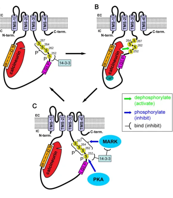
Figure 19: Regulation of the TRESK channel by calcineurin and phosphorylations. 58 Figure 20:TREK1 is involved in several polymodal pain perception. ... 60
Figure 21: Lysophosphatidic acid (LPA) and two-pore-domain K (K2P) channels. Representation of K2P channels in the membrane.. ... 61
Figure 22: Migraine step by step ... 66
Figure 23: Patways and brain regions involved in the transmission of migraine. ... 69
Figure 24: Diagram showing the various mechanisms involved in migraine at different sites in the nociceptive pathway ... 71
Figure 25: Functional roles of the proteins coded by known FHM genes within a glutamatergic synapse.. ... 73
Figure 26: A schematic representation outlining some of the predicted mechanisms contributing to migraine pathogenesis. ... 76
Figure 27: Schematic topology of the human TRESK subunits showing the position variants. ... 77
17
Figure 28 : Main potential targets of currently utilized preventive drugs and those
under-investigation for migraine.. ... 81
Figure 29 : K2P2.1 Channel may be inhibitory or excitatory, depending at which start codon translation is initiated. ... 84
Figure 30: Alternative translation start sites and variants of alternative open reading frames within eukaryotic mRNA. ... 84
Figure 31: Co-translational assembly of protein complexes ... 87
Figure 32: Schematic diagrams for passive conductance and Gβγ-dependent glutamate release ... 90
Figure 33: Schematic diagram of the SiMPull principle of HA-K2Px with another K2P subunit. ... 94
Figure 34: Electronic Von Frey aesthesiometer apparatus with the grid and plastic boxes ... 99
Figure 35: Drawing of the mouse paw. ... 99
Figure 36: Procedure used for targeting rat trigeminal ganglia ... 100
Figure 37: Cartoon of the subunit counting with X. laevis oocytes………105
Figure 38: Interacting K2P partners………115
Figure 39: TREK1-PCS for functional heteromerization study. ... 115
Figure 40: MDCK cells expressing mCherry-KCNQ1del572-576-GFP ... 179
Figure 41: Translation of a second protein is not at the origin of a dominant negative effect on KCNQ1. ... 180
Figure 42: Schematic representation of the SiMBeads. ... 182
Figure 43: Interaction with inhibitors can make a loss of fluorescent spots observed in SiMPull. ... 243
Tables Table 1: Summary of studies investigating the effect of altered K+ channel expression and function on acute, neuropathic, and inflammatory pain phenotypes. ... 63
Table 2: Heterodimers of two-pore-domain potassium (K2P) channels . ... 91
Equation Equation de Nernst………19
18
I. Introduction
When the principal concepts of electrophysiology were developed (Hodgkin and Huxley, 1952), the importance of K+ currents for cellular excitability has been revealed. Nevertheless, at this point, the molecular entities responsible for K+ current through the membrane were not known. Ion channels are fundamental elements for the signalization in the nervous system. They generate electric signals in neurons, regulate neurotransmitters secretion and switch electric signals toward sensorial stimuli as nociceptive stimuli, to which neurons are submitted. Ion channels properties also are linked with long term modifications in signalization, at the origin of the nervous system plasticity.
During the last 40 years, new techniques such as electrophysiology, biochemistry and molecular biology made up a revolution in the study of these channels. In 1970, only few voltage sensitive ion channels were known, along with some neurotransmitters receptors. Nowadays the number of classified ion channels has been largely increased (more than 300). To understand the role of each of these channels in the body and more specifically in the nervous system, the study of their structure, their subcellular localization along with their electrophysiological and pharmacological properties is fundamental.
Nevertheless, among this diversity, principles have emerged. Thus, many channels are built on a same model, where they delimitate a central pore in which ions can get through the plasma membrane. Furthermore, we can sort these channels in families, in which we have a well describe model for the molecular structure and function.
The resting membrane potential
All prokaryote and eukaryote cells have plasma membranes. The plasma membrane (also known as the cell membrane) is the outermost cell surface, which separates the cell from the external environment. The plasma membrane is composed primarily of lipids, especially phospholipids, and proteins. The lipids occur in a bilayer. We observe, for each eukaryotic cell at rest in our body, a resting membrane potential (RMP) which is negative. This RMP can be explained by the difference of the unequal distribution of ions across the membrane and the relative permeability for the different ions. K+ concentration being more important inside the cell, the situation for Na+, Ca2+
19
is opposite. These gradients of concentration, are due to a differential membrane permeability for the different ions and their passive transport by diffusion. Their movement is motivated by the concentration and also, as ion is a charged particle, by the existence of a transmembrane electric field. Thus, for each ion specie, the equilibrium condition will not be obtained by equalization of concentrations as it is the case in the simple diffusion of a neutral solute, but is obtained by an electrochemical gradient across the membrane. The membrane potential for the equilibrium of an ion is given by the Nernst equation.
𝐸𝑖𝑜𝑛 = 𝑅 ∗ 𝑇
𝑧 ∗ 𝐹∗ 𝐿𝑛
[𝑖𝑜𝑛]𝑒𝑥𝑡 [𝑖𝑜𝑛]𝑖𝑛𝑡
Equation de Nersnt in which R is the universal gas constant: R = 8.314472(15) J K−1 mol−1, T is the temperature in kelvin, F is the Faraday constant, the number of coulombs per mole of electrons: F = 96 485 C.mol−1, z is the number of electrons transferred in the cell reaction.
At rest the membrane is principally permeable to K+ and therefore the RMP is close to Ek. This K+ permeability at rest is due to K2P channels along with some other K+ channels. Two-pore-domain potassium channels are leaky meaning they are opened at resting membrane potential and let the potassium to reach out of a cell. This leak current of potassium is maintained until the equilibrium state is reached (Ek).
This state is mandatory for the cellular health. In mammal, more than 80 genes have been found to encode for potassium channels. They are involved in a wide diversity of physiological functions and their dysfunction lead to many pathologies (Shieh et al., 2000). These dysfunctions can either be genetically or acquired due to autoimmune diseases. The pathologies involving the ion channels are called channelopathies.
Potassium channels: structure and function
Potassium channels are ubiquitously expressed by nearly all kingdoms of life and provide a wide range of functions. The x-ray crystallographic structure of KcsA was the first prokaryotic potassium channel structure characterized (Doyle et al., 1998). Since, the mechanism of gating and functionality of potassium channels has been well studied. K+ channels are made of transmembrane helices, anchoring the lipid bilayer, and pore domains. According to their structure (see Figure 1) and function, they are sorted in four major classes:
20
- The voltage-gated (Kv), and calcium-sensitive (KCa), made of either six or seven transmembrane domains,
- the inwardly rectifying (Kir), with two transmembrane domains, - the two-pore-domain (K2P), made of four transmembrane domains.
Figure 1: Potassium channel structures. Voltage-gated channels (red) possess 6 transmembrane domains and one pore domain (P). Some of them present a 7th transmembrane segment. The inward rectifier channels possess 2 transmembrane domains and one pore domain (P). The two-pore-domain potassium channels possess 4 transmembrane domains and 2 pore domains (P1 and P2).
To be functional, potassium channels have to be associated in a complex of 2 or 4 subunits called alpha (α), forming the pore. This complex can be made of the same subunits, forming homomers, or of different subunits, called heteromers. Ancillary subunits, named beta (β), sometimes combine with the channel ( subunit) to change its biophysical properties.
These α-subunits possess a sequence called P loop, highly conserved among species and mandatory to form the selectivity filter of potassium channel. Four pore-forming domains comprise a pore through which the potassium moves. Intra and extracellular domains possess several regulation sites, which can play a role on the gating or the conductivity of the channel.
In this part I will first describe the general architecture of the pore, describe the different potassium channel families and give some explanation about their general gating and more precisely about K2P gating.
The pore
The ions move into a pore formed by four pore-forming domains which is conserved among the K+ channels. Doyle et al. described in 1998 the structure of the pore-forming domain of a K+ channel from Streptomyces Lividans (Doyle et al., 1998). The pore forming domain consists of the outer helix, loop regions, pore helix, selectivity
21
filter and inner helix. The inner and outer helixes represent the intern cavity. The pore helix (P) plays a structural role in the filter stabilization. The active site of K+ channels is made of the selectivity filter (SF). This last is made of a conserved signature sequence TxGY/FG (Heginbotham et al., 1994). K+ ions are conducted very efficiently in this selectivity filter, at near diffusion-limited rates (107ions channel−1.s−1) (Kuang et
al., 2015). K+ channels are highly selective and more permeant for K+ than sodium ions (1000 K+ for 1 Na+).
Selectivity and conductivity
The potassium enters into the central water-filled cavity (Sc) through the intracellular side via the helical bundle of KcsA channel (see Figure 2). Then passes the selectivity filter (SF) (from S4 to S1) to reach the extracellular entryway (S0 and Sext), following the electrochemical gradient. Hydration state of the potassium ions changes during these steps, going from hydrated in the Sc, dehydrated in the SF, to end-up hydrated in the Sext (Doyle et al., 1998).
22
Figure 2: The transmembrane part of KcsA. a. The atomic structure of KcsA in the conductive state (PDB: 1K4C) viewed along the membrane plane. The pore-forming domain consists of the outer helix (magenta), loop regions (green), pore helix (blue), SF (yellow), and inner helix (orange). The conducted K+ ions are represented by purple balls with surrounding water molecules in red. EC is extracellular and IC is intracellular. b, c. The enlarged view of the boxed area in (a) containing the SF and the extracellular entryway. The different states of occupation are depicted. For clarity, only two monomers opposite to each other are shown (from Kuang et al., 2015).
The filter has two states of occupation of its four potassium binding sites. The K+ binding sites are formed by the carbonyl oxygens of the SF signature sequence TVGYG and the side chain of threonine. These four site are not all occupied at the same time by ions, indeed there would be alternation between a molecule of water and a K+ ion. There are two conformations: 2,4 (S1/H2O, S2/K+, S3/H2O, S4/K+) and the 1, 3 (S1/K+, S2/H2O, S3/K+, S4/H2O) (Figure 2b and 2c respectively). The conduction mechanism consists of a concerted exchange between the two configurations until the arrival of a third ion on one side of the filter, causing the output of a K+ ions on the other side. This structure of the SF minimizes the transfer energy, thus maximizes ionic conduction velocity (Bernèche and Roux, 2001). This mechanism called knock-on, may not be the only one. In 2104, Kopfer et al proposed another conduction mode, the direct coulomb knock-on, where the ions would preferentially occupy S2 and S3, going out to the extracellular site when another ion arrives at S1 (Köpfer et al., 2014).
Selectivity could be explain by the tridimensional structure of this region. Hydrated ions or smaller ones cannot be well addressed into the channel. However, under physiological conditions, two K+ ions residing in the SF prevent Na+ ions conduction
(Valiyaveetil et al., 2006).
Voltage-gated potassium channels
The action potential generated by neurons involves several types of voltage-gated ion channels. Whereas voltage-gated sodium channels are opened for a short period followed by a quick inactivation, the voltage-gated potassium channels (Kv) are activated and remain open for a longer period (Hodgkin and Huxley, 1952).
The majority of Kv channels opens when the membrane is depolarized and closes when the membrane is hyperpolarized and inactivates for maintained depolarization. They are made of 6 transmembrane domains and one P loop. To be functional, they need to form tetramers. All subunits possess a voltage sensitive domain (VSD, S1 to
23
S4, S4 being the sensor). S5 and S6 delimitate the pore domain. S4 is composed by positively charged residues such as lysine or arginine every 3 amino acids, allowing a conversion of electric energy toward a mechanic one, rearranging S5 and S6 domains leading to the channel opening (Mannuzzu et al., 1996).
Inactive at resting membrane potential, they will open when the activation threshold is reached during a depolarization. K+ massive efflux will result in the repolarization of the membrane to the resting membrane potential.
The 40 mammalian members of this family are sorted in 12 subfamilies depending on their sequence homology.
2.a) Shaker TYPE
The first channel to have been cloned in Drosophila melanogaster by the Jan’s lab (Papazian et al., 1987). Its name is given by the loss of function of the gene KCNA1 coding for this protein, leading to a shaking phenotype in the fly’s paw. In human they are encoded by 8 genes KCNA1-8 coding for the protein Kv1.1-8.
These channels have been found in the brain, the heart, the skeletal muscle, the lung, the retina and pancreas. They are sensitive to 4-aminopyridine (4-AP) and some toxins (Browne et al., 1994; Gutman et al., 2005; Scheffer et al., 1998).
In sensory neurons of the trigeminal somatosensory system, Kv1.1 and Kv1.2 underlie the excitability brake current of cold thermoreceptor neurons. The slow-inactivating outward K+ current produced by Kv1 is involved during the action potential phase of repolarization. It prevents the unspecific activation by cold. Mutations in the channel Kv1.1 (Maylie et al., 2002) give rise to truncated forms of the protein witch destabilize the open state and thus are associated to disease (González et al., 2017).
2.b) Shab, Shaw, Shal
Shab or KV2.1-2, Shaw or Kv3.1-4 and Shal or Kv4.1-3 are expressed in the brain, smooth muscle, lung, heart, testis and pancreas. They have been found by sequence homology in the drosophila. As Shaker channels, they are inhibited by tetraethylammonium (TEA) and 4-minopyridine (4-AP) (Gutman et al., 2005).
24
These five channels named Kv7.1-5 (or KCNQ1 to KCNQ5) are expressed in the brain, the heart, inner ear, skull muscle and pancreas. In neurons, they show a slow activation when associated with β-subunits and thus are involved in the fast after hyperpolarization phase, right after the action potential.
KCNQ channels are involved in pathologies affecting heart and brain. Mutations in the Kv7.1 channel, expressed in the heart results in a modification of the potassium current leading to long QT syndrome which is characterized by a prolonged QT interval, resulting in a heart beating abnormally and an increased risk of arrhythmias and sudden death.
2.d) Ether-a-gogo
As the Shaker family, this family was first discovered in drosophila mutants that showed a shaking phenotype in the paw, under ether anesthesia. They are three subfamilies, called eag (or Kv10.1-2), erg (for eag related gene or Kv11.1-3) and elk (for eag-like K+ channels or Kv12.1-3). They produce a current with a slow activation. In the heart and the brain, they are involved in the action potential repolarization. As KCNQ1, mutations in human erg channel Kv11.1 are involved in long QT syndrome, via its involvement in ventricular repolarization.
2.e) “Modifier” subunits
Four of the Kv families (Kv5, 6, 8, and 9) encode subunits that act as modifiers.
Although these do not produce functional channels on their own, they are able to assemble with Kv2 family subunits forming heterotetrameric channels with unique
properties increasing the functional diversity within Kv2 channel family. They modulate the activation and/or inactivation phase of the channels.
2.f) Ancillary subunits and partners of Kv channels
A variety of other peptides has also been shown to associate with Kv tetramers and modify their properties, including several β subunits (which associate with Kv1 and Kv2 channels), KCHIP1 (Kv4), calmodulin (Kv10), and minK (Kv11).
2.g) Kca channels, activated by calcium
These tetrameric channels are activated by an intracellular increase of calcium concentration and voltage for some. This family can be divided in two distinct
25
subfamilies. The first is made of BK channels, for big K+ conductance (Wei et al., 1994) and the channels possess 7 TM. As Kv channels, the BK channels are voltage-dependent. The second is made of 6 TM channels and is divided in SK channels (slow conductance) and IK channel for intermediate conductance, both insensitive to potential.
(1) BK channels
These channels are both calcium and voltage dependent. Each of the subunit is composed of 7 TM, with a S4 domain sensitive to potential, and another transmembrane domain called S0, making the N-terminus to be on the extracellular side. The calcium sensor is localized in the C-terminus and rely on hydrophobic negatively charged residues that bound the calcium (Schreiber and Salkoff, 1997). The calcium binding shifts the IV curve toward more hyperpolarized potential favoring BK activation (Sun et al., 1999).
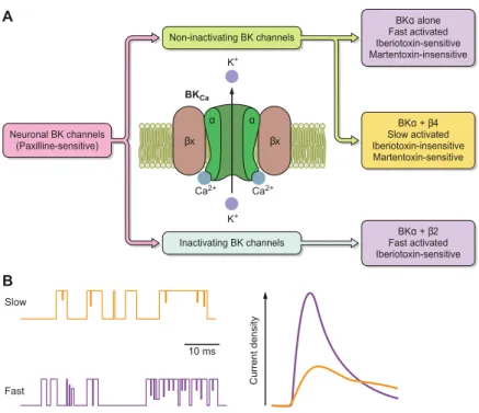
BK channels are ubiquitous in the SNC and are involved in the rapid/medium phase of the hyperpolarization after an action potential. Their sensitivity to calcium involves them in many physiological functions notably in the nervous system, BK channels have been involved in neurotransmitter release (Faber and Sah, 2003).
Ancillary subunits
Most of the BK channels are associated with a β or γ subunits. β subunits are made of 2 transmembrane domains. Four different β subunits (β1–β4) have been cloned and identified in mammals. They act on the calcium and voltage sensitivity of the BK channels, as well as the activation and their pharmacology (see Figure 3 below). In the nervous system, BK channels have been involved in neurotransmitter release. In the cells where they are expressed, depending on the α and β subunit assembling, the resulting currents and functions will not be the same.
26
Figure 3: BK channels and its anciliary subunits are pivotal in neuronal excitability. A: diagram chart of BK neuronal channels, pharmacology of scorpion toxins, and general gating features. B: illustration of single-channel recordings and current densities for fast and slow activated neuronal BK from (Latorre et al., 2016).
In trigeminal ganglia, the combination of BK channel with β4 subunit modulates glutamate release, and CGRP release is inhibited by activation of this combined channels (Samengo et al., 2014).
The four γ subunits are encoded by LRRC genes and also play on biophysical and pharmacological properties of BK channels (Yan and Aldrich, 2010). In testes, γ2 (LRRC52) increases BK current and is linked to fertility deficit (Zeng et al., 2015).
(2) SK channels
SK channels or KCa2.1-3 possess a small conductance to potassium from where they have been named. They are made of 6 TM and one pore domain. Unlike BK channel, they are not voltage sensitive and do not directly interact with the calcium but are modulated by calmodulin that directly acts on the C-terminus domain (Maylie et al., 2004).
They are expressed throughout the central nervous system. Their activation limit the firing frequency of action potential (Faber and Sah, 2007), playing a role in medium afterhyperpolarization following an action potential and in synaptic plasticity (Stackman et al., 2002).
27
(3) IK channels
IK or KCa3.1 channel (or SK4) for intermediate conductance is not sensitive to membrane potential. They share the same sensitivity to intracellular calcium concentration as SK channels via calmodulin biding in the C-terminus. In cancer types, this channel has been found to be important in cell proliferation (Thurber et al., 2017).
Inwardly rectifying potassium channels
Kir channels have diverse physiological functions in the cell, depending on their type and location, and are modulated by various mediators, such as ions, phospholipids, and proteins binding (for revue see Hibino et al., 2010). The fifteen members can be divided into seven subfamilies based on their mediators and the properties of ion conduction. They have the ability to form in vivo homo or hetero tetramers. These seven subfamilies can be classified into four main groups:
- Classical Kir channels (Kir2.1-4) are constitutively activated, are involved for instance in cardiac myocytes where Kir2.1 and Kir2.2 heteromer stabilizes membrane potential close to Ek (Zobel et al., 2003).
- G protein-gated Kir (Kir3.1-4) are regulated by G-protein coupled receptors (GPCR). In the brain, GIRK channels expressed in hippocampal neurons will generate post-synaptic inhibitory potentials induced by GABAB receptor activation (Lüscher et al., 1997) in concert with TREK channels (Sandoz et al., 2012).
- ATP-sensitive K+ channels (Kir6.1-2), linked to cellular metabolism. KATP are inhibited by ATP, but in complex with sulfonylurea receptor in presence of ADP, they will be activated. They regulate the membrane potential of pancreatic β cells thus modulating insulin secretion (Ashcroft and Rorsman, 1989).
- K+ transport channels (Kir1.1, Kir4.1-2, Kir5, and Kir7). Mostly expressed in the apical membrane of epitheliums where they are involved in Na+ and K+ homeostasis.
One characteristic of these channels is the inward rectification resulting from the pore block by the magnesium ion (Mg2+) on the intracellular side for depolarized membrane potential (Figure 4). The same allosteric block is observed with
28
polyamines. The rectification will be weaker or stronger depending on the expressed α subunit.
Another characteristic of these channels is their structure. Each subunit being made of two TM, they need to form tetramers to be functional. They can either homo- or heteromerize.
Figure 4: IV curves and structure of Kir channels.a. Outward rectification is characterized by a current intensity that increases with the values of depolarized potentials (red). Inward rectification is characterized by a decrease in the outgoing current with the values of depolarized potentials (blue). b, c. Representation of the blocking of the KIR channels by the magnesium ion. When the membrane potential is lower than potassium reversion potential, K+ ions (pink) can get through (b). When the membrane potential exceeds the potential potassium equilibrium Mg+ ion (yellow) blocks the pore of the channel, thus preventing the outward current (c). Modified from Hsieh et al., 2015.
K2P channels
K2P channels are made up of two subunits, each contains 2 pairs of transmembrane segments, each flanking a pore domain (in the order: TM1, P1, TM2, TM3, P2, TM4). At rest, the activity of these channels drives the membrane potential toward the K+
29
equilibrium potential and therefore reduces cellular excitability. Fifteen members of the K2P channel family have been cloned in mammals which can be divided into 6 subfamilies based on sequence homology and biophysical properties (Figure 7 see below page 36). The members of the TREK channel subfamily, TREK1, TREK2 and the more distant TRAAK channels, are widely expressed in the nervous system. The TREK channels display low basal activity but can be stimulated by various stimuli including an increase in temperature, mechanical stretch and cell swelling, intracellular acidification, arachidonic acid and other polyunsaturated fatty acids (PUFAs), external alkalinization, lysophospholipids and phosphatidylinositol‐4,5‐bisphosphate, and pharmacological agents such as volatile anesthetics and riluzole. TRESK channel is the only member of the TRESK subfamily. It has a sensitivity to intracellular calcium. My thesis project focused exclusively on K2P channels, especially TREK and TRESK subfamilies. I will develop extensively the properties and characteristics of these channels in the chapters following the chapter 5 “Gating“.
Gating
Voltage dependent K+ channels usually have three states: resting, activated, and inactivated. The channels are usually closed in the resting state, and opened after a stimulus activation, followed by turning to the nonconductive state.
There are three types of mechanisms in K+ channels to describe the gating one for the activation and two for the inactivation.
Activation will rely on the intracellular helixes –the bundle crossing gating–, inactivation involves the selectivity filter –SF gating. The two gates will be coupled differentially in Kv channels and two-pore-domain potassium channels. A third mode of gating, N-type or ball-and-chain inactivation, reflects block of the open pore by a portion of the amino terminal of the same protein (Loots and Isacoff, 1998). In fact, the blockage is caused by a "ball" of a N-terminal sequence which binds to the inner vestibule within the channel. This blockage causes inactivation of the channel by stopping the flow of ion. This mechanism is responsible of the fast inactivation of KV channels and has not been
reported in K2P channels.
30
The voltage sensing for activation relies mainly on the membrane potential and is called the bundle-crossing gating. The voltage dependence of voltage-gated channels is mediated by the mobile charge-containing fourth transmembrane segment, S4, in the voltage sensor domain (VSD, S1 to S4 see the structure below Figure 5). When the channel is closed, there is a physical block by the inner helixes, prohibiting the ion to get through from the intracellular side (Loots and Isacoff, 2000). Following a depolarization, the positively-charged residues on the S4 domains move toward the exoplasmic surface of the membrane, thank of a hinge glycine residue, inducing a conformational change of the activation gate allowing opening of the channel (Figure 6 and Blunck et al., 2006).
Figure 5 : Structure and conformational change of the potassium channel. (a) Crystal structure of Kv1.2 potassium channel (PDB accession number: 2A79). Each subunit is in a different colour. The pore domain is surrounded by four sensor domains. (b) Structure of a single subunit showing the position of S4 (red), selectivity filter (P), and gate-forming S6 (magenta). (c) Schematic representation of the conformational change that occurs on gating. Blue and red represent the position in the resting and activated state, respectively (from Yoshimura and Sokabe, 2010).
In K2P, it remains poorly understood how stimuli such as phospholipids, G-protein receptor stimulation or pressure can regulate the channel activation. However, the questioning about a bundle crossing mechanism is opened in K2P. Ben-Abu et al. have
31
demonstrated that chimera constructs between a Kv subunit, and the P1 region of a drosophila version of a K2P channel possess a more stable activation gate than Kv channels. Activation of this construct by voltage could rely on the action of the voltage-sensor on the K2P activation gate, suggesting that inner helixes could be involved in K2P channel gating (Ben-Abu et al., 2009).
(2) SF gating
The SF gating is involved in the inactivation and occurs near the selectivity filter region. This mechanism is also called C-type inactivation. It has been highlighted using the pore inhibitor tetrahethylammonium (TEA) along with its dependence on extracellular K+ and its sensitivity to mutation of residues on the SF vicinity. Structural work on KcsA channel shows that when removing the K+, the channel undergoes a conformational change (from conductive to nonconductive state), leading to a reduction in the number of bound ions at the SF. This mechanism has been observed on prolonged depolarization-promoted opening Kv channel and has been linked to the slow inactivation process.
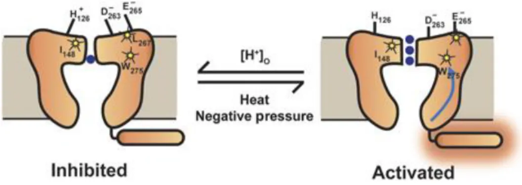
This mechanism has been found in K2P channels, there is a gating control in the selectivity filter via changes of conformation in helical transmembrane domains. Several subfamilies of K2P channels possess a pH sensitivity, given by residues such as histidine, in the vicinity of the selectivity filter (see the cartoon model in Figure 6, Bagriantsev et al., 2011; Cohen et al., 2008; Sandoz et al., 2009). Another argument that pH sensing is similar to C-type inactivation of Kv channels supporting this view is that a high external K+ concentration known to slow C-type inactivation alters pH inhibition of human (Cohen et al., 2008) and mouse TREK1 (Sandoz et al., 2009), and pH activation of TREK2 (Sandoz et al., 2009).
Figure 6: Cartoon model of K2P2.1 (TREK‐1) C‐type‐like gating by extracellular acidosis, heat, and pressure. Low‐activity (inhibited) state and a high‐activity (activated) state that involves a C‐type‐like gate. As suggested earlier (Sandoz et al,
32
2009), external acidification causes structural rearrangements in the pore triggered by electrostatic interactions between the protonated extracellular pH‐sensing His126, and the negatively charged region in the P2‐loop, which includes Asp263 and Glu265. Authors propose that Ile148, Leu267, and Trp275 (indicated with stars) are crucial elements of the C‐type‐like gate. As suggested elsewhere (Honore, 2007), temperature and mechanical stress have their sensing elements located in the intracellular C‐terminal domain. Activated C‐terminal domain (indicated with the orange halo) induces movements of the M4 transmembrane segment and affects channel activity through the C‐type gate. The blue arrow indicates the putative pathway. For clarity, only one of the two cytoplasmic C‐termini is depicted (from Bagriantsev et al., 2011).
Furthermore, Tucker and Baukrowitz have shown in 2011 using quaternary ammonium (QA) derivates – known to be allosteric blockers of potassium channels selectivity filter – that the selectivity filter regulation in K2P does not rely on the physical closure observed in the bundle crossing mechanism, since QA are still able to accede the selectivity filter, no matter the protonation state of the channel (Piechotta et al., 2011).
Since 2012, Dan Minor’s team is deeply trying to understand the mechanism by which, the Carboxy-terminal tail (C-tail) and the selectivity filter can be coupled (Bagriantsev et al., 2011, 2012, Lolicato et al., 2014, 2018; Pope et al., 2018). Mutations in the selectivity filter abolish intracellular regulation by the C-tail and vice versa. Furthermore, mutation in the TM4 helix or between TM4 and C-tail induce a loss of the coupling. The main conclusion is that the gating of TREK1 involves C-type like gate, but also involves TM4 flexibility. Zhuo and collaborators provide a role of a glycine hindge G312 in M4 involved in the allosteric regulation of the C-tail (Zhuo et al., 2016). In 2017, Minor group identified a druggable K2P site that stabilizes the C-type gate ‘leak mode’ (Lolicato et al., 2018).
(3) Hydrophobic gating in K2P
In line with these experiments, playing with hydrophobic residues, Lesage’s team has shown in TWIK1 that mutation of leucine located in the M2 domain (possessing a hydrophobic side chain) L146 to aspartic acid (a negatively charged residue) would increase the usually low current (Chatelain et al., 2012).
More recently, an explanation has been provided by Tucker’s team. They presented a new mechanism in TWIK1 channel termed hydrophobic gating (Aryal et al., 2014). Molecular dynamic studies revealed that the region represents a hydrophobic barrier, leading to a dehydration of the pore region, thus decreasing the ionic conduction, to finally downregulate TWIK activity. Replacing these hydrophobic residues to positively
33
charged residues such as arginine will increase the observed current. In conclusion, amino acids composition of the intern cavity will play a role on ion efflux.
K2P channels family
Mammalian K2P channels all share the same topology and a main role which is to establish and maintain the resting membrane potential. The 15 members have been cloned in mammalian and are classified (see Figure 7) depending on their sequence homology and their functional characteristics in 6 subfamilies. Surprisingly, even though they share the same function, apart from the pore domain, the sequence is poorly conserved between members, leading to particular characteristics.
Figure 7: Dendrogram of the K2P potassium channel family and their characteristic functional features. The effects of different stimuli on the currents of the channels within each K2P subfamily are indicated. PUFA, polyunsaturated fatty acids; PKC, protein kinase C; AA, arachidonic acid. TWIK ("Tandem of P domains in a Weak Inwardly rectifying
34
potassium (K+ ) channel") TREK ("TwikRElated K+ channel") TASK (Twik-related Acid-Sensitive K+ channel) TALK (Twik-related ALkalin pH activated K+ channel) THIK (Twik-related Halothane Inhibited K+ channel) TRESK (Twik-RElated Spinal cord K+ channel) from (Decher et al., 2017a).
Generalities
This family of potassium channels is the last one to have been uncovered and cloned. The first mammalian one, TWIK1, was cloned in 1996 in Nice (Lesage et al., 1996). TWIK is the acronym for Tandem of pore domains in a Weak Inwardly rectifying potassium (K+) channel. The channel was at the origin of the leak current described by Hodgkin and Huxley in 1952 in squid giant axon.
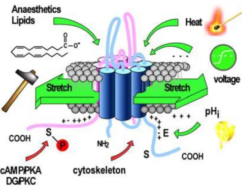
Leak potassium channels have been described as being constitutively open at rest. However, these channels are polymodal and respond to a wide range of stimuli, such as pH, temperature, membrane stretch, fatty acids and anesthetics (Lesage and Lazdunski, 2000).
They are ubiquitous, and thus play a role in the brain, the cardiovascular system, immune system and the somatosensory system. They exhibit the functional properties expected for K+ leaky channels: opening near the physiological voltage range to pass K+ selective currents, showing few or no voltage/time dependency.
Electrophysiological properties
In the 50’, Hodgkin and Katz have postulated that the cell membrane at rest is permeable to potassium (Hodgkin and Katz, 1949). General assumptions were made to establish the Goldman-Hodgkin-Katz (GHK) flux equation:
- The membrane is a homogeneous substance
- The electrical field is constant so that the transmembrane potential varies linearly across the membrane
- The ions access the membrane instantaneously from the intra- and extracellular solutions
- The permeant ions do not interact
- The movement of ions is affected by both concentration and voltage differences Allowing one to predict the current/voltage plot of potassium shown in Figure 8. Most K2P channels lack voltage sensitivity, and any kind of activation or inactivation kinetic. The rectification can thus be approximated by GHK equation in asymmetric K+
35
conditions, which rely on the electrochemical gradient of K+ ion, for most of K2P channels (Duprat et al., 1997).
Figure 8: Background potassium currents described by the GHK equation. In physiological condition the current-potential curve (IIV curve) in blue crosses the x-axis at current-potential value close to the equilibrium current-potential of the K+ ion (E
K =-80mV). In symmetrical K+ solution, IV curve (red) crosses the x-axis at 0mV.
If membrane potential is over the potassium potential equilibrium, the electrochemical gradient is in favor of a potassium output. Otherwise, there will be a potassium entry. The IV curve will thus be characterized by an outward rectification in physiological conditions. This outward rectification has important implications in the role of K2P in the central and peripheral nervous system by i) stabilizing the resting membrane potential, and ii) enabling action potential (AP) generation in absence of Kv channels by repolarization of the membrane after AP thank of their conductance (MacKenzie et al., 2015).
However, some K2P channels such as TREK1 or TASK3 do not respect this equation (Fink et al., 1996). TREK1 for instance possesses a voltage- and time-dependant activation due to an increased open probability for depolarized potential (Schewe et al., 2016).
Structure
K2P channels are characterized by the presence of two pore forming regions and four trans-membrane (TM) regions named TM1 to TM4 (or M1 to M4) in each channel subunit and, unlike other classes of K+ channels, are functional as dimers (not
36
tetramers, see Figure 9) (Lesage et al., 1996; Levitz et al., 2016). TM1 and TM2 are linked together via an extracellular loop containing two alpha helixes termed C1 and C2 and a pore domain named P1. The second extracellular loop joins together TM3 and TM4 and contains the second pore domain P2.
Figure 9: Cartoon showing the topology of a two-pore-domain potassium channel K2P and its assembly in dimer.
TWIK1 and TRAAK crystallographic structures have been released in 2012 and gave clues about the understanding of structure-function relationships in K2P channels (Brohawn et al., 2012; Miller and Long, 2012). C1 and C2 form an extracellular cap, of about 35 Å that could be involved in dimerization. In TWIK1 channel and other K2P (TREK1, TREK2…) two subunits are connected via a covalent disulfide bound that may help in the structure stabilization (Lesage et al., 1996). TM2 and TM4 helixes flanking the channel pore pass through the membrane obliquely. The more external helixes, cross the membrane vertically (Renigunta et al., 2015).
Crystallographic model of TRAAK revealed in 2012 by Brohawn et al. gave new clues about the path taken by the ions in the selectivity filter (Brohawn et al., 2012). There is a lateral window made of helix M2 of one subunit and helix M4 of another subunit that gives a direct access to the intern cavity in the intracellular side. These helixes also act on the conductivity and ion selectivity.
37
Figure 10: The structure of K2P-channels. a. Topology of K2P-channels. b. Sketch of the structure of the N-terminal part of the two subunits (including the M1, C1, C2, P1 and M2 domains). c. Sketch of the structure of the C-terminal part of the two subunits (including the M3, P2 and M4 domains). The helices are not drawn to scale. For clarity, the pore helices are relatively small (from Renigunta et al., 2015).
More recently, TREK2 structure has been characterized (Dong et al., 2015), allowing one to better understand the gating mechanism of K2P channels.
Up and down state and opening mechanism
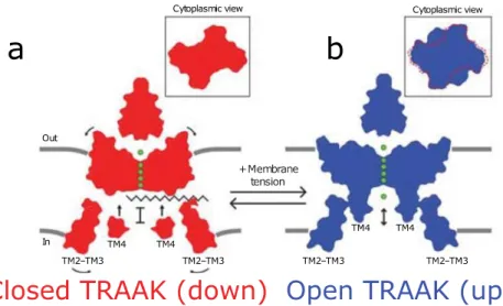
Two states, termed “up” and “down,” are known from x-ray structural crystallographic studies and have been suggested to differ in conductance. However, the structural details of the gating behavior are largely unknown. According to MacKinnon group, in the up configuration, TRAAK channel offers to potassium a passage open and direct between the cytoplasm and the extracellular environment (Brohawn et al., 2012,
38
2014). In the down state, the passage of ions is blocked by the phospholipid chain of the plasma membrane.
Figure 11: Cartoon model of TRAAK gating and mechanosensitivity. The down conformation (red) is a closed channel state and the up conformation (blue) is an open channel state. In the down conformation, a lipid acyl chain accesses the central cavity through intra membrane openings to block ion (green) conduction. In the up conformation, lipid is sealed from the cavity to permit ion conduction. Shape changes upon opening can explain the mechanosensitivity of TRAAK and TREK channels. Area expansion and reduced mid plane bending create an energy difference between channel states that promotes force activation, from (Brohawn et al., 2014).
The down state suggested to be the closed one, has been conflictual. Indeed, Minor’s group determined the down state using a constitutively activated TRAAK channel as the opened and activated state (Lolicato et al., 2014). Subsequent structure of TREK2 channel, provided an insight into the relevance of up and down states (Dong et al., 2015; McClenaghan et al., 2016) and its involvement in mechanosensitivy.
39
Figure 12: The down state, lower conductivity conformations are shown in orange, with the fenestrations indicated in yellow. The up state conformations are shown in blue. Inhibitors such as norfluoxetine are represented by a red triangle. (A) Overall scheme for K2P channel gating. Higher and lower conductivity states are shown for both conformations as the filter may gate independently of these larger changes, though with a different probability. Conformations for which we have crystal structures are indicated with an asterisk. (B) Schematic of activation of TREK channels by mechanical stretch. The direct interaction of TREK-2 with lipids in the membrane may allow lateral forces to facilitate conversion from the down to the up state which is fluoxetine insensitive. (C) Association of the C-terminal domain (CTD) with the membrane. The diverse cytoplasmic C-terminal extensions of K2P channels provide an additional site for modulation of channel activity. This regulation could operate in part though association of the CTD with the membrane after post-translational or pH changes that would favor the more active up state (from Dong et al., 2015).
Membrane tension can provide the channel energy to switch from one configuration to another (Brohawn, 2015). The transition between the two open conformations, "open-down" stabilized by pH and "open-up" stabilized by the pressure, would be more energetically favorable than the transition from a closed conformation to an open conformation (McClenaghan et al., 2016).
40
TWIK1 was the first K2P channel to be cloned in mammalian in 1996 in Nice (Lesage et al., 1996). TWIK is the acronym for Tandem of pore domains in a Weak Inwardly rectifying potassium (K+) channel. TWIK channel family is made up of 3 members, TWIK1, TWIK2 and KCNK7. In heterologous expression system, they produce very small or no currents. This is partially due to their subcellular localization. Indeed, KCNK7 is especially found in the endoplasmic reticulum (Salinas et al., 1999), while TWIK1 is located in recycling vesicle (Feliciangeli et al., 2010).
TWIK1 is expressed in the central nervous system (hippocampus, thalamus, and cerebellum) and in periphery in the heart, lungs, and kidneys (Arrighi et al., 1998). TWIK1 channels are selectively expressed in DRG neurons and show a reduction of expression after neuropathic injury. The maintenance of this reduction weeks after the injury suggest a major role for TWIK1 in the maintenance of chronic neuropathic pain (Mao et al., 2017; Pollema-Mays et al., 2013). However, TWIK1 only provides a small background current when expressed in heterologous expression systems, and seems to be in majority retained in intracellular compartment (Feliciangeli et al., 2010; Lesage et al., 1996).
Human TWIK2 also generate currents with very small amplitude in heterologous system (Patel et al., 2000). However, the rat homologous TWIK2 channel creates 15 times more current. Beside this difference, they possess the same particular electrophysiological properties. Its role is not well known and its tissue distribution could be linked to its silent activity.
As TWIK2, KCNK7 does not generate any current when expressed in heterologous system, in which it is retained in the endoplasmic reticulum (Salinas et al., 1999). Nothing is known about the function and the pathophysiological implication of this channel in human, where it is expressed in dorsal root ganglia and cerebellum (Medhurst et al., 2001).
Would TWIK1 role in pain rely on the formation of heteromers in the brain with other K2P channels?
Astrocytes express TWIK1. These cells show a passive conductance resulting in a low membrane resistance. This resistance seems to implicate leak potassium current, but the responsible are not yet identified. Indeed, it was hypothesized by Zhou et al that
41
TREK1 and TWIK1 independently participate in this passive conductance (Zhou et al., 2009), however, the same group suggested that mice KO for TWIK1 did not show significant differences in astrocytic membrane passive conductance. Independently, Park group showed that passive conductance membrane in astrocytes that expressed TWIK1 specific shRNA was completely reduced (Hwang et al., 2014). Furthermore, they have shown that TWIK1/TREK1 heteromeric channels were formed, and these last carry the passive conductance in astrocytes along with a fast glutamate release upon Gβγ binding (Hwang et al., 2014). This last characteristic of the pore leading to the transport of glutamate upon activation however remains unknown. We have tested the ability of TWIK1 and TREK to heteromerize and did not observed this association in our assay.
TASK family
TASK1 and TASK3 are two pore domains K+ channels that are pH sensitive. The current they generate show little time-dependence and weak rectification while TASK5 is found to be inactive in heterologous expression system. TASK1 and TASK3 are both inhibited by extracellular acidification (Duprat et al., 1997) and local anesthetics (Maingret et al., 2001). They also are inhibited by hormones and transmitters that act via Gq/11-coupled receptors, such as muscarinic M3 receptors. They are activated by volatile anesthetics (halothane and isoflurane). These channels are non-sensitive to TEA and 4-AP as well as Barium and Cesium that usually block K+ channel.
These two channels are highly expressed in the central nervous system. TASK5 is also found in the brain, in the central auditory system nervous system, but is thought to be non-functional, however it may works in association with an ancillary subunit (Karschin et al., 2001).
42
Figure 13 : Differential distribution of mRNA coding for TASK-1 and TASK-3 in the rat CNS. Sagittal (top) and horizontal (bottom) sections from rat brain were hybridized with radioactive probes specific for TASK-1 and TASK-3 (Talley and others 2001). On the left are grayscale images of the resulting film autoradiograms. To generate the colorized panels shown on the right, the grayscale images were converted to a single color (TASK-1: green, TASK-3: red) and the respective images were merged. Only regions with moderate-to-high signal intensity were included. Areas of overlapping expression are shown in yellow. Particularly evident in this regard are granule cells in the cerebellum and olfactory bulb, and brainstem motoneurons (Talley et al., 2003)
Figure 14: TASK channels regulations.
TASK currents can be inhibited in two ways by protein G coupled receptors. Notably, after Gq activation, the activation of phospholipase C (PLC) induces depletion in PIP2 in plasma membrane reducing channel activity (Chemin et al., 2003; Czirják and