CHARGE-BASED TRANSPORT AND DRUG DELIVERY INTO
CARTILAGE FOR LOCALIZED TREATMENT OF DEGENERATIVE ARCHIVES
JOINT DISEASES
I
by
Ambika Goel Bajpayee
OF TECHNOLOLGY
APR 15 2015
LIBRARIES
BEng in Manufacturing Processes & Automation Engineering, University of Delhi, 2006
MEng in Mechanical Engineering, Massachusetts Institute of Technology, 2007
Submitted to the Department of Mechanical Engineering
In Partial Fulfillment of the Requirements of the Degree of
Doctor of Philosophy in Mechanical Engineering
At the
MASSACHUSETTS INSTITUTE OF TECHNOLOGY
February 2015
Massachusetts Institute of Technology
All Rights Reserved
Signature redacted
A uthor...
Department of Mechanical Engineering
January 20, 2015
Signature redacted
Certified By...
Alan J. Grodzinsky
Professor o
iological, Electrical & Mechanical Engineering
Thesis Supervisor
Signature redacted
A ccepted B y...
..
...
David E. Hardt
Chairman, Department Committee on Graduate StudentsCHARGE-BASED TRANSPORT AND DRUG DELIVERY INTO CARTLAGE FOR
LOCALIZED TREATMENT OF DEGENERATIVE JOINT DISEASES
By
Ambika Goel Bajpayee
Submitted to the Department of Mechanical Engineering In Partial Fulfillment of the Requirements of the Degree of
Doctor of Philosophy in Mechanical Engineering
Abstract
Traumatic joint injuries significantly increase synovial fluid levels of pro-inflammatory cytokines that can initiate cartilage degeneration leading to osteoarthritis (OA). Articular cartilage is a highly negatively charged, avascular tissue, which relies on synovial fluid convection and electro-diffusion to transport proteins and therapeutics to tissue chondrocytes. No OA drug has yet passed the safety criteria of clinical trials due to ineffective intra-articular (i.a.) delivery methods, which require very high drug doses that cause systemic toxicity. There is a need to design local delivery mechanisms that can enable drugs or drug carriers to rapidly diffuse into the cartilage extracellular matrix to achieve intratissue therapeutic levels before these drugs are cleared from the joint space via lymphatics and synovium vasculature.
This dissertation investigates the effects of size and charge of solutes on their penetration, binding and retention within negatively charged tissues such as cartilage. Based on this understanding we selected Avidin, a globular protein, as a drug carrier owing to its optimal size and high positive charge (66,000 Da,
pI 10.5). Avidin resulted in a six-fold upward Donnan partitioning factor at the synovial fluid-cartilage
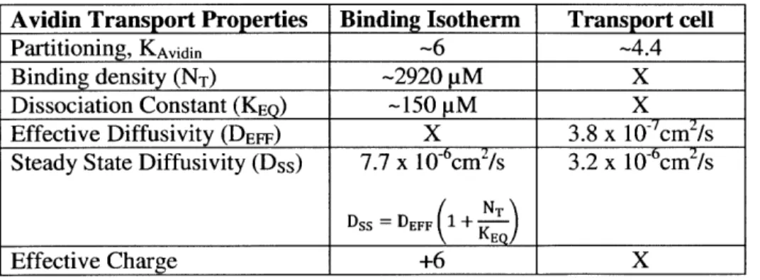
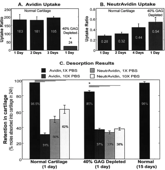
interface, had a 400-fold higher uptake than its electrically neutral counterpart (Neutravidin), and remained bound within cartilage for at least 15 days. Competitive binding experiments revealed that despite Avidin's weak and reversible ionic binding (dissociation constant, KD -150 pM) to the negatively
charged glycosaminoglycans, its long term retention was facilitated by large intratissue binding site density (NT- 2,920 pM). Thus, structures like Avidin are ideal candidates for local i.a. drug delivery into cartilage.
In vivo animal studies revealed that Avidin retained inside the joint space for extended time periods resulting half-life of 154h in rabbit cartilage which was 5-6 times longer than that in the thinner rat cartilage. This was confirmed to be consistent with the concept that diffusion-binding kinetics scale as the square of tissue thickness, emphasizing the necessity of using larger animal models for studying joint space transport and pharmacokinetics. Avidin's neutral counterpart (Neutravidin) was completely cleared from the joint space of both rats and rabbits within 24h.
We then conjugated Avidin with the glucocorticoid, dexamethasone, using chemical linkers to enable its sustained release. Avidin delivered dexamethasone into cartilage deep zones where majority of chondrocytes reside thereby successfully inhibiting cytokine-induced catabolic activity in cartilage explants in-vitro. A single i.a. injection of Avidin-conjugated drug can thereby enable sustained drug delivery in low doses and therefore has the potential to replace the current clinical practice of using
multiple injections of high dose glucocorticoids in patients. The biological efficacy of this system in rescuing degenerative mechanisms of OA is currently being validated in a well-accepted rabbit model of post-traumatic OA as part of a preclinical study.
Thesis Supervisor: Title:
Alan Jay Grodzinsky
Professor of Biological, Electrical & Mechanical Engineering Massachusetts Institute of Technology
Other thesis committee members: Paula T. Hammond
Professor of Chemical Engineering, MIT
Rohit N. Karnik
Associate Professor of Mechanical Engineering, MIT
Christopher H. Evans
Professor of Orthopaedics, Mayo Clinic
Dr. Eliot H. Frank
Principal Research Engineer, Grodzinsky Lab, MIT
To my teachers
Acknowledgements
I have been fortunate to be surrounded by some of the most intellectually stimulating yet humble and nice
people during my doctoral work at MIT, and I am truly grateful to all of them.
I would like to thank my advisor, Professor Alan Grodzinsky for being a wonderful mentor, teacher and
an inspiration. I have learned many lessons from Al both on a professional and a personal level. Al has been one of the best teachers I have had during my academic training. I have learned immensely from his unfettered dedication to teaching and mentorship, and his systematic approach of presenting and organizing blackboards to effectively impart scientific knowledge to his students and scientific communities with diverse backgrounds. His style of supervision provided me with a high level of freedom while he always remained approachable regardless of day or time. The four years spent in Al's lab and the many hot toddies we've had together at Area 4 are unforgettable. I hope that I can emulate his humility, his positive attitude and that my association with him will remain life-long.
I would also like to thank my thesis committee for their support and feedback throughout the course of
this work. Prof. Rohit Karnik's suggestions and feedback were critical especially during the early stages of this work. Prof. Chris Evans provided the clinical perspective, which was very useful while designing and conducting animal studies. Prof. Paula Hammond and her lab guided me through the chemistry side of the project. Dr. Eliot Frank has provided thorough feedback on several aspects of experiments and modeling.
I have been fortunate to have attended lectures by inspiring teachers like Professors Roger Kamm and
Tomasz Wierzbicki, who have played a critical role in advancing my knowledge of biomechanics.
Prof. Ryan Porter's lab at the Beth Israel Deaconess Medical Center was key to all the animal work. It has been an absolute pleasure to work with Ryan and his lab members and orthopedic surgeons, Dr. Max Scheu and Dr. Rodolfo Vega.
I am grateful to all members of the Grodzinsky lab for a friendly and a collaborative environment. I
would like to specially thank Dr. Yang Li and Yang Wang for helping me with biology related questions and techniques. Yang Wang and I have spent some memorable times together in lab, at ORS conferences, and also outside of work. Undergraduate students Nathan Varady and Isabel Yannatos provided direct assistance in experimental work. My very caring and funny MechE friends, Sergio Castellanos, Bavand Keshavarz, and Michela Geri are like a family; starting from studying for qualifying exams together till now, our friendships have strengthened and blossomed. Being part of student organizations like GAME, MEGAWomen and GSW has further enriched my MIT experience. I would like to thank Leslie Regan for her continuous support and approachability, and for taking care of a lot of things that I didn't have to worry about.
I would like to thank the Deshpande Center for Technological Innovation for funding this project. Leon
Sandler, the center's executive director, has been a great mentor. He introduced us to Dr. Barry Berger, whose guidance has been critical for this work. I would also like to acknowledge the Legatum Center and the World Bank for their financial support.
My two younger brothers, Shakti Amar Goel and Arpit Amar Goel, have checked on me from time to
time, supported me throughout and taken keen interest in what I am doing. We have gone through a lot together, and they hold a special place in my heart. I am grateful to my parents for instilling in me a strong work ethic, discipline and importance of hard work. My nanny (I call her Pappy) has played an important role in my upbringing and I can't thank her enough for teaching moral values and emphasizing
the simplicity of life. I owe a lot of my upbringing to the teachings of Vedanta and Bhagavad Gita, which have played a critical role in my thinking process and shaping my philosophy for life. I have to thank my Sanskrit and Hindi language teachers from Delhi Public School, Noida for equipping me to interpret and elaborate their meaning.
Finally, the most important and the greatest man in my life is my best friend and my husband, Anurag Bajpayee who gives meaning to my existence. His unconditional love, support and interest in anything and everything I have desired to do is inexpressible in words. Despite his busy schedule, he has stayed up many nights to give me company in lab, listened to my endless talking about research and many failed experiments that no one will hear about, cheered me up, cooked me delicious meals and always been there for me. Having him in my life makes me yearn for nothing but our eternal togetherness. Nothing is better than having a partner who I can discuss any topic from science, engineering, cosmology, spirituality and life. MIT is even more special because this is where we met. I am very lucky to have him. -Ambika Goel Bajpayee
Lead me from the unreal to the real;
from darkness (ignorance) to light (knowledge);
and from death to immortality.
Table of Contents
Chapter 1 Introduction 12
1.1. Osteoarthritis, a degenerative joint disease 12 1.2. Intra-articular (i.a.) therapy and the need for intra-tissue drug delivery 14
1.3. Structure of cartilage 14
1.4. Charge based mechanisms for local intra-tissue drug delivery 15
1.5. Thesis outline 17
1.6. References 18
Chapter 2 Avidin as a model for charge driven transport into cartilage and drug delivery for
treating early stage post-traumatic osteoarthritis 24
2.1. Introduction 25
2.2. Materials and Methods 26
2.3. Results 30
2.4. Discussion 35
2.5. Conclusion 38
2.6. References 38
2.7. Supplementary Material 47
Chapter 3 Charge Based Drug Delivery Mechanisms and a Mathematic Model of Joint Space
Transport Kinetics 51
3.1. Introduction 51
3.2. Charge based drug delivery mechanisms 51
3.2. Joint space transport kinetics 54
3.3. Summary 56
3.4. References 57
Chapter 4 Electrostatic interactions enable rapid penetration, enhanced uptake and retention of
intra-articular injected Avidin in rat knee joints 60
4.1. Introduction 61
4.2. Methods 62
4.3. Results 64
4.4. Discussion 66
Chapter 5 On the choice of animal model for intra-articular drug delivery relevant to treatment of post-traumatic osteoarthritis 77 5.1. Introduction 78 5.2. Methods 79 5.3. Results 80 5.4. Discussion 82 5.5. References 84
Chapter 6 Targeted intra-tissue sustained delivery of dexamethasone using Avidin as a
nano-carrier for treating degenerated cartilage 92
6.1. Introduction 93
6.2. Materials and Methods 94
6.3. Results and Discussion 99
6.4. Conclusion 104
6.5. References 105
Chapter 7 In-vivo efficacy of single dose Avidin-DEX treatment using rabbit ACL transection
model 116
7.1. Design of Animal Study 116
7.2. Methods 116
7.3. Results and Discussion 118
Chapter 8 Summary and Future Direction 125
8.1. Summary of Thesis 125
List of Figures
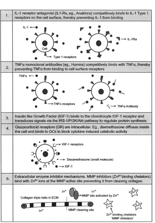
Figure 1.1 Examples of mechanisms of interaction between some of the potential OA therapeutics and their intracellular or extracellular target binding sites. ... 22 Figure 1.2 Anatomy of the knee joint and depth dependent properties of articular cartilage ... 23 Figure 2.1 Design of transport chamber used for understanding size dependent transport of solutes inside
cartilage ... 42
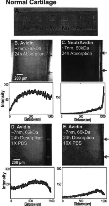
Figure 2.2 Confocal images of time dependent concentration profiles inside cartilage explants...42 Figure 2.3 Confocal images of the concentration profile of 15 nm quantum dots inside normal cartilage explants ... 43
Figure 2.4 Transport chamber showing visual evidence of significantly higher uptake for Avidin
compared to NeutrAvidin over a 24h period ... 44
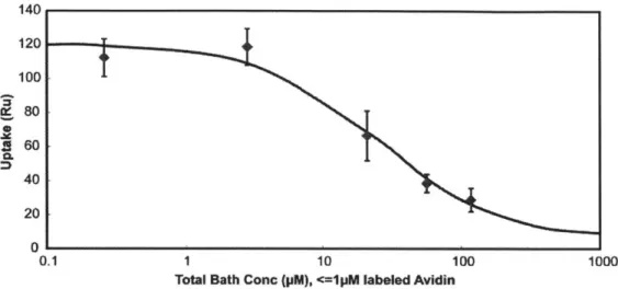
Figure 2.5 Uptake ratios measured for Avidin and NeutrAvidin after
1
to 4 day equilibration periods for normal and 40% GAG-depleted cartilage explants...45 Figure 2.6 Binding isotherm for Avidin's transport inside cartilage ... 46Figure 2.7 Non-equilibrium diffusive transport of Avidin-FITC across cartilage explants...46 Figure 2S.1 Avidin binding isotherm re-plotted with the x-axis normalized to KEQ using the best fit
parameter values data of Fig. 2.6 (K-6, KEQ- 150 pM and NT- 3000 pM)...47 Figure 2S.2 Upper bound estimate of Avidin charge assuming only electrostatic interactions and no b inding ... 50 Figure 3.1 Three classes of particle based drug delivery systems for local i.a. intra-tissue delivery of drugs...57
Figure 3.2 Kinetics and concentration profiles of intra-articular injected drug carrying particles inside the
joint
space ... 58 Figure 4.1 Images of rat knee joint following injection with Texas Red-conjugated Avidin...70 Figure 4.2 Confocal images of tissue specimens extracted from rat knee joints 6h after intra-articularinjection of Avidin-Texas Red...71
Figure 4.3 Avidin and Neutravidin uptake and retention in different tissue types from rat knee joints after 6 h, 24 h, 4 days and 7 days (Av) or after 24 h (Nu)... 72 Figure 4.4 sGAG concentration measured using DMMB assay and calculated Avidin half-lives in
different rat knee tissues. ... 73
Figure 4.5 Toluidine blue staining of naive rat knee tissues ... 74
Figure 4.6 Images of fluorescently stained bovine cartilage explants after treatment with increasing dose
of Avidin to check for chondrocyte viability ... 75 Figure 4.7 Cumulative sGAG loss from bovine cartilage and biosynthesis rates with a one time dose of
increasing concentration of Avidin...76
Figure 5.1 Images of tissues harvested from the rabbit's knee joints after 24h following injection with
Texas Red-conjugated Avidin...87 Figure 5.2 Avidin uptake and retention in different tissue types from rabbit knee joints after
1
day, 4 days and 7 days ... 88 Figure 5.3 Safranin-O staining of joint tissues harvested from the rabbit knee from the contralateralcontrol group ... 89 10
Figure 5.4 sGAG concentration measured using DMMB assay and calculated Avidin half-lives in
different rabbit knee tissues ... 90 Figure 5.5 Avidin half-lives for different tissue types plotted against as a function of sGAG
concentration* tissue thickness square. ... 91
Figure 6.1 Schematic representation and chemical structures of the four Avidin (Av) conjugated
dexamethasone (DEX) compounds formulated ... 108
Figure 6.2 Titration curve of 2,6 ANS-fluorometric assay for stoichiometry of biotin-PEG with Avidin in PBS and SDS PAGE (4-12%) of Avidin and PEGylated Avidin under reducing conditions stained with C oom assie B lue ... 111 Figure 6.3 In vitro DEX release profile for Av+DEX, PEG Av+DEX, PEG Av-ester-DEX in PBS (pH
7.4) at 37*C and PEG Av-hydrazone-DEX at pH 7.4 (diamonds) and at pH 4 (squares)...111
Figure 6.4 Images of fluorescently stained bovine cartilage explants to check for chondrocyte viability
after 48h incubation with Avidin conjugated DEX compounds ... 113 Figure 6.5 Effect of one dose vs. continuous dose of IOOnM DEX on IL-I a simulated GAG loss in
bovine cartilage explants...113
Figure 6.6 Biological activity of single dose Av-DEX compounds on IL- I
a
(I ng/ml) treated bovinecartilage explants over a period of 22 days...114 Figure 6.7 Images of fluorescently stained IL- Ia treated bovine cartilage explants cultured for 8 days to
check for chondrocyte viability after treatment with one dose of Avidin-DEX compounds...115 Figure 7.1 Design of preclinical rabbit ACLT study to evaluate safety and efficacy of Avidin DEX
com pounds ... 12 1
Figure 7.2 Image showing the experimental configuration for indentation tests using the Dynastat mechanical spectrometer to obtain biomechanical properties of rabbit tibial plateau cartilage and lateral m eniscus...12 1
Figure 7.3 mRNA levels of IL-i
I,
MMP1, ACAN and MMP13 in pooled articular cartilage from therabbit medial femoral condyle and the medial tibial plateau ... 122 Figure 7.4 Mean equilibrium modulus and dynamic stiffness measured in the lateral tibial plateau cartilage...123
Figure 7.5 sGAG and collagen concentration measured in rabbit lateral tibial plateau cartilage and patella cartilage...124
Chapter 1
Introduction
1.1. Osteoarthritis, a degenerative joint disease
Osteoarthritis (OA) is a complex debilitating disease that affects millions of people worldwide, causing loss of productivity, quality of life, and loss of joint function. OA affects over 150 million people worldwide, about 25 million people in the US and this number is expected to double by 2020 (Source: Disease, Incidence, Prevalence & Disability; WHO 2009, data based on census in 2004) making it the fourth leading cause of disability by 2020. Yet, there is no cure for it.
IPrevalence of Osteoarthritis
Life years lost due to Osteoarthritis
Disability-adjusted lifeyear(DALY) forOA per 100,000 inhabitants
DALY =YLL+YL(ye..sf i. t+ye. irwedwiI disWmty *no0fai 28O.Mi0 *360.38
024.20 * 3 0380
It is now accepted that OA is a disease of the entire joint, eventually affecting all joints tissues including cartilage, bone, ligaments, menisci, the joint capsule, synovial membrane, muscles and neural tissue [1,2]. Distinct subtypes of OA are associated with the varying risk factors that mediate OA initiation and progression; these risk factors include improper joint mechanics, gender, age, obesity, genetic and metabolic factors, and acute joint injury leading to post-traumatic OA (PTOA) [3, 4]. PTOA accounts for 12% of the total OA population [5]. Approximately 50-80% of young active individuals who
12
suffer traumatic joint injuries (e.g., rupture of the anterior cruciate ligament or meniscus) progress to PTOA within 10-20 years [6, 7]. Following acute joint injury, there is an immediate increase in synovial fluid levels of inflammatory cytokines (e.g., IL-I, IL-6, TNFa) which can diffuse into cartilage and rapidly initiate proteolysis and loss of cartilage matrix [8, 9]. By the time of clinical (radiographic) diagnosis, irreversible changes to cartilage and other joint tissues have often occurred [6]. Since the cause and time of the initial trauma is known, there exists a unique opportunity for early drug intervention to prevent further degeneration of cartilage and other tissues, and to reverse the course of PTOA by inducing repair [6].
While there are disease-modifying anti-rheumatic drugs (DMARDS) for rheumatoid arthritis and several related rheumatic diseases (such as the TNFa-blockers [10], Table 1.1), no efficacious disease-modifying osteoarthritis drugs (DMOADS) are yet available, i.e., drugs which alter or halt the progression of OA [1,11,12]. Current therapies provide only short term relief of pain and inflammation (e.g., analgesics, hyaluronic acid lubricants, etc.), but afford no protection against further degeneration of cartilage, the hallmark of end-stage OA [3], leading to the need for joint replacement. Several anti-catabolic and pro-anabolic drugs have been identified as potentially useful to reverse or prevent PTOA-associated breakdown of cartilage, including anti-catabolic glucocorticoids (e.g., dexamethasone) and pro-anabolic growth factors (e.g., IGF-1, FGF-18, and BMP-7) [11,13-15]. Consistent with the concept that generalized OA involves the whole joint, DMOAD development and associated clinical trials are now targeting cartilage breakdown (e.g., protease and cytokine inhibitors), bone remodeling (e.g., bisphosphonates, BMP-7, calcitonin), and synovial and inflammatory mediators (e.g., cytokine blockers)
[11]. Table 1.1 provides a list of therapeutics currently being considered for OA treatment delivered via
intra-articular injections. Biological agents such as monoclonal antibodies for inhibiting IL-l1 (Canakinumab), TNFa (Infliximab, Adalimumab) and other anti IL- or TNFa agents (such as, Anakinra and Enbrel) have been successfully used for systemic treatments for rheumatoid arthritis and are being considered for i.a. therapy for OA treatment. However, no drug candidates have yet passed the safety/efficacy hurdle, and systemic drug side-effects have been a major safety concern causing several trial failures to-date [12]. Anakinra (IL-IRa) showed only short term benefit in its phase II evaluation as an i.a. treatment for OA owing to rapid clearance from the joint due to its small size (l7kDa). Thus, it is important to develop appropriate drug delivery methods to administer potentially efficacious drugs or drug combinations directly to selected target tissues, such as cartilage thereby eliminating any systemic adverse effects [16].
1.2. Intra-articular (i.a.) therapy and the need for intra-tissue drug delivery
Intra-articular injection has enabled local administration of drugs into the joint space and thus reduced systemic toxicity and improved drug bioavailability; yet it remains inadequate due to rapid exit of drugs from the joint space. Small molecules exit via the rich synovial capillary network while the larger macromolecules (e.g., hyaluronan) are cleared by the lymphatic system, which is located in the subsynovium [17, 18]. Short mean half-lives of NSAIDs (Non-Steroidal Anti-Inflammatory Drugs) in the synovial fluid have been reported as 1-4 h [19, 20]. Furthermore, simple i.a. injection can require very high drug doses to achieve a concentration gradient high enough for drug diffusion into cartilage. Such high doses can cause deleterious effects; even low-dose sustained release in the synovial fluid may still cause unwanted drug exposure to other joint tissues.It is important to note that most therapeutics listed in Table 1.1 (except hyaluronan and many proteinase inhibitors) such as glucocorticoids, and growth factors have to be delivered to the chondrocytes, most of which reside in the deep zones of cartilage. Fig. 1.1 shows example mechanisms of how therapeutics interact with their intracellular or extracellular target binding sites to inhibit catabolic effects and restore normal biosynthesis levels. High molecular weight, hyaluronan (clinically approved, e.g., Sanofi's Synvisc is 6000 kDa in MW) or lubricin-like products restore properties of synovial fluid in the joint space and make a film about
1
m thick on cartilage surfaces. Thus their target sites are in the superficial surfaces of cartilage, synovial membrane and in the synovial fluid. Synovial fluid is essential for joint lubrication and mechanical sliding that enables diffusion of nutrients into the avascular cartilage.New approaches have focused on delivery of functionalized drugs directly into the joint space-synovial fluid using micro and nanoparticles [21-25], drug releasing peptides and gels [26-28]. Most of these systems are aimed at increasing the residence time of drugs in synovial fluid but are unable to transport drug/drug carriers directly into the deep zones of cartilage where the majority cell/tissue targets reside. Entry of macromolecules into cartilage is hindered by its complex architecture (discussed in section 1.3) of dense collagen fibrils and negatively charged aggrecan proteoglycans. Thus, cartilage is often considered as a 'barrier to drug/drug carrier entry'.
1.3. Structure of cartilage
Articular cartilage is a highly complex, avascular, alymphatic and aneural tissue made of a dense network of collagen fibrils (constituting 50-60% dry weight of tissue), aggrecan proteoglycans that contain highly negatively charged glycosaminoglycan (GAG) chains (30-35% tissue dry weight) and
many additional extracellular proteins which are continuously synthesized by a low density of chondrocytes (1-5% tissue dry weight). Type II collagen is the most abundant type present and its mesh pore size is 60-200nm.The superficial zone (SZ) of cartilage (the most superficial and well lubricated region that provides gliding surface and is about 10-20% of total cartilage thickness) has collagen fibrils aligned parallel to the surface. The collagen fibrils are oriented randomly in the middle zone (MZ,
40-60% tissue thickness) and are perpendicular to the subchondral bone in the deep zone (DZ, 30-40% tissue
thickness). The calcified zone provides a mechanical transition between the soft cartilage tissue and the stiff subchondral bone. The collagen network is space-filled with 300MDa aggregates of aggrecan, having sulfated GAG chains that are only 2-3nm apart from each other. The aggrecan aggregates have a bottle brush like structure comprised of a long central hyaluronan (HA) GAG chain that links electrostatically to 3MDa aggrecan monomers at their Gl binding domains. This binding is further stabilized by link protein (Fig. 1.2), which has a structure analogous to that of the aggrecan GI domain.
The aggrecan and chondrocyte densities increase with depth into cartilage from the superficial zone, which further reduces the pore size and restricts the ability of solutes to penetrate and diffuse within cartilage. There is a lack of understanding of what size range of solutes can penetrate through full thickness of dense cartilage ECM (extracellular matrix), and this will be explored further in Chapter 2.
In summary, there are two competing rates of transport inside the joint space for drug carrying particles: (i) the lumped rate of transport of particles into soft tissues such as cartilage (i.e., the diffusive flux of entry into the soft tissues, QEnt) and (ii) rate of exit from the lymphatics (the diffusive flux of exit
from the joint space, QExit-) QEntry should be such that it can enable fast enough transport of drug carriers
inside the desired target tissue to achieve intra-tissue therapeutic levels before the majority of the drug carriers are cleared from the joint space (QExit). Solute flux (Q) in a given medium is related to the concentration (Ci) gradient and its diffusivity by Fick's first law of diffusion:
Qi =
-DiVCi
+
Civ,
where Di is the effective solute diffusivity in the medium and v is the synovial fluid velocity. Joint space kinetics are further explored in Chapter 3.
1.4. Charge based mechanisms for local intra-tissue drug delivery
The high density of heavily negatively charged GAGs inside cartilage provides a unique opportunity to use electrostatic interactions to augment transport rate, uptake and binding of drug carriers
inside the tissue. In fact, the negatively charged aggrecan proteoglycans are also present in other soft tissues such as menisci, ligaments and in small concentrations in tendons (See Chapters 4 and 5). The synovial fluid (SF) also has a high density of the negatively charged hyaluronic acid (HA) and lubricin. The viscosity of SF is attributed to these two molecules, which provide effective hydrodynamic lubrication necessary for healthy functioning of joints. SF forms a thin layer on cartilage surfaces, synovial membrane and fat pads, providing retention pockets for cationic drug carriers. The infrapatellar fat pad (Hoffa) is the largest fat pad in the joint and also contains sulfated GAGs as well as non-sulfated
HA [29].
The high fixed charge density (FCD) within the ECM of such tissues provides a natural reservoir for cationic drug carriers, yet this attribute of joint tissues has not been fully exploited for local i.a. drug delivery. This aspect, however, has been effectively used in physiological imaging techniques such as delayed gadolinium enhanced medical resonance (MR) imaging of cartilage (dGEMRIC) and sodium MRI for differentiating degenerated cartilage from normal tissue. Both techniques work on the principle that after cartilage degradation, proteoglycan loss occurs, which reduces the tissue's negative FCD. Consequently concentration of positively charged sodium ion (Na') declines inside the tissue, and this attenuated Na' signal is captured by sodium MRI. Similarly, the negatively charged Gd (DTPA)2 molecules in dGEMRIC technique accumulate in high concentration in areas lacking in GAG and in low concentrations in GAG-rich regions [30]. In the following section, this electrically driven solute equilibration in the negatively charged articular cartilage is described using the principle of Donnan equilibrium partitioning.
1.4.1. Donnan partition coefficient: The partition coefficient is defined as the equilibrium
concentration of unbound or free solutes inside cartilage normalized by the solute concentration in the equilibration bath. The partition coefficient of a solute depends on its size, charge as well as on the composition of cartilage-matrix. A neutral solute which is not sterically hindered by cartilage ECM can have a partition coefficient of 1, which means equal solute concentrations in the tissue and the surrounding bath at equilibrium. The negative charge of GAGs in cartilage interacts with charged solutes and can cause exclusion of negatively charged solutes and enhanced uptake of the positively charged ones. This charge interaction between freely moving charged solutes and charged tissue is explained by Donnan theory, which is based on assumptions that (1) all freely moving charged species will partition into a charged tissue according to the Boltzmann statistics and, (2) the net charge in the tissue is zero by electroneutrality (i.e. the sum of tissue fixed charge density and the mobile carrier concentrations). This
means that at physiological conditions, Na' ions partition up inside the cartilage while Cl- ions will partition down.
Transport of large sized solutes in to tissue is sterically hindered and therefore will result in partition coefficients smaller than 1. Thus the size and charge of the solute as well as tissue fixed charge density are important parameters affecting partition coefficients. For example, Maroudas et al. [31,32] showed that serum albumin (MW 69kDa, diameter=7nm, pI 4.7) is sterically hindered in normal human cartilage, with a partition coefficient less than 0.05, which increased in OA cartilage owing to tissue fibrillation and loss of negatively charged groups.
1.4.2. Equilibrium binding: Binding of solutes to intra-tissue sites (for example, proteins binding to their cell surface receptors, or drugs/drug carriers functionalized to bind with ECM) can significantly slow down diffusion kinetics inside the tissue. It is, however, important for drugs/drug carriers to penetrate full thickness of tissue to reach their target sites and yet have a long residence time. The weak and reversible binding that can accompany electrostatic interactions provides a distinctive advantage by allowing drug carriers to penetrate through the full thickness of tissues. It is important to note that, while all charged solutes will partition within cartilage according to Donnan Equilibrium, electrostatic interactions do not necessarily cause "binding" of cationic solutes to negatively charged
ECM macromolecules. The potential for binding must be determined experimentally on a case by case
basis for each cationic solute (e.g., drug carrier) of interest. This is further discussed in Chapters 2 and 3.
1.5. Thesis outline
With the goal of developing particle-based drug delivery mechanisms for local treatment of OA, the objectives of Chapter 2 are (i) to determine the size range of solutes that can penetrate and diffuse through normal and osteoarthritic cartilage, and (ii) to investigate the effects of electrostatic interactions on solute uptake, partitioning and binding within cartilage by using a positively charged protein, Avidin. Avidin, due to its optimal size and positive charge exhibited ideal characteristics of a nanoparticle for drug delivery into negatively charged cartilage.
Based on our understanding of size and charge dependent transport of particles inside cartilage, we propose three particle based drug delivery mechanisms in Chapter 3. The effects of increasing net positive charge on cationic particles on their diffusion kinetics and retention inside cartilage are also discussed. Furthermore, governing equations to model transport kinetics inside the joint are presented.
Next, our goal was to use Avidin as a drug delivery vehicle and thus we quantified its transport kinetics, uptake and retention (half-lives) in different knee joint tissues of healthy small (rats) and large (rabbits) animals in vivo. Chapters 4 presents data from small rat models and also highlights the importance of using larger animals with thicker cartilage for understanding transport mechanisms as diffusion and diffusion-reaction kinetics is a function of the square of the tissue thickness. Chapter 5 discusses Avidin kinetics in rabbits, a larger animal model. We also evaluate the dose dependent response of Avidin on chondrocyte viability, GAG content and matrix biosynthesis to determine a safe useable dosage for delivering drugs.
We used dexamethasone (DEX) as an example drug for OA treatment, and in Chapter 6, we present synthesis and characterization of Avidin conjugated DEX structures using chemical linkers to enable both fast and slow DEX release. Fast release of drugs will provide patients with immediate pain and inflammation relief while the slow and sustained drug release is critical to suppress OA catabolic effects and restore normal biosynthesis levels. Their bioactivity is also tested in a cytokine (IL-1) challenged bovine cartilage explant model involving 3 weeks of in vitro organ culture.
Chapter 7 presents the experimental design for preclinical evaluation of safety and efficacy of single dose Avidin-DEX compound using anterior cruciate ligament transection (ACLT) injury model of post traumatic osteoarthritis in mature rabbits. This study uses 60 rabbits for evaluating efficacy at two time points: 3 weeks and 9 weeks post-surgery. This work is currently ongoing but we will present some data from the 3 weeks study. Chapter 8 will outline major conclusions from this thesis, highlight outstanding questions and also identify potential future directions.
1.6. References
[1] Le Graverand-Gastineau M-PH. 2010. Disease modifying osteoarthritis drugs: facing development challenges and choosing molecular targets. Curr Drug Targets. 5, 528-35.
[2] Felson DT, Neogi T. 2004. Osteoarthritis: is it a disease of cartilage or of bone? Arthritis Rheum.
50, 341-4.
[3] Wieland HA, Michaelis M, Kirschbaum BJ, et al. 2005. Osteoarthritis - an untreatable disease? Nat Rev Drug Discov. 4, 331-44.
[4] Bay-Jensen A-C, Hoegh-Madsen S, Dam E, et al. 2010. Which elements are involved in reversible and irreversible cartilage degradation in osteoarthritis? Rheumatol Int. 30, 435-42.
[5] Brown TD, Johnston RC, Saltzman CL, et al. 2006. Posttraumatic Osteoarthritis: A First Estimate of Incidence, Prevalence, and Burden of Disease. Journal of Orthopaedic Trauma. 20, 739-44
[6] Anderson DD, Chubinskaya S, Guilak F, et al. 2011. Post-traumatic osteoarthritis: improved understanding and opportunities for early intervention. J Orthop Res. 29, 802-9.
[7] Lohmander LS, Englund PM, Dahl LL, et al. 2007. The long-term consequence of anterior cruciate ligament and meniscus injuries: osteoarthritis. Am J Sports Med. 35, 1756-69.
[8] Irie K, Uchiyama E, Iwaso H. 2003. Intraarticular inflammatory cytokines in acute anterior cruciate ligament injured knee. Knee. 10, 93-6.
[9] Kapoor M, Martel-Pelletier J, Lajeunesse D, et al. 2010. Role of proinflammatory cytokines in the pathophysiology of osteoarthritis. Nat Rev Rheumatol. 7, 33-42.
[10] Aaltonen KJ, Virkki LM, Malmivaara A, et al. 2012. Systematic Review and Meta-Analysis of the
Efficacy and Safety of Existing TNF Blocking Agents in Treatment of Rheumatoid Arthritis. PLoS
ONE. 7, e30275.
[11] Hunter DJ. 2011. Pharmacologic therapy for osteoarthritis--the era of disease modification. Nat Rev
Rheumatol. 7,13-22.
[12] Matthews GL, Hunter DJ. 2011. Emerging drugs for osteoarthritis. Expert Opin Emerg Drugs. 16,
479-91.
[13] Lu YC, Evans CH, Grodzinsky AJ. 2011. Effects of short-term glucocorticoid treatment on changes
in cartilage matrix degradation and chondrocyte gene expression induced by mechanical injury and inflammatory cytokines. Arthritis Research & Therapy. 13,R142.
[14] Nixon AJ, Brower-Toland BD, Bent SJ, Saxer RA, et al. 2000. Insulin like growth factor-I gene therapy applications for cartilage repair. Clin. Orthop. Relat. Res. 379 Suppl, S201-213.
[15] Miller RE, Grodzinsky AJ, Cummings K, et al. 2010. Intra articular injection of heparin-binding
insulin-like growth factor
1
sustains delivery of insulin-like growth factor1
to cartilage through binding to chondroitin sulfate. Arthritis Rheum. 62, 3686-94.[16] Gerwin N, Hops C, Lucke A. 2006. Intraarticular drug delivery in osteoarthritis. Adv. Drug Deliv.
Rev. 58, 226-42.
[17] Larsen C, Ostergaard J, Larsen SW, et al. 2008. Intra-articular depot formulation principles: role in
the management of postoperative pain and arthritic disorders. J. Phann. Sci. 97, 4622-4654
[18] Evans CH, Kraus VB, Setton LA, et al. 2014. Progress in intra-articular therapy. Nat. Rev.
Rheumatol. 10, 11-22
[19] Day RO, McLachlan AJ, Graham GG et al. 1999. Pharmacokinetics of nonsteroidal
anti-inflammatory drugs in synovial fluid. Clin. Phannacokinet. 36, 191-210
[20] Owen SG, Francis HW, Roberts MS. 1994. Disappearance kinetics of solutes from synovial fluid after intra-articular injection. Br. J. Clini. Pharmacol. 38, 349-355
[21] Butoescu N, Seemayer CA, Foti M, et al. 2009. Dexamethasone-containing PLGA
superparamagnetic microparticles as carriers for the local treatment of arthritis. Biomaterials 30,
1772-1780.
[22] Rothenfluh DA, Bermudez H, O'Neil CP, et al. 2008. Biofunctional polymer nanoparticles for intra-articular targeting and retention in cartilage. Nat. Mater. 7, 248-254.
[23] Horisawa E, Kubota K, Tuboi I, et al. 2002. Size-dependency of DL-lactide/glycolide copolymer
particulates for intra-articular delivery system on phagocytosis in rat synovium. Pharm. Res. 19,
132-139.
[24] Tungay M, Calis S, Kas HS, et al. 2000. In vitro and in vivo evaluation of diclofenac sodium loaded albumin microspheres. J. Microencapsul. 17, 145-155.
[25] Quan L, Zhang Y, Crielaard BJ, et al. 2014. Nanomedicines for inflammatory arthritis:
head-to-head comparison of glucocorticoid-containing polymers, micelles, and liposomes. ACS Nano 8, 458-466.
[26] Whitmire RE, Wilson DS, Singh A, et al. 2012. Self-assembling nanoparticles for intra-articular
delivery of anti-inflammatory proteins. Biomaterials 33, 7665-7675.
[27 ] Vemula, PK, Boilard E, Syed A, et al. 2011 .On-demand drug delivery from self-assembled
nanofibrous gels:a new approach for treatment of proteolytic disease. J. Biorned. Mater. Res. A 97,
103-110.
[28] Betre H, Liu W, Zalutsky MR, et al. 2006. A thermally responsive biopolymer for intra-articular
drug delivery. J. Control. Release Off J. Control. Release Soc. 115, 175-182.
[29] Nakano T, Wang YW, Ozimek L, et al. 2004. Chemical composition of the infrapatellar fat pad of
swine. J. Anat. 204, 301-306
[30] Braun HJ, Gold GE. 2012. Diagnosis of osteoarthritis: Imaging. Bone. 51, 278-288
[31] Maroudas A. 1976. Transport of solutes through cartilage: permeability to large molecules. J Anat. 122, 335-47.
[32] Snowden JM, Maroudas A. 1976. The distribution of serum albumin in human normal and
degenerate articular cartilage. Biochim. Biophys. Acta. 428, 726-40.
Monoclonal antibodies against Nerve growth factors
(Anti-NGF)
Receptor antagonists
-iouproten (Acv11, morten) -Naproxen (Aleve)
-Celecoxib (Celebrex) -Tanezumab (Pfizer) -Fluranumab (J&J)
TNFa inhibitors*
-Infliximab (Remicade, Janssen Pharm) -Adalimumab (Humira, Abbot Labs) IL11P inhibitor*
-Canakinumab (Ilaris, Novartis)
ILI Receptor Antagonist*
-ILl-Ra (Anakinra, Amgen) TNFa inhibitor*
-Etanercept (Enbrel, Amgen) (fusion of TNF receptor & antibody
150Kda
initoit tne uux enzymes, which are located in blood vessels of joint capsule and subchondral bone
NGF is produced by OA
synovial cells &
chondrocytes, and acts directly on sensory neurons. Anti NGF binds to and
inhihitq NGF
Prevents TNF trom binding with its cell surface
receptors by competitively
binding with TNF
Competitively binds with IL-1 or TNFa cell surface receptors thereby preventing these cytokines to bind
v ascuiature or tne joint capsule (synovial lining) and cartilage-bone interface
Free nerve endings in soft tissues like patella ligament and below the synovial layer
Close to the chondrocyte surface. Majority
chondrocytes found in the deep zones of cartilage
2.
IL-i +-0
.
0 Cell -+ Type I receptors -+ IL-1RaPr
0 0TNFa monoclonal antibodies (eg., Humira) competitively binds with TNFa, thereby 2. preventing TNFa from binding to cell surface receptors
TNFo
4-.WO
TNFa receptors + TNFo
Antibody
3 Insulin like Growth Factor (IGF-1) binds to the chondrocyte IGF-1 receptor and
transduces signals via the IRS-1/Pl3KIAkt pathway to regulate protein synthesis
Glucocorticoid receptors (GR) are intracellular Eg., dexmethasone diffuses inside
the cell and binds to GCs to block cytokine induced catabolic activity IGF-1 receptors
0* -+ Dexamethasone (small molecule)
he - iF-1
Exracellular enzyme inhibitor mechanisms. MMP inhibitors (Zn2*bindng chelators) bind with Zn2+ ions at the MMP active site preventing it from cleaving coltagen
Collagen triple helix in ECM MMP site activated by Zn 2
7a. MMP cleaving site
Zn2* binding chelators
'MMP inhibitors'
Figure 1.1 Examples of mechanisms of interaction between some of the potential OA
therapeutics and their intracellular or extracellular target binding sites.
Medial side Lateral side
Superficial zone (SZ)
r Chondrocytes
Increasing concentration of aggrecans with depth
Meniscus
D
Anterior & Posterior
Cruciate Calcified zone
Ligaments
Chondroitin sulfate chains -25kDa
Link tein t
Core protein
Keratan 1-2 nm
sulfate chains
Aggrecan length - 200-400 nm
Hyaluronan (HA) MW-3 MDa
Aggrecan Aggregate 300 MWa
Figure 1.2. Depth dependent properties of articular cartilage: densities of chondrocytes and negatively charged aggrecan aggregates increase with depth into the tissue from the superficial zone (SZ). SZ forms 10-20% of the total tissue thickness, MZ (middle zone) is 40-60% and the DZ (deep zone) is 30-40% of total tissue thickness. Aggrecan aggregate is a large 300MDa macromolecule comprising of HA as the core protein to which 3MDa aggrecan monomers are attached non-covalently at the Gi domain. Each aggrecan monomer has negatively charged chondroitin and keratan sulfate GAG chains that are separated from one another along the core protein by 2-3 nm, causing large electrostatic repulsion forces since the electrical Debye length is -1 nm at physiological ionic strength. Also shown is the anatomy of the knee joint and different soft tissues.
Chapter 2
Avidin as a model for charge driven transport
into cartilage and drug delivery for treating early stage
post-traumatic osteoarthritis*
Local drug delivery into cartilage remains a challenge due to its dense extracellular matrix of negatively charged proteoglycans enmeshed within a collagen fibril network. The high negative fixed charge density of cartilage offers the unique opportunity to utilize electrostatic interactions to augment transport, binding and retention of drug carriers. With the goal of developing particle-based drug delivery mechanisms for treating post-traumatic osteoarthritis, our objectives were, first, to determine the size range of a variety of solutes that could penetrate and diffuse through normal cartilage and enzymatically treated cartilage to mimic early stages of OA, and second, to investigate the effects of electrostatic interactions on particle partitioning, uptake and binding within cartilage using the highly positively charged protein, Avidin, as a model. Results showed that solutes having a hydrodynamic diameter 10 nm can penetrate into the full
thickness of cartilage explants while larger sized solutes were trapped in the tissue's superficial zone. Avidin had a 400-fold higher uptake than its neutral same-sized counterpart, NeutrAvidin, and >90% of the absorbed Avidin remained within cartilage explants for at least 15 days. We report reversible, weak binding (KD -150 pM) of Avidin to intra-tissue sites in cartilage. The large effective binding site density
(NT - 2920 pM) within cartilage matrix facilitates Avidin's retention, making its structure suitable for
particle based drug delivery into cartilage.
* This chapter is an edited version of the following publication:
AG Baipayee, CR Wong, MG Bawendi, EH Frank, AJ Grodzisnky, "Avidin as a model for charge driven
transport into cartilage and drug delivery for treating early stage post traumatic arthrtitis, Biomaterials 2014; 35(1):538-49
2.1. Introduction
As discussed in Chapter 1, it is essential for a drug delivery system to enable rapid drug penetration throughout cartilage and to facilitate retention and sustained delivery to specific cell and matrix targets within the tissue. Recent research has focused on drug-encapsulating polymeric particles for use by intra-articular injection [1, 2]. Their effectiveness depends on their ability to enter the dense extracellular matrix (ECM) of cartilage and to be retained over time. A variety of particles have been explored in vitro and in vivo [3-7] but the effects of particle size and surface morphology on their penetration, binding and retention within cartilage are less well understood [8]. The relative utility of 15 nm micelles vs. 138 nm liposomes was recently reported, showing the need to further differentiate between size and structure [9].
The present study focuses on developing particle based drug delivery mechanisms for treating PTOA by investigating the effects of particle size and surface charge on transport, binding and retention within cartilage. Cartilage is an avascular tissue having a dense ECM of collagen fibrils, aggrecan proteoglycans containing highly negatively charged glycosaminoglycan (GAG) chains, and many other extracellular proteins which are continuously synthesized by the low density of chondrocytes. The type II collagen network mesh size is 60-200 nm [10], while the distance between GAG chains on aggrecan is only 1-2 nm apart from each other [11]. Aggrecan density increases with depth into cartilage from the surface (superficial) zone and restricts the ability of solutes to penetrate and diffuse within cartilage [12]. Maroudas et al. [12, 13] showed that serum albumin (MW 69 kDa, diameter-7 nm, pI- 4.7) was sterically hindered in normal human cartilage, with a partition coefficient less than 0.05, which increased in OA cartilage. Immunoglobulin (IgG) antibodies (MW-160 kDa) were sterically excluded by cartilage ECM. However, Fab antibody fragments (e.g., an anti-IL-6 Fab, 48 kDa [14]) can diffuse into bovine and human cartilage over 3 days. Fab uptake was higher in the superficial compared to deeper zones, suggesting that transport is dependent on the aggrecan content. These reports suggest the need for investigating nano-sized particles for delivery into cartilage.
While transport of solutes is size and shape dependent, binding within cartilage ECM is also modulated by particle surface properties including charge. The high negative fixed charge density of cartilage is known to regulate Donnan partitioning and binding of proteins, growth factors and other macromolecules. For example, negatively charged albumin partitions downward [12], while the positively charged growth factor IGF-1 (pI 8.5) partitions upward into cartilage [15]. Here, we investigate Avidin, a globular and highly glycosylated protein, as an example of a structure that due to its size (MW 66 kDa,
diameter -7 nm) and high positive charge (pI 10.5) may offer unique advantages for rapid uptake and binding within the tissue. Avidin has been previously investigated for targeted delivery into tumors [16]; results showed enhanced tissue and cellular uptake and binding due, in part, to electrostatic interactions.
With the goal of developing particle-based drug delivery mechanisms for treating PTOA, the objectives of this study were (1) to determine the size range of a variety of solutes that could penetrate and diffuse through normal and enzymatically treated cartilage to mimic early stages of OA, and (2) to investigate the effects of electrostatic interactions on solute uptake, partitioning and binding within cartilage by using the highly positively charged protein, Avidin.
2.2.
Materials and Methods
In a series of transport studies, cartilage disks were incubated in medium containing a range of fluorescently tagged solutes of varying size and charge. Cross-sections of the cartilage were then imaged using confocal microscopy to determine the depth of penetration and the spatial distribution of each solute type within the tissue. In separate experiments to obtain a measure of total solute uptake, cartilage disks were equilibrated in solutions of selected solutes and then desorbed into phosphate buffered saline (PBS) baths. The measured fluorescence in the absorption and desorption baths were used to quantify the equilibrium uptake ratio, partition coefficient, and equilibrium binding properties of these solutes within the tissue. Additional studies of non-equilibrium transport through cartilage disks enabled estimation of the effective diffusivity of selected solutes within cartilage.
2.2.1. Bovine cartilage harvest and culture
Cartilage disks were harvested from the femoropatellar grooves of 1-2 week old bovine calf knee joints (obtained from Research 87, Hopkinton, MA) as described previously [17]. Briefly, cylindrical cartilage disks (3 mm or 6 mm diameter) were cored using a dermal punch and then sliced to obtain the top
1mm
of cartilage with intact superficial zone. Cartilage disks for all treatment groups were matched for depth and location along the joint surface. The disks were then pre-equilibrated in PBS (withoutCa2+/Mg2+) supplemented with protease inhibitors (Complete Protease Cocktail tablet in 50 mL PBS,
Roche Applied Science, IN) in a 370C, 5% CO2 incubator for 24-48h.
2.2.2. Solutes types
a) Size exclusion studies: We used solutes having a wide range of sizes from -0.9 nm to 15 nm
diameter: (i) fluorescein isothiocyanate (FITC, 389.3 Da, diam -0.9 nm), (ii) FITC-dextran (8 kDa,
hydrodynamic diameter 4.3 nm), (iii) FITC-dextran (40 kDa, diameter -10 nm (all from Sigma Aldrich, MO); (iv) FITC-conjugated NeutrAvidin, an electrically neutral globular protein at pH 7 (60 kDa, diameter -7 nm; Invitrogen, CA) and (v) Cd-Se Quantum Dots 15 nm in diameter (Red, synthesized at MIT [18]).
b) Binding/Retention studies: Effects of electrostatic interactions on solute transport, uptake and
binding were investigated by using (i) FITC-conjugated and non-labeled Avidin (pI 10.5, 66 kDa, diameter -7 nm, Invitrogen, CA), the positively charged counterpart of NeutrAvidin, and (ii) amine functionalized 15 nm diameter Cd-Se quantum dots (QDs) (Qdot @565, Green, Invitrogen, CA, USA). FITC-dextran (8kDa) was dialyzed using 1 kDa MW cut off dialysis tube (Float-A-Lyzer G2, SpectrumLabs Inc., CA) and all other solutes were dialyzed using 3 kDa cutoff MW centrifugal filter (Amicon Ultra-4, Millipore Corp, MA) to determine the amount of free FITC; the fluorescence readings of these solutions after dialysis indicated negligible amounts of free FITC. The solute types with their physical properties are listed in Table 2.1.
2.2.3. Transport configuration for confocal microscopy imaging
A special poly(methyl methacrylate) (PMMA) transport chamber was designed to study one-way
diffusion of solutes entering into cartilage from the tissue's superficial zone (SZ) (i.e., transport in the X direction in Fig. 2.1). The chamber walls were treated with casein to block non-specific binding of solutes to PMMA surfaces. Pre-equilibrated cartilage disks (6 mm diameter,
1
mm thick) were first cut in half, and the half-disk specimens were placed within holding slots machined into the chamber (Fig 2.1A). The upstream chamber side facing the superficial zone was filled with 45 pl of a known concentration of solute in lX-PBS solution supplemented with protease inhibitors (Roche Applied Science, IN); the downstream chamber side was filled with 45 pl of1X-PBS
containing protease inhibitors alone. The chamber was then placed in a petri dish containing DI water, covered (to minimize evaporation), and placed on a slow-speed rocker inside an incubator at 37"C to minimize stagnant layers at cartilage surfaces.After 24-96 h, the cartilage half-disks were removed from the bath, gently rinsed in IX PBS, and surface fluid along with any non-absorbed solutes were gently removed with Kimwipes. Using a scalpel, a slice (100-200 pm thick) was then cut from the center of each disk (Fig. 2.1B). The middle region of the slice (shown by the dotted boundary) was imaged in the X-Y plane using a confocal microscope (Nikon
TE2000-U) at lOX magnification to identify the penetration and X-directed solute concentration profile
settings used for imaging. For desorption studies, the solute solution was removed from the chamber of Fig. 2.1 and replaced with IX or
loX
PBS containing protease inhibitors. To ensure proper image comparison, solute concentrations were chosen such that the FITC concentration in each solution was identical, thereby giving equal fluorescence intensities. Nominal concentrations for the absorption baths were 2.5 ptM (FITC), 125 pM (FITC-dextran, 8 kDa), 25 pM (FITC-dextran, 40kDa), 18 pM (Avidin), and 30 MM (NeutrAvidin). 100 pM (FITC-Dextran, 40 kDa) was also used for a separate 24-96 h transport study (Fig. 2.2D-F). The concentrations for the two types of QD solutions were chosen such that they exhibited equal fluorescence intensity.2.2.4. Quantitative analysis of solute uptake into cartilage
a) Quantum Dot Uptake using Induced Coupled Plasma Measurement: The total uptake of
QDs into cartilage half disks was measured via quantification of the amount of cadmium ("'Cd) present in the tissue and the absorption/desorption baths that were collected immediately after each QD uptake experiment. (Cd is present in the core of QDs). Inductively coupled optical-emission spectrometry
(ICP-OES) was performed using a Horiba Jobin Yvon Activa ICP OES (Horiba Scientific, NJ) to quantify the
amounts of "..Cd using a previously published method [19]. The sum of final amounts of Cd in the bath and the cartilage half disks corresponded to the initial amount of Cd in the starting 45 pl of QD-PBS upstream solution. The Cd amounts were converted into QD concentrations using calibration plots made for each QD studied. The background amount of Cd in fresh, untreated cartilage was measured to be zero.
b) Equilibrium uptake of Avidin and NeutrAvidin: 3 mm diameter, 1 mm thick cartilage
explants were incubated for specific times in 300 pl of known concentration (3pM) of FITC-Avidin and FITC-NeutrAvidin, supplemented with protease inhibitors at 37C in a 96 well plate format. After removal from the absorption baths, the disks were rinsed, gently wiped and then incubated in IX or lOX PBS supplemented with protease inhibitors for 24 h or longer as specified. At the end of the experiment, the surfaces of each disk were quickly blotted with Kimwipes and the wet weight was measured. The disks were then
lyophilized
and the dry weight was measured; the water weight was calculated from the tissue wet and dry weights. The fluorescence signal in the absorption and desorption baths was quantified using a plate reader (1400 Wallace Victor, PerkinElmer, MA); the solute content inside the cartilage disk was determined from the difference between the fluorescence reading of the absorption/desorption baths before and after incubation. In establishing standard curves, the fluorescence intensities and solute concentrations for both FITC-Avidin and FITC-NeutrAvidin were found to be linear with bath concentration. The solute uptake ratio was calculated as the concentration of the FITC-solute in thecartilage (per intra-tissue water weight) normalized to the concentration of FITC-solute in the equilibration bath.
2.2.5. Effect of sGAG depletion on solute uptake
To understand the effects of the negatively charged glycosaminoglycan (GAG) chains within cartilage matrix on solute uptake and binding, groups of cartilage disks (3 mm diameter,
1
mm thick) were treated with either chondroitinase-ABC (Sigma Aldrich, MO, USA), or trypsin (Invitrogen, CA). Chondroitinase-ABC digests and removes GAG chains (predominantly the chondroitin sulfate GAG chains of the highly abundant aggrecan proteoglycans in cartilage) while the protease, trypsin, cleaves the core proteins of aggrecan and other GAG-containing proteoglycans and glycoproteins. However, both treatments leave cartilage's collagen network intact [20]. The dimethyl-methylene blue (DMMB) dye binding assay [21] was used to quantify the content of sulfated GAG (sGAG) remaining in the disks after enzyme treatment as well as that lost to the medium as previously described [22], and the percentage ofGAG removed by specific enzyme treatments was thereby calculated. For one series of experiments, a 24
h chondroitinase-ABC treatment (0.lU/ml in 0.15 M NaCl, 0.05 M Na phosphate, pH 7.2 for 24 h at
37*C) was used, resulting in 38.6% (-40%) depletion of sGAG, primarily from the outer tissue surfaces,
which mimics the initial GAG loss caused by traumatic joint injury in vivo [23] and in models of cartilage injury in vitro [24]. A second group of disks was treated with trypsin (1 mg/ml, in 0.15 M NaCl, 0.05 M Na phosphate, pH 7.2 for 24 h at 37*C). Previous studies showed that treatment of similar bovine calf cartilage disks with
1
mg/mI trypsin caused nearly complete loss of measureable sGAG by 24 h [25]. After enzyme treatments, the disks were washed three times in fresh PBS. Uptake experiments were then conducted using solute-PBS solutions containing protease inhibitors to minimize any additional protease activity. The transport and binding properties were then compared with that in the normal cartilage.2.2.6. Transport measurements for effective diffusivity
Real-time measurement of diffusive transport of Avidin and NeutrAvidin through young bovine cartilage disks (with intact superficial zone) was measured using a diffusion chamber consisting of two compartments as described previously [15]. Groups of three cartilage disks (6 mm diameter, 400 pm thick) were clamped by