The cell-secreted microenvironment: shaping
embryonic stem cell self-renewal and differentiation
by
Laralynne M. Przybyla
B.S., Biochemistry and Molecular Biology, Purdue University 2006
SUBMITTED TO THE DEPARTMENT OF BIOLOGY IN PARTIAL FULFILLMENT OF THE REQUIREMENTS FOR THE DEGREE OF
DOCTOR OF PHILOSOPHY AT THE
MASSACHUSETTS INSTITUTE OF TECHNOLOGY JUNE 2012
© 2012 Massachusetts Institute of Technology All rights reserved.
Signature of Author______________________________________________________________ Laralynne Przybyla Department of Biology May 10, 2012 Certified by____________________________________________________________________ Joel Voldman Associate Professor of Electrical Engineering and Computer Science Thesis Supervisor
Accepted by___________________________________________________________________ Robert T. Sauer Salvador E. Luria Professor of Biology Co-Chair, Biology Graduate Committee
The cell-secreted microenvironment: shaping embryonic stem cell
self-renewal and differentiation
by
Laralynne M. Przybyla
Submitted to the Department of Biology on May 10, 2012 in partial fulfillment of the requirements for the degree of Doctor of Philosophy in Biology
ABSTRACT
The objective of this work is to obtain an in depth understanding of how embryonic stem cell-secreted signals contribute to their identity. We analyze the contribution of broad and specific signals present in the cell-secreted microenvironment using techniques that can easily be applied to studies of other cell types and signaling systems. Determining the effects of external signals produced endogenously by stem cells is important for understanding fundamental biological processes regarding cell communication and for implementing more sophisticated manipulation protocols for future clinical applications. Harnessing the ability of stem cells to generate specific cell types is necessary for many regenerative medicine and tissue engineering applications and would be enhanced by a more thorough understanding of the signaling pathways required to maintain stem cell self-renewal and to initiate an exit from the self-renewing state.
In this thesis, we describe work showing that mouse embryonic stem cell (mESC)-secreted signals are required to maintain self-renewal, as cells enter a primed, epiblast-like state of early differentiation when microfluidic perfusion is used to deplete soluble cell-secreted signals. We show that this phenotypic change can be used to our advantage for directed differentiation, and further demonstrate that remodeling the endogenous extracellular matrix halts the exit from the self-renewing state that occurs in mESCs growing under perfusion. Matrix remodeling is then shown to be both necessary and sufficient for maintaining mouse embryonic stem cell self-renewal in the absence of other external cues, and we demonstrate a method for assessing the relative contributions of soluble versus matrix-based cues.
Together, our data indicate the importance of mESC-secreted factors in contributing to cell survival, self-renewal, and differentiation in normal cultures. Beyond furthering our understanding of intrinsic signaling mechanisms, this information can be used to devise better culture systems for directed differentiation of pluripotent cells. In addition, the techniques developed and implemented here for assessing the contributions of endogenous signals can all be applied generally to any adherent cell type for studies of how the cell-secreted microenvironment contributes to signaling processes and ultimately to cell phenotype.
Thesis Supervisor: Joel Voldman
Acknowledgements
The work described in this thesis would not have been possible without the help and support of a great many people. First, I want to thank Dr. Joel Voldman, who agreed to let me join his lab despite my lack of experience and always enthusiastically supported my research. I admire and value his willingness to simultaneously pursue several diverse projects, which has given me the opportunity to learn about fields of research I may never have encountered otherwise. Thanks also to my thesis committee, Dr. Frank Gertler, Dr. Alex Meissner, and especially Dr. Laurie Boyer, who has graciously allowed me to attend and present at her group meetings for the past four years and whose lab has provided me with valuable input and essential experimental advice. Many members of the Voldman lab have made my experience in grad school more meaningful and enjoyable. Thanks to Lily Kim and Alison Skelley for first introducing me to stem cells and microfluidics. Also thanks to Lily and Katarina Blagovic for helping with design of the perfusion device that was used in many of the experiments described here. In addition to the device, Katarina also contributed significantly to the lab environment and my growth as a scientist, as did Mike Vahey, Salil Desai, and Pat Sampattavanich. I also had experimental help from Joseph Kovac and Tao Sun, who helped me with polymerization experiments, and Yi-Chin Toh who shared her expert knowledge on immunofluorescence staining. Thanks also to Brian Taff, Nick Mittal and Melanie Hoehl, who I am glad to have worked with.
Lab members who are currently contributing to making the Voldman lab an interesting and collegial environment include my officemate Catherine Lo, the ever-enthusiastic Hao-Wei Su, the lively post-docs Marc Castellarnau and Javier Prieto, Sarvesh Varma, who shares in my continuous frustration over perfusion but is friendly and accommodating nonetheless, and Burak Dura, Aalap Dighe, and Thibault Honegger, all of whom I wish I had a chance to overlap with more in lab. I owe special thanks to everyone mentioned in this paragraph for not only putting up with me in lab during the last few months of finishing my thesis, but also for being very supportive and helpful as I looked to the next stage in my academic career.
Beyond the scientific help already mentioned above, I have also worked directly with some people for generating portions of this thesis. I would like to thank Eloise Shaw for being a fantastic summer student who did great work trying to separate ESC colonies based on morphology, found in Chapter 6.3. In addition, Thor Theunissen, a post-doc from Rudolf Jaenisch’s lab, helped with some experiments described in Chapter 7 involving epiblast stem cells and with in vivo experiments. The Flow Cytometry and BioMicro center core facilites have been invaluable, especially Fugen Li who helped with analyses of RNA sequencing data. Finally, the Boyer lab has provided many reagents, including the H2A-GFP cell line used in this work.
Beyond scientific support, I have also been lucky to have great friend and family support without whom my time here at MIT would not have been nearly as rewarding. My entering graduate class still includes many great friends who I feel fortunate to have shared this experience with. My parents John and Michelle Przybyla have been nothing but supportive of my personal and professional choices, which I do and will continue to value. My siblings Ben, Becki, and Stephanie Przybyla are three of the cleverest, most talented people I know and I appreciate the competitive spirit I’m sure they helped instill in me. And finally, thanks to Luke, for everything.
Table of Contents
Chapter 1 Introduction... 10
1.1 Significance ... 10
1.2 Embryonic stem cells ... 11
1.3 Cell-secreted microenvironment ... 14
1.4 Embryonic stem cell and in vivo microenvironment: soluble and ECM-based signals ... 16
1.5 Manipulating extracellular signaling ... 20
1.6 Microfluidic approaches to study cell-secreted signaling ... 23
1.7 Thesis specific aims and overview ... 25
Chapter 2 Effects of manipulating soluble ESC signaling under perfusion ... 28
2.1 Introduction ... 28
2.2 Microfluidic device specifications and transport parameters ... 29
2.3 Minimal requirements for ESC growth under microfluidic perfusion ... 32
2.4 Adjusting heterogeneity by manipulating exogenous signaling ... 34
2.5 Exit from self-renewing ESC state under perfusion ... 38
2.6 Cells enter a primed epiblast-like state under perfusion ... 43
2.7 Discussion ... 49
2.8 Methods ... 51
Chapter 3 Controlling ESC fate in the neutral background of perfusion ... 54
3.1 Introduction ... 54
3.2 Using perfusion for directed differentiation ... 55
3.3 Identifying and modeling ligands removed under perfusion ... 58
3.4 Discussion ... 62
3.5 Methods ... 64
Chapter 4 Depletion of soluble versus extracellular matrix-based signaling ... 65
4.1 Introduction ... 65
4.2 Extracellular matrix remodeling under perfusion ... 66
4.3 Contributions of soluble versus ECM-based signals... 70
4.5 Discussion ... 76
4.6 Methods ... 78
Chapter 5 Matrix remodeling maintains ESC self-renewal in static cultures ... 80
5.1 Introduction ... 80
5.2 Feeder cells secrete MMPs that functionally influence ESC self-renewal ... 81
5.3 MMP1/collagenase maintain ESC self-renewal ... 83
5.4 Long term LIF-independent culture of ESCs is possible in the presence of MMP1 ... 87
5.5 Pathways not implicated in MMP-mediated self-renewal ... 89
5.6 MMP acts by releasing a gp130 ligand that signals through Stat3 ... 91
5.7 Discussion ... 96
5.8 Methods ... 98
Chapter 6 Manipulating ESC organization and signaling ... 101
6.1 Introduction ... 101
6.2 Patterning signals around existing ESC colonies ... 103
6.3 Morphological assessment of cell fate ... 107
6.4 Discussion ... 109
6.5 Methods ... 111
Chapter 7 Conclusions ... 113
7.1 Contributions ... 113
7.2 Future directions ... 115
Figures and Tables
Figure 1-1 Transcriptional regulation of Nanog ... 13
Figure 1-2 Functional autocrine-acting signals in ESCs... 18
Figure 1-3 Paracrine signals involved in developmental specification in the early embryo ... 19
Figure 1-4 Methods for investigating autocrine/paracrine signaling ... 21
Figure 2-1 Microfluidic device setup. ... 30
Figure 2-2 Images of cells growing in a device ... 32
Figure 2-3 ELISA measurements of VEGF collected from mESCs ... 33
Figure 2-4 mRNA expression levels after 3 days of growth ... 34
Figure 2-5 MEK inhibition increases homogeneity of mESC cultures ... 35
Figure 2-6 Growth and proliferation of cells grown with MEK inhibition ... 37
Figure 2-7 Homogeneity of mESCs grown under perfusion ... 38
Figure 2-8 Growth characteristics of cells under perfusion ... 39
Figure 2-9 ESCs exit their stable state under long-term perfusion ... 40
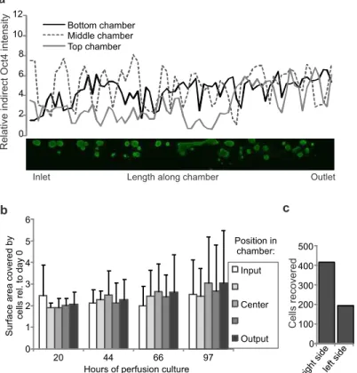
Figure 2-10 Spatial analysis and physical manipulations of cells grown under perfusion ... 41
Figure 2-11 Control experiments under perfusion ... 42
Figure 2-12 An epiblast-like state is attained upon cell-secreted factor removal ... 44
Figure 2-13 Signaling characteristics of perfused cells ... 48
Figure 2-14 Self-renewal ability of cells grown under perfusion ... 49
Figure 2-15 Perfusion blocks secreted factors that maintain self-renewal ... 50
Figure 3-1 Adding proteins under perfusion ... 56
Figure 3-2 Induction of mesoderm genes under perfusion ... 57
Figure 3-3 Representative fluorescent images of Brachyury-GFP embryoid bodies ... 58
Figure 3-4 Perfusion chamber modeling results ... 60
Figure 3-5 Comprehensive steady-state modeling results ... 61
Figure 3-6 Inhibiting ERK signaling under perfusion ... 62
Figure 4-1 Disruption of heparan sulfation inhibits differentiation ... 68
Figure 4-2 Effects of collagenase addition under perfusion ... 69
Figure 4-3 mRNA expression levels of structural ECM genes ... 70
Figure 4-4 mRNA expression levels in the absence of LIF and BMP4. ... 71
Figure 4-5 Fold increase in growth of replated cells ... 72
Figure 4-6 Embryoid body differentiation after manipulation of exogenous signaling ... 73
Figure 4-7 mRNA expression levels of MMPs ... 74
Figure 4-8 MMP production and secretion ... 75
Figure 4-9 MMPs are functional in mESC cultures ... 76
Figure 4-10 ECM-based and secreted endogenous signals ... 76
Figure 5-1 MMP secretion by feeder cells ... 82
Figure 5-3 Acute effects of collagenase addition ... 84
Figure 5-4 Contributions of other MMPs ... 85
Figure 5-5 High-throughput RNA-sequencing data ... 86
Figure 5-6 Inducible MMP1a-overexpressing cell line ... 87
Figure 5-7 Long-term maintenance of self-renewal by MMP1 addition ... 88
Figure 5-8 Embryoid body mRNA expression level timecourse ... 89
Figure 5-9 Signaling pathways not involved in MMP-mediated self-renewal ... 90
Figure 5-10 Wnt signaling is not involved in MMP-mediated self-renewal ... 91
Figure 5-11 Signals directly downstream of LIF are active in MMP-mediated self-renewal ... 92
Figure 5-12 Stat3 is activated with addition of MMP ... 93
Figure 5-13 Stat3 signaling is required for MMP-mediated self-renewal ... 94
Figure 5-14 Upstream ligand is not a known gp130 ligand ... 96
Figure 5-15 Model depicting the effect of matrix remodelling on mESCs ... 97
Figure 6-1 Selective tethering of proteins to hydrogels ... 104
Figure 6-2 Optimization of concentrations and times for hydrogel preparation ... 105
Figure 6-3 PEGDA structures around mESC colonies ... 105
Figure 6-4 Quantification of Oct4 fluorescence in the presence or absence of tethered LIF ... 106
Figure 6-5 Techniques for sorting adherent cells by morphology ... 107
Figure 6-6 Radical-activated sorting of mESCs. ... 108
Figure 6-7 Polymerization-activated sorting of mESCs.. ... 109
Commonly used abbreviations
BMP4 – Bone morphogenetic protein 4EB – Embryoid body ECM – Extracellular matrix
ERK – Extracellular signal-regulated kinase ESC – Embryonic stem cell
EpiSC – Epiblast stem cell FGF – Fibroblast growth factor GFP – Green fluorescent protein GSK3 – Glycogen synthase kinase 3 HSPG – Heparan sulfate proteoglycan ICM – Inner cell mass
JAK – Janus kinase
LIF – Leukemia inhibitory factor MEF – Mouse embryonic fibroblast MMP – Matrix metalloproteinase
PD03 – PD0325901, a MEK/ERK inhibitor PDMS - Polydimethylsiloxane
PEGDA – Polyethylene glycol diacrylate
STAT3 – Signal transducer and activator of transcription 3 TGFβ – Transforming growth factor, beta
Chapter 1
Introduction
1.1 Significance
Cell phenotype is defined in part by the extracellular signals encountered by the cell, whether it is a cell growing in culture, a cell growing in the body performing normal functions, or an abnormal cell that exploits extracellular signals to disrupt cellular functions. The in vivo extracellular microenvironment, or niche, consists of basement membranes and extracellular matrix, cell-cell contacts, and soluble signals that travel from other cells throughout the body. The niche is particularly important for stem cell microenvironments, as subtle signal changes can lead to significant downstream fate adjustments. Understanding the composition and contributors to the in vivo niche is required for successful recapitulation of stem cell microenvironments in vitro.
In recent years, the ability to grow and maintain many diverse types of cells in culture has significantly advanced due to technical developments allowing for more sophisticated cell culture systems. Tools that can successfully manipulate the in vitro microenvironment can be used to learn more about how extracellular signals act to regulate fate choice and thus provide platforms to further our understanding of the biological and functional impacts of particular signals. This information can be useful for generating in vitro models of in vivo events and for establishing conditions for maintenance of stem cells as multipotent entities in vitro.
In this thesis, we use embryonic stem cells (ESCs) as an unbiased model stem cell system and apply various methods to disrupt the endogenous extracellular microenvironment in an effort to learn more about how embryonic stem cell-secreted signals contribute to the remarkable ability of these cells to self-renew indefinitely while retaining the ability to differentiate into any cell type found in an adult organism. Our results highlight the importance of the cell-secreted microenvironment on stem cell growth, self-renewal, and differentiation, and show that cell fate can be strongly influenced by altering microenvironmental cues. The utility of this work lies in its broad applicability to other stem cell systems, its use as a potential tool to direct differentiation or maintain self-renewal, and its uncovering of novel endogenous mechanisms used by stem cells in culture to balance differentiation and self-renewal cues.
1.2 Embryonic stem cells
ESCs are pluripotent cells—they are able to differentiate into all lineages—derived from the inner cell mass (ICM) of a blastocyst. In addition to being pluripotent, they are also able to self-renew by dividing while maintaining their pluripotent state, or to differentiate by dividing and exiting their pluripotent state to adopt other phenotypes. ESCs have been derived from several mammals (Evans and Kaufman, 1981; Martin, 1981; Thomson et al., 1998, 1995; Iannaccone et al., 1994; Hayes et al., 2008; Schneider et al., 2007), but mouse (Evans and Kaufman, 1981; Martin, 1981) and human (Thomson et al., 1998) ESCs (mESCs and hESCs, respectively) are most commonly studied. ESCs are readily available from cell banks and can be derived de novo, are relatively straightforward to culture, and can divide indefinitely in culture without losing their pluripotency. Pluripotent stem cells can also be derived from somatic cells by reprogramming, which has been shown for both mouse (Takahashi and Yamanaka, 2006; Wernig et al., 2007; Okita et al., 2007) and human (Yu et al., 2007; Takahashi et al., 2007). These cells, called induced pluripotent stem (iPS) cells, share most features with ESCs and can be derived from patient-specific backgrounds. Because they can be generated from cells with specific disease backgrounds, iPS cells provide an opportunity to study disease progression from development onward. The ability of pluripotent cells to differentiate into all tissues in the adult explains their clinical significance, in that they have the potential to form cells or tissues for regenerative medicine.
Self-renewal is the most fundamental process that ESCs undergo, and is interesting both from a basic cell biological view (i.e., how do ESCs decide to stay as ESCs) and biotechnological (i.e., how do we design bioprocesses that allow expansion of ESCs to therapeutic scales while maintaining pluripotency). Thus, significant effort has been expended over the last 30 yrs to understand ESC self-renewal, resulting in the identification of exogenous signals important for this process. ESCs are derived from mouse at embryonic day 3.5 (E3.5), at which point the inner cell mass is formed but not organized and implantation has not yet occurred. By E4.5, the embryo has segregated to form the trophectoderm, epiblast, and primitive endoderm, and begins the process of attachment and implantation. By E6.5, the embryo is implanted and gastrulation begins, whereby the epiblast forms the primitive streak and generates cells of the mesoderm and endoderm lineages. The epiblast itself goes on to generate cells of the ectoderm lineage, which
differentiate into epidermal and neuronal cells, while the mesoderm forms cells of the circulatory system and muscle, and endoderm forms the gut, liver, pancreas and lungs.
It was recently demonstrated that pluripotent cells could be isolated from the mouse later than E3.5 when epiblast stem cells (EpiSCs) were isolated from the post-implantation epiblast (E5.5-5.75) (Tesar et al., 2007; Brons et al., 2007). While EpiSCs can differentiate into tissues from all three germ layers, they cannot contribute to all tissues in the developing organism after injection into a blastocyst, indicating that they are not fully pluripotent. mEpiSCs and mESCs also have different exogenous requirements for maintaining their self-renewal due to their different origins, and mEpiSCs more closely resemble hESCs in terms of morphology and self-renewal requirements, indicating that hESCs may be derived from a human developmental stage equivalent to the E5.5 stage in mice.
The work done in this thesis is centered around mESCs, which are conventionally maintained on a mouse embryonic fibroblast feeder layer with addition of leukemia inhibitory factor (LIF) and serum (Nichols et al., 1990). Many ESC lines have been adapted to not require a feeder layer, but are grown in media with LIF and serum. Various types of serum-free media have also been developed, and the variant I refer to in this thesis is N2B27 media supplemented with LIF and bone morphogenetic protein 4 (BMP4) (Ying et al., 2003b). The common component among all these methods for maintaining mESCs in a self-renewing state is LIF, a cytokine originally isolated based on its ability to inhibit growth and induce differentiation into macrophages in the mouse leukemic cell line M1 (Hilton et al., 1988). LIF was found to share this ability with a previously identified cytokine, IL-6, and it was thus found that the two signals shared a common receptor component in gp130 (Gearing et al., 1992; Ip et al., 1992). Along with gp130, the LIF receptor forms a heterodimer that allows the LIF signal to be transmitted internally. This family of cytokine receptors was found to activate tyrosine kinase activity when stimulated, and the specific tyrosine kinases that bind to dimerized gp130 are in the Janus kinase (JAK) family (Silvennoinen et al., 1993). JAK family tyrosine kinases then go on to phosphorylate signal transducer and activator of transcription (Stat) family proteins, the first identified being Stat3, which is phosphorylated and translocates into the nucleus within minutes after gp130 activation (Wegenka et al., 1993). For mESC maintenance, Stat3 is the most relevant Stat family member downstream of LIF, as it acts as a transcription factor to regulate genes necessary for the
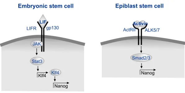
maintenance of mESC self-renewal, including Klf4 and Nanog (Darnell, 1997; Jiang et al., 2008) (Figure 1-1).
In contrast to the signaling elements that act to maintain mESC self-renewal, the primary extracellular signaling requirements for both mEpiSCs and hESCs are fibroblast growth factor 2 (FGF2) and Activin/Nodal/TGFβ family ligands. FGF2 is typically added to mEpiSC cultures to maintain their self-renewal, and EpiSCs have been shown to also require autocrine Activin/Nodal signaling, which signals through Smad2/3 to activate self-renewal signals, including Nanog (Greber et al., 2010) (Figure 1-1). FGF2 is also added to hESC cultures, where it may act to reinforce autocrine production of FGF2 (Eiselleova et al., 2009). TGFβ/Activin signaling has also been proposed as an autocrine loop for maintenance of hESC self-renewal (Xu et al., 2008), and Activin addition may induce autocrine FGF2 production in hESCs (Xiao et al., 2006). GDF3, also in the TGFβ superfamily, is secreted from hESCs and acts to block BMP and thus inhibit differentiation (Levine et al., 2009), while autocrine levels decrease during differentiation (Levine and Brivanlou, 2006). Though the extracellular regulatory processes are different in mouse and human ESCs, likely due to differences in the developmental stage at which they are isolated, both types of cells share some elements of the core self-renewal regulatory circuitry, including autoregulatory control by the transcription factors Oct4, Sox2, and Nanog (Boyer et al., 2005).
Nanog gp130 JAK LIFR Stat3 Klf4 Klf4
Embryonic stem cell Epiblast stem cell
LIF Nanog Smad2/3 Activin ALK5/7 ActRII
Figure 1-1 Model figure depicting major pathways by which transcription of the crucial self-renewal transcription factor Nanog is upregulated in mouse embryonic (left) and epiblast (right) stem cells.
Neither mouse nor human ESCs are homogenous under normal culture conditions. Part of the mechanism by which exogenous FGF2 acts to maintain hESC cultures is thought to be due to a paracrine loop that forms after some fraction of the population differentiates to a more fibroblast-like state that then secretes IGF-II in response to exogenous FGF2, which in turn serves to keep the remaining hESCs in a self-renewing state (Bendall et al., 2007). mESC cultures are also heterogeneous, with cells maintaining a balance between naïve and primed states, the latter of which allows the cells to be ready for differentiation. Cells in the naïve state express specific ESC markers such as Klf4 and Rex1, while primed cells have markers more similar to that of the epiblast stem cell state, including FGF5 and Dnmt3b (Lanner et al., 2010; Kunath et al., 2007). This heterogeneity has been shown to be functional, as in the case of Rex1 high- and low-expressing mESC populations with different differentiation potentials after FACS separation (Toyooka et al., 2008), which was also seen for the self-renewal marker Nanog (Kalmar et al., 2009) and the visceral endoderm marker Hex (Canham et al., 2010). Maintaining a balance between the naïve and primed states in mESC culture is thought to be important for maintenance of pluripotency, as cells trapped in a naïve state are unable to properly differentiate to all three germ layers (Kunath et al., 2007; Lanner et al., 2010).
Besides addition of LIF, other culture systems have also been shown to maintain mESC self-renewal, the best characterized being addition of inhibitors of the FGF4-ERK signaling pathway and glycogen synthase kinase 3 (GSK3) (Ying et al., 2008), but the maintenance of self-renewal under these conditions is still enhanced by addition of LIF, a media formulation known as 2i/LIF. We will not fully understand the process of self-renewal until we determine the signals that are both necessary and sufficient to maintain self-renewal. Embryonic stem cells are a useful model cell type in which to study microenvironmental cues, as these cells have a dynamic and robust capacity for intercellular signaling, and functional assays can be performed to convincingly test both necessity and sufficiency of a particular signaling environment to maintain self-renewal and pluripotency.
1.3 Cell-secreted microenvironment
To test sufficiency of signals for any cellular process, it is imperative to either know the identity of or develop the ability to control the relevant signals secreted from the cells themselves. One
important type of cell-secreted signals are soluble signals that travel through the culture media to signal to other cells in the environment, but cells can also signal through direct contact with other cells or by extracellular matrix-based signals.
Soluble signals consist of autocrine or paracrine signals, which canonically refer to signals produced by cells to which they respond (autocrine) or to which neighboring cells respond (paracrine) (Sporn and Todaro, 1980). Here, I will use the term autocrine signaling to refer to signals secreted by a cell that may bind to that cell or to a neighboring cell of similar phenotype, while paracrine signaling refers to signals produced by a cell to which that cell type cannot respond, but other cell types can. These soluble signals can include growth factors, which are defined as having a positive effect on proliferation and/or differentiation, cytokines, which constitute other signaling molecules with diverse functions in intercellular communications, and hormones, which are generated at specific sites in vivo and typically act in a limited range with tightly controlled secretion levels. Different types of soluble signals also have different rates of uptake, as the number of cytokine receptors on the cell surface is usually on the order of 102 to 103, a hundred times less than that of hormone or growth factor receptors (Kishimoto, 2005). Contact-mediated signals, also known as juxtacrine signals, require two adjacent cells to be in contact, as these types of signals are not secreted extracellularly from the producing cell. Such signals can be transmitted through transmembrane receptors or through membrane channels. Notch signaling is an example of a juxtacrine mechanism in which the ligand and receptor are both transmembrane proteins such that cells must be in direct contact for signaling to occur. Notch is activated by Delta, Jagged, or Serrate proteins in an adjacent cell and is then cleaved, at which point it translocates to the nucleus and binds and activates transcription factors (Schroeter et al., 1998). Small soluble signals (less than 15 kD) can also be directly transmitted between cells through gap junctions made up of connexin proteins that create a pore between cells through which ions can freely pass (Peracchia and Dulhunty, 1976).
Once adherent cells are attached to a substrate, they begin to produce an extracellular matrix (ECM) consisting of many types of signals that become an important part of the extracellular microenvironment. The ECM includes structural proteins, connective proteins, glycoproteins, signaling proteins, etc., all contained in a dense and dynamic structure. Fibronectin is a large
glycoprotein that serves as a general adhesive molecule, linking cells to their substrate and to each other (Yamada and Olden, 1978). The extracellular matrix present in vivo surrounding epithelial tissue generally includes a dense region called the basal lamina, which consists of laminin and collagen along with glycoproteins such as heparan sulfate proteoglycans (Sanes et al., 1990; Jenniskens et al., 2000). Heparan sulfate proteoglycans (HSPGs) are important ECM components that are thought to act as a reservoir of signaling proteins such as growth factors (Hynes, 2009), and also affect the signaling of molecules such as Wnt, vascular endothelial growth factor (VEGF) and fibroblast growth factor (FGF) (Rosen and Lemjabbar-Alaoui, 2010). The developmental signal sonic hedgehog binds vitronectin in the ECM (Pons and Martí, 2000), and core ECM components such as integrins and laminins have been shown to act as signaling molecules themselves. Integrins are essentially transmembrane fibronectin receptors that bind to cytoskeletal proteins on the inside of a cell, thus integrating the extracellular and intracellular scaffolds (Tamkun et al., 1986).
In addition to acting as a substrate on which cells can grow and directly or indirectly providing signals from its structural molecules or by binding growth factors, the ECM has physical properties that can also influence cells. For uncommitted progenitor cells, the stiffness of the matrix has been shown to influence differentiation trajectory in a variety of systems, including for mesenchymal stem cells (Engler et al., 2006), neural stem cells (Keung et al., 2011), and embryonic stem cells (Chowdhury et al., 2010), and increased matrix stiffness has also been implicated in cancer cell migration and proliferation (Ulrich et al., 2009). Manipulating the rigidity of the ECM can alter availability of autocrine signals, as seen with myofibroblasts and autocrine TGFβ accessibility (Wells and Discher, 2008). ECM composition is also affected endogenously by cell-secreted proteinases, including matrix metalloproteinases (MMPs), a disintegrin and metalloproteinase with thrombospondin motifs (ADAMTS) family proteins, and serine proteases, which have the potential to affect any of the ECM-based signaling mechanisms described above. Of these, the MMP family includes the major secreted proteins that dynamically regulate the ECM both in vitro and in vivo during development (Vu and Werb, 2000), the significance of which will be described in the next section.
1.4 Embryonic stem cell and in vivo microenvironment: soluble and ECM-based signals
Proper embryonic development requires specific regulation of a series of signaling events emanating from within the embryo or maternally. At early stages of development, many of these signals are identical to the ones used for maintenance or early differentiation of embryonic stem cells cultured in vitro. Here, we detail what is known about the functionality of ESC in vitro endogenous extracellular signals and then describe several relevant examples of signaling events required for proper development in the early embryo.
Embryonic stem cells divide rapidly and secrete high levels of proteins, both in terms of soluble ligand secretion and extracellular matrix formation. Several mESC autocrine factors have been identified that influence proliferation (Figure 1-2), including Activin, Nodal and Cyclophilin A (Ogawa et al., 2007; Mittal and Voldman, 2011). LIF has also been shown to act in an autocrine fashion (Davey et al., 2007; Davey and Zandstra, 2006; Zandstra et al., 2000), though not at levels sufficient to maintain self-renewal, while autocrine Wnt signaling has been shown to be required but not sufficient for mESC self-renewal (ten Berge et al., 2011). Autocrine differentiation-inducing signals have also been identified, the most studied example being FGF4, which signals through extracellular signal-regulated kinase 1 and 2 (ERK1/2). FGF4-null mES cells were found to be more likely to express pluripotency markers and less likely to express differentiation markers, and a similar phenotype was observed in ERK2-/- cells (Kunath et al., 2007), while disruption of both FGF4 and ERK signaling in the absence of LIF caused cells to stagnate in a reversible primitive ectoderm state in which no further differentiation is possible (Stavridis et al., 2007). At the transcriptional level, the ERK pathway has been implicated in the repression of the pluripotency-related transcription factor Nanog (Hamazaki et al., 2006). Many of the functional identified autocrine signals acting in hESCs differ from those in mESCs, which have been described in chapter 1.2 and are also summarized in Figure 1-2.
Autocrine factor Function Works through
Fgf4 Differentiation ERK Activin/Nodal Growth Smad2/3 Cyclophilin A Growth Unknown Wnt Self -renewal, Inhibition
of differentiation
GSK3 inhibition
LIF Self -renewal Stat3
Human embryonic stem cell Mouse embryonic stem cell
production of supportive autocrine/paracrine
factors
self-renewal differentiation
growth self-renewal differentiation
Fgf4
Wnt LIF
Gdf3
Figure 1-2 Functional autocrine-acting signals that have been identified in mESCs (a) and hESCs (b) and their roles. While a variety of natural and synthetic matrices have been used to influence ESC self-renewal or differentiation, and ESCs also deposit a rich matrix of their own, the mechanisms behind how extracellular matrix affects cell fate are largely unexplored. Autocrine FGF4 has been shown to require ECM-based HSPGs for signaling to ESCs, and disruption of FGF4 binding leads to increased self-renewal of mESCs (Lanner et al., 2010). Other autocrine signals such as Wnts have also been shown to bind in the ECM and to HSPGs (Schryver et al., 1996; Fuerer et al., 2010), but no functional role for the ECM in mESC autocrine Wnt signaling has been conclusively shown. Mechanically, it has been shown that softer matrices, at similar stiffnesses to mESCs themselves, are able to maintain mESCs in the absence of LIF for multiple passages (Chowdhury et al., 2010), indicating the possibility for a direct mechanical role of ECM in ESC self-renewal, but the mechanism behind this phenomenon is unknown. A direct role for ECM in maintaining renewal has been further characterized in hESCs, where their survival and self-renewal in defined media requires growth on an exogenous ECM, mediated by the binding of endogenous integrins with exogenously supplied vitronectin (Braam et al., 2008). ESCs will differentiate into heterogenous cultures if not passaged regularly, signifying that regular breakdown and rebuilding of the ECM is important in maintaining a more homogenous undifferentiated state.
Autocrine factor Function Works through
Wnt Inhibition of differentiation ß-catenin, potentially others Gdf3 Pluripotency, Inhibition of differentiation BMP inhibition Fgf2 /IgfII (paracrine loop) Differentiation to maintain self -renewal ERK, others
Tgf ß/Activin Self -renewal Smad2/3 CypA Activin/Nodal/TGF Fgf2 Activin/ Nodal Wnt a b ß
Although ESCs do not directly correspond to cells found in a developing embryo, uncovering autocrine signals present in vitro is important for understanding early embryonic development, as the pluripotent state in vivo is transient and proper development requires an exit from this state as a result of maternal and embryonic autocrine and paracrine signaling (Figure 1-3). Much is known about the signals generated in vivo that contribute to embryonic development, implantation, and gastrulation, but many of these studies have been performed in non-mammalian systems, so this section will highlight the relevant soluble and ECM-based signals that contribute to early mammalian embryonic development.
Inner cell mass
Trophoblast Endometrium Paracrine signals Paracrine signals
Figure 1-3 Origin of paracrine signals involved in developmental specification in the early embryo.
In terms of soluble signaling, paracrine communication between cells that make up the inner cell mass and the trophectoderm or between extraembryonic cells and adjacent epiblast cells has been shown to be an important part of early mouse embryonic growth (Murohashi et al., 2010; Mesnard et al., 2011), and blastocyst implantation requires paracrine LIF expression in the uterus (Stewart et al., 1992). Signals within the embryo itself are also important, for example, it has been shown that the anterior visceral endoderm inhibits Nodal signaling to ensure proper formation of the primitive streak during gastrulation (Bertocchini and Stern, 2002; Perea-Gomez et al., 2002). FGF signaling is also important within the embryo, as it is in ESC differentiation. Embryos lacking FGFR1 die at gastrulation (Deng et al., 1994; Yamaguchi et al., 1994), and both FGF4 and FGF8 are required for early embryonic development, as their disruption results in abortive postimplantation development and disrupted gastrulation, respectively (Feldman et al., 1995; Sun et al., 1999). Endogenous FGF signaling has recently been more specifically identified as the signal required for segregation of the primitive endoderm and epiblast cells during ICM maturation (Yamanaka et al., 2010).
Direct cell contacts such as gap junctions are also important in development, for example, before the blastocyst even forms, the first eight blastomere cells are connected by gap junctions, forming physiological compartments within the developing embryo (Kalimi and Lo, 1988). The gap junctions regulate the compaction of the blastomeres to form the blastocyst, as inhibiting connexins inhibits further embryonic development (Lo and Gilula, 1979). Defects in cell-cell adhesion in the early mammalian embryo due to knockout of a specific myosin chain affected production of epiblast-derived cells in the post-implantation embryo and resulted in embryonic death by E7.5 (Conti et al., 2004).
Cell adhesion and migration are essential mechanisms involved in embryonic implantation and gastrulation, and these mechanisms depend on the ability of cells to attach to extracellular matrices. Cell-cell and cell-ECM adhesion molecules that have been shown to act during gastrulation in mouse include E-cadherins and integrins (Hammerschmidt and Wedlich, 2008), and both their extra- and intracellular domains are required for proper gastrulation movements (Lee and Gumbiner, 1995; Kühl et al., 1996). Integrin interaction with fibronectin plays a large role in cell migration that occurs during gastrulation, while other known integrin ligands such as collagens and laminins are expressed at the end of or after this process (Bökel and Brown, 2002). Preceding this, the development of the epiblast prior to implantation requires proper basement membrane assembly between epiblast cells and their underlying epithelial layer (Murray and Edgar, 2000), and integrin- and laminin-deficient cells are unable to form basement membranes and thus do not undergo epiblast differentiation (Li et al., 2002). While ECM may not directly contribute to intracellular signaling during development, it is an important regulator of signals such as those from the TGFβ, Wnt, and Hedgehog protein families, all important secreted mediators of cell patterning and fate determination in early embryos (Brown, 2011). Matrix remodeling is important in vivo as it is in vitro, as MMPs and MMP inhibitors were found to be expressed at high levels in mouse blastocysts and play a role in embryonic implantation (Alexander et al., 1996), with a significant increase in MMP production during the peri-implantation period (Chen et al., 2007).
1.5 Manipulating extracellular signaling
Cell-secreted autocrine and paracrine factors comprise a significant fraction of available soluble signals, particularly in serum-free cultures, but their identity and significance are challenging to study. When specific factors or receptors that are part of an autocrine/paracrine signaling pathway are known, the best way to investigate the contributions of the pathway is via inhibition with knockout cell lines or specific inhibitors. Many small molecule inhibitors have been identified that are specific to receptors or downstream signaling molecules, and blocking antibodies can be developed to target known proteins or receptors. Cell lines can also be derived with specific secreted proteins or receptors knocked out. With these reagents, one can perform the definitive experiments to identify and characterize an autocrine loop (Figure 1-4a). By measuring ligand in the media and characterizing phenotype with and without receptor-blocking antibody, one can determine that (1) the cells are secreting ligand, (2) the ligand binds to the receptor, and (3) ligand binding alters phenotype (DeWitt et al., 2001; Joslin et al., 2007). Importantly, all these methods are limited to studies of known factors.
phenotype phenotype
normal loop blocked
reaction diffusion convection source sink a b Figure 1-4 Methods for investigating autocrine/paracrine signaling. (a) As autocrine factors are secreted from a cell, they bind to receptors to trigger a downstream response (left), unless the receptor is blocked, in which case autocrine signals accumulate in the surrounding media (right). (b) Transport modes for cell-secreted soluble signals, including diffusion, reaction, and convection of ligands from sources to sinks.
What is currently known about autocrine signaling in ESCs has primarily been determined using these methods. For example, when autocrine Wnt signals were identified to be necessary for mESC self-renewal, it was found that addition of either a Wnt antagonist or an inhibitor of Wnt signal production were able to halt self-renewal, an effect that could be reversed with exogenous Wnt addition (ten Berge et al., 2011). An alternative to varying cell density is to use conditioned media assays. Experiments in which media that has been exposed to one population of cells to condition it (i.e., to load it with cell-secreted factors) and then transferred to a separate cell population have been used in studies of both autocrine and paracrine signaling. In studies of autocrine signaling, conditioned media can be used as a surrogate for cell density, as cells can be
grown sparsely and then conditioned media added to simulate culture at high density; this approach has been used to find that autocrine factors are important for maintenance of a short G1 cell cycle phase in hESCs (Becker et al., 2010). Conditioned media studies are even more powerful when studying paracrine signaling. Many in vitro protocols for differentiation of ESCs rely on conditioned media or on co-culture with other cell types (Banerjee et al., 2011; Kawasaki et al., 2000; Lam et al., 2010). One common method involves use of a transwell, an insert that allows paracrine signals to pass between cells cultured in a single well but separated by a protein-permeable membrane. Though useful, conditioned media assays may suffer from inconsistency, as the complement of growth factors present may vary based on cell seeding density, growth time, and preparation and storage of conditioned media.
In addition to these conventional cell culture methods, microtechnologies that enable cell patterning and organization have also been adopted to investigate cell-cell signaling, either within colonies of cells or between colonies of the same or different cell types. Micropatterning has been used to study density-dependent autocrine signaling in ESCs by allowing for control over colony size, which in turn affects ligand source and sink levels. Modulating signaling by altering colony size can help to remove source/sink variations in autocrine signals while also indicating whether cell fate is density-dependent. For example, Peerani and colleagues patterned hESCs into different-sized colonies using microcontact printing and assessed the ESCs’ phenotype using quantitative immunocytochemistry, ultimately implicating endogenous BMP2 and GDF3 as modulators of self-renewal (Peerani et al., 2007). Related studies with mESCs patterned at different colony sizes indicated the importance of endogenous Stat3 activation on self-renewal and showed that transcription downstream of Stat3 can be regulated by colony size (Peerani et al., 2009). Control of cell placement has also been used to manipulate and study contact-mediated signaling with mechanical control and micrometer precision (Hui and Bhatia, 2007), a technique that could feasibly be applied to studies involving stem cells.
In addition to their utility in placing cells to alter levels of secreted ligands, patterning techniques can also be used to pattern islands of specific matrix-associated molecules to limit their exposure within a population. ECM binding of cell-secreted factors limits their diffusion, which can be mimicked by attaching such factors to the surface on which cells are growing. For example, Shh attached to a polymer hydrogel surface was shown to promote the ostogenic differentiation of
mesenchymal stem cells (Ho et al., 2007), while attachment of EGF was able to sustain ERK signaling in these cells to promote cell spreading and survival (Fan et al., 2007). In addition to signaling molecules, structural ECM proteins can also be patterned and shown to be functional, as was shown for neuronal stem cells, which showed enhanced neuronal and astrocytic differentiation on immobilized fibronectin molecules but not laminins (Nakajima et al., 2007). Cell and substrate patterning techniques have also been combined to create experimentally convenient in vitro models of vivo environments. Bio-flip chip cell patterning creates patterns by overturning a cell-loaded microwell array onto a recipient substrate, whereupon the cells fall out of the well and onto the recipient substrate while maintaining their arrangement (Rosenthal et al., 2007), and this technique has been combined with stenciling to pattern mESCs along with other cell populations found in the early embryo to create developmental models to study early embryonic patterning events in vitro (Toh et al., 2011).
The endogenous extracellular matrix can itself be disrupted to determine its broad roles or the roles of specific molecules or classes of molecules, though relatively few studies exist that specifically probe endogenous matrix functionality. The disruption of proper heparan sulfate proteoglycan sulfation has been shown to cause neonatal lethality in vivo and to inhibit the ability of mESCs to differentiate properly in vitro (Ringvall et al., 2000; Lanner et al., 2010). As mentioned above, HSPG sulfation is known to be important for its binding function of many cytokines and growth factors (Bernfield et al., 1999), and the inability of ESC developmental progression in the absence of this function was primarily attributed to a lack of FGF4 signaling (Lanner et al., 2010). In this case, matrix disruption caused a matrix-based signal to no longer signal properly. Conversely, matrix remodeling can allow for proteins trapped within the matrix to be released, thereby increasing levels of available cell-secreted signals in the extracellular signaling environment (Taipale and Keski-Oja, 1997).
1.6 Microfluidic approaches to study cell-secreted signaling
While conventional approaches provide methods for uncovering the presence of autocrine and paracrine signaling pathways, revealing their importance, and identifying specific intercellular molecules involved, recent technological advances in microfluidic technology have enabled a more precise quantitative understanding of spatial and temporal parameters, thus allowing for
more controlled studies of the cell-secreted signaling environment. This section is adapted from a recently published review article (Przybyla and Voldman, 2012a).
To control cell-secreted signaling, the modes by which secreted molecules are transported in liquids need to be considered. In general, ligand is produced by “source” cells at some rate (molecules/sec), and then can bind to cell surface receptors (reaction sink), diffuse away, or be convected away (e.g., by fluid flow) (Figure 1-4b). To directly control transport of ligand in the media, nondimensional numbers can be used to compare different modes of transport, and thus determine the appropriate microfluidic operation regime. A diffusion velocity can be estimated by D/L, where D is the ligand diffusivity (for a ~20 kD cytokine, D ~ 10-6 cm2/s), and L is a characteristic length (e.g., the chamber height). Similarly, a reaction velocity can be defined as
konRs, where kon is the ligand binding on-rate (in M-1s-1), Rs is the receptor density (in mol/m2), and the convection velocity is simply v, the characteristic fluid velocity in the system. Ratios of these values lead to previously defined nondimensional numbers known as the Peclet number (convection/diffusion, vL/D), the Damkohler group I number (reaction/convection, konRs/v) and the Damkohler group II number (reaction/diffusion, konRsL/D). By altering these transport phenomena, one can alter the balance between diffusion, convection, and reaction, and in turn modulate the activity of autocrine loops to discover their effects on cell state.
Microfluidics allows a decrease of L and the application of v, and thus allows tuning of both diffusion and convection. To decrease soluble signaling, one wishes to decrease the effect of reaction, which can be accomplished by increasing convection. The fundamental requirement of microfluidic systems used for removing soluble signals is that they have some mechanism for exchanging the medium in the culture chamber. Thus, these systems are typically comprised of polydimethylsiloxane (PDMS) microfluidic chambers with inlets and outlets, and often have valves (King et al., 2007; Unger et al., 2000) and debubblers (Kang et al., 2008) to provide additional functionality.
Several microfluidic platforms have been described for the culture of ESCs, primarily to minimize reagent volumes for screens (Kamei et al., 2009; Villa-Diaz et al., 2009). Flow has also been used in microscale cultures to periodically replace the media in cell cultures to minimize nutrient depletion while allowing periodic accumulation of secreted factors, as was shown for
hESCs grown on a feeder layer that required a short pulse of media every 2-4 hours (Korin et al., 2008). Determination of the cell-secreted signals that are sufficient and necessary to maintain ESC self-renewal can also be aided by the precise control afforded by microfluidics. The use of microfluidics to control soluble factor mass transport has been demonstrated for both hESCs (Cimetta et al., 2009) and mESCs (Kim et al., 2006; Blagovic et al., 2011). For hESCs, a system was developed that could be tuned to operate in either a convection- or diffusion-dominated regime, resulting in different percentages of differentiated cells (Cimetta et al., 2009). This effect was primarily attributed to the effects of shear in the convection-dominated regime, but also to a decrease in soluble signaling due to the fact that the relative amount of differentiation was density-dependent.
In our lab, microfluidic techniques for controlling mESC soluble cell-secreted microenvironment were initially developed by Lily Kim, who demonstrated the first continuous, logarithmically scaled perfusion of mESCs for several days and showed that different perfusive flow rates affected mESC growth and colony size (Kim et al., 2006). A perfusion device designed by Lily Kim and Katarina Blagovic for growing mESCs at a single flow rate with two separate media conditions in triplicate chambers was then used to differentiate mESCs toward a neuroectodermal fate under perfusion (Blagovic et al., 2011). The viability of mESCs during this process was found to require cell-secreted factors beyond autocrine-acting FGF4. These studies laid the groundwork for using microfluidic perfusion to test the sufficiency and necessity of cell-secreted factors on the self-renewal of mESCs, which is the basis of much of the work described below.
1.7 Thesis specific aims and overview
In this thesis, I will discuss my work involving manipulating the endogenous embryonic stem cell extracellular signaling environment and assessing the effects on embryonic stem cell fate. These studies involve implementation of conventional techniques and development of novel methods for disruption of ESC-secreted signals, combined with downstream assays of the resulting phenotypic and functional consequences. The work can be broadly categorized into three specific aims, as follows:
1. Determining the effects of globally depleting soluble mESC-secreted signals and exploiting these effects to control ESC fate
2. Assessing the independent contributions of soluble and matrix-based endogenous signals with broad manipulations
3. Identifying and mechanistically analyzing functional endogenous components of the ESC extracellular matrix
In chapter 2, I describe the design and implementation of a microfluidic perfusion device for growth of mESCs under a continuous depletion of cell-secreted factors in serum-free culture, as part of aim 1. I discuss the motivation behind the device’s initial application as studying how cell-secreted signaling influences heterogeneity in mESC cultures. I then show that mESCs exposed to decreased levels of cell-secreted soluble signals exit their self-renewing state and exhibit marker expression and signal responsiveness indicative of a more primed epiblast-like state.
Chapter 3 continues aim 1 by using the information gained from a depletion of ESC signals to manipulate and further characterize cells growing under perfusion. I first show results indicating that rapid directed differentiation is possible under perfusion given the right culture conditions, and I then describe a method for modeling and identifying the signals being removed under perfusion.
In chapter 4, I turn towards different cell-secreted signals, those emanating from the extracellular matrix. To address aim 2, I disrupt soluble signaling using perfusion in combination with broad disruption of the extracellular matrix, and show that these two types of signaling act in generally different pathways and adjust cell fate in different ways. While disrupting soluble signals causes cells to exit their self-renewing state, disrupting ECM-based signals does the opposite, and these opposing actions can be reconciled by realizing the importance of cell-secreted signals that act to disrupt the matrix in normal cultures.
An interesting conclusion from chapter 4 is further probed in chapter 5, as the sufficiency of matrix remodeling is demonstrated by exogenously adding matrix-remodeling proteins and
showing that they are able to maintain long-term LIF-independent mESC self-renewal, a result that I go on to explore mechanistically to address aim 3.
Chapter 6 describes a novel technique and a new application for an existing technique, both aimed at assessing the origin and significance of heterogeneity in mESC populations and within mESC colonies by manipulating or exploiting different aspects of the cell signaling microenvironment, to further address aim 2.
Finally, chapter 7 includes the broad conclusions that can be drawn from this work as a whole and describes several future directions that can be pursued using results and evidence described in the previous chapters.
Chapter 2
Effects of manipulating soluble ESC signaling
under perfusion
2.1 Introduction
Determining the precise contributions of autocrine or paracrine signals to a particular process is difficult if the full complement of signals involved is unknown. Currently, such studies involve varying cell density or patterning cells at specific locations to find density-dependent responses (Lauffenburger and Cozens, 1989; Peerani et al., 2007). However, these methods are often incomplete due to the fact that autocrine loops can be self-sufficient even at clonal density (Van Zoelen et al., 1989). Further complicating matters, fluctuations in autocrine ligand concentration both within colonies and between colonies occur in a culture dish, leading to different levels of downstream pathway activation. Such differences in local concentrations of extracellular signals can cause and be exacerbated by population-wide heterogeneity.
In mESC cultures, heterogeneity is due in part to the fact that ESCs are constantly shifting between naïve and primed states (Chambers et al., 2007; Enver et al., 2009), which have different ligand and receptor expression and production levels. While naïve ESCs are primarily dependent on LIF signaling through Stat3 to maintain self-renewal (Darnell, 1997; Jiang et al., 2008), primed ESCs are closer to being in an epiblast-like state in which they may be more dependent on autocrine-acting Activin for maintenance of self-renewal (Greber et al., 2010). These opposing states cause heterogeneity in ligand production and uptake within mESC cultures, which can lead to difficulties when studying specific cell-secreted signaling pathways. Heterogeneity has been assessed within ESC colonies via specific marker expression (Singh et al., 2007; Toyooka et al., 2008), and this has been quantified in terms of how radial position within a colony relates to marker expression (Davey and Zandstra, 2006; Peerani et al., 2007). Building on these studies, we developed and tested a method to quantify the extent to which diffusible signaling affects heterogeneity in mESC cultures, showing that inhibition of MEK/ERK signaling causes mESC cultures to be more homogenously naïve. Because heterogeneity is due in large part to local differences in the cell-secreted extracellular signaling microenvironment, we sought to deplete cell-secreted signaling in general to determine whether other signals also contribute to heterogeneity.
We use a microfluidic perfusion system in which cells can be cultured under continuous media perfusion to remove cell-secreted soluble signals, thus providing a more neutral background with reduced signaling noise. In this chapter, we describe the characterization of this device in terms of transport parameters and practical operation with cells, and then we show that cells can successfully be cultured in the device. In order to analyze the phenotype of cells grown under perfusion, it is essential to compare them to cells grown in static culture under identical conditions. Thus, all experiments performed using perfusion involve cells grown in the device on polystyrene slides coated with gelatin, and these cells are compared to cells grown in gelatin-coated standard polystyrene culture dishes at identical cell density in cells/mm2. To provide cells with a neutral signaling background, it is also important to use defined culture media, as use of feeder cells or addition of serum would create a noisier background that could obscure the contributions of cell-secreted factors.
Using this device affords us the opportunity to test how a global depletion of ESC-secreted factors affects cells in terms of their fundamental characteristics. We use a combination of population-wide assays to assess mRNA levels and single-cell measurements to quantify relative intracellular levels of a protein of interest to assess the resulting phenotype after cells have undergone perfusion, and compare this to the phenotype seen in static cultures. We can also use this system to add in factors individually or together to test their specific effects in the neutral background of perfusion. Here, we describe how depleting cell-secreted soluble signals in mESC populations compares to inhibiting the specific intracellular signal ERK, and go on to further characterize the cells that result from several days of growth under continuous perfusion.
Sections 2.2, 2.3, 2.5, and 2.6 of this chapter are adapted from Przybyla and Voldman (Przybyla and Voldman, 2012b).
2.2 Microfluidic device specifications and transport parameters
The microfluidic perfusion device used in all subsequent perfusion studies (Figure 2-1) is made from the transparent, biocompatible polymer polydimethylsiloxane (PDMS), a material commonly used for microfluidic cell culture (Meyvantsson and Beebe, 2008) that we have previously shown is suitable for culture of mESCs (Kim et al., 2006). Certain parameters need to be taken into consideration when designing a device and choosing operating conditions in order
to ensure that cell-secreted factors are removed while cells are not being affected in other ways by device operation. To ensure secreted factor removal, we consider the three molecular transport mechanisms that act on secreted molecules, namely convection, diffusion, and reaction (i.e., ligand binding to receptor) (Figure 1-4b). In order for molecules to be removed, convection must dominate over reaction and diffusion. To compare the importance of the different transport mechanisms, we make use of established non-dimensional parameters.
Pe is given by vh/D, where v is a characteristic fluid velocity in the system (in our case, the
average velocity, ~0.0296 mm/s), h is a characteristic length (in our case, half the chamber height, 125 µm), and D is the diffusivity of a relevant molecules (for a ~20 kD cytokine, D ~ 10-6 cm2/s). This results in Pe ~37, where Pe>1 indicates a convection-dominated regime. The ratio of the Peclet number and the Damkohler number Da is given by v/konRs, where kon is the ligand binding on-rate (~106 M-1s-1 for a strong interaction) and Rs is the receptor density (which we take to be ~12 receptors/µm2 for a 8 µm radius cell with ~10000 receptors). This results in Pe/Da of ~1500, indicating that convection dominates over reaction. As medium flows by the autocrine-secreting cells, the axial convection and transverse mass transport will induce a concentration boundary layer above the cells, increasing in thickness along the length of the chamber (Squires et al., 2008). The boundary layer in general will decrease flux to and from the surface, and thus the concentration of secreted factor at the cell surface will be higher at the cell outlet than the inlet. The thickness of the boundary layer in microsystems such as ours generally scales as 1/Pe1/3 and thus will get thinner at higher Pe, while the flux through the boundary layer increases as Pe1/3, therefore motivating operation at high Pe and use of short chambers, along
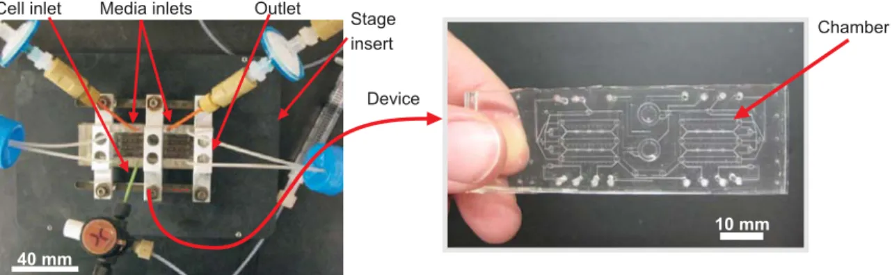
10 mm
Chamber Media inlets Outlet
Cell inlet Stage insert Device { 40 mm
Figure 2-1 Microfluidic device setup. Left panel shows the device, inlets and outlets clamped to an insert for imaging on the microscope. Right panel shows the PDMS device.
with experiments assessing axial heterogeneity. Together, these calculations suggest that secreted proteins that detach from the cell surface will be convected away and not recaptured.
Using these transport parameters, the shear at the culture surface is ~0.007 dynes/cm2, two orders of magnitude lower than what is considered low fluid shear stress for cells (Grabowski and Lam, 1995), and also much lower than the shears of 5-25 dyn/cm2 used to induce ESC-derived endothelial cells to begin expression of endothelial and tight junction markers (Nikmanesh et al.), or to induce endothelial cell-specific genes in mouse embryonic endothelial cells (Egorova et al., 2011). While mESCs have been shown to sense shear stress and respond to it dose-dependently at stresses from 0.016-16 dyn/ cm2 (Toh and Voldman, 2011), ESCs have been grown indefinitely in bioreactors without any effects on self-renewal properties at shears up to 6.1 dyn/cm2 (Cormier et al., 2006; Fok and Zandstra, 2005). In terms of cell removal, the detachment shear stresses for fibroblasts are ~30-50 dyn/cm2 (Crouch et al., 1985), and shear stresses of 5 dyn/cm2 have been applied to an endothelial monolayer for a week without noticeable cell detachment (Dewey et al., 1981), indicating that the shear required to detach adherent cells from substrates are typically >>1 dyn/cm2. For mESCs in particular, removal shear stresses have been reported to be >6.5 dyn/cm2 (Fok and Zandstra, 2005).
Furthermore, our chamber height (250 µm) was chosen to be substantially higher than the colony heights (55 µm) to minimize the effects of cell or colony height or morphology on flow patterns. To account for any flow rate differences in the chambers that could result from the presence of three-dimensional cell colonies, we use a previously described model (Gaver III and Kute, 1998). In general, for a cell or colony whose height is less than 30% of the chamber height (for our 250-µm high chambers, this corresponds to a 75 250-µm high colony), there is only a minor effect (0.05%) on flow rate realized in the system, due to increasing flow resistance from the decreased gap size between the colony and the chamber walls. Cells or colonies present in the chambers will also affect the shear stress, specifically increasing the surface shear stress as compared to the shear stress on a flat surface. However, Gaver and Kute demonstrate that the cells/colonies increase shear by a maximum of 3× with respect to a flat surface when the cell/colony height is <1/4 of the channel height (corresponding to 62 µm for our chambers). We measured colony heights in our chamber using optical microscopy to be 25-55 µm (average 40 µm), smaller than the 62 µm or 75 µm thresholds. Thus, for our chamber geometry, any cells or colonies smaller
than 62 µm in height would negligibly affect the flow and would have surface shear stresses well below shear stresses that have been shown to negatively affect cells.
2.3 Minimal requirements for ESC growth under microfluidic perfusion
To use microfluidic perfusion as a tool with which to characterize the contributions of ESC-secreted signals, it was first necessary to determine the minimal media required to successfully culture healthy cells. As stated above, use of serum-free, defined media was essential to this study, but ESCs generally do not grow as well or as reliably in the absence of serum. While mESCs in static culture can be induced to differentiate into neuronal precursors in defined media known as N2B27 (Ying et al., 2003b), they do not proliferate well in this defined media under perfusion where cell-secreted factors are removed (Blagovic et al., 2011). N2B27 can be used to maintain mESC self-renewal by supplementation with LIF and BMP4, and we found that this minimal defined self-renewing media did allow for growth of mESCs under perfusion (Figure 2-2). Once minimal defined conditions for mESC attachment and survival were established, we went on to show that cell-secreted factors were indeed removed and that cells growing under perfusion did not suffer any acute damage due to culture in the device.
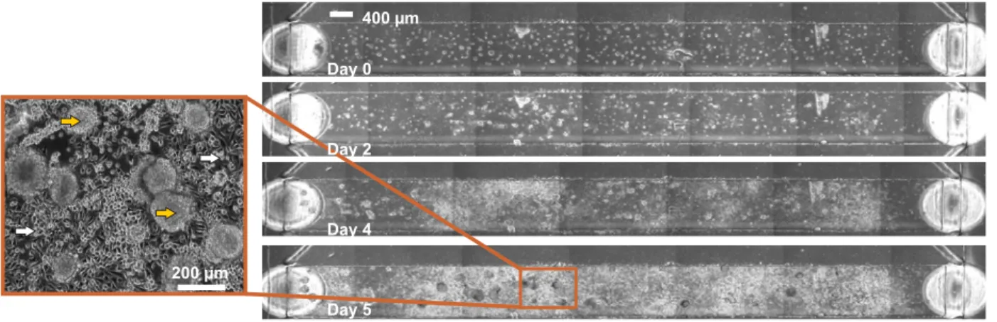
400 µm Day 4 Day 0 200 µm Day 5 Day 2
Figure 2-2 Images of cells growing for the indicated number of days in one chamber of a device. Left panel is a close-up of a portion of the chamber in, where yellow arrows represent colonies with ESC-like morphology, while white arrows represent surrounding differentiated-looking cells.
In order to experimentally verify what our theoretical predictions regarding removal of cell-secreted factors suggest, we sought evidence for soluble factor removal. A molecule known to be secreted at high levels by mESCs (Guo et al., 2006b), VEGF, was collected and levels were measured by ELISA. Interestingly, we found that levels of VEGF collected from cells under perfusion after 30 hours of culture were almost ten-fold higher than those from static cultures, in
both differentiation (N2B27) and self-renewal (N2B27 with added LIF and BMP4) medium (Figure 2-3). The increased VEGF collected from cells under perfusion is consistent with autocrine systems in which the binding of ligand to receptor is blocked (DeWitt et al., 2001). In these systems, where secreted ligand can be recaptured by its receptor, blocking that capture (via blocking antibody/small molecule (Lauffenburger et al., 1998), or in this case, by flow) causes more ligand per cell to be delivered into the bulk media and recovered (Figure 1-4a). These results verify removal and downstream recovery of secreted molecules in this system.
VEGF (fg/cell/day) 0 0.05 0.1 0.15 Perfusion Static differentiation self renewal flow through ** **
Figure 2-3 ELISA measurements of VEGF collected from mESCs cultured in static and perfused systems, either in self-renewal (N2B27+LIF+BMP4) or differentiation (N2B27) environments. Also shown are the VEGF levels measured in systems without cells (“flow through”).
To show that mESCs grown under perfusion do not suffer damage as a result of being grown in the perfusion device, we characterized the cells after three days of perfusion in serum-free self-renewal conditions. We found that cells grew with normal morphology (Figure 2-2), and that expression levels of the early differentiation markers Brachyury and FGF5 were not altered as compared to static self-renewal cultures (Figure 2-4), while those markers did increase in static differentiation conditions (Figure 2-4). Thus, we show that diffusible signaling can be reduced in this system and that cells under short-term perfusion predominantly resemble self-renewing mESCs.
Relative mRNA expression # 0 2 4 50 52 54 Brach Fgf5 Nanog static perfusion static diff # # ##
Figure 2-4 mRNA expression levels of key markers after 3 days of growth in static or perfusion self-renewal culture
or in static differentiation culture.
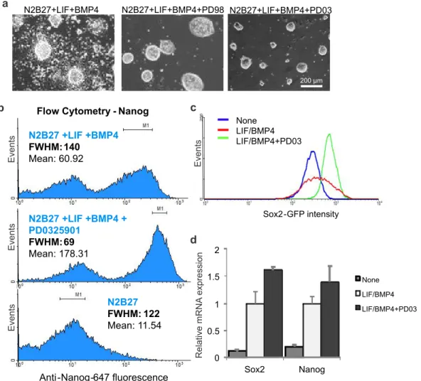
2.4 Adjusting heterogeneity by manipulating exogenous signaling
After establishing conditions appropriate for mESC growth under perfusion, we next wanted to assess the effects of depleting soluble cell-secreted signals. We initially expected such a depletion to cause cells to become more homogenous at a population-wide level, as we assumed the cells would experience less variability in local concentrations of extracellular signals. To develop a system for measuring homogeneity, we initially performed experiments in static culture using the FGF4-ERK signaling pathway as a model system. FGF4 is one of the most well-studied functional autocrine mESC signals, and it signals through FGF receptors to activate ERK, leading to downstream changes consistent with a transition to a primed stem cell state more amenable to ectoderm differentiation (Kunath et al., 2007; Stavridis et al., 2007). It has been shown that blocking intracellular ERK signaling causes mESCs to retain a more undifferentiated morphology and to cause mESC populations to become more homogeneous (Burdon et al., 1999). Consistent with this, continuous blockage of ERK signaling, combined with blockage of signals that inhibit growth, allows for maintenance of mESC self-renewal indefinitely over repeated passage (Ying et al., 2008). We also show that blocking ERK activity with two different MEK inhibitors, PD98059 and PD0325901, reduces mESC morphological heterogeneity, even in serum-free cultures (Figure 2-5a). Because MEK and ERK are the major downstream signals of extracellular FGF4 (Kunath et al., 2007), these data indicate that