HAL Id: hal-02912692
https://hal.archives-ouvertes.fr/hal-02912692
Submitted on 5 Nov 2020HAL is a multi-disciplinary open access archive for the deposit and dissemination of sci-entific research documents, whether they are pub-lished or not. The documents may come from teaching and research institutions in France or abroad, or from public or private research centers.
L’archive ouverte pluridisciplinaire HAL, est destinée au dépôt et à la diffusion de documents scientifiques de niveau recherche, publiés ou non, émanant des établissements d’enseignement et de recherche français ou étrangers, des laboratoires publics ou privés.
Therapeutic potential of prenylated stilbenoid
macasiamenene F through its anti-inflammatory and
cytoprotective effects on LPS-challenged monocytes and
microglia
Veronika Leláková, Sophie Béraud-Dufour, Jan Hošek, Karel Šmejkal, Vilailak
Prachyawarakorn, Phanruethai Pailee, Catherine Widmann, Jiří Václavík,
Thierry Coppola, Jean Mazella, et al.
To cite this version:
Veronika Leláková, Sophie Béraud-Dufour, Jan Hošek, Karel Šmejkal, Vilailak Prachyawarakorn, et al.. Therapeutic potential of prenylated stilbenoid macasiamenene F through its anti-inflammatory and cytoprotective effects on LPS-challenged monocytes and microglia. Journal of Ethnopharmacology, Elsevier, 2020, pp.113147. �10.1016/j.jep.2020.113147�. �hal-02912692�
Therapeutic potential of prenylated stilbenoid macasiamenene F through its anti-1
inflammatory and cytoprotective effects on LPS-challenged monocytes and microglia 2
3
Veronika Lelákováa,b,*, Sophie Beraud-Dufoura, Jan Hošekc, Karel Šmejkald, Vilailak
4
Prachyawarakorne, Phanruethai Paileee, Catherine Widmanna, Jiří Václavíkd, Thierry
5
Coppolaa, Jean Mazellaa, Nicolas Blondeaua,$, and Catherine Heurteauxa,$
6 7
aUniversité Côte d’Azur, CNRS, IPMC, Sophia Antipolis, F-06560, France;
8
lelakova@ipmc.cnrs.fr, beraud@ipmc.cnrs.fr, widmann@ipmc.cnrs.fr, coppola@ipmc.cnrs.fr,
9
mazella@ipmc.cnrs.fr, blondeau@ipmc.cnrs.fr, heurteau@ipmc.cnrs.fr
10
bDepartment of Molecular Biology and Pharmaceutical Biotechnology and dDepartment of
11
Natural Drugs, Faculty of Pharmacy, Masaryk University, Palackého tř. 1946/1, CZ-612 00
12
Brno, Czech Republic; karel.mejkal@post.cz, jivac@seznam.cz
13
cDivision of Biologically Active Complexes and Molecular Magnets, Regional Centre of
14
Advanced Technologies and Materials, Faculty of Science, Palacký University in Olomouc,
15
Šlechtitelů 27, CZ-783 71 Olomouc, Czech Republic; jan.hosek@upol.cz
16
eChulabhorn Research Institute, Kamphaeng Phet 6 Road, Laksi, TH-10210 Bangkok,
17
Thailand; vilailak@cri.or.th, pruethai@cri.or.th.
18 19
$These co-authors share the position of last author.
20 21
*Corresponding author: Veronika Leláková
22
CNRS, IPMC, 660 route des Lucioles, Sophia Antipolis,
23 06560, Valbonne, France. 24 Tel.:+33493953406, +420773105951 25 Email: lelakova@ipmc.cnrs.fr 26
ABSTRACT 27
Ethnopharmacological relevance— Macaranga Thou. (Euphorbiaceae) is a large genus that 28
comprises over 300 species distributed between Western Africa and the islands of the South
29
Pacific. Plants of this genus have a long-standing history of use in traditional medicine for
30
different purposes, including the treatment of inflammation. Fresh and dried leaves of certain
31
Macaranga species (e.g. M. tanarius (L.) Müll.Arg.), have been used to treat cuts, bruises,
32
boils, swellings, sores and covering of wounds in general. Several reports described
33
Macaranga spp. being a rich source of polyphenols, such as prenylated stilbenes and
34
flavonoids, mostly responsible for its biological activity. Similarly, an abundant content of
35
prenylated stilbenes was also described in M. siamensis S. J. Davies, species recently
36
identified (2001) in Thailand. While the respective biological activity of the prenylated
37
stilbenes from M. siamensis was poorly investigated to date, our recent study pointed out the
38
interest as the natural source of several novel anti-inflammatory stilbenoids isolated from this
39
species.
40
Aim of the study—This work investigated the potential anti-inflammatory effects of the 41
stilbenoid macasiamenene F (MF) isolated from M. siamensis S.J. Davies (Euphorbiaceae) on
42
the lipopolysaccharide (LPS)-induced inflammation of monocytes and microglia, major cells
43
involved in the peripheral and central inflammatory response, respectively.
44
Materials and methods—LPS-induced stimulation of TLR4 signaling led to the activation of 45
inflammatory pathways in in vitro models of THP-1 and THP-1-XBlue™-MD2-CD14 human
46
monocytes, BV-2 mouse microglia, and an ex vivo model of brain-sorted mouse microglia.
47
The ability of the stilbenoid MF to intervene in the IкB/NF-кB and MAPKs/AP-1
48
inflammatory cascade was investigated. The gene and protein expressions of the
pro-49
inflammatory cytokines IL-1β and TNF-α were evaluated at the transcription and translation
levels. The protective effect of MF against LPS-triggered microglial loss was assessed by cell
51
counting and an LDH assay.
52
Results—MF demonstrated beneficial effects, reducing both monocyte and microglial 53
inflammation as assessed in vitro. It efficiently inhibited the degradation of IкBα, thereby
54
reducing the NF-кB activity and TNF-α expression in human monocytes. Furthermore, the
55
LPS-induced expression of IL-1β and TNF-α in microglia was dampened by pre-, co-, or
56
post-treatment with MF. In addition to its anti-inflammatory effect, MF demonstrated a
57
cytoprotective effect against the LPS-induced death of BV-2 microglia.
58
Conclusion—Our research into anti-inflammatory and protective effects of MF has shown 59
that it is a promising candidate for further in vitro and in vivo investigations of MF
60
intervention with respect to acute and chronic inflammation, including potentially beneficial
61
effects on the inflammatory component of brain diseases such as stroke and Alzheimer’s
62
disease.
63
64
Keywords: natural stilbenoids, neuroinflammation, microglia, monocytes, prenyl 65
Abbreviations: 67
Aβ, amyloid beta; AD, Alzheimer’s disease; AP-1, activator protein 1; BBB, blood-brain
68
barrier; COXs, cyclooxygenases; DAMPs, damage-associated molecular pattern molecules;
69
HD, Huntington’s disease; IкB, inhibitor of NF-kappa B transcription factor; IL-1β,
70
interleukine 1 beta; LDH, lactate dehydrogenase; LPS, lipopolysaccharide; MAPKs, mitogen
71
activated protein kinases; NF-кB, nuclear factor of kappa light polypeptide gene enhancer in
72
B-cells; NSAIDs, non-steroidal anti-inflammatory drugs; PAMPs, pathogen associated
73
molecular patterns; PD, Parkinson’s disease; RA, rheumatoid arthritis; SEAP, secreted
74
embryonic alkaline phosphatase; TLR4, toll-like receptor 4; TNF-α, tumor necrosis factor
75
alpha.
1. Introduction 77
Inflammation represents an adaptive reaction of body tissues to invading pathogens,
78
injuries, or endogenous signals from stressed, damaged, or dead cells. It is a physiological
79
process that is beneficial, well-coordinated, and self-regulated. However, when the response
80
to inflammatory stimuli becomes uncontrolled and continuous, chronic inflammatory
81
conditions may develop and lead to severe pathologies (Medzhitov, 2008). Non-resolving
82
inflammation contributes to the pathogenesis of both peripheral (neuropathic pain) and central
83
nervous system diseases, such as stroke, traumatic brain injury, multiple sclerosis,
84
amyotrophic lateral sclerosis, Alzheimer’s disease, Huntington’s disease, Parkinson’s disease,
85
or depression (Degan et al., 2018; Miller et al., 2009; Skaper et al., 2018). Moreover, a
86
prolonged exposure to pathogens or an ongoing systemic inflammation of the kind observed
87
in metabolic diseases such as diabetes or obesity can lead to a chronic state of
88
neuroinflammation and ultimately result in neuronal cell death (Nicolas et al., 2017; Skaper et
89
al., 2018).
90
Unrecovered and repeated acute inflammation, as well as long-term moderate
91
exposure to noxious stimuli at the central or systemic level may result in a chronic state of
92
disease. Nowadays, chronic low-grade inflammation is often related to diseases of modern
93
civilization, such as metabolic syndrome, which is often characterized as a cluster of
94
conditions such as hypertension, hyperglycemia, insulin resistance, obesity, and high levels of
95
serum triglycerides, that increase the risk of diabetes mellitus type 2, cerebrovascular disease,
96
and stroke (Lopez-Candales et al., 2017; Minihane et al., 2015). Some inflammatory
97
autoimmune disorders, such as rheumatoid arthritis, are also based on chronic inflammation
98
(Smolen et al., 2018). Systemic and CNS immune system crosstalk during inflammatory
99
response is a critical consideration not to be overlooked (Minihane et al., 2015). Systemic
100
macrophages and brain microglia together with CNS-associated macrophages play a crucial
role in surveillance of the internal environment and control of homeostasis (Ginhoux and
102
Jung, 2014; Yin et al., 2017). Through toll-like receptors (TLRs), they transmit a signal
103
during bacterial infection, injury, or brain damage, including that triggered by
104
hypoxia/ischemia (Lehnardt et al., 2003).
105
Acute inflammation is often associated with a microbial infection that exemplifies
106
itself in a form such as meningitis (Hoffman and Weber, 2009), bronchitis (Wark, 2008),
107
pneumonia (Percopo et al., 2019), myocarditis (Blauwet and Cooper, 2010), or cystitis
108
(Flores-Mireles et al., 2015). Moreover, Gram-negative bacteria such as Klebsiella
109
pneumoniae, Escherichia coli, or Pseudomonas sp., possess in their outer membrane the
110
endotoxin lipopolysaccharide (LPS), responsible for their pathogenic potential and massive
111
clinical manifestations in humans (Sperandio et al., 2013). High peripheral concentrations of
112
LPS can disrupt the blood-brain barrier (BBB) and promote neuroinflammation (Banks et al.,
113
2015). LPS triggers an innate immune system response through TLR4 receptors linked with
114
several inflammatory signaling pathways leading to the production of cytokines and the onset
115
of inflammation (Pålsson-McDermott and O’Neill, 2004). We therefore used LPS from E. coli
116
to induce TLR4-mediated inflammatory cascades in our experimental set up.
117
Stilbenoids are natural phenolic compounds found in foods (grapes, peanuts, berries,
118
passion fruit, rhubarb), beverages (wine, white tea), and medicinal plants, including Morus
119
spp., Artocarpus spp., and the less well known Macaranga spp. (Dvorakova and Landa, 2017;
120
Hošek et al., 2019). Structurally, they share a common diarylethylene core and differ in its
121
substitution, which may include prenyl group. Stilbenoids serve the plant as phytoalexins,
122
protecting it against infection or harm. The strong anti-oxidant and anti-inflammatory
123
properties of stilbenoids have multiple health benefits. Cardioprotective, neuroprotective,
124
anti-diabetic and cancer preventive properties have been reported (Akinwumi et al., 2018;
125
Dvorakova and Landa, 2017; Eräsalo et al., 2018; Hornedo-Ortega et al., 2018). The
neuroprotective potential of stilbenoids has been tested mostly on neurodegenerative
127
disorders, such as AD. For instance, veraphenol, cis-scirpusin A and oxyresveratrol, stilbenes
128
obtained from Smilax spp., showed significant inhibitory effects in vitro against β-secretase,
129
the enzyme crucially responsible for the amyloid beta (Aβ) formation in AD (Jeon et al.,
130
2007). Other than the most widely known and structurally simple stilbene trans-resveratrol,
131
and its hydroxyderivates oxyresveratrol and piceatannol (found, e.g., in grapes), whose
132
antioxidant properties and therapeutic potential in cardiovascular diseases arouse great
133
interest, have been studied very little to date. Little or no information is available about the
134
biological activities of less-known stilbenoids, including those of the genus Macaranga
135
(Hošek et al., 2019).
136
The genus of Macaranga comprises around 300 species and is native to Africa,
137
Australia, Asia, and various islands of Indian and Pacific Oceans. Some species, such as M.
138
tanarius (L.) Muell. Arg., in Thailand known as "Mek”, have been used in traditional Thai
139
medicine as anti-inflammatory treatment. Whereas the fresh leaves were used to cover
140
wounds, a decoction from the dried root was administered against cough and fever
141
(Kamarozaman et al., 2018; Phommart et al., 2005, Phupattanapong and Wongprasert, 1987).
142
M. sampsonii Hance has been used for the treatment of swellings, cuts and sores (Quynh et
143
al., 2018). The European expeditioners surveying the traditional medicinal plants of Papua
144
New Guinea islands recorded the use of M. aleuritoides F. Muell. fruits and seeds by
145
aborigine people to relieve abdominal pain. Various species of Macaranga were used for the
146
treatment of diarrhea, constipation and stomach complaints in several islands, such as Tonga,
147
Fiji, Java, Philippines, Bougainville Island and Malaysia (Holdsworth et al., 1983; Nick et al.,
148
1995). M. harveyana (Muell. Arg.) Muell. Arg. known as “Loupata” was used in Tonga folk
149
medicine for treatment of obstetric and gynecological disorders, such as secondary
150
amenorrhea, post-partum hemorrhage and abdominal pain (Singh et al., 1984).
Several studies trying to describe the phytochemical profile of Macaranga species
152
identified this genus as a rich source of polyphenols such as prenylated and geranylated
153
stilbenes and flavonoids, terpenes, tannins, steroids and coumarins. Most of the described
154
biological activities are attributed to stilbenes and flavonoids found in high amounts
155
(Magadula, 2014; Ngoumfo et al., 2008).
156 157
M. siamensis S. J. Davies is a large-leaved plant, common in central and northern
158
Thailand, firstly identified in 2001. The first phytochemical investigation provided by Pailee
159
et al., 2015 demonstrated the high content of novel stilbenes, including macasiamenenes A, B,
160
F, K, L, and P that have been isolated from dichloromethane extract of leaves and twigs. But,
161
little information about their biological effects has been reported. Based on our previous study
162
suggesting the anti-inflammatory properties of several prenylated stilbenoids contained in M.
163
siamensis (Hošek et al., 2019), by the present work we undertook to compare the
anti-164
inflammatory effects of seven compounds obtained from Macaranga to identify the
165
therapeutic potential value of the most promising candidate, macasiamenene F (MF), for
166
treatment of both systemic and central inflammation. The characterization of MF led us to
167
describe for the first time its anti-inflammatory effects on microglial inflammation.
168 169
2. Materials and methods 170
171
2.1. Animals 172
Seven week-old male C57BL/6JRj mice (Janvier, France) were housed at 22°C with a
173
12-h light-dark cycle and free access to water and food. All animal care and use were
174
performed in accordance with the policies of European Community Directive 86/609/EEC.
The ex vivo experiments were approved by the local committee. Every effort was made to
176
minimize the number of animals used and their suffering.
177 178
2.2. Isolation of compounds and treatments 179
Pailee et al. (2015) isolated the macasiamenene F (MF) along with L (1), K (2), P (3),
180
A (5), B (6) and 2,6-diprenyl-resveratrol (4) from leaves and twigs of M. siamensis S. J.
181
Davies (Euphorbiaceae) by chromatographic methods and identified them by spectroscopic
182
methods, including NMR analysis. The voucher specimen (No. CRI 466) was deposited in the
183
Laboratory of Natural Products, Chulabhorn Research Institute, Thailand (Pailee et al., 2015).
184
The plant name has been checked with http://www.theplantlist.org. Compound 4,
2,6-185
diprenyl-3,5,4´-trihydroxystilbene (4) was already known (Verotta et al., 2009), but
186
compounds MF, 1–3, 5, and 6 were isolated for the first time. The purity of compounds was
187
confirmed to be more than 98% using HPLC (Figure S1). DMSO was used as a vehicle; its
188
concentration in cell culture never exceeded 0.1%. Apart from the dose-response studies,
189
treatments were performed using appropriate serum-free medium at a concentration of 1
190
µmol/L (0.1 µmol/L for compound 1), which would not affect cell viability. At these
191
concentrations, the viability measured by WST-1 was 100 % compared to non-treated cells
192
(Fig. 1B). LPS from E. coli 0111:B4 (Sigma-Aldrich) freshly dissolved in PBS was used at a
193
concentration of 1 µg/mL (Taka et al., 2015; Triantafilou et al., 2001).
194 195
2.3. Cell culture and differentiation 196
The THP-1 human monocytic leukemia cell line was purchased from the European
197
Collection of Authenticated Cell Cultures (Salisbury, UK). The BV-2 murine microglial cells
198
were generated from primary microglial culture by infection with v-raf/v-myc oncogene
199
carrying J2 retrovirus (Blasi et al., 1990). The THP-1 and THP-1-XBlue™-MD2-CD14
(Invivogen, San Diego, CA, USA) cells were cultured in RPMI 1640 medium containing
201
stabilized 2 mmol/L L-glutamine (Biosera, France), whereas the BV-2 cells were cultured in
202
DMEM (4.5 g/L glucose, 4 mmol/L L-alanyl-glutamine, and w/o sodium pyruvate (Sigma).
203
All of the cultures were supplemented with 10 % fetal bovine serum (FBS, Gibco) and
204
antibiotics [100 U/mL penicillin and 100 µg/mL streptomycin (Gibco)]. Cells were grown in a
205
water-saturated atmosphere of 5% CO2 at a temperature 37°C. The experiments were
206
performed on passages ranging from 3 to 16 for all types of cell lines. The THP-1 monocytes
207
were differentiated into macrophages by adding 50 ng/mL of phorbolmyristateacetate (PMA)
208
stimulation (Leláková et al., 2019). All subsequent experiments with all cell lines were
209
performed after 2 h of incubation in serum-free medium.
210 211
2.4. Sorting of brain immune cells 212
Immune cells were isolated from the brains of adult mice using an Adult Brain
213
Dissociation Kit (Miltenyi Biotec, USA) according to the manufacturer’s instructions. Brain
214
homogenates were filtered using 70 µm cell strainers (BD Biosciences) and centrifuged (10
215
min, 2,000 rpm). The resulting cell pellets were then re-suspended and washed with PBS at
216
pH 7.2 containing 0.5 % BSA and 2.5 mM EDTA. They were then labeled with CD11b+
217
MicroBeads (Miltenyi Biotec, USA) and incubated for 15 min at 4°C. CD11b+ mouse
218
microglia and CNS associated macrophages were isolated using LS columns (Miltenyi Biotec,
219
USA) in a magnetic field according to the manufacturer’s instructions.
220 221
2.5. Ex vivo culture of microglia 222
Brain-sorted microglia were seeded into 96-well tissue culture plate in DMEM
223
(Gibco) containing 10 % fetal bovine serum (FBS, Gibco) and antibiotics [100 U/mL
224
penicillin and 100 µg/mL streptomycin (Gibco)] at a density of 7 × 10⁴ cells/well. Microglial
cells were cultured on poly-L-lysine-covered plates at 37°C in an atmosphere containing 5 %
226
CO2 for 18 h. Thirty minutes after cell plating, MF at a concentration of 1 µmol/L, which has
227
been determined to be non-toxic for both monocytes (Fig. 1) and microglia (Fig. 2), was
228
added together with LPS (1 µg/ml) as a co-treatment. The supernatants and cell contents were
229
collected and used subsequently to measure the levels of cytokines after18 h.
230 231
2.6. Cell viability: Dose-response relationships 232
Undifferentiated floating THP-1 cells in serum-free RPMI 1640 medium were seeded
233
into 96-well plates (5 × 10⁴ cells/well). After 2 h, compounds MF, and 1–6 were added at
234
concentrations ranging from 0.125 to 20 µmol/L. The cell viability was assessed 24 h later
235
using Cell Proliferation Reagent WST-1 kit (Roche Diagnostics, Basel, Switzerland). Based
236
on the resulting viability curves, the IC50 values were calculated according to four parameters
237
logistic (4PL) analysis. In the case of adherent BV-2 cells, the dose-response assays were
238
carried out in exponential phase of growth established by a pilot study in 24-well plates.
239
Briefly, BV-2 cells were seeded in the evening in complete DMEM medium at a density 1.5 ×
240
105 cells/well, the next day washed by PBS, and the medium was replaced by serum-free
241
medium. Cells were treated after 2 h with MF at concentrations ranging from 1 to 15 µmol/L.
242
The mitochondrial activity and cell proliferation were evaluated after 24 h of incubation using
243
a Cell Titer 96® Aqueous One Solution Cell Proliferation Assay (MTS; Promega, France)
244
and manual counting of the living cells excluded from Trypan Blue (Sigma) staining,
245
respectively. Cell mortality was assessed after 24 h using a Cytotoxicity Detection Kit (LDH;
246
Roche) and represented as the percentage of lactate dehydrogenase (LDH) released by
247
damaged cells as compared to cells treated with only the vehicle.
248 249 250
2.7. Determination of NF-κB/AP-1 activity 251
The activity of nuclear factor κB (NF-κB) and activator protein-1 (AP-1) was
252
measured on THP-1-XBlue™-MD2-CD14 cells (Invivogen, CA, USA) derived from THP-1
253
human monocytes. This cell line stably expresses the NF-κB and AP-1 inducible secreted
254
embryonic alkaline phosphatase (SEAP) reporter gene, the activity of which was detected
255
using QUANTI-Blue™ reagent (Invivogen, San Diego, USA). Cells in serum-free medium
256
RPMI 1640 medium were seeded (5 × 10⁴ cells/well, 96-well plate) and incubated for 2 h at
257
37°C. Compounds MF, 2–6, and prednisone (P) used as a positive control, were added at the
258
non-toxic concentration of 1 µmol/L (0.1 µmol/L was used for compound 1), together with
259
vehicle DMSO (0.1 % (v/v)). After 1 h of incubation, the cells were treated with LPS
260
(1 µg/mL) to activate NF-κB/AP-1 and produce SEAP. After 24 h, 20 µL of the supernatant
261
was withdrawn, mixed with 175 µL of QUANTI-Blue™ reagent and incubated for 30 min at
262
37°C. Spectrophotometric measurements at 655 nm were carried out in a Fluostar Omega
263
Microplate Reader (BMG Labtech, Ortenberg, Germany).
264 265
2.8. Molecular docking 266
All of the simulations of ligand-protein docking on NF-κB were performed with
267
AutoDockVina (Trott and Olson, 2010) using a PyRx virtual screening tool. Molecular
268
dynamics were performed with a CUDA version of NAMD (Phillips et al., 2005) and VMD
269
was used to prepare the molecular visualization and protein preparation. Docking simulations
270
were performed using system with Core I7 and NVIDIA GTX 850 in the same manner we
271
described previously (Leláková et al., 2019). The crystallographic coordinates of NF-ĸB
272
bound to DNA were obtained from the RCSB Protein Data Bank with ID 3GUT. Only chain
273
A (p65) and B (50) were used for simulations. All tested compounds meet the conditions of
274
Lipinski rule of five, suggesting that tested compounds can penetrate cell membranes, pass
into cell compartments, and have several chemical groups forming hydrogen bonds. All
276
compounds formed complexes with target protein structure with high binding energy (ΔG)
277
around -7.0 kcal/mol.
278
2.9. Western blot analysis of TLR4-mediated signaling pathways 279
THP-1-XBlue™-MD2-CD14 cells in 6-well plates at a density of 3 × 10⁶ cells/well
280
were treated with MF or the vehicle (Veh). After 1 h, LPS (1 µg/mL) was added to the cells.
281
Cells not stimulated with LPS represented the control (Ctrl). After 30 min, the cells were
282
collected, washed in PBS, and homogenized in cold lysis buffer (50 mmol/L Tris-HCl, pH
283
7.5; 1 mmol/L EDTA; 1mmol/L EGTA; 1 mmol/L sodium orthovanadate; 5 mmol/L sodium
284
pyrophosphate; 50 mmol/L sodium fluoride; 270 mmol/L sucrose) with protease inhibitors
285
(Roche Diagnostics, Basel, Switzerland). Equal amounts of proteins (9 µg) were separated in
286
12 % polyacrylamide gel and transferred to a PVDF (polyvinylidene fluoride) membrane.
287
This membrane was then incubated overnight at 4°C with the following monoclonal primary
288
antibodies: anti-IĸB-α (1:500; Cell Signaling, USA, product No. 4814), anti-β-actin (1:5000;
289
Abcam, Cambridge, UK, No. 8226), anti-SAPK/JNK (1:1000; Sigma-Aldrich, USA,
290
SAB4200176), antiphospho-p44/42 MAPK (p-ERK1/2, 1:2000, No. 4370), anti-p44/42
291
MAPK (ERK1/2, No. 4695), anti-phospho-p38 MAPK, and anti-phospho-SAPK/JNK
292
(1:1000; Cell Signaling, MA, USA, No. 4511 and 4668) and polyclonal antibody: anti-p38
293
MAPK (1:1000; Cell Signaling, MA, USA, 9212). Each membrane was then probed with the
294
appropriate anti-mouse or anti-rabbit IgG secondary peroxidase-conjugated antibodies
295
(1:2000, from Sigma-Aldrich, USA, No. A0168 and A0545). The ECL signal (Bio-Rad,
296
USA) was detected using a Syngene PXi4 chemiluminescence imaging system
297
(Cambridge,UK). Densitometric analysis was performed using AlphaEaseFC 4.0.0 software
298
(Alpha Innotech, USA). The relative effects of MF and vehicle (Veh) on IĸB-α were
299
compared after normalization to β-actin.
301
2.10. Isolation of RNA and qRT-PCR 302
Total RNA was extracted using TRI Reagent® (Sigma), followed by treatment with
303
TURBO DNAse (Invitrogen). First-strand cDNAs were synthetized from 0.5 µg of total RNA
304
using 200U/µL SuperscriptTM IV Reverse transcriptase (Invitrogen) in reaction buffer in the
305
presence of 2.5 µmol/L oligo d(T)20 primers (Eurogentec, France), 0.5 mmol/L
306
desoxyribonucleotide triphosphate (dNTP) mix, and 5 mmol/L dithiothreitol (DTT). The
307
reaction was incubated for 5 min at 65°C, then for 60 min at 50°C, and it was finally
308
inactivated by raising the temperature to 70°C for 15 min. Quantitative RT-PCR was
309
performed using a LightCycler® 480 SYBR Green I Master with a LightCycler® 480 sequence
310
detector (Roche Diagnostics). RPLO and GAPDH were used as reference genes for
311
normalization. The following sequences of primers were used: mIL-1β (forward
5´-312
TGGTGTGTGACGTTCCCATT-3´, reverse 5´-CAGCACGAGGCTTTTTTGTTG-3´),
313
mTNF-α (forward 5´-CATCTTCTCAAAATTCGAGTGACAA-3´, reverse
5´-314
TGGGAGTAGACAAGGTACAACCC-3´), mRPLO (forward 5´- 315
ACTGGTCTAGGACCCGAGAAG-3´, reverse 5´-TCCCACCTTGTCTCCAGTCT-3´), and
316
mGAPDH (forward 5´-CCAGTGAGCTTCCCGTTCA-3´, reverse
317
GAACATCATCCCTGCATCC-3´) (Eurogentec, France).
318 319
2.11. Immunodetection of inflammatory cytokines 320
Differentiated THP-1 macrophages were pretreated for 1 h with the test compounds or
321
prednisone 1 µmol/L (in the case of compound 1 0.1 µmol/L) dispersed in 0.1 % DMSO or
322
with a 0.1 % DMSO solution alone (the vehicle). The release of TNF-α and IL-1β was
323
evaluated by testing the supernatant after 24 h, using Human TNFα and IL-1β ELISA kits
324
(Diaclone, France). For BV-2 cells, the expression of TNF-α and IL-1β was determined by
testing cell contents and supernatants, using AlphaLISA mIL1β (AL503C) and mTNFα
326
(AL505C) kits (PerkinElmer, MA, USA). The Amicon® Ultra 0.5mL filters (10K) for protein
327
concentration (Merck Millipore, France) were used to concentrate supernatants before
328
detection of IL-1β released from BV-2 cells. The total quantities of cytokines produced were
329
calculated and normalized to the amounts of total proteins determined by Bradford protein
330
assay (Biorad, France).
331 332
2.12. Statistical analysis 333
Statistical analysis was carried out using GraphPad Prism 6.00 software. Data were
334
expressed as the mean±SEM. Statistical analyses of differences between groups were
335
performed using the parametric (n>30) Student’s t-test (for comparisons of 2 groups) and
336
non-parametric (n<30) Mann-Whitney (for 2 groups), and Kruskal-Wallis analysis, together
337
with Dunn’s post-hoc test (for more than 2 groups). Values of p less than 0.05 were
338
considered statistically significant.
339 340
3. Results 341
342
3.1. Effects of Macaranga stilbenoids on the viability of THP-1 cells 343
The prenylated stilbenoid MF has been found in the plant M. siamensis S. J. Davies,
344
isolated by chromatographic methods, and identified by NMR analysis and other spectral
345
methods (Pailee et al., 2015). We are the first to evaluate its effects on the viability of THP-1
346
human monocytes. Figure 1B shows that MF displays one of the safest profiles. Its IC50 was
347
calculated at 5.7±1.1 µmol/L. The IC50 values of compounds 1–6 were 1.8±1.1, 3.3±1.1,
348
4.9±1.1, 12.8±1.1, 2.7±1.1, and 3.9± 1.1 µmol/L, respectively (Hošek et al., 2019).
According to previous studies on humans evaluating the impact of oral supplementation of
350
stilbenoids on their circulating concentration in plasma, targeting the concentration of 1
351
µmol/L that is accepted for the most studied stilbene resveratrol as lowest dose with a
352
significant anti-inflammatory effect on peripheral cells (Chen et al., 2018) is a
353
pharmacologically and clinically relevant (Boocock et al., 2007; Smoliga et al., 2011).
354
Therefore to identify the most interesting anti-inflammatory candidate at this dose, we
355
performed a non-exhaustive screening of the anti-inflammatory activities on stimulated
356
macrophages of the seven compounds isolated from the plant M. siamensis.
357 358
3.2. MF reduces the secretion of inflammatory cytokines by acting on the IкBα/NF-кB 359
pathway 360
We first evaluated and compared to prednisone, a corticosteroid used in the
361
management of inflammatory conditions or diseases, the effect of the seven compounds
362
isolated from the plant M. siamensis at the concentration of 1 µmol/L on the release of the
363
pro-inflammatory cytokine TNF-α (Fig. 1C) and IL-1β (Fig. 1D) in PMA-differentiated
THP-364
1 macrophages. Interestingly, MF reduced the secretion of TNF-α from macrophages
365
stimulated by LPS by 22%, which was more than any of the other prenylated Macaranga
366
stilbenoids (range of 3-16%) tested by Hošek et al. (2019). This anti-inflammatory effect of
367
MF was comparable to that of prednisone (24 %), a routinely used corticoid
anti-368
inflammatory drug (Fig. 1C). Moreover, a similar anti-inflammatory effect of MF was also
369
observed on the secretion of IL-1β from macrophages stimulated by LPS (Fig. 1D).
370 371
372 373
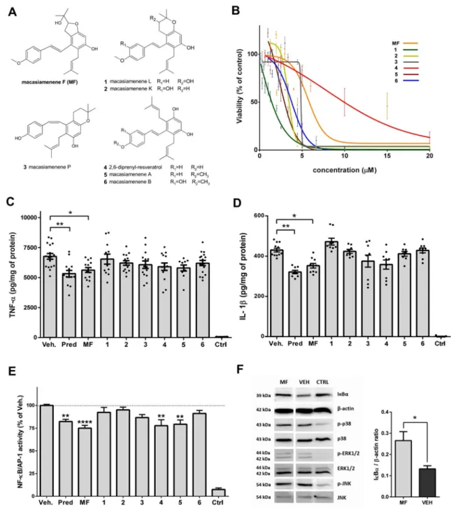
Figure 1: Effects of prenylated M. siamensis stilbenoids on NF-κB signaling of LPS-stimulated 374
THP-1 macrophages and THP-1 XBlue™-MD2-CD14 monocytes. A, Test stilbenoids MF, 1-6; B, 375
Dose-response curves showing viability of THP-1 after a 24 h incubation with compounds MF, 1–6; 376
C, Effect on release of Tα protein; D, Effect on release of IL-1β protein; E, Activation of NF-377
кB/AP-1 after 24 h; F, MF upstream regulation of IкBα. Data are presented as the mean±SEM, n=3, 378
*indicates a significant difference of *P<0.05, **P<0.01, ****P<0.0001 versus only with the vehicle 379
treated cells (Veh) analyzed by the non-parametric tests Kruskal-Wallis in the case of C and D, and the 380
Mann and Whitney for E. 381
We next focused on the regulation of TNF-α and IL-1β upstream, particularly on the
382
transcription factors NF-кB and AP-1, which concomitantly control the expression of several
383
pro-inflammatory cytokines, including TNF-α and IL-1β. The canonical inflammatory
384
pathway that was triggered by TLR4 and led to overexpression of Tα involved
NF-385
кB/AP-1 activation (Lawrence, 2009; MacIntyre et al., 2014). Again in this case MF showed
386
one of the strongest inhibitory effects of the tested prenylated M. siamensis stilbenoids (Fig.
387
1E), with a therapeutic potential greater than that of prednisone. Overall at the clinically
388
transferable dose of 1 µmol/L, the anti-inflammatory activities of compounds 4 and 5 are
389
weaker than MF justifying it to be the most promising candidate for further evaluation of
anti-390
inflammatory and protective effects. Then, to gain some mechanistic insights we investigated
391
MF effect on the upstream activation of NF-кB/AP-1 which includes IкBα (Lawrence, 2009)
392
and MAPKs (MacIntyre et al., 2014). Interestingly, MF reduced the degradation of IкBα, a
393
direct inhibitor of NF-κB, but did not inhibit the activation (phosphorylation) of MAPKs
394
known for leading to the activation of AP-1 (Fig. 1F). Therefore it is tempting to believe that
395
MF could inhibit the inflammation in LPS-stimulated macrophages primarily through
396
IкBα/NF-кB signaling, rather than by acting via the MAPK/AP-1 pathway.
397
Since inflammatory response of NF-κB is driven by its nuclear translocation and its
398
binding activity, interfering with each of these steps or with both may account for MF
anti-399
inflammatory effect. Since IкBα primary role is to bind to NF-κB for preventing its
400
translocation to the nucleus, our results showing MF potential to inhibit IкBα degradation
401
suggests that it could interfere with NF-κB nuclear translocation. Therefore we further
402
investigated whether MF could also directly interfere with NF-κB to dampen its binding
403
activity.
405 406
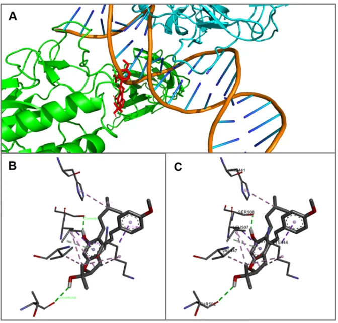
Figure 2: Molecular docking pose of MF interactions with NF-κB (PDB 3GUT). A, The bond of 407
MF to p50 subunit of NF-κB DNA binding pocket. Red - MF, green ribbon – p50 subunit, blue ribbon 408
– p65 subunit, orange double helix with colored sticks - DNA fragment; B, Interactions of MF with 409
p50 NF-κB depicting hydrogen bonds (green–classical, grey–non-classical) and hydrophobic 410
interactions (dark and light purple); C, MF forms interactions with p50 residues Thr502, Ser508 411
(classical hydrogen bonds), Leu507 (non-classical hydrogen bond and hydrophobic interactions), 412
His441, Lys444, and Val447 (hydrophobic interactions). 413
414
Thus, we evaluated the interaction of MF with NF-кB DNA binding site by simulating a
415
ligand-protein docking into DNA binding pocket of the NF-κB heterodimer p65/p50
416
according the method by (Trott and Olson, 2010) (Figure 1A). The hydroxyl groups of MF
tend to form conventional hydrogen bonds (O˗H…O) with Thr502 and Ser508 residues.
Non-418
classical hydrogen bond (N˗H…π) was observed with Leu507. The alkyl hydrophobic
419
interactions were detected with residues His441, Lys444, Val447, and Leu507. Two phenyl
420
rings of MF typical for stilbene core tend to form π-alkyl hydrophobic bonds with Lys444 and
421
Leu507 (Figure 2B and 2C). Overall MF displayed a high affinity (ΔG = -6.7 kcal/mol) for
422
κB DNA binding site similarly to other biologically active stilbenes inhibiting the
NF-423
κB/AP-1 pathway (Leláková et al., 2019). Altogether our results suggest that MF may not
424
only inhibit NF-κB activity by indirectly targeting its translocation preventing IкBα
425
degradation but also directly by interfering with the DNA binding site of NF-κB.
426 427
3.3. Anti-inflammatory dose of MF does not alter the proliferation and viability of BV-2 428
microglial cells 429
We then tried to determine if this anti-inflammatory effect observed on monocytes
430
could be extended to microglia, the immune cells of the brain. To evaluate whether the
anti-431
inflammatory effect of MF could be obtained on the safe side within a concentration range
432
similar to the active one used for monocytes, we first performed a dose-response study to
433
assess whether MF had an effect on the proliferation of BV-2 microglia (Fig. 2A) and their
434
corresponding mitochondrial activity (Fig. 2B), that are accepted indicators of BV-2 vital
435
cellular functioning. Not only changes in mitochondrial activity reflect an alteration of the
436
energy homeostasis of the cell physiology often associated with a switch form physiological
437
to pathological states, but mitochondria alteration is known for triggering inflammation and to
438
play a central role in pro-inflammatory signaling and inflammatory response (Meyer et al.,
439
2018). Therefore it was crucial to know whether and at which dose MF may alter the
440
physiological functioning of BV-2. The high dose of 15 µmol/L altered the proliferation and
441
activity of BV-2, but doses below 6 µmol/L had no negative effect (Fig. 2A-B).
443 444
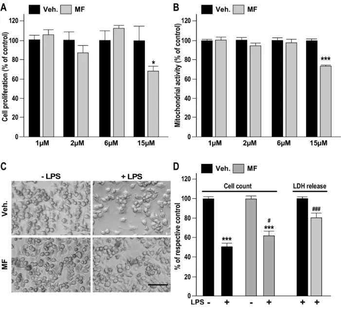
Figure 2: Determination of the dose-limiting toxicity of MF and its protective effect against 445
inflammatory challenge in BV-2 cells. A, Effect of a 24 h-application of MF at concentrations of 1, 446
2, 6, and 15 µmol/L on the proliferation of cells assessed by cell counting, * indicates P<0.05, 447
significant difference in comparison with BV-2 cells treated only with the vehicle (Veh.); B, Effect of 448
a 24 h-application of MF on the mitochondrial activity, *** indicates P<0.001 compared to Veh.; C, 449
Representative snapshots (Scale bar = 150 µm) showing the MF-induced improved survival of BV-2 450
cultures injured by a 24 h-LPS challenge; D, Quantitative assessment of the beneficial effect of MF on 451
BV-2 cultures injured by a 24 h-LPS challenge, as determined by cell counting and the extent of LDH 452
release, *** indicates P<0.001 compared to the respective LPS non-stimulated control, #P<0.05 and 453
###P<0.001 indicate significant differences between MF+LPS vs. Veh.+LPS. Data are presented as 454
the mean±SEM; n=6 measured in sextuplicates, and analyzed by Student’s t-test. 455
We therefore chose the concentration 1 µmol/L, previously identified and used to reduce
457
the LPS-induced inflammatory response in monocytes, and used it for all subsequent
458
experiments to evaluate the protective and anti-inflammatory potential of MF on microglia
459
challenged by LPS (Kacimi et al., 2011) At this concentration, pretreatment with MF
460
decreased the LPS-stimulated loss of microglia by 20% relative to the vehicle after 24 h (Fig.
461
2C and D). This cellular protection was accompanied by a 20% reduction in the release of
462
LDH by cells pretreated with MF as compared to cells treated only with the vehicle (Fig. 2D).
463 464
3.4. Pretreatment with MF dampens the LPS-induced pro-inflammatory response of 465
BV-2 microglial cells 466
The important anti-inflammatory properties of MF at 1 µmol/L, established on
LPS-467
stimulated monocytes, along with its protective action for BV-2 brain immune cells, led us to
468
investigate its anti-inflammatory action in reducing the production of IL–1β and TNF-α. MF
469
reduced the TNF-α and IL–1β protein expression in both intracellular content and release of
470
proteins at the basal level, with no harmful stimuli (Fig. 3A).
471
Moreover, BV-2 cells pretreated with MF showed an anti-inflammatory effect in the
472
form of dampened BV-2 response to LPS-induced inflammation (Fig. 3D). Indeed, the
473
intracellular content and release of both cytokines were also substantially reduced in cells
474
pretreated with MF as compared to those treated only with the vehicle (Fig. 3D). This effect
475
was correlated with a reduced level of gene expression for both pro-inflammatory cytokines:
476
1 h of pretreatment of BV-2 cells with MF significantly reduced the expression of
LPS-477
stimulated TNF-α and IL-1β mRNA by 26 % and 20 %, respectively (Fig. 3C).
478 479
480
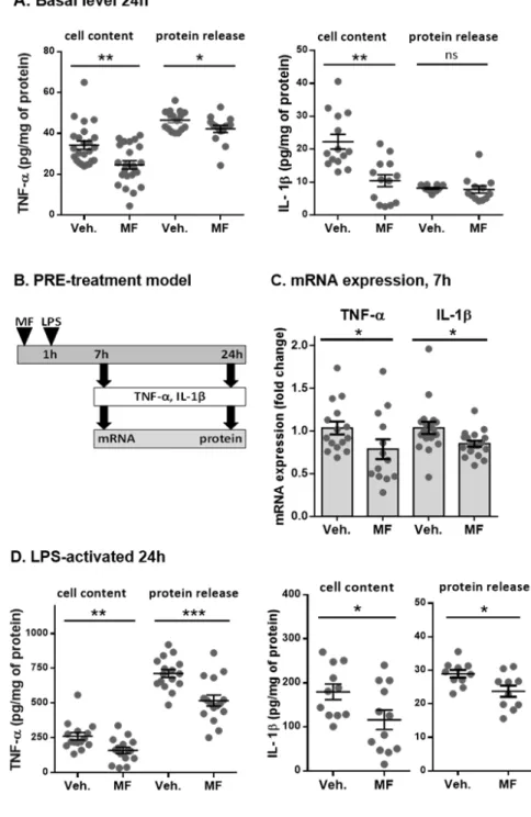
Figure 3: Anti-inflammatory effects of MF pre-treatment on the expression of TNF-α and IL-1β. 481
A, MF reduced the basal expression and release of TNF-α and IL-1β by BV-2 cells; B, Schema 482
illustrating the protocol of pre-treatment of BV-2 cells with MF (1 µmol/L) and the timing of 483
evaluation of its effect on the mRNA and protein expression: C, MF reduced the LPS-stimulated 484
expression of TNF-α and IL-1β mRNA; D, MF reduced the LPS-induced expression and release of 485
TNF-α and IL-1β by BV-2 cells. *P<0.05, **P<0.01, ***P<0.001 vs. Veh. group. Data are expressed 486
as the mean±SEM, n=3, measured in sextuplicates, analyzed by the non-parametric Mann and 487
Whitney test for two independent groups. 488
489
3.5. MF dampened the early stage of inflammation in a co-treatment with LPS 490
The anti-inflammatory effect of pretreatment with MF is of great interest for
491
nutritional intervention and the development of functional foods. But the fact that
492
pretreatment with MF would need to be applied pre-emptively limits its clinical usefulness,
493
particularly for the situations arising without warning. Therefore, we investigated the possible
494
therapeutic use of MF as an anti-inflammatory drug during (a cotreatment protocol) or after (a
495
posttreatment protocol) the establishment of inflammation.
496
To study the effect of MF in the early phase of inflammation, we assessed the cellular
497
content and protein release of the inflammatory cytokines TNF-α and IL–1β in a culture
498
medium of BV-2 cells after 6 h and 24 h of cotreatment with MF and LPS. Co-application of
499
MF and LPS reduced both the intracellular level and the release of TNF-α which are normally
500
induced by the application of LPS (Fig. 4). Marked inhibition (33 %) of the LPS-induced
501
TNF-α up-regulation was already observed after 6 hours of cotreatment. This
anti-502
inflammatory effect clearly influenced the level of TNF-α release, which was decreased by
503
26 % after 24 h (Fig. 4B). Similarly, severe inhibition of the IL-1β response to LPS was
504
reflected in both the cell content and the culture medium after 6 and 24 h of cotreatment (Fig.
505
4C). We then determined the extent to which the beneficial effects observed in the BV-2 cell
506
line could be extended to brain-sorted primary microglia (Fig. 4D). Whereas under the applied
507
experimental conditions cotreatment with MF seemed not to be efficiently reduce the level of
508
TNF-α in a statistically significant manner. Cotreatment with MF lowered the level of IL-1β
509
in primary microglia by factor of five, as compared to the vehicle (Fig. 4D).
511
Figure 4: Anti-inflammatory effects of MF cotreatment with LPS in the BV-2 cell line and the ex 512
vivo culture of microglia cells. A, Schema illustrating the cotreatment of BV-2 cells and primary 513
mouse microglia with MF (1 µmol/L) co-treatment with LPS (1µg/mL) together with the times at 514
which the cytokines were measured: 6 h and 24 h for the BV-2 cell line, and 18 h for the ex vivo 515
culture of microglia cells; B, TNF-α expressed in the cell content and protein release from the BV-2 516
cells at 6 and 24 h; C, IL-1β expression in cell content and protein release from BV-2 cells at 6 and 517
24 h; D, TNF-α and IL-1β protein contents in brain-sorted mouse microglia after 18 h of cotreatment 518
of MF with LPS or only the vehicle. Data are expressed as the mean±SEM, n=3, measured in 519
sextuplicates, analyzed by the Mann-Whitney test. *P<0.05, **P<0.01 vs. Veh. group.
520 521
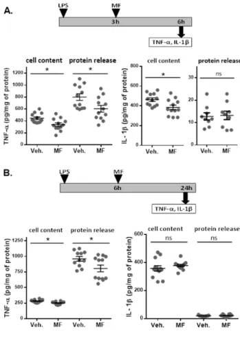
3.6. Post-treatment with MF for resolving LPS-induced inflammation 522
Establishing the proof of the anti-inflammatory MF efficiency of MF as post-treatment
523
is a mandatory steep toward clinical translation. Given as post-treatment, anti-inflammatory
524
drugs are expected to resolve an ongoing inflammatory state by lowering the levels of
525
cytokines such as TNF-α and IL-1β. After LPS challenge, the expression of such
pro-526
inflammatory molecules is been initiated within a 3-6h time window, that also correlates with
527
their in vivo expression, observed in severe acute CNS pathologies with massive
528
inflammatory reaction, like stroke (Le Thuc et al., 2016; Nilupul Perera et al., 2006). As
529
inhibition of inflammation is of interest to change the course of the pathology and improve
530
stroke outcome, especially if starting the treatment after the recommended therapeutic
531
window for thrombolysis with tPA (after 3-4.5 hours after the onset of stroke), we therefore
532
investigated the effects of administering MF as a post-treatment, 3 or 6 h after the onset of
533
LPS-induced inflammation (Figs. 5A and 5B, respectively) on TNF-α and IL-1β
534
inflammatory response, observed after 6 h or 24 h, respectively. 535
The amount of TNF-α were found to be reduced by approximately 20 % in both the
536
cellular content and external medium, whether MF was applied 3 or 6 h after LPS (Fig. 5A).
537
Treating the inflammation with MF 3 h after its induction reduced the IL-1β response to LPS
538
found after 6 h, but treatment delayed by 6 h did not influence IL-1β levels 24 h after the
539
onset of inflammation (Fig. 5B). This result may be explained by the fact that as IL-1β is
540
known to be expressed rapidly during the first hours after LPS stimulation (Kwon et al., 2010)
541
the association of late post-treatment and a low dose of MF would be too stringent conditions
542
to show any anti-inflammatory effect. Therefore, increasing the dose and/or shorten the
543
timing of administration may be anticipated option to further investigate for revealing and
544
improving the MF anti-inflammatory effect on this particular cytokine. Moreover while
545
several group have shown that BV-2 cells produce detectable quantity of interleukin, in our
settings, IL-1β was hardly induced and secreted as compared to TNF-α at the selected
547
measurement times (6 and 24 h). In these stringent settings, it is most likely that the difference
548
in MF effects at the dose of 1 µmol/L on TNF-α and IL-1β could be explained by the timing
549
and level of the protein release in response to LPS challenge.
550 551
552
Figure 5: Anti-inflammatory effects of posttreatment with MF. Schema illustrating the protocol of 553
inducing inflammation with LPS (1 µg/mL) in BV-2 cells and treating them 3 h (A) or 6 h later (B) by 554
applying MF post-treatment (1 µmol/L). A, Effect of MF applied 3 h after the induction of 555
inflammation by LPS. MF post-treatment reduced both the protein content and release of the TNF-α 556
and the IL-1β as evaluated 6 h after the application of LPS. B, When the MF posttreatment was 557
effectuated at 6 h after the induction of inflammation by LPS, the anti-inflammatory effect of reduced 558
TNF-α protein content was still found 24 h after the application of LPS, but that of the IL-1β protein 559
was not. That transitory expression of IL-1β apparently occurred entirely before the post-treatment 560
was applied. Data are expressed as the mean±SEM, n=3, measured in sextuplicates, analyzed by the 561
Mann-Whitney test. *P<0.05, **P<0.01, ***P<0.001 vs. Veh. group. 562
563
1. Discussion 564
Inflammation accompanies many acute and chronic pathologies at both the peripheral
565
and CNS levels. Acute inflammatory diseases are mostly infectious, but inflammation may
566
also be a part of non-infectious pathologies, such as stroke, traumatic brain injury (Skaper et
567
al., 2018), and hemorrhage (Goerge et al., 2008). Chronic inflammation is usually
568
characterized as a slowly progressing mild inflammatory reaction that is linked with several
569
long-term disorders, including atherosclerosis (Libby et al., 2002), inflammatory bowel
570
diseases (Vochyánová et al., 2015), chronic obstructive pulmonary disease (Barnes et al.,
571
2015), osteoarthritis (Robinson et al., 2016), or autoimmune disorders, such as allergic asthma
572
(Barnes et al., 2015) or rheumatoid arthritis (Smolen et al., 2018).
573
Monocytes and microglia are major phagocytes of the innate immune system,
574
providing the first line of defense. The presence of Toll-like receptors (TLRs) enables them to
575
respond to different noxious stimuli, exogenous pathogen-associated molecular patterns
576
(PAMPs) and some endogenous damage-associated molecular pattern molecules (DAMPs).
577
TLR4 is a major receptor implicated in the inflammatory response recognizing bacterial LPS
578
(Zhu and Mohan, 2010). Activation of TLR4 signaling leads to the expression of several
pro-579
inflammatory cytokines, including TNF-α and IL-1β, mediated by the activation of NF-κB
580
signaling pathway (Pålsson-McDermott and O’Neill, 2004). Both TNF-α and IL-1β are potent
581
regulators of the innate immune system important for host defense. Especially, there is an
582
increasing evidence pointing out a prominent role of IL-1β in acute neuronal injuries and
583
many chronic brain diseases (Allan and Rothwell, 2003). Targeting the TLR4/NF-κB pathway
584
therefore represents a potential therapeutic strategy for both acute and chronic
inflammation-585
based diseases that are linked with abnormal secretion of cytokines (Kuzmich et al., 2017).
Plant-derived stilbenoids have demonstrated a promising ability to attenuate peripheral
587
(Dvorakova and Landa, 2017; Eräsalo et al., 2018; Leláková et al., 2019) and CNS
588
inflammation (Akinwumi et al., 2018; Hornedo-Ortega et al., 2018) in vitro and in vivo.
589
However, clinical trials to investigate their potential in humans are still needed. Pterostilbene,
590
a natural derivative of trans-resveratrol, that has frequently been investigated, has shown the
591
ability to inhibit platelet aggregation ex vivo (Messina et al., 2015), improve high fat-induced
592
atherosclerosis inflammation via NF-κB signaling in vivo (Zhang and Zhang, 2016), protect
593
against myocardial ischemia-reperfusion (I/R) injury by reducing oxidative/nitrosative stress
594
and the inflammatory response to it (Yu et al., 2017), and reduce the size of a myocardial
595
infarction (Wu et al., 2017). At the CNS level, pterostilbene protects against hyperglycemia-
596
(Yang et al., 2017) and glutamate-induced neuronal oxidative injury (Wang et al., 2016). In
597
contrast, the stilbenoids of Macaranga spp. have not yet been thoroughly investigated.
598
Among these compounds, MF displays a safe profile for therapeutic use, with low cytotoxic
599
effects on THP-1 cells and on the BV-2, MOLT-3, HepG2, HuCCA-1, and A549 cell lines
600
(Pailee et al., 2015). We focused on the activity of the transcriptional factors NF-кB and
AP-601
1, both of which have been shown to concomitantly control the expression of various
602
inflammatory cytokines during inflammatory response (MacIntyre et al., 2014). Whereas
NF-603
кB is mostly responsible for the gene and protein expressions of TNF-α and IL-1β and is
up-604
regulated by its direct cytoplasmic inhibitor IкBα (Lawrence, 2009), AP-1 is controlled
605
predominantly by ERK1/2, p38, and JNK, members of the family of mitogen-activated
606
protein kinases (MAPKs) (Zhou et al., 2018). In the present work, we reveal the promising
607
potential of MF against peripheral inflammation by demonstrating its ability to: 1) intervene
608
in the IкBα/NF-кB pathway and inhibit the degradation of IкBα, which leads to decreased
609
activation of the transcriptional factor NF-кB, and 2) reduce secretion of inflammatory
610
cytokine TNF-α, which is under the transcription control of NF-кB. This result is in
agreement with the ability of non-prenylated stilbenoids to reduce the phosphorylation of
612
MAPKs (Leláková et al., 2019), whereas prenylated stilbenoids have been shown to affect the
613
degradation of IкBα (Hošek et al., 2019).
614
Like monocytes at the peripheral level, microglia and CNS infiltrated monocytes are
615
responsive to neuronal damage. Their activation is an alarming sign of the brain pathology
616
that can occur in conditions such as microbial infection, traumatic brain injury, or stroke, and
617
also degenerative disorders of the CNS (Dheen et al., 2007). In this work, we demonstrate the
618
ability of MF to improve the in vitro native state of microglia by reducing the basal release of
619
cytokines. This beneficial effect is associated with protection against LPS-induced loss of
620
microglia.
621
Moreover, under inflammatory conditions, MF dramatically inhibits the
LPS-622
stimulated gene and protein expressions of both TNF-α and IL-1β, whatever the stage of
623
treatment (pre-, co-, and post-treatment). The greatest reduction of cytokines (up to 48 %) is
624
achieved when the microglia are pretreated with MF before inflammation is induced.
625
Although the up-stream signalization, such as level of NF-κB and IκB was not investigated in
626
BV-2 cells, due to the phenotypic similarity of monocytes and microglia (Orihuela et al.,
627
2016), it is tempting to anticipate that the response in mouse microglia may be similar. These
628
results rivet attention on the potential for developing MF as a nutraceutical or dietary
629
supplement, aimed at preventing any type of systemic and/or CNS inflammatory disorder, and
630
thereby maintaining or improving organ function, preventing chronic disorders, promoting
631
good health, or even delaying the process of aging (Nasri et al., 2014; Tauskela et al., 2016).
632
In the acute and chronic states of inflammatory disease, the ability of MF to reduce the
633
secretion of the cytokines TNF-α and IL-1β may be of particular interest, because these
634
cytokines are known to be involved in the activation of resident microglia and astrocytes
635
during infection, inflammation, and the development of brain injury (Norden et al., 2016).
Their dysregulation and sustained activation can create a toxic circle, elevating the release of
637
inflammatory factors that endanger the functioning and survival of neurons (Ramesh et al.,
638
2013).
639
Our results show, that MF could be interesting as a supplementary compound used
640
against chronic inflammatory diseases that would correspond to the so called “early phase of
641
inflammation” in the present work. Low-grade systemic inflammation is like CNS
642
inflammation (neuroinflammation), a pathological process that accompanies many persistent
643
diseases and also the normal aging process nicknamed “inflammaging”. Inflammaging is
644
possibly related to chronic activation of the innate immune system and changes in the
645
functions of monocytes (Benedetto et al., 2017). Altered activation of microglia, characterized
646
by elevated levels of TNF-α and IL-1β, is associated with neurodegenerative disorders, such
647
as AD, Parkinson’s disease (PD), and Huntington’s disease (HD), and is a reaction to the
648
presence of cellular aggregates and the misfolded proteins β-amyloid, α-synuclein, and
649
huntingtin, respectively. Reducing neuroinflammation could be a way to slow the progress of
650
these diseases (Sastre et al., 2014). Inflammation underlying neurodegeneration is also present
651
in multiple sclerosis (MS), amyotrophic lateral sclerosis (ALS), systemic lupus erythematosus
652
(SLE), neuromyelitis optica (NMO), diabetes mellitus (DM), chronic bacterial meningitis,
653
Lyme neuroborreliosis, and HIV-1 encephalitis (Amor et al., 2010; Ramesh et al., 2013).
654
Some stilbenoids, such as trans-resveratrol, isorhapontigenin, gnetol, and pinosylvin, have
655
shown significant potential to inhibit butyrylcholine esterase (BChE) enzyme, the increased
656
activity of which is associated with the formation of Aβ plaque in patients with AD
657
(Sermboonpaisarn and Sawasdee, 2012). Oxyresveratrol exerts neuroprotective effects against
658
Parkinsonian mimetic 6-hydroxydopamine (6-OHDA)-induced neurotoxicity in
659
neuroblastoma cells (Chao et al., 2008). Studies of the “Mediterranean diet”, which is based
660
on vegetables, fruits, whole grains, nuts, fish, and seafood, healthy fats, and olive oil, with
moderate consumption of red wine, and is rich in polyphenols, including stilbenoids, have
662
reported beneficial effects, mitigating age-related and other neurodegenerative pathologies
663
(Hornedo-Ortega et al., 2018).
664
The anti-inflammatory effects of MF may also be of interest for acute conditions such
665
as cerebral ischemia, in which reduced blood flow and oxygen supply (hypoxia) lead to
666
cerebral infarction. This condition triggers an intravascular inflammatory cascade that extends
667
into the surrounding tissues of the injured brain, and is characterized by elevated levels of
668
inflammatory cytokines, chemokines, adhesion molecules, reactive oxygen species, nitric
669
oxide, extracellular proteases, and other substances flowing into and accumulating in immune
670
cells, such as microglia (Le Thuc et al., 2015). A therapeutic strategy that intervened in the
671
inflammatory process of cerebral ischemia might slow down and limit the brain damage
672
caused by ischemia. The potential of stilbenoids to act against cerebral ischemia has been,
673
however not thoroughly, investigated. It is noteworthy that a study using a transient rat middle
674
cerebral artery occlusion model has shown that oxyresveratrol reduces the size of volume of
675
an infarction and thus prevents neurological deficits induced by the ischemia/reperfusion (I/R)
676
injury, inhibits the release of cytochrome c, and prevents the activation of caspase-3 (Andrabi
677
et al., 2004). This indicates that as strong anti-oxidant and anti-inflammatory agents,
678
stilbenoids could be effective in the treatment of I/R-injury, a devastating pathology without
679
therapeutic opportunity.
680
Despite their relatively wide range, most of the anti-inflammatory drugs available on
681
the market are linked with many side effects when used in the long term. The therapeutic and
682
adverse effects of the most commonly prescribed non-steroidal anti-inflammatory drugs
683
(NSAIDs) are related to the inhibition of cyclooxygenases (COXs). Their gastrointestinal,
684
renal, and cardiovascular toxicity, together with numerous drug interactions, represent a risk
685
especially for elderly patients who take multiple medications (Wongrakpanich et al., 2018).
Steroidal drugs can have pleiotropic and severe adverse effects (Aljebab et al., 2017).
687
Biological treatment in the form of anti-TNF-α and anti-IL-1β antibodies has been shown to
688
be highly effective for chronic disorders such as RA, especially when treatment is started in
689
the early phase of the disease. However, this treatment represents an enormous financial
690
burden for the health care system and is not accessible to all patients (Dinarello et al., 2012;
691
Monaco et al., 2015). Thus, nutritional intervention and the development of functional food
692
based on natural molecules that are not known to have profound side effects alter promise of
693
preventing inflammation caused by several chronic and acute diseases. Many good candidates
694
may be hidden in natural sources. MF would be among the most promising thanks to its
695
ability to counteract both peripheral and CNS inflammation. MF is structurally a small
696
molecule, effective at a pharmacologically relevant concentration. Synthesizing it in the
697
laboratory could lower its production cost as compared to existing commercial treatments.
698
MF and its derivatives could fulfill the goal of finding a novel anti-inflammatory
699
substance that is potent in prevention and/or supplementary treatment of inflammation-based
700
pathologies of both systemic and central character and safe for long-term use.
701
702
2. Conclusion 703
Our current study shows that stilbenoid macasiamenene F isolated from a plant M.
704
siamensis has significant anti-inflammatory effects. It is therefore an interesting candidate for
705
further investigation using in vivo models where inflammation in the brain plays a major role,
706
e.g., cerebrovascular diseases. The increasing prevalence of chronic inflammatory disorders
707
and more frequent occurrence of acute inflammatory diseases make the need for safe and
708
effective anti-inflammatory therapeutic strategies. Other promising candidates may yet be
709
found in natural sources, but with respect to the current state-of-the-art in nutraceuticals, MF
is the most likely to prove being effective for the prevention and treatment of
inflammation-711
related pathologies, including secondary brain injury following a stroke, for which a
712
therapeutic opportunity is severely lacking.
713 714