European Heart Journal (1996) 17: 1572-1575
Arm-leg pressure gradients on late follow-up after
coarctation repair
Possible causes and implications
J. Guenthard, U. Zumsteg and F. Wyler
Departments of Cardiology and Endocrinology, University Children's Hospital of Basel, Basel, Switzerland
Seventeen years after coarctation repair, 36 patients were studied by magnetic resonance imaging and exercise testing to measure residual anatomical stenosis and hormonal response to exercise, and to evaluate their effect on arm-leg gradients and on exercise hypertension. The systolic arm pressure, leg pressure and arm-leg gradient were measured at rest and during exercise. Active renin and catecholamines were measured in the plasma at rest and after peak exercise. On magnetic resonance imaging 18 patients had residual stenosis of less than 30% (group I) and 18 had residual stenosis of equal to or more than 30% (group II). At peak exercise, the arm pressure was 235 (133-296) mmHg in group I and 241 (157-286) mmHg in group II (ns), the leg pressure was 138 (111-173) mmHg in group I and 114
(75-154) mmHg in group II (/>=0-002). The adrenalin increase from rest to exercise was 32-7 ± 91 pg . ml" ' in the patients with exercise hypertension and 31 ± 4-7 pg. ml ~ ' in the patients who remained normo-tensive during exercise (/>=002). In conclusion, residual anatomical stenosis leads to a pressure drop in the legs, which influences the arm-leg gradient. Arm hypertension is not related to anatomical narrowing but to interaction of enhanced sympathetic nerve activity and structural and functional abnormality of the precoarctation vessels. (Eur Heart J 1996; 17: 1572-1575)
Key Words: Coarctation repair, arm-leg gradients, residual anatomical stenosis, sympathetic nerve activity.
Introduction
Long-term complications after coarctation resection mainly consist of hypertension of the upper body at rest or on exercise, and residual stenosis, which is usually evaluated non-invasively by measuring systolic arm-leg pressure gradients. However, there is still some contro-versy about the relationship of the arm—leg gradient to anatomical narrowing11'21. Moreover, the relationship of arm hypertension to the arm-leg gradient has led to the assumption that late hypertension after coarctectomy is caused by residual anatomical stenosis'341. We studied the interaction of isthmic anatomy and hormonal re-sponse to exercise after coarctation repair and evaluated its effect on arm-leg gradients and on arm hypertension.
Methods
Thirty-six patients aged 13—35 (mean 23) years were studied 10-29 (mean 17) years after coarctation
re-Revision submitted 17 October 1995, and accepted 13 November 1995
Correspondence: Joelle Guenthard, MD, Romergasse 8, CH-4OO5
Basel, Switzerland.
section. Age at operation varied from 1 month to 15 years (mean 6 years). Patients with a bicuspid aortic valve and with mild stenosis were included in the study. None of them was taking cardiac or antihypertensive medication and all were leading a normal life. According to the guidelines of the institutional ethical committee, the study participants or their parents were carefully informed and oral consent was obtained.
After lOmin of supine resting, simultaneous blood pressure measurement of the arm and leg was performed using an automated oscillometric device (Colin BP 8800 Carbamed Ruegge, Baden, Switzerland) with a cuff of adequate size for the upper arm and the ankle. Blood samples for determination of active renin and catecholamines were taken from an indwelling catheter, which had been placed 15 min earlier in the antecubital vein of the arm not used for blood pressure recordings. The samples were collected without the need of a tourniquet and were stored on ice in chilled tubes. The pressure cuffs were left in place and the patients were elevated to 45°, in order to perform leg exercise similar to that normally undertaken in the upright position. They then underwent a graded exercise test on an electronically braked bicycle ergometer (Ergoline computerergometer 900L, Bitz, Germany) according to the James protocol'51. The peak exercise blood pressure
Arm-leg pressure gradients late after coarctectomy 1573
of the arm was measured using the RR method, by temporary replacement of the pressure cuff with a computerized microphone (Ergoline ergometrics 900, Bitz, Germany). A second blood sample was taken at peak exercise. Towards the end of exercise the patients were set back to the supine position while they were still pedalling and exercise was stopped at exhaustion. Within 30 s of exercise conclusion blood pressure of the arm and leg was measured simultaneously to determine the exercise leg pressure and arm-leg gradient. Blood samples were centrifugated at 4° and frozen at — 20° until analysis.
In a second session, magnetic resonance imaging of the thoracic aorta was performed in the supine position with a 1-5 T MR scanner (Magnetom SP 4000 Siemens) using the body coil and ECG triggering. Transverse and oblique sagittal T1W SE images were obtained (TR 650 ms, TE25ms, matrix 128x256, 2 acquisitions, slice thickness 6 mm). Magnetic resonance imaging data were analysed by two independent observers and the aortic diameter was measured at the anastomosis and at the diaphragm level. Percent stenosis was calculated as 100 x (1-(diameter anastomosis/ diameter 10 cm distal to the anastomosis))'61.
Active renin was directly measured in the plasma by an immunoreactive assay sandwich technique using Renine Active Pasteur kits (ERIA Diagnostics Pasteur, Marnes La Coquette, France). Sensitivity: 1-5 pg . ml~ ', intra-assay and inter-assay variability less than 10%. Catecholamines were measured by radioimmunoassay after enzymatic and chemical derivatization using the Amicyl-Test, KATCOMBI kits (IBL, Hamburg, Germany). Sensitivity: l - 2 p g . m l ~ ' plasma, intra-assay and inter-intra-assay variability less than 15%.
The patients were classified into two groups according to the degree of isthmic stenosis on magnetic resonance imaging similar to the findings of Huysmans et al. late after coarctation repair*71. Group I: stenosis less than 30% (n=18), group II: stenosis equal to or greater than 30% (n= 18).
Statistical analysis
Results were expressed as mean ± 1 SEM and range. Comparative statistics were performed by non-parametric unpaired t-test. Significance level was taken at P<005.
Results
All patients reached at least stage 3 of the James protocol on ergometry. Maximal work intensity ranged from 115-220 (mean 161) watt, exercise heart rate ranged from 140-176 (mean 161) beats, min"1 and exercise time varied from 6-24 (mean 13) min. The residual stenosis at the anastomosis level on magnetic resonance imaging was 14% (range 0-29%) in group I and 42% (range 30-63%) in group II (P<00001).
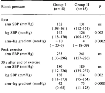
Table I Blood pressure data in group I and II (mean and
full range)
Blood pressure Group I (n=18) Group II (n=18) Rest arm SBP (mmHg) leg SBP (mmHg) arm-leg gradient (mmHg) Peak exercise arm SBP (mmHg)
30 s after end of exercise arm SBP (mmHg) leg SBP (mmHg) arm-leg gradient (mmHg) 132 (108-161) 142 (118-170) -10 (-23-3) 235 (133-296) 180 (112-226) 138 (111-173) 42 (0-65) 131 (112-151) 126 (105-153) 4 (- 18-39) 241 (157-286) 189 (135-237) 114 (75-154) 75 (11-128) ns 0002 00002 ns ns 0002 00008
SBP=systolic blood pressure.
Table 1 summarizes blood pressure data at rest and during exercise in groups I and II. The groups were similar as regards exercise performance. At rest there was no statistical difference in systolic arm pressure between the groups, but systolic leg pressure was signifi-cantly lower and the arm-leg gradient signifisignifi-cantly higher in group II than in group I (P=0-002 and P=0-0002). During and 30 s after exercise there was no statistical difference in systolic arm pressure between the groups. However, 30 s after exercise the systolic leg pressure was significantly lower and the arm-leg gradient significantly higher in group II than in group I (P=0002 and P= 50-0008).
Active plasma renin was 12-6 ± 7 pg . ml" ' in group I and 5-1 ± 1 pg . ml ~ ' in group II (ns) at rest. At exercise the values rose to 19-4 ±12 in group I and 16-4 ± 9 in group II (ns). Plasma adrenalin at rest was 24-8 ± 4 pg . ml ~ ' in group I and 23-8 ± 2 pg . ml " ' in group II (ns). At exercise there was a rise to 58-2 ± 11 pg . ml" ' in group I and to 50-6 ± 10 pg . ml ~ ' in group II (P=5ns). Plasma noradrenalin at rest was 217 ± 37 pg . ml" ' in group I and 219 ± 29 pg . ml~ ' in group II (ns). At exercise there was a rise to 304± 31 pg . m l " ' in group I and to 359±52 pg. m l "1 in group II (ns).
The patients were divided into two further groups according to the blood pressure response to exercise. For young subjects, hypertension at exercise is defined as follows: systolic arm pressure over 225 mmHg at a working intensity of 230 watt for males and over 210 mmHg at a working intensity of 150 watt for females'81. Using these criteria, we defined two groups: exercise normotensive patients (n=9) and exercise hypertensive patients (n = 27). There was no difference in maximal work output, kg"1, exercise heart rate and exercise time between the exercise hypertensive and the
1574 J. Guenthard et al.
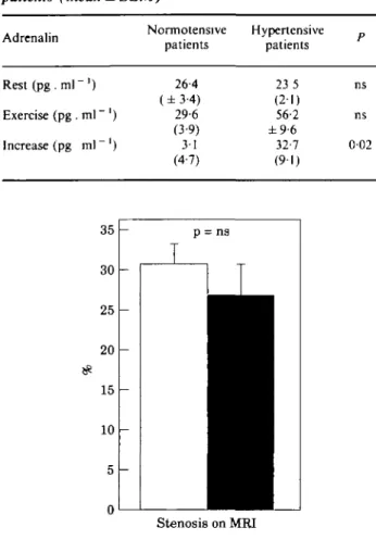
Table 2 Plasma adrenalin at rest and after exercise in the exercise normotensive and exercise hypertensive patients (mean ± SEM)
Adrenalin Normotensive Hypertensivepatients patients
Rest (pg . ml Exercise (pg . Increase (pg ') m l "1) m l "1) 26-4 ( ± 3-4) 29-6 (3-9) 31 (4-7) 23 5 (2-1) 56-2 ±9-6 32-7 (91) ns ns 002 35 30 25 20 15 10 5 n - p = ns
T
-T
^H
Stenosis on MRIFigure 1 Percent stenosis at the anastomosis on
magnetic resonance imaging (MRI) (mean + 1 SEM). D = normotensive (n = 9); • = hypertensive (n = 27).
exercise normotensive patients. The systolic arm blood pressure at peak exercise was 180 (range 133— 204) mmHg in the exercise normotensive and 258 (range 216-296) mmHg in the exercise hypertensive patients (/><00001). The amount of residual stenosis on mag-netic resonance imaging was 31% (range 16-43%) in the exercise normotensive and 27% (range 0-63%) in the exercise hypertensive patients (ns). The plasma adrenalin at rest showed no difference between the exercise normotensive and exercise hypertensive patients. After exercise, the plasma adrenalin was higher in the exercise hypertensive patients, but failed to reveal statistical significance because of the wide range of individual values. However, the individual increase in plasma adrenalin after exercise was significantly higher in the exercise hypertensive (32-7 ± 9 1 p g . m P1) than in the exercise normotensive patients (31 ±4-7 pg . ml~ ') (P=002) (Table 2).
Age at operation and time interval from repair to study had no influence on systolic pressures at rest or during exercise. 45 40 35 30 °- 20 15 10 5 0 -p = 0.02 "
: H
Adrenalin increaseFigure 2 Difference in plasma adrenaline concentration
between rest and exercise (mean + 1 SEM). • = normo-tensive (n = 9); • = hypernormo-tensive (n = 27).
Discussion
Blood pressure response to exercise, which is measured to assess surgical outcome after coarctation repair, is characterized by systolic hypertension of the arms'3'4'91. As restenosis is thought to be one of the main causes of this hypertension, measurement of systolic arm-leg pressure gradients at rest or at exercise are routinely used for non-invasive evaluation of residual stenosis'3'41. However, blood pressure measurement of the legs during exercise is influenced by technical problems due to movement artifacts. Therefore simultaneous arm and leg pressure is usually measured 1-2 min after termin-ation of exercise. Significant pressure changes occur in these 2 min of recovery and leg pressure as well as arm-leg gradients recorded with such a delay cannot be considered as true exercise values'10'"1. Our patients, however, exercised in a 45° position with fixed cuffs, which allowed us to obtain the first blood pressure results within 30 s of the end of ergometry.
Leg blood pressure at rest and at exercise was significantly lower in group II, with residual isthmic stenosis equal to or above 30%, than in group I. Therefore, impairment of blood flow from residual stenosis is responsible for a pressure drop in the legs, as has already been suggested by others'112'. As the systolic arm pressures did not differ between groups I and II, but as the arm-leg gradients were significantly higher in group II, we conclude that part of the gradient is caused by the changes of leg pressures, which in their turn are dependent on residual stenosis. Accordingly, the relationship of arm-leg gradients to anatomical stenosis is mainly determined by the pressure drop in the legs. Arm hypertension, however, seems not to be dependent on anatomical narrowing, since the magnetic resonance imaging results did not differ between exercise hyper-tensive and exercise normohyper-tensive patients. Similar
Arm-leg pressure gradients late after coarctectomy 1575
findings were described by Hanson et al., who studied residual anatomical changes after coarctation repair by angiography1''.
Together with the hypertensive response, we observed a significantly higher increase of plasma adrenaline after exercise in exercise hypertensive patients. Recently, Ross et al. studied 35 patients 17 years after coarctation resection. In a hypertensive group of 19 patients they found elevated plasma norepinephrine and renin activity and concluded that enhanced sympathetic nervous output contributes to hypertension after coarctation repair1'3'. Since plasma
adrenaline is usually a more reliable parameter than plasma noradrenalin for the evaluation of sympathetic nerve activity1'4' and adrenaline has been shown to act
as cotransmitter on sympathetic nerve endings'151, our
results support the idea that enhanced sympathetic nerve activity is involved in late hypertension after coarctectomy. The difference in plasma renin after exercise compared to the results reported by Ross et al. may be due to the great individual variability of this hormone and to the difference of exercise protocol and method of hormone measurement in both studies.
The metabolic demand of working muscles during exercise is triggered by sympathoadrenal activity and an increase of plasma adrenalin'14'. In addition,
enhanced sympathetic nerve activity later after co-arctation repair may be caused by functional changes of the baroreceptors due to structural abnormalities of the precoarctation vessels1'6'. Thus, late hypertension after
coarctation repair could result from the interaction of augmented sympathetic activity and of functional abnormalities of the precoarctation vessels'17"20'.
In conclusion, residual anatomical stenosis pro-duces a gradient caused by lowered arterial pressure in the legs. Arm hypertension, however, as part of the pressure gradient at exercise seems not to depend on residual narrowing, but on increased sympathoadrenal activity. Exercise hypertension after coarctation repair does not lead to reintervention unless there is a signifi-cant gradient across residual stenosis
References
[1] Hanson E, Eriksson BO, Sorensen SE. Intra-arterial blood pressures at rest and during exercise after surgery for coarctation of the aorta. Eur J Cardiol 1980; 11: 245-57. [2] Kappetein PA, Guit GL, Bogers JC et al. Noninvasive long
term follow-up after coarctation repair. Ann Thorac Surg 1993; 53: 1153-9.
[3] Connor TM. Evaluation of persistent coarctation of aorta after surgery with blood pressure measurement and exercise testing. Am J Cardiol 1979; 43: 74-8.
[4] Freed MD, Rocchini A, Rosenthal A, Nadas AS, Castaneda AR. Exercise induced hypertension after surgical repair of coarctation of the aorta. Am J Cardiol 1979; 43: 253-8. [5] James FW, Blomquist CG, Freed MD et al. Standards for
exercise testing in the pediatric age group. Circulation 1982; 66: 1377A-9A.
[6] Stern HC, Locher D, Wallnofer K. et al. Noninvasive assess-ment of coarctation of the aorta: comparative measureassess-ments by two dimensional echocardiography, magnetic resonance and angiography. Pediatr Cardiol 1991; 12: 1-5.
[7] Huysmans HA, Kappetein AP. Late follow-up after resection of aortic coarctation. Z Kardiol 1989; (Suppl 7): 39-41. [8] Nordenfelt I, Adolfsson L, Nilsson JE, Olsson S. Reference
values for exercise tests with continuous increase in load, d i n Physiol 1985; 5: 161-72.
[9] James FW, Kaplan S. Systolic hypertension during submaximal exercise after correction of coarctation of the aorta. Circulation (Suppl II) 1974; 50- II27-II34.
[10] Wendel H, Teien D, Human DG, Nanton MA. Assessment of blood pressures and gradients by automated blood pressure device compared to invasive measurements in patients previously operated on for coarctation of the aorta. Clin Physiol 1992; 12- 155-162.
[11] Palatini P. Blood pressure behaviour during physical activity. Sports Medicine 1988; 5: 353-74.
[12] Schumacher G. Fahrradergometrische Belastungsunter-suchung mit Blutdruckmessung und Bestimmung der Plasma-Renin-Aktivitat. In: Schumacher G, ed. Arterielle Hypertonie bei Aortenisthmusstenose. Georg Thieme Verlag Stuttgart, New York: 1988: 60-77.
[13] Ross RD, Clapp SK, Gunther S et al. Augmented norepinephrine and renin output in response to maximal exercise in hypertensive coarctectomy patients. Am Heart J 1992; 123: 1293-9.
[14] Galbo H. The hormonal response to exercise. Diabetes/ Metabolism Review 1986; 4: 385-^08.
[15] Blankestitijn PJ, Tulen J, Boomsma F et al. Support for adrenalin-hypertension hypothesis: 18 hour pressor effect after 6 hours adrenalin infusion. Lancet 1988; 17: 1386-9. [16] Beekman RH, Katz BP, Moorehead-Steffens C, Rocchini AP.
Altered baroreceptor function in children with systolic hypertension after coarctation repair. Am J Cardiol 1983; 52: 112-7.
[17] Sehested J, Baandrup U, Mikkelsen E. Different reactivity and structure of the prestenotic and poststenotic aorta in human coarctation: implication for baroreceptor functions. Circulation 1982; 65: 1060-5.
[18] Gidding SS, Rocchini AP, Moorehead C, Schork MA, Rosenthal A. Increased forearm vascular reactivity in patients with hypertension after repair of coarctation. Circulation
1985; 71:495-9.
[19] Hanson E, Eriksson B, Sivertsson R. Blood flow resistance in the hand after coarctectomy. Clinical Physiology 1981; 1: 257-62.
[20] Gunthard J, Wyler F. Exercise induced hypertension in the arms due to impaired arterial reactivity after successful coarctation resection. Am J Cardiol 1995; 75: 814-7.