HAL Id: hal-01501453
https://hal.sorbonne-universite.fr/hal-01501453
Submitted on 4 Apr 2017
HAL is a multi-disciplinary open access archive for the deposit and dissemination of sci-entific research documents, whether they are pub-lished or not. The documents may come from teaching and research institutions in France or abroad, or from public or private research centers.
L’archive ouverte pluridisciplinaire HAL, est destinée au dépôt et à la diffusion de documents scientifiques de niveau recherche, publiés ou non, émanant des établissements d’enseignement et de recherche français ou étrangers, des laboratoires publics ou privés.
Vitamin D and multiple sclerosis: An update
Charles Pierrot-Deseilligny, Jean-Claude Souberbielle
To cite this version:
Charles Pierrot-Deseilligny, Jean-Claude Souberbielle. Vitamin D and multiple sclerosis: An update. Multiple Sclerosis and Related Disorders, 2017, 14, pp.35-45. �10.1016/j.msard.2017.03.014�. �hal-01501453�
Review Article
Vitamin D and multiple sclerosis: an update
Charles Pierrot-Deseilligny
a* and Jean-Claude Souberbielle
ba Département de Neurologie, Hôpital de la Salpêtrière, Assistance Publique Hôpitaux de Paris, Université Pierre
et Marie Curie (Paris VI), 47 bd de l’Hôpital, 75013 Paris, France.
b Service d’explorations fonctionnelles, Hôpital Necker-Enfants Malades, Assistance Publique Hôpitaux de Paris,
Université René Descartes (Paris V),149 rue de Sèvres, 75915 Paris, France. *Corresponding author
E-‐mail addresses: [email protected] (C. Pierrot-Deseilligny), [email protected] (J-C.
Souberbielle)
Highlights:
- Genetics has confirmed that hypovitaminosis D is one of the risk factors for MS. - A modulation of the global risk for MS may exist from conception to the disease onset.
- The main mechanism of action of vitamin D in MS appears to be immunomodulatory. - Vitamin D could mainly be active in the inflammatory component of MS.
- Systematic moderate vitamin D supplementation of MS patients is recommended.
Abstract.
The most recent findings linking exposure to sun and vitamin D insufficiency to multiple sclerosis (MS) are reviewed. Due to insufficient sunshine and changing lifestyles, hypovitaminosis D is widespread in temperate countries. Numerous epidemiological studies have strongly suggested that sunshine and vitamin D insufficiency contributes to MS risk in these countries. Moreover, several large genetic studies in MS patients have recently stated unequivocally that diverse abnormalities involving vitamin D metabolism are related to the risk of the disease. The important implications of such results are discussed here. Then, the interactions of hypovitaminosis D with the other genetic and environmental protective and risk factors, such as the allele HLA DRB1*1501, Epstein-‐Barr virus infection, obesity, smoking and sexual hormones, are summarized. Vitamin D insufficiency and sufficiency could be a risk and a protective factor, respectively, among many other factors possibly continuously modulating the global MS risk from the mother’s pregnancy to the triggering of MS in adulthood. However, many interactions between these different factors occur more particularly between conception and the end of adolescence, which corresponds to the period of maturation of the immune system and thymus and may be related to the dysimmune nature of the disease. The main mechanisms of action of vitamin D in MS appear to be immunomodulatory, involving the various categories of T and B lymphocytes in the general immune system, but neuroprotector and neurotrophic mechanisms could also be exerted at the central nervous system level. Furthermore, several controlled immunological studies performed in MS patients have recently confirmed that vitamin D supplementation has multiple beneficial immunomodulatory effects. However, there is still an enduring absence of major conclusive randomized clinical trials testing vitamin D supplementation in MS patients because of the quasi-‐insurmountable practical difficulties that exist nowadays in conducting and completing over several years such studies involving the use of a vitamin. Nevertheless, it should be noted that similar robust statistical models used in five different association studies have already predicted a favorable vitamin D effect reducing relapses by 50-‐70%. If there is now little doubt that vitamin D exerts a beneficial action on the inflammatory component of MS, the results are as yet much less clear for the progressive degenerative component. Lastly, until more information becomes available, vitamin D supplementation of MS patients, using a moderate physiological dose essentially correcting their vitamin insufficiency, is recommended.Key words: environment; genetics; multiple sclerosis; supplementation; vitamin D
Abbreviations: anti-EBNA1, antibodies against EBV antigen nuclear 1; CIS, clinically isolated syndrome; CNS, central nervous system; CYP27B1, enzyme 1α-hydroxylase; EAE, experimental autoimmune encephalopathy; EBV, Epstein–Barr virus; HLA, human leukocyte antigen; MS, multiple sclerosis; RCTs, randomized clinical trials; UVB, ultraviolet B rays; VDR, vitamin D receptor; VDRE, vitamin D responsive element; Tregs, T regulator lymphocytes
Vitamin D and multiple sclerosis: an update, Multiple Sclerosis and RelatedDisorders, 2017, in press, DOI :
http://dx.doi.org/10.1016/j.msard.2017.03.014
1. Introduction
The key role of vitamin D in bone metabolism has been known for a long time, but our knowledge has developed considerably during the past 15 years about the physiological extra-‐skeletal actions of this vitamin, in particular its immunodulatory effects (Hayes et al., 2015), which have implications in some inflammatory and autoimmune diseases such as multiple sclerosis (MS). In parallel, great advances have also been made in identifying a number of protective factors and risk factors for MS, including exposure to sun and vitamin D insufficiency, which complexly interact with one another (Ascherio and Munger, 2016). The main recent findings linking vitamin D to MS are reviewed in this update.
Figure 1. Unequal solar ultraviolet radiation in the world. UVB reach the ground and skin only
when the sun is sufficiently high in the sky for UVB to penetrate all the layers of the atmosphere. This physical mechanism can only exist in the middle of the day and, beyond the 40th parallel in mid and high latitude regions, only for a few months of the year, essentially between the middle of spring and the end of summer. Thus, a vast portion of the northern hemisphere – comprising almost all of Europe and a large part of North America and of the former Soviet Union – as well as a few countries in the southern hemisphere – are involved. In all these countries, exposure to sun includes UVB only during these relatively reduced time slots in the day and year. These regions correspond to those where the highest multiple sclerosis prevalence is observed. By contrast, subtropical and tropical regions, where 80% of the world’s population lives, have enough sunshine to provide the major source of natural vitamin D almost throughout the year. 40°, 40th parallel; UVB, ultraviolet B rays; kWh/m2 kilowatt/hour per
square meter. Adapted from Meteonorm values.
Insufficient UVB for more than 6 months/y
Insufficient UVB for 3-6 months/y Equator 40°$ 40°$ Insufficient UVB for 3-6 months/y
Insufficient UVB for more than 6 months/y
(including)almost)all)of)Europe)and)a)large)part) )of)North)America)and)the)former)Soviet)Union)) (including)New)Zealand) Tasmania)and)Patagonia)) UVB almost throughout the year 500! 1000! 1500! 2000! 2500! Yearly$total$ in$kWh/m2$
2. General and physiological considerations on sunshine and vitamin D
Vitamin D is principally produced by the action of ultraviolet B rays (UVB) in the skin, providing 80–90% of human requirements (Holick, 2011; Wacker and Holick, 2013), but this main natural solar source of vitamin is unequally distributed in the world (O’Neil et al., 2016), as shown in Figure 1. The metabolism of vitamin D is nowadays relatively well known and is summarized in Figure 2 (Norman and Bouillon, 2010; Pike and Meyer, 2012). 25-‐OH-‐D is the vitamin D metabolite usually measured in the blood since it is representative of the vitamin D store in the organism (Souberbielle et al., 2010). The optimal levels for serum 25-‐OH-‐D concentration vary according to the target population (Hoel et al., 2016; Holick et al., 2011; Ross et al., 2011). For immune health and more generally for patients, the target 25-‐OH-‐D range usually accepted nowadays (30-‐ 60 ng/mL or 75-‐150 nmol/L) has been derived from multiple metabolic and bone clinical data (Binkley and Krueger, 2008; Holick et al., 2011; Pierrot-‐Deseilligny and Souberbielle, 2013): these findings are very different in nature but, quite remarkably, they all indicate the critical feature of the lower limit (30 ng/mL), below which a pathological significance is reached. This is why those norms are nowadays often recommended (Hoel et al., 2016), even though there is not yet a total consensus (Holick et al., 2011; Pludowski et al., 2013; Ross et al., 2011; Souberbielle et al., 2010).
Figure 2. Schematic representation of vitamin D metabolism. Vitamin D3 is mainly synthesized at the skin level thanks to UVB and, after a double hydroxylation in the liver and the kidney, is transformed into calcitriol, i.e. the active metabolite. Calcitriol can also be directly synthesized in the cell cytoplasm from the circulating 25-‐OH-‐D and links at this level to a specific receptor (VDR), which is present in numerous cells of the organism, including immune and nervous cells. Then, this complex penetrates into the nucleus, forms a heterodimer with the receptor RXR and this heterodimer finally binds to VDRE, which constitutes a specific sequence of DNA within the promoter region of the target genes. The whole regulates (by activation or suppression) gene transcription and expression and, ultimately, protein synthesis in approximately 5–10% of the genome. Thus, for immune cells, which are particularly involved in MS pathogenesis, the synthesis of the diverse favorable and unfavorable cytokines related to inflammatory mechanisms could be either increased or decreased depending on the genes and the vitamin D status at the time (see below in the text). Note that the allele HLA DRB1*1501, which is particularly deleterious in terms of MS risk and taken here as example, possesses a VDRE area. C, calcitriol; CYP27B, enzyme 1α-‐hydroxylase; CYP2R, enzyme 25-‐ hydroxylase; HLA, human leukocyte antigen; PTH, parathyroid hormone; RXR, retinoid X receptor; UVB, ultraviolet B rays; VDR, vitamin D receptor; VDRE, vitamin D responsive element.
7"dehydrochole
sterol-
UVB-
Skin- Cholecalciferol- (vitamin-D3)-Oral%intake% (marginal%in% food)%CYP2R1-
25"OH"D-
(calcidiol)-
1,25"OH"2D-
(calcitriol)K i d n e y - CYP27B1-C-Cytoplasm%
VDR-RXR-N u c l e u s %
Heterodimer%Gene%
Promoter% region%Proteins:--(cytokines,%etc.)%
VDRE-
VDR-Cel
l-Main natu ral source o f vitamin D Immune and nervous cells, etc. C- CYP27B1,- bypassing- the-renal- step-PTH
L i v e r
- e.g.-HLA-DRB1*- 1501- Vitamin--D3- C- VDR-C-To reach a 25-‐OH-‐D serum level above 30 ng/mL and then permanently maintain it within the physiological zone (30-‐60 ng/mL), one must receive a daily vitamin D dose of between 2000 and 4000 IU (3000 IU on average) (Bischoff-‐Ferrari et al., 2012; Hall et al., 2010; Wacker and Holick, 2013), which is naturally observed only in outdoor workers during the summer (Azizi et al., 2012; Bager-‐Lux and Heaney, 2002) or in tropical countries (Luxwolda et al., 2012). Indeed, food, even in a well-‐ balanced diet, provides only about 100 IU/d and, if fortified, not more than 300-‐400 IU/d, which is in any case insufficient (Holick, 2012). Moreover, the organism does not durably store vitamin D, which progressively disappears within 6-‐8 weeks after a single solar or oral intake. The so-‐called ‘toxic’ area, potentially generating hypercalcemia, seems to be located largely above 150 ng/mL, corresponding to a daily intake far higher than 10,000 IU/d (Hathcock et al., 2007).
These multiple considerations – physical, geographical, societal and physiological – explain why vitamin D insufficiency is widespread nowadays in the general population of temperate countries, and how this problem has progressively worsened during the last hundred years as a corollary of the development of the modern lifestyle which has progressively taken these populations away from direct exposure to sunshine (Pierrot-‐Deseilligny and Souberbielle, 2010).
3. Role of vitamin D in the risk for MS
3.1. Epidemiology of latitude and solar radiation effects on the MS risk
It has long been known that latitude influences MS risk, the prevalence of the disease being minimal at the equator and increasing with North or South latitude (Simpson et al., 2011). Furthermore, MS prevalence may change after migrations that occurred during the second decade of life, with a beneficial effect for people who migrated from a high-‐latitude region (with a high MS prevalence) to a sunnier, lower-‐latitude region, with a low MS prevalence (Handel et al., 2010; McLeod et al., 2011). It has also been reported that the MS prevalence of a region is inversely correlated to the level of solar radiation of this region, always with a high level of significance, on a world scale (Sloka et al., 2011), in the USA (Beretich and Beretich, 2009), in France (Pierrot-‐Deseilligny and Souberbielle, 2010) and the United Kingdom (Ramagopalan et al. 2011), thus explaining the well-‐known link between MS risk and latitude. It is also known that the time spent outdoors during childhood and adolescence has an impact on MS risk: people who practiced many outdoor activities during their youth, and thus received more UVB, have a significantly lower risk later for MS (Bjørnevik et al., 2014; Laursen et al., 2016a; Van der Mei et al., 2003). In spite of methodological debates (Fides et al., 2014), the month of birth does also appear to influence the MS risk: people born at the end of autumn, i.e. whose mothers could have had a relatively good exposure to sun in the major part of pregnancy, have a significantly reduced MS risk, whereas those born at the end of spring, whose mothers likely had much less exposure to sun in the major part of pregnancy, have an increased MS risk, these findings existing in both hemispheres, in which seasons are reversed (Balbuena et al., 2016; Dobson et al., 2013; Rodriguez Cruz et al., 2016).
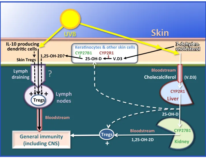
3.2. Specific UVB effects on the MS risk
From a general point of view, vitamin D is likely a major intermediary link between exposure to sun and the MS risk since UVB physiologically produce directly this vitamin in the organism. However, it may also be that UVB play a beneficial role in immunity through a specific immunomodulatory effect that could be independent of the vitamin D synthesis (Breuer et al., 2014; DelLuca and Plum, 2017; Hart and Gorman, 2013) (see Figure 3). Although the respective immunological vitamin D and UVB effects from the skin and its different outlet pathways are not easy to differentiate and delineate in humans, the possibly specific UVB action may be exerted in parallel to the immunomodulatory effect of vitamin D (Lucas et al. 2015), which is currently far more widely documented (see below). In any case, these two mechanisms do not exclude each other and their possibly different specific actions have even been differentiated in a few epidemiological studies (Bäärnhielm et al., 2012; Lucas et al., 2011).
Figure 3. The two main pathways through which UVB may influence immunity. The first, classical
pathway (right) starts in the skin under the influence of UVB with the transformation of 7-‐ dehydrocholesterol into cholecalciferol (vitamin D3), which then passes through the bloodstream into the
liver and the kidneys and results in calcitriol (the active metabolite) stimulating Tregs, having a beneficial immunomodulatory effect on the general immunity (see below in the text). The second pathway (left), as yet much less well known, could be partly independent of the vitamin D synthesis: the UBV could also stimulate skin-‐derived tolerogenic dendritic cells, producing IL-‐10 (a favorable cytokine), resulting in stimulation of local Tregs and also, after moving through lymph draining, of Tregs located in the lymph nodes. Tregs finally join through the bloodstream the general immune system, in which they could also have a beneficial action. However, it should be noted that calcitriol may also be locally produced within the skin from 7-‐dehydrochlesterol or the circulating 25-‐OH-‐D in the keratinocytes and other skin cells, which have a complete enzymatic equipment to do this (Lehmann, 2009). Furthermore, calcitriol could be used in the skin (McCully et al., 2015; Schuessler et al. 2001) and captured, through lymph draining, by the lymph nodes (Gorman et al., 2007), where it might also stimulate the local Tregs. Thus, this possible further pathway involving vitamin D within the skin itself complicates the study of a specific, independent role of UVB in immunity. 1,25-‐OH-‐2D, calcitriol; 25-‐OH-‐D, calcidiol; CNS, central nervous system; CYP27B1, enzyme 1α-‐hydroxylase; CYP2R1, enzyme 25-‐hydroxylase; Tregs, T regulator lymphocytes; UVB, ultraviolet B rays; V.3, vitamin D3.
3.3. Epidemiology of vitamin D effects on the MS risk
In fact, the direct epidemiological link between vitamin D and the MS risk has been suggested in studies in which the consumption of vitamin D supplements during youth (Munger et al., 2004), cod liver oil during adolescence (Cortese et al., 2015) or fatty fish (rich in vitamin D) (Bäärnhielm et al., 2014) appeared to provide significant protective effects for the MS risk later in life. In another study, young American soldiers who had a spontaneous 25-‐OH-‐D serum level above 40 ng/mL had a 62% lower MS risk than those with less than 25 ng/mL (Munger et al., 2006). More recently, analogous findings were observed in Swedish pregnant women in whom those with a 25-‐OH-‐D serum level above 30 ng/mL had a 61% lower MS risk than those with less than 30 ng/mL (Salzer et al., 2012). Vitamin D status during
UVB$
1,25%OH%2D)Skin)
Lymph))
nodes)
General)immunity)
(including)CNS))
Cholecalciferol)
)))))))(V.D3))7%dehydro%)
cholesterol)
?$
25%OH%D)+)
IL%10)producing)
)dendriKc)cells)
Tregs)
Liver)
Kidney)
Lymph)
draining)
1,25%OH%2D?) Skin)Tregs) Bloodstream) Bloodstream) Bloodstream)+)
+)
Tregs)
CYP27B1) CYP2R1) KeraKnocytes)&)other)skin)cells) CYP27B1)))))))CYP2R1) 25%OH%D)))))))))))V.D3)pregnancy also played a significant role in the MS risk of offspring in two other studies, in the USA (Mirzaei et al., 2011) and in Finland (Munger et al., 2016). Lastly, in a recent Danish study, the 25-‐OH-‐D serum level of newborns appeared to influence their MS risk 15-‐30 years later, with the observation that for each increase of 20 ng/mL in the serum level the MS risk was decreased by 60% (Nielsen et al., 2017). Accordingly, the results of these multiple studies strongly suggest that an insufficiency in exposure to sun and/or in vitamin D during the first part of life constitutes a major risk for MS, with as a corollary a consistent quantified reduction of about 60% of this risk resulting from a normal vitamin D serum level.
3.4. Contribution of genetic abnormalities of the vitamin D metabolism to the MS risk
Lastly, vitamin D insufficiency contributes to MS susceptibility as both an environmental and a genetic risk factor. Genetic studies in MS patients are numerous but we will focus here only on the major recent advances that have linked in a non-‐ambiguous way diverse specific genetic errors in the vitamin D metabolism to MS risk. Abnormalities of the gene of enzyme 1α-‐hydroxylase (CYP27B1) (Sawcer et al., 2011), which specifically controls calcitriol synthesis, significantly influenced the MS risk in studies testing the variants rs703842 (Karaky et al., 2016; Simon et al., 2011; Sundqvist et al., 2010) or rs10877013 (Zhuang et al., 2015). Genetic abnormalities of the CYP24A1 gene, which encodes the enzyme responsible for initiating calcitriol degradation, also influenced MS risk (Ramasamy et al., 2014). Moreover, two vast studies recently aimed to link genetically low 25-‐OH-‐D serum levels to the MS risk: the first study, in Canada (Mokry et al., 2015), and the second one, in both the USA and Sweden (Rhead et al., 2016), comprising further methodological criteria, involved thousands of MS patients and control subjects, who were tested using mendelian randomization techniques from several single nucleotide polymorphisms corresponding to different stages of vitamin D metabolism. In the results, very strong links existed between these vitamin D abnormalities and the risk for MS. Accordingly, such studies, in which there was methodologically very little room for reverse causality and confounding factors, state that a genetically induced hypovitaminosis D is a risk factor for MS. This finding has several important implications. First, even if such a relatively rare type of genetic hypovitaminosis D represents a very minor part of currently known forms of MS heritability, dominated by dysimmune abnormalities, it nevertheless provides evidence that hypovitaminosis D per se is a risk factor. Second, it may be inferred from such evidence that the most widespread vitamin D insufficiency, which is not genetically induced but results from unfavorable environmental conditions, is also an MS risk factor, as strongly suggested by the epidemiological studies reviewed above in which this environmental part of the risk seemed even to be a major component. Third, since these genetic studies specifically involve vitamin D metabolism, they also confirm that UVB do not play an exclusive beneficial role in immunity (see above).
4. Other risk factors for MS and interactions with vitamin D
4.1. Role of the human leukocyte antigen system
For MS there are in fact many other risk factors and protective factors, both genetic and environmental, which could cumulate or compensate one another. Among the other genetic risk factors for MS, the allele DRB1*1501 in the human leukocyte antigen (HLA) system accounts for 11% of heritability of the disease, whereas other alleles such as HLA-‐A*0201 could be protective (Sawcer et al., 2011). There is a vitamin D responsive element (VDRE) zone in the promoter region of HLA DRB1*1501 gene which strongly suggests an involvement in mechanisms linked to vitamin D, even if these mechanisms are as yet little known (Ramagopalan et al., 2009). Furthermore, in a recent pediatric study, the allele HLA DRB1*1501 was significantly associated with the 25-‐OH-‐D serum level and with the relapse rate (Graves et al., 2016), whereas in another study this allele was related to low exposure to sun and to obesity (Laursen et al., 2015). Lastly HLA DRB1*1501 is over-‐represented in Caucasian populations and in women and it has been suggested that certain HLA alleles could have evolved during previous generations thanks to epigenetic mechanisms influenced by favorable or unfavorable environmental conditions, in which the vitamin D status could figure (Handunnetthi et al., 2010; Koch et al., 2012) (Figure 4).
Figure 4. Schematic continuous modulation of protective and risk factors in MS. For more details see text. A
schematic curve of the global MS risk, fluctuating with time, is shown in the middle. Hypothetical risk factors for MS (bottom) and protective factors for the disease (top) are both genetic and environmental. The numerous interactions between these different factors may occur (from right to left) in adulthood, in adolescence, in childhood, during the mother’s pregnancy and even during previous generations, through epigenetic mechanisms that could shape protective or deleterious alleles in the HLA system depending on favorable or unfavorable environmental conditions. The evolution of the MS risk (left to right) starts at conception, where protective and deleterious genetic factors are transmitted, in particular HLA DRB1*1501 and genes involved in abnormalities of vitamin D metabolism (bottom). Then, a birthday in spring, a high vitamin D serum level during pregnancy, in the newborn or later during the first part of life, but also outside activities during youth (with UVB and/or vitamin D) and migration to a sunny country in adolescence would all be protective elements, whereas the reverse conditions could increase the MS risk. Infection with EBV, responsible for infectious mononucleosis, appears to be a major and possibly founding risk factor for MS, in particular if primo-‐infection occurs late, after childhood, and/or if it leaves a high level of anti-‐EBNA1, whereas a negative serologic status to EBV could be protective (5% of population). The viruses HHV6 and HERVs could also have deleterious effects in terms of MS risk, maybe more particularly during the disease course. By contrast, numerous common infections or parasitic infestations during childhood could be protective, according to the hygiene hypothesis, as would also be positive serology for CMV. Sexual hormones, i.e. testosterone in men and estrogens and progesterone in women, could be protective. Obesity, in particular in adolescence, and cigarette smoking are other risk factors for MS, whereas coffee (at high dose), ALA and other substances could be protective. All these protective and risk factors interact between conception and the triggering of MS, usually occurring in young adulthood, leading to the concept of a continuous modulation of the global MS risk throughout the first part of life. However, many of these interactions intervene between pregnancy and the end of adolescence, in a period corresponding to that of the maturation of the immune system and thymus, which could be related to the dysimmune nature of the disease. Certain combinations of deleterious risk factors could finally trigger the disease, whereas diverse protective effects could prevent it durably. If the disease starts (right), some risk factors could continue to have deleterious effects by worsening its course and favoring relapses, whereas certain protective factors could contribute to remissions. ALA, α-‐linoleic acid; anti-‐EBNA1, antibodies against EBV antigen nuclear 1; CMV, cytomegalovirus; EBV, Epstein–Barr virus; HERV, human endogenous retrovirus; HHV6, human herpes virus of type 6; HLA, human leukocyte antigen; M, men; MS, multiple sclerosis; Nl, normal; UVB, ultraviolet B rays; V.D, vitamin D; W, women.
Migration to the sun
T e s t o s t e r o n e insufficiency? Coffee? ALA? Etc.
Numerous infections (hygiene hypothesis) e.g. HLA DRB1* 1501 = 11% of deleterious heritability Favorable conditions of environment Unfavorable conditions of environment Remission(
(UVB-VD) Almost no outdoor activities
Obesity (V.D?) (passive) (active) Smoking
Adolescence
Childhood
Previous generations e.g. HLA A*0201 (protec- tive)Vitamin D
insufficiencyVitamin''''''D''''''suf,iciency''''
'
'''''''
(≥'40'ng/ml)'Frequent outdoor activities (UVB V.D)
G e n e t i c risk factors : Caucasian ; HLA DRB1 *1501; siblings of MS patients (3%);
g e n e t i c a b n o r m a l i t i e s o f v i t a m i n D metabolism, etc. Nl testosterone (M)? Estrogens (W)
No EBV (5%) CMV+ status?
Pro tective genetic factors : dark skin; HLA A*0201, etc.
Low V.D in mother Nl V.D in mother Birthday in autumn High(neo, natal(V.D( Mother’s pregnancy Epigene5cs( Epigene5cs(
late occurrence high anti-EBNA1 level HHV6-HERV?
Low(neo,( natal(V.D( Birthday in spring
Epstein,Barr(
virus&infec+on:&
Relapses( P(r(o (t (e(c(t (i (o (n ((((( W(((o (((r(((s(((e((n (((i (((n (((g(Early Adulthood
t
No(
(
(
MS(
!!!!!Maturat!!ion!!!of!!!the!!!immune!!!!!!system!!!&!!!thymus!MS(
C
o
n
ce
p
ti
o
n
PR O T EC T IVE F A C T O R S F O R MS RISK F ACT ORS FOR MS
4.2. Role of viruses and infections
Among the other environmental risk factors, Epstein-‐Barr virus (EBV) infection might play a major role (Ascherio and Munger, 2016; Olsson et al., 2017; Salzer et al., 2013; Waubant et al., 2016) (see more details in Figure 4). In MS patients, genetic arguments for interactions between EBV infection and vitamin D have been reported (Ricigliano et al., 2015), with, furthermore, associations existing between HLA DRB1*1501, the 25-‐OH-‐D serum level and the serum level of antibodies against EBV antigen nuclear 1 (anti-‐EBNA1) (Wergeland et al., 2016), or between these last two factors (Salzer et al., 2013). Moreover, vitamin D supplementation appears to decrease the anti-‐EBNA1 serum level (Røsjø et al., 2017), which might be a beneficial effect. Infections with human herpes virus of type 6 or human endogenous retrovirus could also be MS risk factors and/or factors worsening the disease (Tao et al., 2017). By contrast, the accumulation of numerous common infections and/or infestations with parasites during childhood appears to be protective from the risk of autoimmune diseases occurring later, including MS (Correale and Gaetan, 2015; Gustavsen et al., 2014): this constitutes the ‘hygiene’ hypothesis, in which a particular development of the protective Tregs could be favorable. Furthermore, links between the mechanisms involved in the hygiene hypothesis and vitamin D may exist, through the vitamin D receptor (VDR) and gut microbiome (Clark and Mach, 2016), the importance of which is beginning to be discovered for the pathogenesis of autoimmune diseases, including MS (Budhram et al., 2016; Mielcraz and Kasper, 2015; Tremlett et al., 2016). Serologic positivity to cytomegalovirus could also be protective (Makhani et al., 2016; Sundqvist et al., 2014; Waubant et al., 2011) due to complex immunological competition with EBV (Chidrawar et al., 2009), in which the 25-‐OH-‐D serum levels may also be associated (Mowry et al., 2011).
4.3. Role of other risk and protective factors
Obesity has recently been identified as another risk factor for MS, in particular if it exists in childhood or adolescence (Gianfrancesco et al., 2017; Mokry et al., 2016; Munger et al., 2013; Wesnes et al., 2015). Although obesity and vitamin D are partly linked since this vitamin is liposoluble and sequestered in fatty tissues and consequently less available in the blood (Earthman et al., 2012), these two factors could also be partly independent in the MS risk (Gianfrancesco et al., 2017). Sexual hormones, i.e. testosterone in men (Bove et al., 2014) and estrogens and progesterone in women (Patas et al., 2013), could also be protective, with possible links between vitamin D, estrogens and progesterone (see below). Lastly, there are other environmental risk factors for MS, such as cigarette smoking (Ascherio and Munger, 2016; Olsson et al., 2017), even when intoxication is passive in children, whereas the α-‐ linolenic acid (Bjørnevik et al., 2017), nicotine (if not smoked), coffee (at high doses), alcohol (at moderate doses) (Olsson et al., 2017) could rather be protective. All these MS risk and protective factors, the list of which is likely not yet exhaustive, appear to be more or less independent of each other and could cumulate or compensate (Ascherio et al., 2012; Ramien et al., 2014).
4.4. General interactions between protective and risk factors in MS
Thus, vitamin D insufficiency is only one of many risk factors for MS. It is generally accepted that MS results from the association of several genetic and environmental risk factors and it may be assumed that multiple different deleterious combinations between these diverse potential risk factors could lead to its triggering. Likewise, vitamin D sufficiency appears to be one of many protective factors, including diverse genetic, infectious and pharmacological protective factors. Lastly, as emphasized above, all these MS risk and protective factors could interact at different times between conception and MS triggering, which generally occurs during adulthood. This leads to the relatively recent concept of a continuous modulation of the global MS risk taking place throughout the first part of life (Figure 4). However, it should be noted that determining events appear to happen more particularly between the mother’s pregnancy and the end of adolescence. This period could correspond to that of the maturation of the immune system and thymus (Tamblyn et al., 2015; Tulic et al., 2012; Vijayendra Chari et al., 2015), during which favorable or unfavorable immunological transformations could be durably constituted, which is likely related to the dysimmune nature of the disease and is reviewed in the next section.
5. Mechanisms of action of vitamin D in MS
5.1. Immunological mechanisms
There are potentially multiple mechanisms by which vitamin D influences MS, before or after the disease is triggered. The general immunological effects of this vitamin are currently the best known and are likely involved at the inflammatory period of the disease, but other, central neurological mechanisms could also exert an effect in parallel. MS is a general dysimmune disease, which exclusively involves the central nervous system (CNS). Numerous dysimmune mechanisms have been discussed in MS (Yadav et al., 2015) and it should be noted that a majority of the genes involved in the disease are related to such mechanisms (Sawcer et al., 2011). The general immunomodulatory role of vitamin D has been abundantly documented in recent years, involving the different categories of T and B lymphocytes and several cytokines (Allen et al., 2012; Cantorna et al., 2015; Danner et al., 2016; Fawaz et al., 2016; Rolf et al., 2014; Sommer and Fabri, 2015; Von Essen et al., 2010). It is likely through these diverse immunomodulatory mechanisms that vitamin D intervenes in MS, as recently confirmed in numerous immunological studies performed in MS patients (Correale et al., 2009; Grau-‐Lopez et al., 2012; Lysandropoulos et al., 2011; Smolders et al., 2009; Smolders et al., 2010) that cannot be detailed here but in some of which vitamin D supplementation resulted in multiple beneficial immunological effects: in particular, with stimulation of Tregs (Murris et al., 2016c) and of the favorable IL-‐10 cytokine (Ashtari et al., 2015; Mosayebi et al., 2011), diminution of the pro-‐inflammatory Th17 lymphocytes (da Costa et al., 2016) and the deleterious cytokine IL-‐17 (Sotirchos et al., 2016), and attenuation of B-‐cell immunoreactivity (Haas et al., 2016). Although these studies usually comprised small numbers of patients, with a follow-‐up of less than 6 months and without analysis of clinical parameters, they were nevertheless usually controlled and thus have allowed it to be stated unequivocally that vitamin D has positive immunological effects in MS.
The same immunomodulatory mechanisms also appear to be involved in experimental autoimmune encephalopathy (EAE) (Cantorna et al., 2015), which is the best experimental model of MS. The numerous studies on vitamin D in EAE published during the past 25 years cannot be detailed here but they have generally shown a preventive and curative action of this vitamin, more particularly in female mice (Cantorna et al., 2015). Moreover, it was experimentally reported that vitamin D and estrogens act synergistically in stimulating different favorable immunomodulatory effects (Spanier et al., 2015). An analogous synergy between progesterone and vitamin D could also stimulate immunomodulation (Thangamani et al., 2015). In humans, it is known that estrogens (Nadkami and McArthur, 2013) and progesterone (Tan et al., 2015) have specific immunomodulatory actions and that pregnancy has rather a protective effect in terms of risk for MS development, most likely due to the physiologically elevated hormonal levels reinforcing general immunomodulation (Nair et al., 2017). In the context of the widespread hypovitaminosis D in countries with little sunshine, a phenomenon that has increased over the past tens of years due to changing lifestyles (Looker et al., 2008), a decrease in immunomodulatory synergies between vitamin D and female hormones, without a similar potentiation existing between vitamin D and testosterone (Bove et al., 2014; Zhao et al., 2017), could have had a more deleterious impact in women than in men and thus might have contributed to the well-‐known and increasing female predominance in MS (Spanier et al., 2015). Furthermore, such immunomodulatory synergies could account for a more beneficial effect of vitamin D supplementation observed in women (Kragt et al., 2009).
5.2. Other possible mechanisms
Vitamin D, among its diverse extra-‐skeletal effects, likely has other non-‐immunomodulatory actions. In particular, this vitamin enters the different types of cells in the CNS – neurons, astrocytes, microglia and oligodendrocytes, which all have VDR – and it could exert at the central neurological level other modes of action, e.g. neuroprotector, neurotrophic, remyelinating, etc. (Smolders et al., 2011). Some experimental results have already been reported in this field (de la Fuente et al., 2015; Shirazi et al., 2015), and a link between axonal degeneration and vitamin D seems to exist in MS patients (Sandberg et al., 2016). However, these other possible central mechanisms remain largely to be explored in human physiology and pathology, i.e. in MS as in other central neurological degenerative diseases.
6. Clinical data on vitamin D in MS
6.1. Practical difficulties of clinical studies using vitamin D supplementation in MS patients
Although there is currently very little doubt that vitamin D insufficiency constitutes one of the risk factors for MS before the triggering of the disease, important questions and doubts remain regarding its action once the disease has started. Many studies using vitamin D supplementation have been performed but they were insufficiently powered, most often without a long-‐lasting follow-‐up and/or with diverse methodological bias, to be able to produce really conclusive results. A few other, longer, phase 2 or 3 randomized clinical trials (RCTs) are still ongoing but they may also prove to be under-‐powered due to their already known baseline characteristics and because of the diverse, quasi insurmountable practical difficulties that exist nowadays with conducting and/or completing studies of this type using vitamin D. Indeed, since patients being enrolled in such RCTs testing vitamin D supplementation will necessarily be aware before they sign their initial consent form that they have an insufficiency of a ‘vitamin’, they often prefer not to participate in the study and, if they do participate, they quit it more or less rapidly or even change their own behavior in regard to vitamin D intake – going out in the sun more frequently and/or using more fortified food and diverse vitamin D supplements than previously – ultimately downsizing the studies or introducing major methodological bias. Only the already published and most reliable clinical results are discussed here.
6.2. Vitamin D serum levels in MS patients
Low 25-‐OH-‐D serum levels (around 20 ng/mL) are usually observed in MS patients as early as the beginning of the disease, i.e. at the stage of the clinically isolated syndrome (CIS) or of the first relapses (Ascherio et al., 2014; Behrens et al., 2016; Kuhle et al., 2015; Martinelli et al., 2014; Pierrot-‐Deseilligny and Souberbielle, 2010). However, spontaneously normal serum levels (above 30 ng/mL) may also be observed in rare MS patients since the disease is multifactorial. The vitamin D insufficiency in MS patients, compared to matched control subjects, could be partly explained by abnormalities in the vitamin metabolism existing in patients (Bhargava et al., 2016). Later in the disease, vitamin D serum levels have a clear tendency to decrease even more, but two associated factors may then favor such a decrease (Pierrot-‐Deseilligny and Souberbielle, 2013): first, the Uthoff phenomenon, where patients tend to avoid direct sunlight because heat aggravates their symptoms, then disability, with fewer outdoor activities and, consequently, less exposure to UVB. If these two factors can indeed contribute to a reverse causality, in which the disease itself widens the vitamin D deficit, it should not be forgotten that a marked vitamin D insufficiency often already exists as early as the beginning of the disease in young and non-‐disabled patients in whom such factors cannot yet have played any significant role.
Reference Cohort MS form Number of Age Follow-‐up Associated Associated Decrease in relapses location patients (years) (mean, years) treatment vitamin D for each increase of
20 ng/mL in 25-‐OH-‐D
Mowry et al., USA RR-‐CIS n = 110 15 ± 3 1.7 No No − 68%
2010 Simpson et al., Australia RR n = 145 44 ± 10 3 IMT No − 50%
2010 Pierrot-‐Deseilligny Paris, France RR n = 156 39 ± 10 2.5 IMT Yes − 68%
et al., 2012 Ascherio et al., Multicentric CIS n = 336 35 ± 9 2 IMT No − 57%
2014 Laursen et al., Denmark RR n = 134 41 ± 9 1 Natalizumab Yes − 70%
2016b
Table 1. Association studies between relapses and 25-‐OH-‐D serum levels in MS patients. These five studies were comparable
in terms of their methodologies, the statistic models used (linear regression, with multivariate analyses), cohort size and the duration of follow-‐ups. Note that the first study was pediatric and that the third and fifth studies were association-‐intervention studies with vitamin D supplementation. In these five studies, statistical models predicted a reduction of 50% to 70% of relapses, depending on the study, for an increase of 20 ng/mg in the 25-‐OH-‐D serum level (right column). For more details, see text. CIS, clinically isolated syndrome; IMT, immunomodulatory treatment; MS, multiple sclerosis; RR, relapsing-‐remitting form of MS.