European Heart Journal (1991) U {Supplement B), 95-98
Treatment of mitral stenosis
D . BURCKHARDT, A . HOFFMANN AND W . KlOWSKI
Division of Cardiology, University Hospital Basel/Switzerland
KEY WORDS: Mitral stenosis, commissurotomy.
In patients with mitral stenosis the need for therapeutic intervention can be assessed by clinical and non-invasive data. Mitral valve replacement is indicated when marked dyspnoea on mild exertion, dyspnoea at rest or pulmonary oedema, haemoptysis, atrial fibrillation, recurrent systemic emboli or right ventricular failure occur in a patient with a mitral valve area of <l-5cm2, as measured by Doppler echocardiography. This treatment will entail life-long anticoagulation in the majority of patients.
Closed commissurotomy is no longer considered a valid therapeutic alternative due to its limited success rate but open commissurotomy and balloon valvotomy may be performed in patients with no significant calcification of valve cusps and no major concomitant mitral regurgitation. Preservation of the subvalvular apparatus and left ventricular geometry can be considered the most important advantages of these techniques. More severe chronic symptoms are generally required as indication for mitral valve replacement because of the additional long-term imponderabilities imposed by an implanted artificial device. Therefore, in patients with mitral stenosis different symptoms and clinical findings will eventually lead to different interventions.
Introduction
Since Bailey's first report of closed commissurotomy in 19491'1 several procedures, such as open
commis-surotomy, mitral valve replacement and lately percutaneous mitral balloon valvotomy have been advocated as interventions in patients with significant mitral stenosis. Each procedure has its own indications and merits which shall be discussed in this report.
Natural history of mitral stenosis
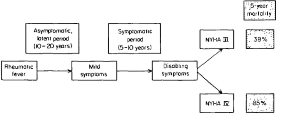
Following an episode of rheumatic fever patients with mitra] stenosis are usually asymptomatic for 10 to 20 years. When symptoms first appear about another 5 to 10 years will pass until disabling symptoms develop121. Once major symptoms are present life
expectancy is shortened and 5-year survival is 62% in patients with NYHA class III symptoms and only 15% with NYHA class IV"'4' (Fig. 1). Furthermore, a
one-year mortality rate of close to 50% was reported by Rapaport"1 in patients with mitral stenosis
developing congestive right heart failure. The most common causes of death are pulmonary oedema, pulmonary hypertension with right ventricular failure, systemic embolism and infective endocarditis.
Haemodynamic background of symptoms in mitral stenosis
With increasing obstruction of the mitral valve there is a concomitant rise in left atrial pressure, and in patients with mitral stenosis a close correlation
Comspondenct: Dieter Burckhardt, MD, Professor of Cardiology, Division of Cardiology, University Hospital Basel, CH-4031 Basel/Switzerland.
between mitral valve area and symptomatology can be observed, although some variability in exercise tolerance in patients with the same degree of stenosis is not an infrequent finding (Table 1). If left atrial and pulmonary capillary pressures exceed 30 mmHg, capillary hydrostatic pressures exceed oncotic press-ures resulting in transudation of plasma into the intrastitial tissue and the intra-alveolar space leading to marked shortness of breath and pulmonary oedema. This mechanism is accentuated by exercise since a two-fold increase in mitral flow produces a four-fold increase in pressure-gradient across the valve. Accordingly, left atrial pressure rises steeply during exercise. The mechanism preventing pulmonary capillary pressures rising above pulmonary oedema level is reduction in diastolic mitral flow and cardiac output. This reduction in flow and cardiac output is accomplished by an increase in pulmonary vascular tone within the smaller arteriolar branches of the pulmonary circulation leading to active pulmonary hypertension. The resulting low cardiac output is responsible for symptoms such as increased fatigue and a marked decrease in exercise tolerance. Furthermore, chronic elevation of pressures within the left atrium will lead to dilatation and finally to atrial fibrillation. Chronic, permanent atrial fibrillation is generally not seen until mitral stenosis is hemodynam-ically significant.
Data from the Framingham study1" showed that the
cerebrovascular accident rate was increased six-fold with isolated atrial fibrillation and 18-fold when atrial fibrillation was associated with mitral valve disease. Since most of these cerebro-vascular accidents were due to embolism, it is not surprising that in 43% of patients with significant mitral stenosis and in 8-5% of patients with mitral regurgitation left atrial thrombi
96 D. Burckhardt et al. Asymptomatic, latent pertod (10-20 years) Rheumatic fever Symptomatic period (5-KD years) Mild symptoms Disabling symptoms NYHA IH
X
NYHA m. ":5-year : mortality :.' 38% ' :-85% . iFigure 1 National history of mitral stenosis.
Table 1 Mitral valve area (MVA) and symptomatic status in patients with mitral stenosis
MVA (cm2) Symptoms
>2-0 Asymptomatic at rest and during exercise 2-0-1 -5 Asymptomatic at rest; shortness of breath
during heavy exercise
1-5-1-2 a- or oligo-symptomatic at rest; shortness of breath with mild exercise
<l-2 Nocturnal cough, PND, orthopnoea, haemoptysis, RV failure.
PND, paroxysmal nocturnal dyspnoea, RV = right ventricular.
were found during surgery121. Approximately 20% of
patients with rheumatic mitral valve disease ex-perience systemic embolization at some time during the course of the illness and in 25% the embolism is recurrent"1. Fifty percent of detected emboli appear in
the cerebral circulation.
With regard to natural history and haemodynamics the following signs and symptoms are of clinical importance in patients with mitral stenosis: marked shortness of breath with mild exercise, paroxysmal nocturnal dyspnoea, orthopnoea, history of pulmo-nary oedema, haemoptysis, increased fatigue, marked decrease in exercise tolerance, systemic embolism, right ventricular failure and atrial fibrillation.
Indications for interventions in patients with mitral stenosis
Signs and symptoms in which an intervention should be considered in patients with mitral stenosis are listed in the following section with their causes in parentheses. Asymptomatic patients with the clinical picture of mitral stenosis should undergo exercise testing in order to objectively assess their exercise tolerance.
(A) SYMPTOMS
These comprise any form of dyspnoea (elevated pulmonary capillary wedge pressure). More severe
chronic symptoms are required as indications for mitral valve replacement than for percutaneous mitral balloon valvotomy because of the additional long-term imponderabilities imposed by the artificial device. Other symptoms include increased fatigue or marked decrease in exercise tolerance (low cardiac output with pulmonary arteriolar hypertension); haemoptysis (pulmonary venous hypertension).
(B) CLINICAL FINDINGS
These comprise systemic embolism (enlargement of left atrial appendage, atrial fibrillation); signs of right ventricular failure (pulmonary hypertension); atrial fibrillation (dilatation of left atrium).
(C) ECHOCARDIOGRAPHIC FINDINGS
These comprise pressure half time (t/2) > 150 ms, i.e. MVA < 1-5 cm2; mean diastolic pressure gradient
at rest >5 mmHg. (D) HAEMODYNAMIC DATA
These comprise mean diastolic pressure gradient at rest >5 mmHg; pulmonary capillary wedge pressure >20mmHg; systolic pulmonary artery pressure >50mmHg; mitral valve area <l-5cm2; pulmonary
arteriolar resistance > 3 units.
Interventions in patients with significant mitral stenosis
There are four possible interventions in patients with haemodynamically significant mitral stenosis: (a) closed commissurotomy; (b) open commissurotomy; (c) mitral valve replacement (with mechanical or bioprosthetic valves); (d) percutaneous mitral balloon valvotomy.
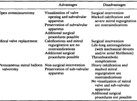
Comparative studies from Farhat et al.m have shown that open commissurotomy improved haem-odynamic values to a greater extent than closed commissurotomy, both at rest and during exercise. Therefore, in industrial countries closed commissuro-tomy is no longer performed and will not be described in this report. Each of the remaining three procedures has its advantages (Table 2) and there is a need for proper patient selection.
Mitral stenosis treatment 97
Table 2 Advantages and disadvantages of different procedures in mitral stenosis
Advantages Disadvantages Open commissurotomy Visualization of valve
opening and subvalvular apparatus
Preservation of subvalvular apparatus
Additional surgical procedures possible Calcifications and mitral
regurgitation are no contraindications Additional surgical
procedures possible Percutaneous mitral balloon Non-surgical intervention
valvotomy Preservation of sub-valvular apparatus
Mitral valve replacement
Surgical intervention Marked calcification and
severe mitral regurgitation are contraindications
Surgical intervention Life-long anticoagulation
(with mechanical devices and/or atrial fibrillation) Thromboembolic
complications Heavy calcification and
marked mitral regurgitation are contraindications No visualization of mitral
valve and sub-valvular apparatus
Additional surgical procedures not possible
(A) PREREQUISITE FOR OPEN COMMISSUROTOMY181
These comprise (1) Pliable valves (loud S, and presence of opening snap); (2) Absent or only slight calcification of mitral valve (reduced risk of dislodgement of calcific material); (3) Absent or only slight mitral regurgitation; no severe subvalvular disease (repair of valvular and subvalvular apparatus possible); (4) No previous commissurotomy; (5) Concomitant other valvular heart disease and/or coronary artery disease.
Left atrial thrombi are no contraindication (reduced risk of intra-operative embolism).
(B) PREREQUISITE FOR MJTRAL VALVE REPLACEMENT These comprise (1) Marked calcification of valve; (2) Moderate to severe concomitant mitral regurgita-tion; (3) Chronic atrial fibrillation with marked dilatation of left atrium together with atrial thrombi; (4) Significant involvement of the subvalvular apparatus; (5) Concomitant other valvular heart disease and/or coronary artery disease.
(C) PREREQUISITE FOR PERCUTANEOUS MITRAL BALLOON VALVOTOMY1"
These comprise (1) Pliable valves (loud S, and presence of opening snap); (2) Absent or minimal calcification; (3) Absent or mild mitral regurgitation; (4) No thrombi in left atrium and no recent previous thromoembolic events; (5) Absence of other con-comitant valvular heart disease and/or coronary artery disease.
Conclusions
In patients with mitral stenosis the need for therapeutic intervention can be assessed by clinical and non-invasive data. The indication for a given
intervention depends mainly on the status of the mitral apparatus, which can best be evaluated by transoesophageal and transthoracic echocardiography. Closed commissurotomy is no longer considered a valid therapeutic alternative due to its limited success rate but open commisurotomy and balloon valvotomy may be performed in patients without significant calcification of valve cusps and no major concomitant mitral regurgitation. Preservation of the subvalvular apparatus and left ventricular geometry can be considered the most important advantages of these techniques. More severe chronic symptoms and contraindications either to balloon valvotomy or commissurotomy are generally required as indications for mitral valve replacement because of the additional long-term imponderabilities imposed by an implanted artificial device. Therefore, in patients with mitral stenosis different symptoms and clinical findings will eventually lead to different interventions.
Finally, for certain special indications such as endstage mitral stenosis, severe pulmonary hyperten-sion, major involvement of the subvalvular apparatus or symptomatic mitral stenosis during pregnancy the optimal type of intervention is still a matter of debate.
References
[1] Bailey CP. The surgical treatment of mitral stenosis (mitral commissurotomy). Dis Chest 1949; 15: 377-84.
[2] Hell RJC. Rheumatic mitral valve disease. In: Julian DG, Camm AJ, Fox KM, Hall RJC, Diseases of the heart. Poole-Wilson, eds. London: Bailliere Tindall, 1989. [3] Bland EF, Jones TD. Rheumatic fever and rheumatic heart
disease. A 20 year report on 1000 patients followed since childhood. Circulation 1951; 4: 836-43.
[4] Oleson KH. The natural history of 271 patients with mitral stenosis under medical treatment. Br Heart J 1962; 24: 349-57.
98 D. Burckhardt et al.
[5] Rapaport E. Mitral stenosis. Chapter 38. In: Pannley WW, Chatterjee K., eds. Philadelphia: J. B. Lippincott, 1989. [6] Kannel WB, Abbot RD, Savaje DD « al. Epidemiologic
features of chronic atria] fibrillation: The Framinghara study. N Engl J Med 1982; 306: 1018-22.
[7] Farhat MB, Boussadia H, Gandjbakhch I et al. Closed vs open mitral commissurotomy in pure noncalcific mitral stenosis: Haemodynamic studies before and after operation.
J Thorac Cardiovasc Surg 1990; 99: 639-44.
[8] Kirklin JW and Barratt-Boyes BG. In: Cardiac Surgery. John Wiley & Sons, 1986: 323-73.
[9] Vahanian A, Michel PL, Cormier B, Vitoux B, Michel X, Slama M, Sarano LE, Trabelsi S, Ben Ismail M, Acar J. Results of percutaneous mitral commissurotomy in 200 patients. Am J Cardiol 1989; 63: 847-52.