Off-pump transapical mitral valve-in-ring implantation
Yu Zou
a,*, Enrico Ferrari
band Ludwig K. von Segesser
ba Department of Cardiac Surgery, The First Affiliated Hospital of Medical College, Zhejiang University, Hangzhou, China b Department of Cardiovascular Surgery, University Hospital of Lausanne, Lausanne, Switzerland
* Corresponding author. Service de Chirurgie CardioVasculaire, University Hospital of Lausanne-CHUV, Rue du Bugnon 46, CH-1011 Lausanne, Switzerland. Tel: +41-21-3142280; fax: +41-21-3142278; e-mail: [email protected] (Y. Zou).
Received 6 April 2012; received in revised form 5 June 2012; accepted 8 June 2012
Abstract
OBJECTIVES: The study aimed to evaluate the feasibility of off-pump transapical mitral valve-in-ring implantation and to test the per-formance of a custom-made self-expandable stent valve, in comparison with the standard SAPIEN valve.
METHODS: Acute experiments were performed infive pigs. Animals (mean weight 58.4 ± 7.3 kg) underwent mitral valve annuloplasties
under cardiopulmonary bypass using 26-mm rings (SJM™). Then, a 30-mm custom-made self-expandable stent valve or a 23-mm
balloon-expandable transcatheter heart valve (Edwards SAPIEN XT™) was deployed within the annuloplasty rings through a transatrial
access and under direct vision. Subsequently, the stent valves were inserted transapically underfluoroscopic guidance and off pump.
RESULTS: The procedural success of transatrial and transapical mitral valve-in-ring procedures was 100% (10 of 10). Mean transatrial and transapical procedure time was 2.0 ± 1.1 and 22.0 ± 5.7 min, respectively. Haemodynamic status during transapical implantation
remained stable, and differences in data collected before and after the stent-valve deployment were not statistically significant. Mean
mitral annulus diameter and mean mitral orifice area in the group of self-expandable stent valves were 2.60 ± 0.02 cm and 4.16 ± 0.48
cm2, respectively, whereas in the SAPIEN group they were 1.95 ± 0.18 cm and 2.26 ± 0.20 cm2, respectively. Trace or mild regurgitation
was detected only in the self-expandable stent-valve group. Mean gradients were 4.1 ± 4.5 mmHg across the self-expandable stent
valves and 1.0 ± 0 mmHg across the SAPIEN valves. Postmortem examination confirmed adequate positioning of the self-expandable
valves and the SAPIEN valves within the annuloplasty ring.
CONCLUSIONS: Off-pump transapical mitral valve-in-ring implantation is safe and feasible. Transapical access may represent the ideal option for valve-in-ring procedures in cases of recurrent mitral regurgitation after mitral valve repair, in high-risk patients. Owing to the
supra-annular profile of the valve components, our custom-made nitinol stent valve provides nearer to normal functional area than the
SAPIEN valve.
Keywords:Transcatheter• Mitral valve • Transapical • Valve-in-ring
INTRODUCTION
Mitral valve repair using an annuloplasty ring is the optimal surgical therapy for mitral regurgitation (MR). Some clinical studies have revealed the superiority of mitral valve repair over
replacement [1,2], including elderly patients [3]. Although
ac-ceptable early mitral valve repair results were demonstrated, recent studies have called into question the durability of mitral valve repair in some patients. They have documented the de-velopment of recurrent MR after repair for degenerative
aeti-ologies to be 2–4% per year [4,5]. Some centres reported that
recurrence of severe mitral valve regurgitation following valve
repair is up to 30% at 6 months [4,6,7], and some of them
require redo cardiac surgery. However, not only the surgeons but also the patients have to take into consideration the chal-lenge of redo surgery, especially in the case of elderly high-risk patients.
Recently, transcatheter valve replacement techniques have been developed to offer patients less invasive alternatives to open heart surgery. Transfemoral and transapical implantation of aortic and pulmonary stent valves have shown promising clinical results. Moreover, transcatheter mitral valve replacement is also under evaluation and, based on this development, some recently
published clinical reports and animal studies have confirmed the
feasibility of transcatheter mitral valve-in-valve or valve-in-ring
implantation [8–11]. Thus, the valve-in-ring technique turns out
to be a potential therapeutic technique for high-risk patients with mitral repair failure.
This study was designed to confirm the feasibility of transapical
transcatheter mitral valve-in-ring replacement without cardiopul-monary bypass (CPB) support using a custom-made self-expandable nitinol stent valve, and to evaluate the haemodynamic performances compared with the conventional SAPIEN heart valve (Edwards Lifesciences, Irvine, CA, USA), in a porcine model.
© The Author 2012. Published by Oxford University Press on behalf of the European Association for Cardio-Thoracic Surgery. All rights reserved.
BASIC
S
CIENCE
MATERIALS AND METHODS
The stent valve
Two different stent valves were employed, as follows.
(i) A 30-mm self-expandable stent with a double-crown design
was manufactured as described in a previous article [12]. It is
composed of two self-expandable nitinol Z-stents covered
by an ultra-thin polytetrafluoroethylene membrane, that
were sutured together like two opposite crowns, for the
annularfixation. A 30-mm diameter Dacron tube is attached
at the centre of the fixation system and it accommodates a
unidirectional trileaflet semilunar valve created in-house with
commercial porcine pericardium (Vascutek, Swillington, UK;
Fig.1A). All stent valves were testedin vitro prior to the
ex-perimentin vivo and then stored in glutaraldehyde solution.
(ii) A 23-mm Edwards SAPIEN XT™ balloon-expandable
trans-catheter heart valve (Edwards Lifescience, Irvine, CA, USA;
Fig.1B).
The delivery system
(i) A self-constructed Teflon sheath delivery system was
employedin vivo. It consisted of a 30-cm-long sheath and a
pusher with a blunt tip. The sheath, including the
stent-loading capsule, was made based on a modified 18
French commercial introducer sheath (B. Braun Melsungen Ag, Melsungen, Gemany). The distal end of the sheath was
dilated up to a 30 French inner lumen diameter by inflating
a 10-mm balloon (Boston Scientific Corp., Watertown, MA,
USA). On the basis of the length of the stent, a 40-mm
capsule was created [13]. Then, the stent valve was
com-pressed with a commercial crimper and loaded into the capsule. The valve deployment in the appropriate position was performed with a pusher into the sheath and without balloon catheters (nitinol stent). The folded valve measured
10 mm in diameter and 40–43 mm in length (Fig.2A).
(ii) Ascendra2 transapical delivery system (Edwards Lifescience,
Irvine, CA, USA; Fig.2B).
Animal preparation
Animals received care in compliance with the Principles of Laboratory Animals formulated by the National Society of Medical Research and the Guide for the Care and Use of Laboratory Animals prepared by the Institute of Laboratory Animal Resources and published by the US National Institutes of Health (NIH publication no. 85-23, revised 1985). The protocol was approved by the Institutional Committee on Animal Research.
Five pigs (mean weight 58.4 ± 7.3 kg, 9–12 weeks, female)
were included in our study for acute evaluation. After induction
Figure 1: Two different stent valves were used: a self-expandable stent valve (A) and the Edwards SAPIEN balloon-expandable transcatheter heart valve (B).
Figure 2:The custom-made delivery system, which loads the self-expandable stent valve (A), and the Ascendra2 transapical delivery system, which loads the Edwards SAPIEN valve (B).
of general anaesthesia, with tracheal intubation and mechanical ventilation (ketamine 22 mg/kg and atropine 0.8 mg/kg intra-muscularly; thiopental 15 mg/kg intravenously for induction; and
isoflurane 2.5% for maintenance of anaesthesia), the right carotid
artery and internal jugular vein were exposed, and catheters were introduced to monitor the blood pressure (BP) and the central venous pressure (CVP), and for blood sampling and infu-sion. The left carotid artery and the external jugular vein were prepared for cannulation for CPB. The right femoral vein was also exposed for insertion of an intracardiac ultrasonic probe (ICUS; Accuson Navigate, Acuson, Siemens, Munich, Germany). Continuous monitoring of electrocardiography, arterial pressure, central venous pressure and oxygen saturation was routine.
Annuloplasty ring implantation and transatrial
stent-valve implantation
After standard sternotomy and heparinization (Liquemine; La Roche Ltd, Basel, Switzerland; 100 IU/kg), the native mitral annular diameter was measured by ICUS. CPB was instituted with cannulation of the left carotid artery and external jugular vein. After aortic cross-clamping, antegrade cardioplegia was administered and the heart arrested. After opening of the left
atrium from the left side, a 26-mm SJM™ Séguin semi-rigid
annuloplasty ring (St Jude Medical Inc., St Paul, MN, USA) custom-marked by titanium clips was sutured at the standard
position of the mitral annulus (Fig.3). Saline injection tests were
performed to evaluate the mitral valve closure. Transatrial stent-valve implantation was performed under direct vision. The ven-tricular side of the self-expandable stent valve was released by sliding the pusher once the delivery system was placed across the ring; then, we gently pulled the introducer back in order to deploy the atrial side. Regarding the balloon-expandable valve, the delivery system was introduced within the ring and then
inflated at the proper position. The heart was de-aired, and the
aortic cross-clamp was removed after atrial closure. Once off pump, the ICUS was performed. The trans-stent and left ven-tricular outflow tract (LVOT) pressure gradient were measured with a needle. After all measurements were completed, a second cardiac arrest was induced on pump with cardioplegic solution,
and the stent valve was removed. The left atrium was closed, the aortic cross-clamp removed and the CPB stopped with stable haemodynamics.
Transapical mitral stent-valve implantation
Self-expandable stent-valve implantation (n = 4).
A double2–0 purse-string monofilament pledgeted felt suture was placed
at the left ventricular apex. The apex was punctured with a needle followed by a guide wire and an 8 French introducer system (Arrows, Reing, PA, USA) inserted into the left ventricle. Several titanium clips placed on the ring were seen as a dotted
circle underfluoroscopy. A soft-tipped, stiff guidewire (0.89 mm,
180 cm; Boston Scientific, Natick, MA, USA) was introduced into
the left ventricle, through the ring, and positioned in a left pulmonary vein. A pigtail catheter was advanced for exchanging the stiff wire. Then, a super-stiff guidewire (TSMG-35-26O-LES; Cook Medical, Limerick, Ireland) was exchanged using the pigtail catheter. A 30 French Ascendra2 Introducer Sheath Set (Edwards Lifescience) was used to dilate the transapical access and then removed, and the bleeding was controlled by tightening the purse-string sutures. The custom-made delivery system loaded with the self-expandable stent valve was pushed along the rigid guidewire, until the middle of the stent valve reached the mitral ring. Without rapid ventricular pacing, the atrial side of the stent partly expanded by advancing the pusher. We withdrew the
delivery system gradually until the fixation portion of the stent
was at the level of the ring, and then we gently pulled back the
delivery sheath to deploy the ventricular side (Fig.4A–D).
Edwards SAPIEN valve implantation (n = 1).
The transapical access was established as described above. A 23-mm SAPIEN valve was mounted on the delivery balloon and introducedthrough the ring under ICUS andfluoroscopy guidance. Once in
position, the stent was deployed following the standard
technique (Fig.4E and F).
After implantation, the ICUS was used to evaluate the function and competence of the stent valve for at least 30 min. The trans-stent and LVOT pressure gradients were measured directly with a needle. Thereafter, haemodynamic parameters were con-tinuously recorded for at least 1 h. The animals were then killed by an iv injection of Phenobarbital to permit inspection of the position of the stent valves in the rings.
Statistical analysis
Data were analysed with SPSS19 software for Windows. Variables
are reported as means ± standard deviation (SD), and Student’s
pairedt-test was used for comparison.
RESULTS
Annuloplasty rings were successfully implanted in all animals. Mean heart arrest time and CPB time were 19.0 ± 3.1 and 53.6 ± 4.9 min, respectively. The procedural success of transatrial and transapical mitral valve-in-ring was 100%. Custom-made self-expandable nitinol stent valves (30 mm) and 23-mm Edwards SAPIEN XT balloon-expandable transcatheter heart valves were used for comparison. The number of implanted stent valves and
Figure 3:The 26 mm semi-rigid annuloplasty ring was sutured at the mitral annulus under cardiopulmonary bypass (viewed and implanted from the left side). Clips were placed to the ring to be visualized underfluoroscopy.
BASIC
S
the implanted access are showed in Table 1. Mean transatrial and transapical procedure times were 2.0 ± 1.1 and 22.0 ± 5.7 min, respectively. Haemodynamic data before and after
transapi-cal valve deployment are showed in Table1.
The mean diameter of the native mitral annulus was 2.54 ±
0.13 cm, and the mean mitral valve area was 4.81 ± 0.52 cm2in
ICUS evaluation. Comparatively, they were 2.60 ± 0.20 cm and
4.16 ± 0.48 cm2 in the group of self-expandable stents and 1.95
± 0.18 cm and 2.26 ± 0.20 cm2in the group of SAPIEN valves,
re-spectively, after implantation. All valves within the stents opened
and closed completely and functioned well (Fig.5). In the
self-expandable stents, trace or mild central regurgitation was detected. A mild leak between the stent and the ring also existed in one of them. No regurgitation was observed in the
SAPIEN valves (Table2). The mean pressure gradient across the
self-expandable stent was 4.1 ± 4.5 mmHg, and across
the SAPIEN valve the gradient was 1.0 ± 0 mmHg. The
corresponding gradients across the LVOT were 3.0 ± 1.0 and 2.5
± 2.1 mmHg, respectively (Table2).
Postmortem examinations confirmed good positioning of the
stents within the annuloplasty rings in all animals, without LVOT
obstruction (Fig.6).
DISCUSSION
With the development of transcatheter aortic valve replacement, transapical access has become a routine approach for the im-plantation of transcatheter aortic valve prostheses because of its safety, reproducibility and low complication rate. Allowance for implantation of large devices and short access to both the mitral and the aortic valve are considered to be its main advantages
[14]. Despite these superior attributes, the interference of the
mitral valve apparatus with the delivery system still has a
Figure 4:The custom-made self-expandable stent valve was delivered (A–C) and anchored to the annuloplasty ring (white arrows; D). The Edwards SAPIEN valve was accurately positioned within the annuloplasty ring, marked with a titanium clip (white arrows; E and F). The stent retained the‘round’ shape (F).
negative influence in spreading this access to transcatheter mitral
valve replacements [15]. Several teams did some work on it and
reported their clinical and animal studies, including replacement of native mitral valves and mitral valve-in-valve procedures for
degenerated bioprostheses [8,16,17]. Nevertheless, up to now
no experimental studies have been published on a transapical mitral valve-in-ring procedure.
In our study, we successfully performed transapical mitral valve-in-ring implantations using two different types of stent
valves, and there were no significant differences in terms of
haemodynamics, heart rate, blood pressure and CVP before and
after the valve implantation (P > 0.05). This finding attests to the
feasibility of transapical mitral valve-in-ring implantations without CPB.
So far, the available balloon-expandable Edwards SAPIEN transcatheter heart valve has been the only valve used in reports on mitral valve-in-valve or valve-in-ring procedures, and this
reflects its promising results and its excellent low profile and
performing delivery system. The main problem of the
valve-in-ring technique is matching the size between the stent valve and the mitral ring. Joerg Kempfert and colleagues reported in their tests that the 23- and 26-mm SAPIEN valves fitted well within annuloplasty ring sizes up to 26 and 28 mm, respectively. An oversized ring may result in median or severe central MR because the stent may expand excessively and may
become dislodged [9]. In our study, the 23-mm SAPIEN valve
matched the 26-mm ring very well. No central or perivalvular regurgitation was recorded, and dislodgement of the stent valve never happened. On the contrary, a conformational change of
shape in SAPIEN valves from ‘round’ to ‘oval’ when implanted
into the ring was described in previous studies, but this was not
observed in our series [9,10]. In fact, when the stent expanded,
its shape remained‘round’, whereas the ‘D’-shaped annuloplasty
ring became circular (Fig.4F). Postmortem examination also
con-firmed this finding (Fig. 6C), and this contradiction is probably
due to the use of semi-rigid annuloplasty rings in our study. A
benefit resulting from this change of shape is the maintainance
of normal valve haemodynamics and a reduction in the risk of perivalvular and/or intravalvular regurgitation.
We also found that the stent-valve orifice area (2.26 ± 0.20
cm2) after the implantation of a SAPIEN valve into the ring was
significantly lower compared with the surface area of the native
mitral valve (4.80 ± 0.60 cm2; P < 0.05). This result is a relative
valve undersizing and could appear to be suboptimal in clinical cases.
There are some advantages in a self-expanding valve design, such as the potential for the positioning of stent-valve
compo-nents in a supra-annular fashion, allowing for a larger orifice
area. In our study, the resulting orifice area was indeed superior
to the ones measured through the SAPIEN valves, but the trans-valvular gradient pressures were suboptimal because of the
Figure 5:An intracardiac ultrasonic probe (ICUS) was employed to evaluate the performances of the implanted self-expandable stent valve (A) and the Edwards SAPIEN valve (B). Both valve types were functioning well.
Table 1: Procedural and haemodynamic data
Animal no. Weight (kg) Stent HRa (beats/min)
BPa (mmHg)
CVPa (mmHg)
ECG Arrest time (min) CPB time (min) Tap time (min) Tat Tap B A B A B A B A 1 50 C C 50 47 56/23 47/12 6 5 NS NS 20 57 30 2 55 C C 92 102 76/31 76/38 5 8 NS NS 23 53 25 3 65 S C 102 120 73/44 92/35 10 7 NS NS 15 48 20 4 55 C S 103 56 81/45 91/23 6 12 NS NS 17 50 20 5 67 S C 132 50 82/29 71/26 5 5 NS NS 20 60 15
aDifferences between haemodynamics before and after the valve-in-ring implantation were not statistically significant. A: after transapical implantation; B:
before transapical implantation; BP: blood pressure; CPB: cardiopulmonary bypass; C: custom-made stent valve; CVP: central venous pressure; ECG: electrocardiography; HR: heart rate; NS: normal sinus; S: SAPIEN valve; Tap: transapical; Tat: transatrial.
BASIC
S
amount of material within the stent. As described in the previous paragraph, a Dacron tube for accommodating a valve was an extra component in our stent compared with the SAPIEN valve. Moreover, this tube was soft and might be partly deformed
while the bloodflow went through. All of these factors resulted
in higher transvalvular gradient pressures. We will further opti-mize the design of the stent in future experiments.
Based on our previous experience [12], the double-crown
self-expandable valve was modified using porcine pericardium to
replace the previous aortic and pulmonary valve homografts, and several advantages appeared when this prototype was compared with the SAPIEN valve, as follows: (i) a larger (diam-eter > 28 mm) stent could be applied; (ii) the structure of the
double crownfits extremely well into the annuloplasty ring, and
the ring offers an appropriate position for the fixation of the
stent valve; (iii) no rapid pacing is required during the
implantation; and (iv) the orifice valve area after the
implant-ation (4.16 ± 0.48 cm2) approximated the native valve area (4.81
± 0.52 cm2; P = 0.297 > 0.05). We think that our custom-made
double-crown stent valve is more suitable for valve-in-ring pro-cedures than the SAPIEN valve. However, in one case we detected a mild central and persistent regurgitation with high transvalvular pressures (15 mmHg); the probable reason for high gradients in this case was that the regurgitation resulted in an in-crease in the left ventricular end-diastolic pressure.
In conclusion, the present study demonstrated the feasibility of transapical mitral valve-in-ring implantations without extracor-poreal circulation. The transapical technique developed for the Edwards SAPIEN stent-valve implantation shows promising performances in valve-in-ring procedures. Moreover, new self-expandable stent valves implanted through transapical access show good haemodynamic results with better functional valve areas.
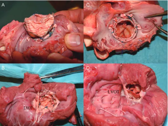
Figure 6:At postmortem examination, the custom-made self-expandable stent (A, left atrial side; B, left ventricular side) and the Edwards SAPIEN valve (C, left atrial side; D, left ventricular side) were positioned accurately within the annuloplasty ring, without left ventricular outflow tract (LVOT) obstruction. The ‘D’-shaped annuloplasty ring became near to circular in the presence of the fully expanded SAPIEN stent valve (C).
Table 2: Implanted stent-valve function
Self-expandable valves Edwards SAPIEN valves
Stent number n = 7 n = 3
Pressure gradient across the valve (mmHg) 4.1 ± 4.5 1.0 ± 0 Pressure gradient across the left ventricular outflow tract (mmHg) 3.0 ± 1.0 2.5 ± 2.1 Mitral regurgitation Trace or mild No
Native Stent Native Stent Valve diameter (cm) 2.54 ± 0.13 2.60 ± 0.02 2.53 ± 0.13 1.95 ± 0.18 Valve area (cm2) 4.81 ± 0.52 4.16 ± 0.48 4.80 ± 0.60a 2.26 ± 0.20a aThere was a significant difference between the area of the implanted Edwards SAPIEN valves and the native mitral valve orifice area.
LIMITATIONS
The study was an acute test in animals. The long-term durability and haemodynamics of both stent valves in rings are unknown.
The fixation stability of stents in mitral rings needs to be
con-firmed by long-term studies.
ACKNOWLEDGEMENTS
We thank Marko Burki and perfusionists involved in this study for their technical assistance during the animal experiments, which contributed to the success of this procedure.
Funding
This work was supported partially by National Natural Science Funding of China (81070264).
Conflict of interest: none declared.
REFERENCES
[1] Mohty D, Orszulak TA, Schaff HV, Avierinos JF, Tajik JA, Enriquez-Sarano M. Very long-term survival and durability of mitral valve repair for mitral valve prolapse. Circulation 2001;104:I1–7.
[2] Suri RM, Schaff HV, Dearani JA, Sundt TM 3rd, Daly RC, Mullany CJet al. Survival advantage and improved durability of mitral repair for leaflet prolapse subsets in the current era. Ann Thorac Surg 2006;82: 819–26.
[3] Medved I, Anic´ D, Ostric´ M, Zrnic´ B, Ivancic´ A, Tomulic´ V. Is mitral valve repair safe procedure in elderly patients? Coll Antropol 2010;34 (Suppl 2):213–5.
[4] Flameng W, Herijgers P, Bogaerts K. Recurrence of mitral valve regurgita-tion after mitral valve repair in degenerative valve disease. Circularegurgita-tion 2003;107:1609–13.
[5] Hoashi T, Bove EL, Devaney EJ, Hirsch JC, Ohye RG. Mitral valve repair for congenital mitral valve stenosis in the pediatric population. Ann Thorac Surg 2010;90:36–41.
[6] Ciarka A, Braun J, Delgado V, Versteegh M, Boersma E, Klautz Ret al. Predictors of mitral regurgitation recurrence in patients with heart failure undergoing mitral valve annuloplasty. Am J Cardiol 2010;106: 395–401.
[7] Galloway AC, Schwartz CF, Ribakove GH, Crooke GA, Gogoladze G, Ursomanno Pet al. A decade of minimally invasive mitral repair: long-term outcomes. Ann Thorac Surg 2009;88:1180–4.
[8] Webb JG, Wood DA, Ye J, Gurvitch R, Masson JB, Rodes-Cabau Jet al. Transcatheter valve-in-valve implantation for failed bioprosthetic heart valves. Circulation 2010;121:1848–57.
[9] Kempfert J, Blumenstein J, Chu MW, Pritzwald-Stegmann P, Kobilke T, Falk Vet al. Minimally invasive off-pump valve-in-a-ring implantation: the atrial transcatheter approach for re-operative mitral valve replace-ment after failed repair. Eur J Cardiothorac Surg 2009;35:965–9; discus-sion 69.
[10] Shuto T, Kondo N, Dori Y, Koomalsingh KJ, Glatz AC, Rome JJet al. Percutaneous transvenous Melody valve-in-ring procedure for mitral valve replacement. J Am Coll Cardiol 2011;58:2475–80.
[11] Casselman F, Martens S, De Bruyne B, Degrieck I. Reducing operative mortality in valvular reoperations: the‘valve in ring’ procedure. J Thorac Cardiovasc Surg 2011;141:1317–8.
[12] Ma L, Tozzi P, Huber CH, Taub S, Gerelle G, von Segesser LK. Double-crowned valved stents for off-pump mitral valve replacement. Eur J Cardiothorac Surg 2005;28:194–8; discussion 98–99.
[13] von Segesser LK, Marty B, Tozzi PG, Corno A. In situ introducer sheath dilatation for complex aortic access. Eur J Cardiothorac Surg 2002;22: 316–8.
[14] Blumenstein J, Van Linden A, Arsalan M, Moellmann H, Liebtrau C, Walther Tet al. Transapical access: current status and future directions. Expert Rev Med Devices 2012;9:15–22.
[15] Lozonschi L, Quaden R, Edwards NM, Cremer J, Lutter G. Transapical mitral valved stent implantation. Ann Thorac Surg 2008;86:745–8. [16] Attmann T, Pokorny S, Lozonschi L, Metzner A, Marcynski-Buhlow M,
Schoettler Jet al. Mitral valved stent implantation: an overview. Minim Invasive Ther Allied Technol 2011;20:78–84.
[17] Lutter G, Quaden R, Iino K, Hagemann A, Renner J, Humme Tet al. Mitral valved stent implantation. Eur J Cardiothorac Surg 2010;38:350–5.
BASIC
S