REVIEW ARTICLE
Arm lengthening after reverse shoulder arthroplasty: a review
Alexandre Lädermann&Tom Bradley Edwards&Gilles Walch
Received: 29 September 2013 / Accepted: 28 October 2013 / Published online: 23 November 2013 # Springer-Verlag Berlin Heidelberg 2013
Abstract
Purpose The purpose of this review is to provide a better understanding of biomechanical changes induced by reverse shoulder arthroplasty (RSA), discuss the different techniques of radiographic assessment of upper limb lengthening after RSA and determine the ideal soft tissue tension that provides the best functional outcome without increasing the risk of complications.
Methods Inclusion criteria were articles in which the primary interest was the technique of measuring upper-extremity lengthening after complications related to lengthening and its role in postoperative function; those written in English, French or German; and those that provided evidence levels I– IV relevant to search terms.
Results Seven articles met our inclusion criteria. Postopera-tively, changes in humeral length varied from minus five to five millimetres, and changes in upper-extremity length varied from 15 mm to 27 mm. The acromiohumeral distance
averaged 23 mm. Humeral and arm shortening increased the risk of dislocation and led to poor anterior active elevation. The type of surgical approach did not play a role in postoper-ative function. Subclinical neurological lesions were frequent. Conclusions Studies in this systematic review indicate that deltoid tensioning by restoring humeral length and increasing the acromiohumeral distance is critical for adequate postoper-ative function and to prevent dislocation. Excessive arm lengthening should be avoided, with zero to two centimetres of lengthening being a reasonable goal to avoid postoperative neurological impairment.
Keywords Reverse Shoulder Arthroplasty . Grammont prosthesis . Lengthening . Arm . Humerus and
acromiohumeral length . Review . Complication . Instability . Acromial fracture . Function
Introduction
During evolution, the development of the permanently upright posture has freed the human shoulder girdle of its quadruped functions. The anterior limbs became the upper limbs with the characteristics of a non-weight-bearing joint [1]. Major bony and muscular adaptations occurred. The rotator cuff is the most common structure that becomes compromised. When detached from the bone, the musculotendinous unit retracts medially [2], and the muscle may atrophy [3,4] or develop fatty infiltration [3,5–8]. In the absence of concavity com-pression and humeral head decom-pression exerted by the rotator cuff, the unopposed contraction of the deltoid creates a force vector that displaces the humeral head superiorly rather than creating abduction. With large rotator cuff lesions, the patient may present with pseudoparalysis [9,10]. To compensate for the loss of rotator cuff function, several options have been proposed. The preferred option, whenever possible, is to
A. Lädermann (*)
Division of Orthopaedics and Trauma Surgery, La Tour Hospital, Rue J.-D. Maillard 3, 1217 Meyrin, Switzerland
e-mail: [email protected] A. Lädermann
Faculty of Medicine, University of Geneva, Rue Michel-Servet 1, 1211 Geneva 4, Switzerland
A. Lädermann
Division of Orthopaedics and Trauma Surgery, Department of Surgery, Geneva University Hospitals, Rue Gabrielle-Perret-Gentil 4, 1211 Geneva 14, Switzerland
T. B. Edwards
Fondren Orthopedic Group, Texas Orthopedic Hospital, Houston TX, USA
G. Walch
Centre orthopédique Santy, Hôpital privé Jean-Mermoz, 24, Avenue Paul-Santy, 69008 Lyon, France
repair the rotator cuff. Good results are obtained in the vast majority of rotator cuff repairs [11–15], with healing of the cuff to the tuberosities [16] and successful reversal of the associated pseudoparalysis [17]. In some circumstances, rota-tor cuff repair is contraindicated, technically impossible or fails. In severe rotator cuff deficiency, the only remaining muscle able to elevate the arm is the deltoid. In order to allow anterior forward elevation above 90°, the abduction role of the deltoid has to be increased. Reverse shoulder arthroplasty (RSA) was developed to medialise and lower the glenohumeral centre of rotation, thereby increasing the lever arm of the deltoid muscle [18]. Deltoid tension, increased by the lower centre of rotation, increases muscle-fibre recruit-ment of the anterior and posterior deltoid, compensating for a deficient rotator cuff [19]. Due to the semiconstrained design of the prosthesis, adequate deltoid tension is critical to avoid dislocation. The lever arm of the deltoid muscle is almost doubled following RSA, and therefore, abduction efficiency of the deltoid increases. Under such tension, the reverse glenoid component provides the stable fulcrum essential for shoulder anterior elevation and prosthesis stability [19]. The increase in compressive force between the humeral and glenoid components also has a stabilising effect [20]. Failure to adequately tension the deltoid may result in prosthetic instability, one of the most common clinically significant complications. Moreover, other complications following RSA, such as neurological lesions, fractures of the acromion or fixed abduction of the arm [19–24], have also been de-scribed and could be related to excessive deltoid tension.
Few studies have been published about biomechanical implications and consequences of upper-extremity and humer-al lengthening following RSA. This article provides a com-prehensive review of current concepts pertaining to upper-extremity lengthening in RSA, including a review of pertinent biomechanical changes induced by the implant, risks related to lengthening and techniques to measure arm and humeral lengthening. Lastly, this article determines recommended del-toid tension to provide the best functional outcome without increasing the risk of complications.
Materials and methods
We identified all studies addressing techniques of measuring upper-extremity lengthening and its effect in RSA by conducting a search on PubMed from January 1970 to April 2013 using the combined terms“reverse shoulder arthroplasty”, “prosthesis”, “biomechanics”, “lengthening”, “complications” and“function”. We did not seek to perform a review of all studies documenting biomechanics but instead included only articles in which the primary interest was the technique of measuring lengthening after RSA, complications related to upper-extremity lengthening and the role of lengthening in
postoperative function. Studies were included in this systematic review if they were published in English, French or German and provided levels I–IV evidence relevant to the search terms.
Results
The literature search identified seven articles that met the inclusion criteria (Table 1). Four articles described both upper-extremity and humeral lengthening following RSA, with its consequences on function and complication rate [25–28]. One article described the relationship between acromiohumeral distance and deltoid lengthening and postop-erative function [29]. Another study limited data to a correla-tion between acromiohumeral distance and postoperative function [30]. One study described a technique of measuring arm length [31]. We also identified one article that reported the relationship of surgical approach on upper-extremity length-ening [25], and one study focused on the relationship between lengthening and postoperative neurological lesions [26].
Factors contributing to upper-extremity lengthening
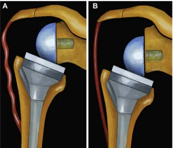
Adequate deltoid tension is accepted as being critical to pros-thetic function and stability [19, 27, 28]. This tension is determined by arm length. Arm length is dependant upon: 1. Position of the glenosphere in the frontal plane (Fig.1) 2. Status of the acromion
3. Size of the glenosphere
4. Use of an eccentric or inferiorly tilted glenosphere 5. Use of an augment or spacer
6. Thickness of the polyethylene 7. Type of stem
Table 1 Description of studies on upper-extremity lengthening after reverse shoulder arthroplasty (RSA)
Study Year No. of RSAs Design Level of evidence Renaud et al. [30] 2001 21 Retrospective cohort IV Boileau et al. [19] 2005 45 Retrospective cohort IV Lädermann et al. [28] 2009 58 Retrospective cohort IV Lädermann et al. [26]
2011 42 Prospective non randomised study II Lädermann et al. [25] 2011 144 Retrospective cohort IV Lädermann et al. [27] 2012 183 Retrospective cohort IV Jobin et al. [29]
2012 49 Prospective cohort design, treatment study
8. Height of the humeral cut and consequent level of stem implantation (Fig.2) [27,28].
Glenosphere position is theoretically fixed, as it should be implanted on the lower part of the glenoid to avoid notching [32–36]. The type of glenosphere (size, eccentricity) allows adjustment of arm length by only several millimeters (about 1 % of arm length). Consequently, the key factor for arm length is humeral length determined by height and type of stem, polyethylene thickness and use of an augment or spacer. Collectively, these factors allow arm lengthening by up to several centimeters (about 10 % of arm length).
Measurement of arm, humerus or deltoid length in RSA Few measurement techniques have yet been validated and can be either radiographic or clinical. Measurements can focus on
upper-extremity (arm) length, humeral length or acromiohumeral distance. Renaud et al. were the first to propose the determination of a “radio-anatomical index” [30]. They described a measuring technique in which anteroposterior (AP) radiographs are compared (Fig.3). This technique reported on acromiohumeral distance only and used radiographs that were not controlled for magnification. In cases of superior escape of the contralateral humeral head, the normal position of the humeral epiphysis was estimated using a horizontal line that passes perpendicular to the centre of the glenoid. The presence of superior glenoid erosion [37] renders this technique inaccurate.
Lädermann et al. presented a technique to determine arm and humeral length using plain radiography [28]. Measurements were taken from bilateral preoperative and postoperative magni-fication and fluoroscopically controlled AP radiographs of the humerus (Fig.4) and were made to determine relative arm length using points along the humerus and the acromion. A similar technique to assess the amount of lengthening of the humerus was subsequently reported by Greiner et al. [31]. Lädermann et al. compared the lengths of the affected and contralateral humeral shafts to determine whether the contralateral humerus may be used reliably as a reference for determining prosthetic height in complex cases with humeral bone loss, or when performing a postoperative assessment in revision cases in which preoperative scaled radiographs of the humerus are unavailable [28]. One disadvantage of this technique is the need to perform magnification-controlled radiographs of the entire humerus. As the X-ray beam is centred on the middle third of the humerus, radiographs do not provide an accurate depiction of the acromiohumeral interval. Consequently, this technique accurate-ly reflects humeral length, but accuracy of acromiohumeral interval measurements is compromised. Moreover, this tech-nique requires drawing an epicondylar reference line, which can be difficult if the humerus is not in neutral rotation.
Jobin et al. recently proposed another technique to evaluate subacromial and deltoid length postoperatively [29]. In their study, complete preoperative and postoperative true AP radio-graphs of the glenohumeral joint in neutral rotation were collected. The subacromial length (acromion to greater
Fig. 1 Influence of glenosphere position in the vertical plane. a A superior implantation of the baseplate or the use of a noneccentric glenosphere does not allow proper deltoid tensioning. b Use of an eccentric glenosphere or inferior positioning of the glenosphere in the vertical plane allows satisfactory deltoid tensioning. From [27], with permission
Fig. 2 Influence of humeral cut on arm length. a Preoperative status with a lack of deltoid tension. b, c Aggressive humeral cut results in low implantation of the stem, with lack of deltoid tension. d, e Minimal
humeral cut leads to high implantation of the prosthetic stem, with adequate deltoid tension. From [27], with permission
tuberosity distance) was measured as the distance from the inferolateral acromial tip to the most prominent superolateral aspect of the greater tuberosity (Fig.5). The middle deltoid length was defined as the distance between the inferolateral tip of the acromion to the midpoint of the deltoid tuberosity with the arm in neutral rotation and 0° abduction, as proposed initially by De Wilde [38]. Length was calibrated by the known diameter of the glenosphere and the fixed bony dis-tances of the humeral shaft width, and the fixed bony distance from the greater tuberosity to the deltoid tuberosity. The technique of Jobin et al. calibrates each radiograph to the glenosphere diameter. Consequently, one inconvenience is the impossibility of determining humeral and subacromial
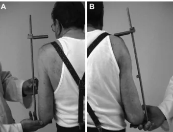
length preoperatively. This technique is therefore not useful in preoperative planning of difficult cases. Furthermore, the greater tuberosity was selected for the proximal reference point. This anatomical landmark may be absent preoperative-ly, or if it is present, it may be difficult to visualise because of arm rotation. Moreover, humeral radiolucencies, stem subsi-dence, radiological signs of stress shielding and resorption of tuberosities are common complications after RSA [34] that can compromise anatomical landmarks used in this technique. Lastly, Boileau et al. measured the postoperative length of the arm relative to the opposite side using a specially designed caliper (Fig.6) [19]. This technique is noninvasive but neither gives information on humeral or subacromial length nor does it allow for preoperative planning.
Results for arm, humerus and subacromial lengthening Postoperative lengthening of the arm, humerus and subacromial space (acromiohumeral interval) is summarised in Table 2. Mean lengthening varied from 15 mm to 27 mm for the arm and from minus five to five millimetres for the humerus. The
Fig. 3 Technique proposed by Renaud et al. Two main lines are placed for measurement: an acromial line that represents the superior cortex of the acromion, and a tangent line to the centre of the prosthetic epiphysis or
to the centre of rotation of the humeral head perpendicular to the first line. The two latter lines represent the acromioepiphyseal distance and are compared to provide a ratio of lengthening. From [30], with permission
Fig. 4 Technique of Lädermann et al. [28]. Preoperative and postopera-tive true anteroposterior, bilateral, magnification-controlled radiographs of the humeri with neutral rotation and the patient standing. An epicondylar line (EL) defined as being between the most lateral part of the medial and lateral epicondyle. The diaphyseal axis (DI) is determined by a line drawn in the centre of the proximal humeral medullary canal. The intersection between the EL and DI represents point C. The inter-section between the DI and the top of the humeral head is point H. Point Ais the intersection between the DI and a perpendicular line passing through the most lateral and inferior point of the acromion (A). A, C and H are represented by small white points; large white points corre-spond to the magnification control marker on the skin of the arm. C condyles, preop preoperative, contra contralateral, EF enlargement factor
Fig. 5 Technique of Jobin et al. Radiographic measurement of deltoid length from the inferolateral acromion tip to the midpoint of the deltoid tuberosity preoperatively (left, d) and postoperatively (right, d’). From [29], with permission
humeral cut was more aggressive when a transdeltoid surgical approach was performed; this was compensated for by an in-crease in thickness of the polyethylene liner. Mean subacromial lengthening reported in two studies was 23 mm [28,29].
Relationship between lengthening and postoperative function Functional outcomes after RSA have shown variable results for range of motion (ROM) [23,39–41]. Poor postoperative ante-rior elevation can be attributed to improper use, poor patient selection and preoperative and postoperative problems [41,42]. Renaud et al. demonstrated a correlation between a subacromial space lengthening of 33–50 % and: (1) Constant score [43] ≥65.5 points (p =0.024), (2) anterior elevation ≥120° (p = 0.001), and (3) gain in abduction ≥60° (p =0.016) [30]. Lädermann et al. compared patients with arm lengthening and those with shortening and found that the postoperative active anterior elevation was significantly greater for arm lengthening (145° vs 122°), with a mean difference of 23° (p <0.001) [27]. Jobin et al. also confirmed that deltoid lengthening correlated
significantly (p =0.002) with active anterior elevation [29]. In their study, deltoid lengthening that achieved an acromion-to-greater-tuberosity distance over 38 mm had a 90 % positive predictive value (PPV) of obtaining 135° of active anterior elevation. These clinical findings confirmed biomechanical studies that demonstrate the crucial role of the deltoid in post-operative function [18,44]. However, arm lengthening showed no relationship to outcome scores, including Constant [43], Disabilities of the Arm, Shoulder and Hand (DASH) [45], American Shoulder and Elbow Surgeons (ASES) [46] or Sim-ple Shoulder Test (SST) [29,31] scores.
Relationship between lengthening and postoperative complications
Dislocation
Dislocation is one of the most common complications after RSA, with rates as high as 14 % and accounting for almost half of the complications in some series [21,41,47–53]. Most cases of dislocation occur during the first few months after implantation and are a result of a technical error [54]. The aetiology of dislocation is multifactorial. It can occur due to: 1. Deltoid insufficiency [41,42]
2. Lack of anterior restraints including subscapularis insuf-ficiency, conjoint tendon weakness [55] and pectoralis major insufficiency
3. Component malpositioning 4. Impingement
5. Infection
Instability is more frequent in cases of revision arthroplasty [56]. Deltoid insufficiency can be caused by preoperative factors [41,42] or result from a postoperative lack of deltoid tension, acromial or scapular spine fracture (Fig.7), polyeth-ylene wear, stem subsidence or postoperative neurological palsy. Interestingly, no previous studies have reported
Fig. 6 Distance between acromion and olecranon with the elbow flexed is determined on a nonoperated and b operated sides. From [19], with permission
Table 2 Mean lengthening of arm, humerus and subacromial space postoperatively in millimetres
Values are mean ± standard devi-ation (range)
DP deltopectoral approach, NA not available, TD transdeltoid approach
a
Compared with contralateral side
b
Compared with ipsilateral side
Study Arm (deltoid lengthening) Humerus Subacromial space (acromiohumeral distance) Renaud et al. [30] NA NA NA Boileau et al. [19] 15±11 (5–40)a NA NA Lädermann et al. [28] 23±12 (4–47)b 20±11 (−2 to 48)a 2±6 (−10 to16)a 23±9 (5–41)b Greiner et al. [31] 17±13 (−10 to 45) NA NA Lädermann et al. [26] 27±18 (0–59)a NA NA Lädermann et al. [25] DP 17±17, TD 12±1.4a DP 5±13, TD−5±10a NA Lädermann et al. [27] 16±19 (−51 to 54)a 2±14 (−47 to 52)a NA Jobin et al. [29] 21±10b NA 23±9b
increased rates of postoperative dislocation after acromial or scapular spine fractures [57, 58]. Lädermann et al. noted a strong correlation (p <0.0001) between preoperative humeral length and postoperative dislocation. Postoperative shortening of the humerus, compared with preoperative or contralateral humeral length, was observed in all cases of dislocation.
Acromial or scapular spine fractures
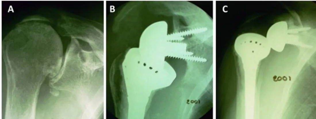
The arm is lengthened by approximately 15–27 mm following RSA (Table2). Biomechanically, tension on the deltoid and acromion is subsequently increased as a result of this length-ening. Preoperative and/or postoperative acromial pathology, which could compromise deltoid function and consequently affect the function of the prosthesis, is of legitimate concern. Postoperative fractures occur in at least in 3 % of cases [59], and their causes are theoretically numerous. Preoperatively, the acromion may be subject to a congenital or acquired abnormality, such as an os acromiale [58]. It may also already be eroded, fragmented or even fractured from the superiorly migrated humeral head in cases of cuff-tear arthropathy or osteoporosis-induced insufficiency. The superior base-plate fixation screw may function as a stress riser that results in acromial fractures (Fig. 8) [60,61]. It seems that the most significant risk factor is preoperative osteoporosis [61].
Neurological lesions
Clinically relevant neurological complications involving the bra-chial plexus or the axillary nerve are considered rare [21,62–65]. A prospective study determined the incidence of peripheral nerve lesions as determined by electromyographical analysis following RSA [26]. If one also takes into account subclinical deterioration of preoperative lesions, 63 % of patients in this study had postoperative neurological lesions. The prevalence of peripheral nerve lesions determined by electromyographical analysis following RSA is thus common, but patients usually recover. Arm lengthening during RSA, because of its nonanatomical design and/or manoeuvre of glenohumeral
reduction, may be a major factor responsible for the increased prevalence of neurological injury.
Discussion
RSA is a commonly performed procedure, and its indications continue to expand. Despite the relatively high complication rate [22,54,66–68], RSA continues to be performed because of the significant postoperative improvement in shoulder function and the high rate of patient satisfaction. Ways to prevent complications associated with RSA require further investigation. A better understanding of the biomechanical implications of inserting an RSA may help avoid some of these complications. Obtaining an improved understanding of the relationship between these biomechanical effects and com-plications was the purpose of this review.
At present, there is no described standardised preoperative planning technique for determining appropriate implant posi-tion based on deltoid tension or length. Intraoperative criteria have been proposed by other authors to assess prosthetic stability. Recommendations are numerous and include:
Fig. 7 a Preoperative anteroposterior X-ray of a right shoulder with an acromial fatigue fracture. b At 2 years of follow-up, a postoperative tilt of the acromion and a grade 4 scapular notch are noted. c Prosthetic dislocation could be related to the lack of deltoid tension
Fig. 8 a Postoperative anteroposterior X-ray of a right shoulder with a scapular spine fracture. b Axial computed tomography scan reveals the superior metaglene fixation screw may function as a stress riser that results in fracture
1. Implanting the prosthesis in such a way that it is difficult to reduce
2. Absence of pistoning of the prosthesis when applying axial traction on the arm
3. Stability throughout a full ROM
4. Passive adduction of the arm to neutral with the elbow at the side
5. Palpation of tension in the conjoint tendon after reduction, with the arm at the side and the elbow extended [19] 6. No asymmetric subluxation or tilting of the proximal
hu-meral component on the glenosphere during adduction [18] 7. Free glenohumeral motion without scapula–thoracic
mo-tion between 0° and 60° of abducmo-tion [69]
These intraoperative criteria, however,are qualitative, sub-jective and depend more on patient relaxation (i.e. depth of anaesthesia and quality of muscle relaxation) and preoperative scar tissue (i.e. post-traumatic arthritis or revision arthroplasty versus primary arthroplasty) than on objective measurements to assess the appropriate length of the deltoid or the arm. Some authors even recommend the use of a“Jedi skill that involves using the Force”, rather than the previously mentioned criteria [70]. A preoperative guide, useful in complex cases such as revision arthroplasty or post-traumatic arthritis where scar tissue and bone loss prevent making an accurate determination of humeral length, has thus been proposed (Fig. 9) [28]. Preoperative planning is probably not necessary in all primary cases; its use in revision cases, however, seems mandatory. To
guarantee the best possible functional results, restoration of the appropriate humeral and arm length should be the goal [27,29,30]. Failure to restore sufficient deltoid tension may be responsible for poor anterior elevation and prosthetic in-stability [27,28,54]. Implantation of the humeral stem at the level of the humeral cut using the thickness of the polyethyl-ene insert to obtain appropriate deltoid tension seems to be a reasonable option [25,28].
Excessive lengthening of the arm may be responsible for neurological lesions, acromial or scapular spine fractures or fixed arm abduction [26,28]. One study demonstrated a high prevalence of acute postoperative subclinical neurological lesions after RSA [26]. Lengthening of the arm during this procedure, because of its nonanatomical design and/or ma-noeuvre of glenohumeral reduction, might be a major factor responsible for the high prevalence of neurological injury. The risk of neurological lesions increases drastically with more than four centimetres of lengthening. An absolute lengthening threshold expressed in centimetres is, however, difficult to determine. Seemingly, a ratio that takes into consideration the total length of the upper limb of the patient, thus representing a percentage of lengthening, would be more accurate. However, this concept must be applied with caution, as lengthening beyond two centimetres compared with preop-erative measurement may increase the frequency of postoper-ative neurological injury [26]. As a result, strategies have been developed to limit upper-extremity lengthening in RSA. In cases with a high risk of dislocation, such as revisions or proximal humeral bone loss, use of larger-diameter glenoid components, a superior approach and prosthetic or bony lateralisation of the glenosphere can be considered to avoid excessive tension [71,72]. Nevertheless, if the preoperatively planned lengthening is over four centimetres, the authors recommend using intraoperative nerve monitoring [73].
Conclusion
Studies in this systematic review indicate that adequate deltoid tension obtained through restoration of humeral length and increase of the acromiohumeral interval is the key for adequate postoperative function preventing instability. Arm lengthening should be controlled, with zero to two centimetres being a reasonable goal to avoid postoperative neurological impair-ment. Current conventional radiographic preoperative planning techniques are inaccurate. Development of new preoperative and intraoperative aides for surgeons, using software, intraop-erative guides and other imaging modalities such as computed tomography and magnetic resonance imaging, are required.
Disclaimer A. Lädermann, his immediate family, and any research foundation with which he is affiliated did not receive any financial Fig. 9 Proposition for determining the height at which the prosthesis
should be implanted by planning of the operation with a 10-cm marker: A = corrected length of contralateral humerus = CHcontra × 10: contra EF=314 mm. B = corrected length of the preoperative humerus = CHipsi × 10: preop EF=264 mm. A-B = corrected length of the missing bone. PHipsi = A-B=50 mm. PHipsi is the exact distance in millimetres that we must measure at the time of implantation between the lateral cortex of the humerus (Hipsi) and the superolateral part of the metallic stem (P). A acromion, C condyles, Hcontra head, EP epicondylar line, DI diaphyseal axis, pre-op preoperative, contra contralateral, EF enlargement factor
payments or other benefits from any commercial entity related to the subject of this article. Tom Bradley Edwards received royalties from, serves as a paid consultant to or is an employee of, or has received research or institutional support from Tornier. Gilles Walch received royalties from Tornier.
References
1. Baulot E, Sirveaux F, Boileau P (2011) Grammont’s idea: the story of Paul Grammont’s functional surgery concept and the development of the reverse principle. Clin Orthop Relat Res 469(9):2425–2431. doi:
10.1007/s11999-010-1757-y
2. Meyer DC, Farshad M, Amacker NA, Gerber C, Wieser K (2012) Quantitative analysis of muscle and tendon retraction in chronic rotator cuff tears. Am J Sports Med 40(3):606–610. doi:10.1177/ 0363546511429778
3. Williams MD, Lädermann A, Melis B, Barthelemy R, Walch G (2009) Fatty infiltration of the supraspinatus: a reliability study. J Shoulder Elbow Surg 18(4):581–587. doi:10.1016/j.jse.2008.12.014
4. Zanetti M, Gerber C, Hodler J (1998) Quantitative assessment of the muscles of the rotator cuff with magnetic resonance imaging. Investig Radiol 33(3):163–170
5. Goutallier D, Postel JM, Bernageau J, Lavau L, Voisin MC (1994) Fatty muscle degeneration in cuff ruptures. Pre- and postoperative evaluation by CT scan. Clin Orthop Relat Res 304:78–83
6. Goutallier D, Postel JM, Bernageau J, Lavau L, Voisin MC (1995) Fatty infiltration of disrupted rotator cuff muscles. Rev Rhum 62(6): 415–422
7. Goutallier D, Postel JM, Lavau L, Bernageau J (1999) Impact of fatty degeneration of the suparspinatus and infraspinatus msucles on the prognosis of surgical repair of the rotator cuff. Rev Chir Orthop Reparatrice Appar Mot 85(7):668–676
8. Kim HM, Galatz LM, Lim C, Havlioglu N, Thomopoulos S (2012) The effect of tear size and nerve injury on rotator cuff muscle fatty degeneration in a rodent animal model. J Shoulder Elbow Surg 21(7): 847–858. doi:10.1016/j.jse.2011.05.004
9. Boileau P, Baque F, Valerio L, Ahrens P, Chuinard C, Trojani C (2007) Isolated arthroscopic biceps tenotomy or tenodesis improves symptoms in patients with massive irreparable rotator cuff tears. J Bone Joint Surg Am 89(4):747–757. doi:10.2106/JBJS.E.01097
10. Lädermann A, Collin P, Walch G (2012) Correlation of the involved compartments of massive rotator cuff tear and loss of active shoulder range of motion. Knee Surg Sports Traumatol Arthrosc 20(Suppl 1): S1–S3. doi:10.1007/s00167-012-1934-5
11. Denard PJ, Jiwani AZ, Lädermann A, Burkhart SS (2012) Long-term outcome of a consecutive series of subscapularis tendon tears repaired arthroscopically. Arthroscopy. doi:10.1016/j.arthro.2012.02.031
12. Denard PJ, Jiwani AZ, Lädermann A, Burkhart SS (2012) Long-term outcome of arthroscopic massive rotator cuff repair: the importance of double-row fixation. Arthroscopy 28(7):909–915. doi:10.1016/j. arthro.2011.12.007
13. Denard PJ, Lädermann A, Burkhart SS (2011) Arthroscopic manage-ment of subscapularis tears. Sports Med Arthrosc Rev 19(4):333– 341. doi:10.1097/JSA.0b013e31822d41c6
14. Lädermann A, Denard PJ, Burkhart SS (2012) Revision arthroscopic rotator cuff repair: systematic review and authors’ preferred surgical technique. Arthroscopy 28(8):1160–1169. doi:10.1016/j.arthro.2012. 01.006
15. Lädermann A, Denard PJ, Burkhart SS (2011) Midterm outcome of arthroscopic revision repair of massive and nonmassive rotator cuff tears. Arthroscopy 27(12):1620–1627. doi:10.1016/j.arthro.2011.08.290
16. Zumstein MA, Jost B, Hempel J, Hodler J, Gerber C (2008) The clinical and structural long-term results of open repair of massive
tears of the rotator cuff. J Bone Joint Surg Am 90(11):2423–2431. doi:10.2106/JBJS.G.00677
17. Denard PJ, Lädermann A, Jiwani AZ, Burkhart SS (2012) Functional outcome after arthroscopic repair of massive rotator cuff tears in individuals with pseudoparalysis. Arthroscopy 28(9):1214–1219. doi:10.1016/j.arthro.2012.02.026
18. Grammont PM, Trouilloud P, Latfay J, Deries X (1987) Etude et réalisation d’une nouvelle prothèse d’épaule. Rhumatologie 39:407– 418
19. Boileau P, Watkinson DJ, Hatzidakis AM, Balg F (2005) Grammont reverse prosthesis: design, rationale, and biomechanics. J Shoulder Elbow Surg 14(1 Suppl S):147S–161S. doi:10.1016/j.jse.2004.10. 006
20. Gagey O, Hue E (2000) Mechanics of the deltoid muscle. A new approach. Clin Orthop Relat Res 375:250–257
21. Boileau P, Watkinson D, Hatzidakis AM, Hovorka I (2006) Neer Award 2005: the Grammont reverse shoulder prosthesis: results in cuff tear arthritis, fracture sequelae, and revision arthroplasty. J Shoulder Elbow Surg 15(5):527–540. doi:10.1016/j.jse.2006.01.003
22. Scarlat MM (2013) Complications with reverse total shoulder arthroplasty and recent evolutions. Int Orthop 37(5):843–851. doi:
10.1007/s00264-013-1832-6
23. Sirveaux F, Favard L, Oudet D, Huguet D, Lautman S (2001) Grammont inverted total shoulder arthroplasty in the treatment of glenohumeral osteoarthritis with massive and non repairable cuff rupture. In: Walch G, Boileau P, Molé D (eds) 2000 shoulder pros-theses: Two to ten year follow-up. Sauramps Medical, Montpellier, pp 247–252
24. Valenti PH, Boutens D, Nerot C (2001) Delta 3 reversed prosthesis for osteoarthritis with massive rotator cuff tear: Long term results (> 5 years). In: Walch G, Boileau P, Molé D (eds) 2000 shoulder prosthe-ses: Two to ten year follow-up. Sauramps Medical, Montpellier, pp 253–259
25. Lädermann A, Lubbeke A, Collin P, Edwards TB, Sirveaux F, Walch G (2011) Influence of surgical approach on functional outcome in reverse shoulder arthroplasty. Orthop Traumatol Surg Res 97(6):579– 582. doi:10.1016/j.otsr.2011.04.008
26. Lädermann A, Lubbeke A, Mélis B, Stern R, Christofilopoulos P, Bacle G, Walch G (2011) Prevalence of neurologic lesions after total shoulder arthroplasty. J Bone Joint Surg Am 93(14):1288–1293. doi:
10.2106/JBJS.J.00369
27. Lädermann A, Walch G, Lubbeke A, Drake GN, Mélis B, Bacle G, Collin P, Edwards TB, Sirveaux F (2012) Influence of arm lengthen-ing in reverse shoulder arthroplasty. J Shoulder Elbow Surg 21(3): 336–341. doi:10.1016/j.jse.2011.04.020
28. Lädermann A, Williams MD, Mélis B, Hoffmeyer P, Walch G (2009) Objective evaluation of lengthening in reverse shoulder arthroplasty. J Shoulder Elbow Surg 18(4):588–595. doi:10.1016/j.jse.2009.03.012
29. Jobin CM, Brown GD, Bahu MJ, Gardner TR, Bigliani LU, Levine WN, Ahmad CS (2012) Reverse total shoulder arthroplasty for cuff tear arthropathy: the clinical effect of deltoid lengthening and centre of rotation medialization. J Shoulder Elbow Surg 21(10):1269–1277. doi:10.1016/j.jse.2011.08.049
30. Renaud P, Wahab H, Bontoux L, Dauty M, Richard I, Bregeon C (2001) Total inverted shoulder prosthesis and rotator cuff insufficien-cy: evaluation and determination of anatomical parameters predictive of good functional outcome in 21 shoulders. Ann Readapt Med Phys: Rev Sci Soc Fr Reeducation Fonctionnelle Readaptat Med Phys 44(5):273–280
31. Greiner SH, Back DA, Herrmann S, Perka C, Asbach P (2010) Degenerative changes of the deltoid muscle have impact on clinical outcome after reversed total shoulder arthroplasty. Arch Orthop Trauma Surg 130(2):177–183. doi:10.1007/s00402-009-1001-y
32. De Biase CF, Ziveri G, Delcogliano M, de Caro F, Gumina S, Borroni M, Castagna A, Postacchini R (2013) The use of an eccentric glenosphere compared with a concentric glenosphere in reverse total
shoulder arthroplasty: two-year minimum follow-up results. Int Orthop 37(10):1949–1955. doi:10.1007/s00264-013-1947-9
33. Levigne C, Garret J, Boileau P, Alami G, Favard L, Walch G (2011) Scapular notching in reverse shoulder arthroplasty: is it important to avoid it and how? Clin Orthop Relat Res 469(9):2512–2520. doi:10. 1007/s11999-010-1695-8
34. Mélis B, DeFranco M, Lädermann A, Mole D, Favard L, Nerot C, Maynou C, Walch G (2011) An evaluation of the radiological chang-es around the Grammont reverse geometry shoulder arthroplasty after eight to 12 years. J Bone Joint Surg Br 93(9):1240–1246. doi:10. 1302/0301-620X.93B9.25926
35. Mizuno N, Denard PJ, Raiss P, Walch G (2013) Reverse total shoul-der arthroplasty for primary glenohumeral osteoarthritis in patients with a biconcave glenoid. J Bone Joint Surg Am 95(14):1297–1304. doi:10.2106/JBJS.L.00820
36. Nyffeler RW, Werner CM, Gerber C (2005) Biomechanical relevance of glenoid component positioning in the reverse Delta III total shoul-der prosthesis. J Shoulshoul-der Elbow Surg 14(5):524–528. doi:10.1016/j. jse.2004.09.010
37. Sirveaux F, Favard L, Oudet D, Huquet D, Walch G, Mole D (2004) Grammont inverted total shoulder arthroplasty in the treatment of glenohumeral osteoarthritis with massive rupture of the cuff. Results of a multicentre study of 80 shoulders. J Bone Joint Surg Br 86(3):388–395 38. De Wilde LF, Audenaert EA, Berghs BM (2004) Shoulder prostheses treating cuff tear arthropathy: a comparative biomechanical study. J Orthop Res 22(6):1222–1230. doi:10.1016/j.orthres.2004.03.010
39. Dedy NJ, Stangenberg M, Liem D, Hurschler C, Simmen B, Riner M, Marquardt B, Steinbeck J (2011) Effect of posterior offset humeral components on range of motion in reverse shoulder arthroplasty. Int Orthop 35(4):549–554. doi:10.1007/s00264-010-1079-4
40. Ortmaier R, Resch H, Hitzl W, Mayer M, Blocher M, Vasvary I, Mattiassich G, Stundner O, Tauber M (2013) Reverse shoulder arthroplasty combined with latissimus dorsi transfer using the bone-chip technique. Int Orthop. doi:10.1007/s00264-013-2139-3
41. Wall B, Nove-Josserand L, O’Connor DP, Edwards TB, Walch G (2007) Reverse total shoulder arthroplasty: a review of results ac-cording to etiology. J Bone Joint Surg Am 89(7):1476–1485. doi:10. 2106/JBJS.F.00666
42. Lädermann A, Walch G, Denard PJ, Collin P, Sirveaux F, Favard L, Edwards TB, Kherad O, Boileau P (2013) Reverse shoulder arthroplasty in patients with pre-operative impairment of the deltoid muscle. Bone Joint J 95-B(8):1106–1113. doi:10.1302/0301-620X. 95B8.31173
43. Constant CR, Murley AH (1987) A clinical method of functional assessment of the shoulder. Clin Orthop Relat Res 214:160–164 44. Schwartz DG, Kang SH, Lynch TS, Edwards S, Nuber G, Zhang LQ,
Saltzman M (2012) The anterior deltoid’s importance in reverse shoulder arthroplasty: a cadaveric biomechanical study. J Shoulder Elbow Surg. doi:10.1016/j.jse.2012.02.002
45. Germann G, Wind G, Harth A (1999) The DASH(Disability of Arm-Shoulder-Hand) Questionnaire–a new instrument for evaluating up-per extremity treatment outcome. Handchir Mikrochir Plast Chir: Organ Deutschsprachigen Arbeitsgemeinschaft Handchir: Organ Deutschsprachigen Arbeitsgemeinschaft Mikrochir Peripheren Nerven Gefasse 31(3):149–152. doi:10.1055/s-1999-13902
46. Michener LA, McClure PW, Sennett BJ (2002) American shoulder and elbow surgeons standardized shoulder assessment form, patient self-report section: reliability, validity, and responsiveness. J Shoulder Elbow Surg 11(6):587–594. doi:10.1067/mse.2002.127096
47. Cazeneuve JF, Cristofari DJ (2006) Grammont reversed prosthesis for acute complex fracture of the proximal humerus in an elderly population with 5 to 12 years follow-up. Rev Chir Orthop Reparatrice Appar Mot 92(6):543–548
48. Cuff D, Pupello D, Virani N, Levy J, Frankle M (2008) Reverse shoulder arthroplasty for the treatment of rotator cuff deficiency. J Bone Joint Surg Am 90(6):1244–1251. doi:10.2106/JBJS.G.00775
49. rCuff DJ, Virani NA, Levy J, Frankle MA, Derasari A, Hines B, Pupello DR, Cancio M, Mighell M (2008) The treatment of deep shoulder infection and glenohumeral instability with debridement, reverse shoulder arthroplasty and postoperative antibiotics. J Bone Joint Surg Br 90(3):336–342. doi:10.1302/0301-620X.90B3.19408
50. De Wilde L, Sys G, Julien Y, Van Ovost E, Poffyn B, Trouilloud P (2003) The reversed Delta shoulder prosthesis in reconstruction of the proximal humerus after tumour resection. Acta Orthop Belg 69(6):495–500
51. Gohlke F, Rolf O (2007) Revision of failed fracture hemiarthroplasties to reverse total shoulder prosthesis through the transhumeral approach : method incorporating a pectoralis-major-pedicled bone window. Oper Orthop Traumatol 19(2):185–208. doi:
10.1007/s00064-007-1202-x
52. Levy J, Frankle M, Mighell M, Pupello D (2007) The use of the reverse shoulder prosthesis for the treatment of failed hemiarthroplasty for proximal humeral fracture. J Bone Joint Surg Am 89(2):292–300. doi:10.2106/JBJS.E.01310
53. Werner CM, Steinmann PA, Gilbart M, Gerber C (2005) Treatment of painful pseudoparesis due to irreparable rotator cuff dysfunction with the Delta III reverse-ball-and-socket total shoulder prosthesis. J Bone Joint Surg Am 87(7):1476– 1486. doi:10.2106/JBJS.D.02342
54. Farshad M, Gerber C (2010) Reverse total shoulder arthroplasty-from the most to the least common complication. Int Orthop 34(8):1075– 1082. doi:10.1007/s00264-010-1125-2
55. Edwards TB, Williams MD, Labriola JE, Elkousy HA, Gartsman GM, O’Connor DP (2009) Subscapularis insufficiency and the risk of shoulder dislocation after reverse shoulder arthroplasty. J Shoulder Elbow Surg 18(6):892–896. doi:10.1016/j.jse.2008.12.013
56. Zumstein MA, Pinedo M, Old J, Boileau P (2011) Problems, com-plications, reoperations, and revisions in reverse total shoulder arthroplasty: a systematic review. J Shoulder Elbow Surg 20(1): 146–157. doi:10.1016/j.jse.2010.08.001
57. Mottier F, Wall B, Nove-Josserand L, Galoisy Guibal L, Walch G (2007) Reverse prosthesis and os acromiale or acromion stress frac-ture. Rev Chir Orthop Reparatrice Appar Mot 93(2):133–141 58. Walch G, Mottier F, Wall B, Boileau P, Mole D, Favard L (2009)
Acromial insufficiency in reverse shoulder arthroplasties. J Shoulder Elbow Surg 18(3):495–502. doi:10.1016/j.jse.2008.12.002
59. Molé D, Favard L (2007) Excentreed scapulohumeral osteoarthritis. Rev Chir Orthop Reparatrice Appar Mot 93(6 Suppl):37–94 60. Crosby LA, Hamilton A, Twiss T (2011) Scapula fractures after reverse
total shoulder arthroplasty: classification and treatment. Clin Orthop Relat Res 469(9):2544–2549. doi:10.1007/s11999-011-1881-3
61. Otto RJ, Virani NA, Levy JC, Nigro PT, Cuff DJ, Frankle MA (2013) Scapular fractures after reverse shoulder arthroplasty: evaluation of risk factors and the reliability of a proposed classification. J Shoulder Elbow Surg. doi:10.1016/j.jse.2013.02.007
62. Boardman ND 3rd, Cofield RH (1999) Neurologic complications of shoulder surgery. Clin Orthop Relat Res 368:44–53
63. Bohsali KI, Wirth MA, Rockwood CA Jr (2006) Complications of total shoulder arthroplasty. J Bone Joint Surg Am 88(10):2279–2292. doi:10.2106/JBJS.F.00125
64. Lynch NM, Cofield RH, Silbert PL, Hermann RC (1996) Neurologic complications after total shoulder arthroplasty. J Shoulder Elbow Surg 5(1):53–61
65. Plausinis D, Kwon YW, Zuckerman JD (2005) Complications of humeral head replacement for proximal humeral fractures. Instr Course Lect 54:371–380
66. Aldinger PR, Raiss P, Rickert M, Loew M (2010) Complications in shoulder arthroplasty: an analysis of 485 cases. Int Orthop 34(4): 517–524. doi:10.1007/s00264-009-0780-7
67. Flury MP, Frey P, Goldhahn J, Schwyzer HK, Simmen BR (2011) Reverse shoulder arthroplasty as a salvage procedure for failed conventional shoulder replacement due to cuff
failure–midterm results. Int Orthop 35(1):53–60. doi:10.1007/ s00264-010-0990-z
68. Ortmaier R, Resch H, Matis N, Blocher M, Auffarth A, Mayer M, Hitzl W, Tauber M (2013) Reverse shoulder arthroplasty in revision of failed shoulder arthroplasty-outcome and follow-up. Int Orthop 37(1):67–75. doi:10.1007/s00264-012-1742-z
69. Valenti P, Katz D (2005) Comment implanter une prothèse inversée? Maîtrise Orthopédique.http://www.maitrise-orthop.com/viewPage. do?id=796
70. Phipatanakul W, Norris T (2009) Complications and treatment of reverse shoulder prosthesis. In: Dines D, Williams G, Laurencin C (eds) Arthritis and arthroplasty: The shoulder. vol arthritis and arthroplasty. Saunders, Philadelphia
71. Boileau P, Moineau G, Roussanne Y, O’Shea K (2011) Bony increased-offset reversed shoulder arthroplasty: minimizing scapular impingement while maximizing glenoid fixation. Clin Orthop Relat Res 469(9):2558–2567. doi:10.1007/ s11999-011-1775-4
72. Walker M, Brooks J, Willis M, Frankle M (2011) How reverse shoulder arthroplasty works. Clin Orthop Relat Res 469(9):2440– 2451. doi:10.1007/s11999-011-1892-0
73. Nagda SH, Rogers KJ, Sestokas AK, Getz CL, Ramsey ML, Glaser DL, Williams GR Jr (2007) Neer Award 2005: periph-eral nerve function during shoulder arthroplasty using intraop-erative nerve monitoring. J Shoulder Elbow Surg 16(3 Suppl): S2–S8. doi:10.1016/j.jse.2006.01.016
![Fig. 4 Technique of Lädermann et al. [28]. Preoperative and postopera- postopera-tive true anteroposterior, bilateral, magnification-controlled radiographs of the humeri with neutral rotation and the patient standing](https://thumb-eu.123doks.com/thumbv2/123doknet/14810925.611241/4.892.170.722.79.243/technique-lädermann-preoperative-postopera-anteroposterior-magnification-controlled-radiographs.webp)