1
Labelling strategy and membrane characterization of marine
bacteria Vibrio splendidus by in vivo
2H NMR
Zeineb Bouhlel1, 2, Alexandre A. Arnold1, Dror E. Warschawski1,3, Karine Lemarchand2, Réjean
Tremblay2 and Isabelle Marcotte1*
1 Department of Chemistry, Université du Québec à Montréal, P.O. Box 8888, Downtown
Station, Montreal, Canada, H3C 3P8
2 Institut des Sciences de la Mer de Rimouski, Université du Québec à Rimouski, 310 allée des
Ursulines, Rimouski, Canada G5L 3A1
3UMR 7099, CNRS - Université Paris Diderot, IBPC, 13 rue Pierre et Marie Curie, F-75005
Paris, France
*Corresponding author Tel: 1-514-987-3000 #5015 Fax: 1-514-987-4054
2 ABSTRACT
Vibrio splendidus is a marine bacterium often considered as a threat in aquaculture hatcheries where it is responsible for mass mortality events, notably of bivalves’ larvae. This bacterium is highly adapted to dynamic salty ecosystems where it has become an opportunistic and resistant species. To characterize their membranes as a first and necessary step toward studying bacterial interactions with diverse molecules, we established a labelling protocol for in vivo 2H solid-state nuclear magnetic resonance (SS-NMR) analysis of V. splendidus. 2H SS-NMR is a useful tool to
study the organization and dynamics of phospholipids at the molecular level, and its application to intact bacteria is further advantageous as it allows probing acyl chains in their natural environment and study membrane interactions. In this study, we showed that V. splendidus can be labelled using deuterated palmitic acid, and demonstrated the importance of surfactant choice in the labelling protocol. Moreover, we assessed the impact of lipid deuteration on the general fitness of the bacteria, as well as the saturated-to-unsaturated fatty acid chains ratio and its impact on the membrane properties. We further characterize the evolution of V. splendidus membrane fluidity during different growth stages and relate it to fatty acid chain composition. Our results showlarger membrane fluidity during the stationary growth phase compared to the exponential growth phase under labelling conditions - an information to take into account for future in vivo SS-NMR studies. Our lipid deuteration protocol optimized for V. splendidus is likely applicable other microorganisms for in vivo NMR studies.
KEYWORDS
3 ABBREVIATIONS
CHAPS,3-[(3-cholamidopropyl)dimethylammonio]-1-propanesulfonate hydrate; cyC17:0, cyclopropyl heptadecanoic acid; cyC19:0, cyclopropyl nonadecanoic acid; CMC, critical micelle concentration; d31-PA, deuterated palmitic acid; DF, degree of freedom; DPC,
dodecylphosphocholine; F, F-statistic; FA, fatty acid; FAME, fatty acid methyl ester; GC-MS, gas chromatography-mass spectrometry; M2, second spectral moment; MTT,
3-(4,5-dimethylthiazol-2-yl)-2,5-diphenyltetrazolium bromide; OA, oleic acid; OD, optical density; OG,
octylglucopyranoside; LB, Lysogeny broth; P, probability value; PA, palmitic acid; SFA, saturated fatty acid; SS-NMR, solid-state nuclear magnetic resonance; MAS, magic angle spinning; UFA, unsaturated fatty acid.
4 1. INTRODUCTION
Vibrio species are Gram-negative bacteria widely spread in coastal marine and estuarine waters and sediments [1-4]. They might also be found in aquatic animal tissues, causing serious pathologies leading in some cases to economic loss in aquaculture industry [5-8]. To date, most research on marine Vibrio species concerned their effects on aquatic organisms acting on immunological process [8-14]. As a matter of fact, a common trait of all members of the Vibrio genus is their opportunistic nature, which allows them to benefit from the collapse of the immune system of cultured organisms in specific conditions to become virulent [14, 15]. Investigating the virulence/response of these species requires considering numerous biotic and abiotic factors where the physiological responses can remain elusive. For instance, V. splendidus are different from their Vibrio congeners by their high genetic variability between strains, where each strain can have a different opportunistic and/or virulence pattern [8, 15, 16].
Very few studies have so far focused on the membranes of environmental bacteria compared to human pathogens, albeit the importance of the external cell envelopes in biological events. Marine bacteria are of great interest for membrane properties investigation considering their high resistance phenotypes and high adaptability to a large scale of salinity and temperature, attributable to their permanent exposure to changing ecosystems [1, 3, 4, 11, 17-21]. Moreover, marine and coastal environments constitute a dynamic platform for water mixing, thus containing a plethora of molecules such as pollutants, aquatic bioactive components, organic or chemical toxins and industrial antibiotics [22-27]. The cell envelope of indigenous bacteria in such environments represents the first barrier encountered by external molecules which could either cross the membrane and ultimately intracellular sites, or directly affect their structural components [28, 29]. Acquiring knowledge on Vibrio membrane structure and fluidity would help tackling physiological processes inside the cell and gain a better understanding of the interaction mechanisms of these marine bacteria with molecules in their environment. This should contribute to improve investigations of larval infections and therapeutic treatments in aquaculture.
Historically, in vivo nuclear magnetic resonance (NMR) has been associated with the observation of metabolites and metallic ions that were sufficiently abundant so generate an NMR signal [30,
5
31]. Afterwards, thanks to technological progress, new labelling schemes, and genetic manipulations, the term in-cell NMR arose, describing the observation of larger molecules inside whole cells [32-34]. For obvious resolution and sensitivity issues, in cell solution NMR first focused on small and/or mobile molecules [31, 32, 34, 35]. During the last five years, solid-state (SS) NMR made a significant contribution, to include not just large molecules with limited movement, but also heterogeneous biological systems [36]. Today, in vivo NMR generally refers to the study of intact living organisms such as bacteria, microalgae, and small invertebrates, as long as their size allows them to fit into the NMR rotor [37-42].
The objective of this work was to establish a protocol to deuterate the lipid acyl chains in V. splendidus membranes, to enable the in vivo 2H SS-NMR study of this marine bacterium. More specifically we used the indigenous 7SHRW strain isolated from the St. Lawrence Gulf sediments (Canada) which can cause significant mortalities to blue mussels and scallops at larval stages [43, 44], and has the advantage to be easily cultured in laboratory conditions. Like other marine bacteria, very few research on membrane phospholipids of Vibrio sp. are available [45-49] and studies on V. splendidus in this regard are even more sparse. To the best of our knowledge, only one study describes membrane phospholipids of V. splendidus, using a strain living in deep anoxic sediments [50].
Deuterium SS-NMR is an excellent tool to investigate the structure and dynamics of membrane lipids at the molecular level [51-53]. Warnet et al. showed that magic-angle spinning (MAS) can be used in 2H in-cell SS-NMR studies to shorten the acquisition time by a factor of 10 while simultaneously maintaining spectral sensitivity, thus favoring the in vivo character of the experiment [54]. However, 2H NMR requires isotopic labelling which can be challenging in biological organisms such as V. splendidus. Notably, deuteration labelling protocols of bacteria phospholipids require the use of surfactants [55-57] to micellize deuterated fatty acids prior to their uptake by the bacteria and use in the phospholipids biosynthesis. These surfactants can possibly affect the membrane. Moreover, the natural saturated/unsaturated lipid ratio in the bacterial membrane should be preserved. A careful optimization of the deuteration protocol is thus necessary to ensure genuine in vivo characterization of these microorganisms.
6
In this work we propose for the first time a protocol for the deuteration of lipids in a model marine bacterium, V. splendidus. To do so, we assessed the effect of different surfactants on the cell growth. We also studied the effect of palmitic and oleic acid on the lipid profile and membrane fluidity by in vivo 2H SS-NMR as a function of cell growth stage. The labelling strategy developed here has the potential to be amenable to the in vivo NMR investigations of a variety of marine and terrestrial bacteria.
2. MATERIALS AND METHODS
2.1 Materials
Triton X-100 and 3-[(3-cholamidopropyl)dimethylammonio]-1-propanesulfonate hydrate (CHAPS), Brij35, oleic (OA) and deuterated palmitic (d31-PA) acids, deuterium-depleted water, as
well as 3-(4,5-dimethylthiazol-2-yl)-2,5-diphenyltetrazolium bromide (MTT), and fatty acid methyl ester (FAME) mix C4-C24 were all purchased from Sigma Aldrich (Oakville, Canada). Dodecylphosphocholine (DPC) and octylglucopyranoside (OG) were obtained from Avanti Polar Lipids (Alabaster, AL). Polyethylene glycol sorbitan monolaurate (Tween® 20) was purchased from BioShop Canada Inc. (Burlington, Canada), whereas LB (Lysogeny broth) Broth Miller was obtained from BioBasic Inc. (Markham, Canada).
2.2 Bacterial growth and 2H labelling protocol
Vibrio splendidus strain 7SHRW were isolated from sediments of Hillsborough River, Prince Edward Island (Gulf of St. Lawrence, Canada) [43]. Cell culture was initiated by adding 100 µl of a frozen cell stock solution (in 40% glycerol at -80°C in 10 mL LB medium (NaCl 10g/L, tryptone 10 g/L, yeast extracts 5 g/L), and incubated at 24.5°C (± 0.5°C) on a rotary shaker (INFORS HT Multitron Pro, USA) operating at 100 rpm. After 3 days of growth, bacteria were transferred into 250 mL Erlenmeyer flasks containing 100 mL of LB 1X (initial OD600nm adjusted to ≈ 0.02) and 1
mL was transferred into a 24-well plate to monitor the growth kinetic with a multiple plate reader (Infinite M200 TECAN, Männedorf, Switzerland). Plates were conditioned to 24.5°C (± 0.5°C) and to an agitation of 87 rpm before measuring the absorbance at 600 nm every 30 min during 48 hours. For each treatment, 3 to 4 wells were used. For isotopic labelling, V. splendidus were grown
7
in the same conditions as described above but in an LB medium enriched with deuterated palmitic acid (d31-PA). Prior to each use, the Vibrio strain was grown for two days and its purity was verified
on LB-agar. The initial bacteria concentration was adjusted to be the same between replicates, i.e., an optical density (OD600nm) of 0.02 at 600 nm wavelengths.
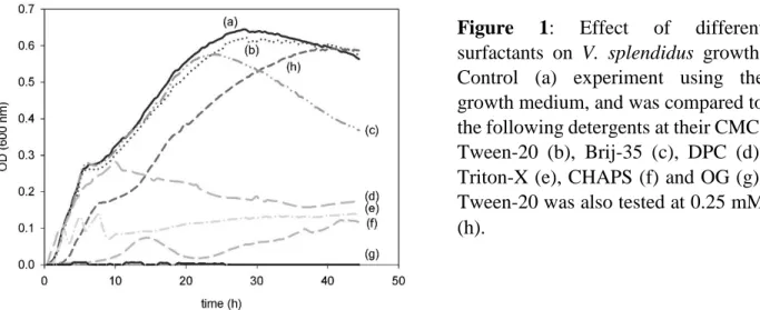
Initially, different commonly used surfactants were tested to micellize palmitic acid (0.3 mM) in culture media, and bacterial growths were monitored. Surfactants were used above their critical micelle concentration (CMC): Brij35 (0.1 mM), Tween20 (0.06 mM), Triton-X (0.4 mM), OG (20 mM), CHAPS (6 mM), and DPC (1.5 mM). The lipid deuteration procedure was optimized as follows: LB culture medium was supplemented with a mixture of d31-PA (0.3 mM) and Tween-20
(0.14 mM), heated at 85°C, and the corresponding solution was flash-frozen and heated again several times until the complete dissolution of d31-PA crystals. Protonated OA was added in the
same proportions (0.3 mM) during bacteria inoculation, to mitigate potential unbalance of the saturated/unsaturated fatty acid (SFA/UFA) ratio within the membrane [54] . Potential effect of the
2H labelling was verified by comparing the growth of bacteria in deuterated and non-deuterated
media. The specific growth rate (µ) was estimated from the slope regression of ln (OD600nm) as a
function of elapsed time [58] 2.3 Fatty acid profile analysis
Fatty acid (FA) profiles were analyzed using gas chromatography combined to mass spectrometry (GC-MS) to obtain the proportion of each FA (deuterated or protonated) in ng.mg-1, and were expressed as relative concentration (weight % relative to total FA contents) as described by Tardy-Laporte et al. [57]. Briefly, starting from three pools of 30 to 60 mg of dry-freezed bacteria, total lipids were extracted using dichloromethane/methanol (2:1 CH2Cl2/MeOH v/v) and
0.88% KCl solution in a Potter glass homogenizer. Neutral and polar lipids were separated by elution through a silica gel column (30×5 mm) hydrated with 6% water. Polar lipids were transesterified using 2 mL of H2SO4 (2% in MeOH) and 0.8 mL of toluene. Final extracts were
diluted in hexane solution and adjusted to a volume of 0.5 mL before GC-MS analysis (Agilent technology-7890A, Santa Clara, CA, USA). FA analyses were performed in parallel on a FAME mix which was used as a standard.
8
2.4 Sample preparation for 2H SS-NMR analysis and viability assays
Freshly collected bacterial cells were centrifuged at 4000 rpm for 10 min to remove the culture medium. Pellets were then suspended in a saline sterile rinsing solution (9‰ NaCl) to remove any residual FAs and detergent molecules, and centrifuged again at 3800 rpm for 5 min. Rinsing was carried out at least 3 times, twice with saline solution prepared with nanopure water, and a final wash with saline solution prepared with deuterium-depleted water. The final pellet was used to fill a 4-mm zirconium oxide rotor, which corresponds to approximately 90 mg of hydrated bacteria. NMR experiments were performed on deuterated bacteria harvested at three different growth times (mid-log, early stationary phase and late stationary phase) and prepared in triplicate.
Following NMR experiments V. splendidus viability was determined using MTT reduction assays [59]. Cell suspensions from NMR samples were diluted in 5 replicates to a final OD600nm of 0.1
each, and were mixed with MTT solution (5mg/ml) to a ratio of MTT/cell suspension of 1:10 (v/v). The mixture was incubated in Eppendorf tubes for 20 minutes at 25°C with open caps until the formation of formazan crystals. Preparations were centrifuged (10.000g ×1min.) and the crystal pellets were dissolved in dimethylsulfoxide and incubated at room temperature for 15 minutes. Optical density was measured at 550 nm and cell viability of bacteria was expressed in relative percentage compared to freshly harvested bacteria before rinse. MTT assays were performed on triplicates of cultures corresponding to the NMR samples.
2.5 In vivo 2H SS-NMR experiments and moment analysis
All 2H solid-state NMR experiments were performed at 25°C on a Bruker Avance HD III wide Bore 600 MHz spectrometer (Billerica, MA, USA) using a double-resonance magic angle spinning (MAS) probe tuned to 92.1 MHz. Sample spinning frequency was set to 10 kHz. Typically, spectra were acquired using a Hahn Echo pulse sequence (90°-t-180°-t) with the following operating conditions: 5 µs 90° pulses separated by an echo delay of 100 µs and a recycle time of 0.5 s. Each spectrum was obtained in approximately 43 min of acquisition time, corresponding to 4096 scans. A total of 32k points were acquired for a spectral width of 500 kHz. Spectra were Fourier transformed after application of a 50 Hz exponential line broadening and zero filling to 64k points.
9
Spectral moment analysis was performed using MestRenova software V6.0 (Mestrelab Research, Santiago de Compostela, Spain) and a macro developed by Pierre Audet (Université Laval). The second moment (M2) was calculated as described in equation 1 [54]:
𝑀2 = 𝜔𝑟2 ∑ 𝑁2𝐴𝑁 ∞ 𝑁=0 ∑∞𝑁=0𝐴𝑁 = 4𝜋2𝜈𝑄2 5 ⟨𝑆𝐶𝐷 2 ⟩ (1)
where ωr is the angular spinning frequency, N the side band number, and AN the area of each
sideband obtained by spectral integration, S2CD is the mean square order parameter, and Q is the
static quadrupolar coupling constant equal to 168 kHz for a C-D bond in acyl chains [60].
3. RESULTS
3.1 Optimized 2H labelling of V. splendidus for in vivo SS-NMR
The enzymatic machinery of bacteria energetically favours the incorporation of exogenous FAs into phospholipids [61], thus enabling the incorporation of deuterated PA chains exclusively in the membranes. We based our 2H labelling procedure of V. splendidus on a previously published protocol for another Gram(-) bacterium, Escherichia coli, which involved micellization of FAs by DPC in the growth medium to facilitate the FA uptake. However, since the presence of surfactants could be harmful to bacteria, and since another surfactant (Brij-58) had been used by other groups [56, 62], we tested a series of detergents to identify the best suited for optimal bacterial lipid deuteration. Therefore, Tween-20, Brij-35, DPC, Triton-X, CHAPS and OG were assessed. To do so, we first monitored bacterial growth in different culture media when mixing protonated palmitic acid (PA) with these non-anionic surfactants above their CMC. Figure 1 shows that Tween-20 is the less harmful detergent for the lipid deuteration of V. splendidus, and that a high concentration of bacteria is measured even at a detergent concentration of 0.25 mM, i.e., well above Tween-20’s CMC (0.06 mM). A concentration of 0.14 mM of Tween-20 was thus employed for the rest of the study because it allows complete solubilization of PA (concentration of 0.3 mM) without affecting the bacterial culture.
10
The deuteration of V. splendidus membrane lipids was then verified by 2H SS-NMR. The second spectral moment M2 was calculated to assess the membrane fluidity. When specific quadrupolar
splittings cannot be measured, M2 values are good reporter of the spectral distribution which
reflects the lipid phases - the greater the M2 value, the greater the lipid ordering. Figure 2B-C shows
that the deuteration protocol used herein was successful as a good signal-to-noise ratio is observed in vivo by 2H SS-NMR when bacteria are sampled in the exponential growth phase. The importance
of the surfactant-mediated micellization step can be seen in Figure 2A which shows that when d31
-PA is used without Tween-20, no side bands can be detected (i.e., there is no labelling). Figure 2C also shows that supplementing the culture medium with oleic acid (OA) in addition to d31-PAleads
to a reduction in side bands intensities, indicating an increase in membrane fluidity further demonstrated by the decrease in M2 value. These findings suggest that both unsaturated (OA) and
saturated (d31-PA) FAs were integrated by the cell. The in vivo NMR conditions during V.
splendidus analysis were confirmed by estimating the bacterial viability, which was 95 ± 5 %.
Figure 1: Effect of different
surfactants on V. splendidus growth. Control (a) experiment using the growth medium, and was compared to the following detergents at their CMC: Tween-20 (b), Brij-35 (c), DPC (d), Triton-X (e), CHAPS (f) and OG (g). Tween-20 was also tested at 0.25 mM (h).
11
Figure 2 : In vivo 2H MAS (10 kHz) SS-NMR spectra of V. splendidus harvested in the mid-log phase. Control experiment corresponding to bacteria labelled with d31-PA without detergent (A),
bacteria labelled with d31-PA in the presence of Tween-20 (0.14 mM) without (B), and with (C)
OA (1:1). Spectra are normalized according to the central peak. Average second spectral moments M2 are indicated (109 s-2).
3.2 Fatty acid composition as a function of cell growth and labelling conditions
Since the labelling protocol was successful, the membrane FA profile was monitored in order to quantify the assimilation of exogenous FAs at different growth stages and assess the potential impact of 2H labelling on the membrane. Lipid analyses were thus performed on labelled and
non-12
labelled bacteria and comparisons established between growth media enriched with d31-PA, with
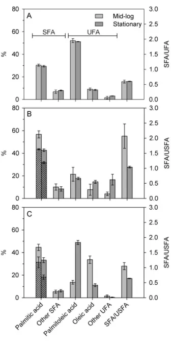
and without OA. Cell growth data are presented in the “Supplementary material” section. Figure 3 shows that the incorporation of d31-PA is indeed successful and that 77% of the PA acyl chains in
the membrane are deuterated during the mid-log phase for V. splendidus grown in the presence of d31-PA (amounting to 44% of all FAs). The deuteration level of PA is maintained as high as 69%
(31% of all fatty acids, numerical values are reported in Supplementary material, Table SI1) when OA is added in the growth medium, in spite of the high incorporation of OA into the membranes lipids. In the early stationary phase, the level of deuterated PA remains high, i.e. 75% of PA (or 32% of all FAs). When OA is added, this deuteration level is reduced to 55% (18% of all FAs), but it is still sufficient to generate a strong 2H-NMR signal (see Figure 4B).
Depending on the growth regimes, V. splendidus FA profile reveals more than 15 different FA chains with three major and recurring components: palmitoleic acid (C16:1), PA (C16), and OA (C18:1) (Fig.3). Our results (Fig. 3A) indicate that deuteration with d31-PA respects the native
composition of V. splendidus. In addition, exogenous OA was highly incorporated during the exponential phase, at the expense of palmitoleic acid - the most naturally abundant UFA in V. splendidus membranes. The choice to enrich the culture with OA as an UFA was to balance as much as possible the incorporation of saturated d31-PA and to allow comparison with previously
published deuteration protocols for other bacteria such as E. coli [54-56]. It also enabled exploring the adaptability of V. splendidus to integrate exogenous FAs and to modulate its FA chain fluidity accordingly.
Indeed bacteria responded to the enrichment with both exogenous FAs by including them in their lipid profile, more strikingly during the exponential phase, without affecting much the proportions of the other FAs (Fig. 3B). Permutational multivariate analysis of variance (Permanova) on Bray-Curtis matrices (Primer 7.0.13) of FAs composition of cell culture from different treatments and growth phases reveal significant interaction between both factors (DF = 2 and 22, Pseudo-F = 4.05, p = 0.005). Pair-wise Permanova comparison test indicates more specifically where differences were observed. Interestingly, the FA compositions were similar to that of the native bacteria membranes during the plateau phase (Fig. 3A), suggesting an adaptation of the bacteria to the
13
growth medium (p = 0.089). In contrast, growing bacteria with only d31-PA leads to a more altered
FA profile (p = 0.002). For instance, the major FA chains for V. splendidus, i.e., palmitoleic acid, was reduced by more than half, and other UFAs such as cyclopropane FAs (cyC17:0 and cyC19:0) were highly synthesizedunder this regime (see Supplementary material).
Figure 3: Fatty acid composition and corresponding SFA/UFA ratio at exponential (mid-log) and early stationary growth phases of V. splendidus in the presence of exogenous FAs. (A) Control sample in LB medium, (B) in presence of d31
-PA, and (C) in presence of d31-PA
and OA (1:1). Other SFAs includes lauric, myristic, pentadecanoic, heptadecanoic, stearic and arachidic acids (see complete assignment in table SI1). Other UFAs include myristoleic, pentadecenoic, stearidonic, -linoleic, eicosanoids,
cyclopropyl heptadecanoic
(cyC17:0), and cyclopropyl nonadecanoic (cyC19:0) acids (see complete assignment in table SI1). Hatched histograms indicate the relative proportion of d31-PA per total
palmitic acid. Saturated/unsaturated fatty acid ratios were calculated from FA content expressed in mol% per total FA (See S.I). Growth temperature was 25°C for all cultures.
14
Under labelling conditions, both with and without OA, the SFA/UFA ratios were higher in the exponential phase than in the stationary phase. This pattern is due to increased biosynthesis of UFAs such as palmitoleic acid (C16:1) or cyclopropane FAs (cyC17:0 and cyC19:0). It should also be noted that the SFA/UFA ratio of bacteria labelled in the presence of OA and during early stationary phase (SFA/UFA = 0.65 ± 0.01) was close to that of unlabelled cultures which was about 0.59 ± 0.05 and 0.60 ± 0.01 during mid-log and early stationary phases, respectively.
3.3 Membrane fluidity characterization by SS-NMR
The 2H SS-NMR spectra obtained for the labelled bacteria (Figure 4) show that membrane rigidity varies with cell division. Under the same growth conditions (d31-PA with OA) and in a
timeframe of 5 hours, a reduction was observed in the sideband intensity and number. Accordingly, the M2 values dropped by half going from the mid-log to the stationary phase. These results are
consistent with the decreased SFA/UFA ratio which reveal an increase in membrane fluidity as a function of growth time under the same growth conditions.
4. DISCUSSION
4.1 2H labelling optimization for in vivo study of membranes by 2H SS-NMR
Nuclear magnetic resonance spectroscopy has gained an appreciated reputation among biologists, notably because of its low radiation energy in the radio frequency range, which causes no detrimental effects on biological tissues [35]. This property has allowed NMR to tackle different living systems (cells or tissues) which is referred to as in vivo NMR [31, 35, 36]. Labelling is a common strategy in NMR and deuterium labelling of lipid chains is a useful approach to explore changes occurring in biological membranes using SS-NMR. However, when studying living organisms, the deuteration protocol could be a disturbing factor to physiological pathways, or induce stress. Previously published work has established the feasibility and pertinence of bacterial membrane deuteration on human pathogens E. coli and B. subtilis [54-57, 63]. Here, a marine bacterium, V. splendidus, is deuterated for the first time while minimizing disturbances on the microorganism. Although V. splendidus and previously characterized E. coli [54, 55] are both
15
Gram(-) bacteria, differences in 2H SS-NMR spectra between these two species were expected, due perhaps to species-specific biological requirements [64, 65].
The capacity of the bacteria to grow under our labelling conditions was first verified. While deuteration of molecules such as FAs does not affect their structure and thermodynamical stability, surfactants used to facilitate the deuterated FA incorporation into the bacteria [66], could have a detrimental effect since detergents are commonly used for biochemical applications such as extraction of membrane proteins. Therefore, the physiological state of the bacteria during labelling procedure was monitored using growth kinetics, and our results showed that Tween-20 had the lowest impact. This result is in agreement with Schuck et al. who compared sensitivities of diverse
Figure 4: 2H MAS (10 kHz) SS-NMR
spectra of intact V. splendidus harvested at three different cell growth times: (A) after 15 h in the mid-log phase (OD600nm ≈ 0.3), (B)
after 22 h (±2h) at the beginning of the stationary stage (OD600nm ≈ 0.5), and (C)
after 30 h at advanced stationary phase
(OD600nm ≈ 0.5). Bacteria were labelled with
d31-PA in presence of OA. Average second
16
cells toward a set of detergents and showed that Tween-20 was always the least effective in provoking membrane resistance, and the best at preserving membrane integrity [67]. Tween-20 has a low CMC, does not affect protein activity, and is rarely used for cell lysis or protein extraction, but rather as a gentle washing agent. [67, 68]. On the other hand, although harmless for the first 10 hours of culture, the zwitterionic detergent DPC used to deuterate E. coli and B. subtilis lipids showed a hampering action on V. splendidus at longer exposure time, which could be explained by its ability to break protein-lipid and lipid-lipid associations [68, 69].
De novo synthesis of FAs is the most energetically expensive mechanism in phospholipid synthesis for bacteria membrane [49]. Because they do not have specialized functional cellular compartments, prokaryotic organisms rely on multiple regulation tools to control their phospholipid composition according to the niche in which they live [47, 49, 50, 70, 71]. Overall, bacteria tend to sustain their membrane homeostasis through adaptation mechanisms by maintaining zwitterionic/anionic, protein/lipid [70] or FA chain ratios [72]. The SFA/UFA ratio is one of the most used reporters of membrane fluidity [73-75] as well as bacterial adaptive strategies [73-77]. Our results showed that when PA was added in the growth medium of V. splendidus without OA, bacteria adapted by synthesizing more cyclic fatty acids (cyC17:0 and cyC19:0) that restored membrane fluidity. Similarly, when OA was also added and despite the fact that OA is not the major UFA in V. splendidus, this FA was highly integrated in the exponential phase, resulting in a FA profile similar to that of unlabelled bacteria. The SFA/UFA ratios determined here confirm the need to enrich the medium with UFAs during labelling with d31-PA in order to avoid extreme
shifts from the natural lipid composition (Fig. 3), in agreement with previous work carried out on E. coli [54-56].
In short, our lipid deuteration protocol on V. splendidus allowed a similar or better labelling rate as compared to previous work on bacteria. Lipid profile analyses showed that at least 69% deuteration of C16:0 acyl chains was observed in the exponential phase. This deuteration level is slightly higher than what was reported for E. coli grown in the presence of d31-PA [54, 55]. Except
for minor differences in the FA incorporation process [47] and a different culture time lapse, the lipid metabolism of Vibrio species is very similar to E. coli’s since they are both Gram(-) bacteria
17
with similar transcriptional regulatory genes for FA metabolism and phospholipid composition [47, 49, 72, 76].
4.2 Membrane evolution during bacterial growth
Our results showed that the membrane fluidity varied with bacterial growth during the labelling process. Indeed bacteria experience the fastest growth rate in the exponential phase during which they produce membrane lipids [71]. Once lipids are synthesized, the lipid profile remains the same until the stationary phase is reached, i.e., when nutrients become limited, DNA replication, protein synthesis and respiration are reduced, and cell lysis begins [78]. Previous in vivo NMR studies have been carried out on bacteria sampled at the mid-log growth stage [54-57] which provides excellent NMR signal-to-noise ratio. However, our results revealed that samples obtained at later stages, especially the early stationary stage, provide sufficient labelling for a good NMR signal-to-noise ratio, as well as additional advantages.
Indeed GC-MS analyses showed that exogenous PA and OA were highly integrated in the membrane at mid-log phase during which they become the most abundant FA chains in the membrane (Figure 3). In subsequent growth stages, PA and OA diminished to reach more “native” proportions (i.e., those measured in the control samples), while the amount of palmitoleic acid increased, through an unknown mechanism that could imply FA conversion or exchange, the addition of a double bond or a CH2, or the synthesis of new FAs. This is consistent with other
investigations on Vibrio species, showing that exogenous FAs are incorporated by direct trans-acylation or after incorporation into the biosynthesis pool [47]. Additionally the SFA/UFA ratio, which decreased as a function of growth time, reaching that of unlabelled bacteria (especially when both PA and OA were added to the culture medium), validate the evolution of the FA profile. The
2H SS-NMR spectra also reveal an increasein membrane fluidity as a function of V. splendidus
growth stages when d31-PA, and to a lesser extent when equal proportions of OA and d31-PA, are
added to the growth medium (Figure 4). In the exponential phase, a more rigid membrane was observed, close to a gel phase (M2 above 20×109 s-2), whereas in later stages the M2 value was
18
and SFA/UFA ratio were closer to their “native” values in the early stationary phase, the lower M2
values would thus report with biological membranes in the fluid phase [79].
The contribution of signal from free d31-PA cannot explain the change in lipid profile during cell
growth. Indeed Figure 2A shows no 2H SS-NMR signal after rinsing. Additionally, free d31-PA is
insoluble in water and would lead to a very broad 2H SS-NMR spectrum with M
2 above 70×109 s -2 (data not shown), which is incompatible with our results. Moreover, the 2H SS-NMR spectrum of
d31-PA crystals would not vary with growth time. In light of these results, the increase in membrane
fluidity with cell growth time would be explained by a gradual adaptation of the microorganism metabolism upon isotopic labelling to cope with possible stress and disturbance of metabolic pathways [73-75] due to chemical isotopic labelling. In most cases, stress is known to lead to an increase in membrane rigidity, although the opposite pattern could happen in the case of marine prokaryotic organisms, and specifically for Vibrio species [46, 75]. The increased fluidity due to increased UFA proportions, and notably cyclopropanes FAs observed when PA was added in the growth medium, seems to prove such environmental stress [75, 80]. However, when both PA and OA were present in the medium, cyclopropane FA proportions were inverted (Table SI1), suggesting that stress was reduced [80]. Similarly, the superimposed growth curves (see Fig. SI1) indicate that V. splendidus metabolism kinetics under isotopic labelling was not affected by labelling stress. Therefore, the FAs turnover would be fast and all exogenous d31-PA transferred
into the membrane lipids at the mid-log phase. Yet, being long and saturated, they would induce large M2 values. In the early stationary phase, bacteria would have had enough time to regulate
their metabolism to the presence of exogenous FAs in the growth medium, generally through adapting their enzymatic machinery [71], and have either converted into or synthesized new UFAs in order to reduce the membrane rigidity, and reach their initial membrane state, as observed by the reduced M2 values.
19 5. CONCLUSION AND PERSPECTIVES
Setting out labelling procedures is an important step towards developing effective and reliable tools for in vivo NMR studies. Here, membranes of intact V. splendidus were characterized for the first time by SS-NMR, as a model of marine Gram(-) bacteria. This work provides a better understanding of changes in bacterial membrane properties as a function of growth time and conditions that have never been studied so far on any biological system by in vivo NMR. Overall, the present study should help in the design of efficient labelling protocols of bacteria, which takes into account not only the technical spectroscopy requirements for analyses (such as signal-to-noise ratio) but also biological considerations. This 2H labelling protocol is useful for 2H SS-NMR, but could also be useful for neutron diffraction, or extended to the labelling of cell membranes by other isotopes such as 13C. Additionally, by characterizing the membrane of V. splendidus, a potential virulent bacterium for marine organisms, this work paves the way towards studying the interactions of its membrane with exogenous molecules in the aquatic environment.
ACKNOWLEDGMENTS
This work was supported by the Natural Sciences and Engineering Research Council (NSERC) of Canada (grant 326750-2013 to I.M. and grant 299100 to R.T.) and the Centre National de la Recherche Scientifique (UMR 7099 to D.E.W.). Z.B. would like to acknowledge the Ressources Aquatiques Québec (RAQ) research network (RS-171172) for the award of scholarships and Pierre Audet for sharing the MestRenova macro. I.M. and R.T. are members of the RAQ.
COMPETING INTERESTS
20 AUTHOR CONTRIBUTIONS
Z.B. designed and conducted all sampling, data collection and analysis, and wrote the first draft of the manuscript. D.E.W. and A.A. contributed to the data analysis and writing of the manuscript. K.L. provided the initial Vibrio strain culture and participated to the revision of the manuscript. R.T. and I.M. designed and supervised the research, contributed to the data analysis and writing of the manuscript.
REFERENCES
1. Urakawa, H. and I.N.G. Rivera, Aquatic environment, in The biology of Vibrios. 2006, American Society of Microbiology. p. 175-189.
2. Thompson, F.L., T. Iida, and J. Swings, Biodiversity of Vibrios. Microbiology and molecular biology reviews, 2004. 68(3): p. 403-431.
3. Cavallo, R.A. and L. Stabili, Presence of vibrios in seawater and Mytilus galloprovincialis (Lam.) from the Mar Piccolo of Taranto (Ionian Sea). Water Research, 2002. 36(15): p. 3719-3726.
4. McDougald, D. and S. Kjelleberg, Adaptive responses of Vibrios, in The biology of Vibrios. 2006, American Society of Microbiology. p. 133-155.
5. DiSalvo, L., J. Blecka, and R. Zebal, Vibrio anguillarum and larval mortality in a California coastal shellfish hatchery. Applied and Environmental Microbiology, 1978. 35(1): p. 219-221.
21
6. Crab, R., A. Lambert, T. Defoirdt, P. Bossier, and W. Verstraete, The application of bioflocs technology to protect brine shrimp (Artemia franciscana) from pathogenic Vibrio harveyi. Journal of applied microbiology, 2010. 109(5): p. 1643-1649.
7. Frans, I., C.W. Michiels, P. Bossier, K. Willems, B. Lievens, and H. Rediers, Vibrio anguillarum as a fish pathogen: virulence factors, diagnosis and prevention. Journal of fish diseases, 2011. 34(9): p. 643-661.
8. Travers, M.-A., K.B. Miller, A. Roque, and C.S. Friedman, Bacterial diseases in marine bivalves. Journal of invertebrate pathology, 2015. 131: p. 11-31.
9. Velji, M., L. Albright, and T. Evelyn, Immunogenicity of various Vibrio ordalii lipopolysaccharide fractions in coho salmon Oncorhynchus kisutch. Diseases of aquatic organisms, 1992. 12(2): p. 97-101.
10. Paillard, C., F. Le Roux, and J.J. Borrego, Bacterial disease in marine bivalves, a review of recent studies: trends and evolution. Aquatic Living Resources, 2004. 17(4): p. 477-498.
11. Pruzzo, C., G. Gallo, and L. Canesi, Persistence of vibrios in marine bivalves: the role of interactions with haemolymph components. Environmental microbiology, 2005. 7(6): p. 761-772.
12. Beaz‐Hidalgo, R., S. Balboa, J.L. Romalde, and M.J. Figueras, Diversity and pathogenecity of Vibrio species in cultured bivalve molluscs. Environmental microbiology reports, 2010. 2(1): p. 34-43.
13. De Decker, S. and D. Saulnier, Vibriosis induced by experimental cohabitation in Crassostrea gigas: Evidence of early infection and down-expression of immune-related genes. Fish & shellfish immunology, 2011. 30(2): p. 691-699.
22
14. Liu, X., C. Ji, J. Zhao, and H. Wu, Differential metabolic responses of clam Ruditapes philippinarum to Vibrio anguillarum and Vibrio splendidus challenges. Fish & shellfish immunology, 2013. 35(6): p. 2001-2007.
15. Gay, M., T. Renault, A.-M. Pons, and F. Le Roux, Two Vibrio splendidus related strains collaborate to kill Crassostrea gigas: taxonomy and host alterations. Diseases of aquatic organisms, 2004. 62(1-2): p. 65-74.
16. Liu, R., L. Qiu, Z. Yu, J. Zi, F. Yue, L. Wang, H. Zhang, W. Teng, X. Liu, and L. Song, Identification and characterisation of pathogenic Vibrio splendidus from Yesso scallop (Patinopecten yessoensis) cultured in a low temperature environment. Journal of invertebrate pathology, 2013. 114(2): p. 144-150.
17. Kogut, M. and N.J. Russell, The growth and phospholipid composition of a moderately halophilic bacterium during adaptation to changes in salinity. Current Microbiology, 1984. 10(2): p. 95-98.
18. Eguchi, M., E. Fujiwara, and N. Miyamoto, Survival of Vibrio anguillarum in freshwater environments: adaptation or debilitation? Journal of Infection and Chemotherapy, 2000. 6(2): p. 126-129.
19. Sarita, G.B., Multiple antibiotic resistances of Vibrio isolates from coastal and brackish water areas. American Journal of Biochemistry and Biotechnology 2005.
20. Masini, L., G. De Grandis, F. Principi, C. Mengarelli, and D. Ottaviani, Research and characterization of pathogenic vibrios from bathing water along the Conero Riviera (Central Italy). Water Research, 2007. 41(18): p. 4031-4040.
23
21. Soto, W., J. Gutierrez, M. Remmenga, and M. Nishiguchi, Salinity and temperature effects on physiological responses of Vibrio fischeri from diverse ecological niches. Microbial ecology, 2009. 57(1): p. 140-150.
22. Phillips, D.J., The use of biological indicator organisms to monitor trace metal pollution in marine and estuarine environments—a review. Environmental Pollution (1970), 1977. 13(4): p. 281-317.
23. Olsen, C.f., N. Cutshall, and I. Larsen, Pollutant—particle associations and dynamics in coastal marine environments: a review. Marine Chemistry, 1982. 11(6): p. 501-533.
24. Nixon, S.W., Coastal marine eutrophication: a definition, social causes, and future concerns. Ophelia, 1995. 41(1): p. 199-219.
25. Baquero, F., J.-L. Martínez, and R. Cantón, Antibiotics and antibiotic resistance in water environments. Current opinion in biotechnology, 2008. 19(3): p. 260-265.
26. Kümmerer, K., Antibiotics in the aquatic environment–a review–part I. Chemosphere, 2009. 75(4): p. 417-434.
27. Riedel, T., M. Zark, A.V. Vähätalo, J. Niggemann, R.G. Spencer, P.J. Hernes, and T. Dittmar, Molecular signatures of biogeochemical transformations in dissolved organic matter from ten world rivers. Frontiers in Earth Science, 2016. 4: p. 85.
28. Hancock, R.E., The bacterial outer membrane as a drug barrier. Trends in Microbiology, 1997. 5(1): p. 37-42.
24
29. Shai, Y., Mechanism of the binding, insertion and destabilization of phospholipid bilayer membranes by α-helical antimicrobial and cell non-selective membrane-lytic peptides. Biochimica et Biophysica Acta (BBA)-Biomembranes, 1999. 1462(1): p. 55-70.
30. Ratcliffe, R.G., In vivo NMR studies of higher plants and algae. Advances in botanical research, 1994. 20: p. 43-123.
31. Serber, Z., L. Corsini, F. Durst, and V. Dötsch, In-cell NMR spectroscopy. Methods in enzymology, 2005. 394: p. 17-41.
32. Selenko, P. and G. Wagner, Looking into live cells with in-cell NMR spectroscopy. Journal of structural biology, 2007. 158(2): p. 244-253.
33. Serber, Z., A.T. Keatinge-Clay, R. Ledwidge, A.E. Kelly, S.M. Miller, and V. Dötsch, High-resolution macromolecular NMR spectroscopy inside living cells. Journal of the American Chemical Society, 2001. 123(10): p. 2446-2447.
34. Maldonado, A.Y., D.S. Burz, and A. Shekhtman, In-cell NMR spectroscopy. Progress in Nuclear Magnetic Resonance Spectroscopy, 2011. 59(3): p. 197.
35. Reckel, S., F. Löhr, and V. Dötsch, In‐Cell NMR Spectroscopy. ChemBioChem, 2005. 6(9): p. 1601-1606.
36. Warnet, X.L., A.A. Arnold, I. Marcotte, and D.E. Warschawski, In-cell solid-state NMR: an emerging technique for the study of biological membranes. Biophysical journal, 2015. 109(12): p. 2461-2466.
25
37. Arnold, A.A., B. Genard, F. Zito, R. Tremblay, D.E. Warschawski, and I. Marcotte, Identification of lipid and saccharide constituents of whole microalgal cells by 13C solid-state NMR. Biochimica et Biophysica Acta (BBA)-Biomembranes, 2015. 1848(1): p. 369-377.
38. Chauton, M.S., O.I. Optun, T.F. Bathen, Z. Volent, I.S. Gribbestad, and G. Johnsen, HR MAS ¹H NMR spectroscopy analysis of marine microalgal whole cells. Marine Ecology Progress Series, 2003. 256: p. 57-62.
39. Simpson, A.J., Y. Liaghati, B. Fortier-McGill, R. Soong, and M. Akhter, Perspective: in vivo NMR–a potentially powerful tool for environmental research. Magnetic Resonance Chemistry, 2015. 53: p. 686-690.
40. Laadhari, M., A.A. Arnold, A.E. Gravel, F. Separovic, and I. Marcotte, Interaction of the antimicrobial peptides caerin 1.1 and aurein 1.2 with intact bacteria by 2H solid-state NMR. Biochimica et Biophysica Acta (BBA)-Biomembranes, 2016. 1858(12): p. 2959-2964.
41. Booth, V., D.E. Warschawski, N.P. Santisteban, M. Laadhari, and I. Marcotte, Recent progress on the application of 2H solid-state NMR to probe the interaction of antimicrobial peptides with intact bacteria. Biochimica et Biophysica Acta (BBA)-Proteins and Proteomics, 2017.
42. Poulhazan, A., A. Arnold, D. Warschawski, and I. Marcotte, Unambiguous Ex Situ and in Cell 2D 13C Solid-State NMR Characterization of Starch and Its Constituents. International journal of molecular sciences, 2018. 19(12): p. 3817.
43. Mateo, D.R., A. Siah, M.T. Araya, F.C. Berthe, G.R. Johnson, and S.J. Greenwood, Differential in vivo response of soft-shell clam hemocytes against two strains of Vibrio splendidus: changes in cell structure, numbers and adherence. Journal of invertebrate pathology, 2009. 102(1): p. 50-56.
26
44. Turcotte, F., J.-L. Mouget, B. Genard, K. Lemarchand, J.-S. Deschênes, and R. Tremblay, Prophylactic effect of Haslea ostrearia culture supernatant containing the pigment marennine to stabilize bivalve hatchery production. Aquatic Living Resources, 2016. 29(4): p. 401.
45. Oliver, J.D. and R.R. Colwell, Extractable lipids of gram-negative marine bacteria: phospholipid composition. Journal of bacteriology, 1973. 114(3): p. 897-908.
46. Oliver, J.D. and W.F. Stringer, Lipid composition of a psychrophilic marine Vibrio sp. during starvation-induced morphogenesis. Applied and Environmental Microbiology, 1984. 47(3): p. 461-466.
47. Byers, D.M., Elongation of exogenous fatty acids by the bioluminescent bacterium Vibrio harveyi. Journal of bacteriology, 1989. 171(1): p. 59-64.
48. Brown, R.N. and P.A. Gulig, Regulation of fatty acid metabolism by FadR is essential for Vibrio vulnificus to cause infection of mice. Journal of bacteriology, 2008. 190(23): p. 7633-7644.
49. Zhang, Y.-M. and C.O. Rock, Transcriptional regulation in bacterial membrane lipid synthesis. Journal of lipid research, 2009. 50(Supplement): p. S115-S119.
50. Freese, E., H. Rütters, J. Köster, J. Rullkötter, and H. Sass, Gammaproteobacteria as a possible source of eicosapentaenoic acid in anoxic intertidal sediments. Microbial ecology, 2009. 57(3): p. 444-454.
51. Davis, J.H., The description of membrane lipid conformation, order and dynamics by 2H-NMR. Biochimica et Biophysica Acta (BBA)-Reviews on Biomembranes, 1983. 737(1): p. 117-171.
27
52. Li, J., T. Vosegaard, and Z. Guo, Applications of nuclear magnetic resonance in lipid analyses: An emerging powerful tool for lipidomics studies. Progress in lipid research, 2017. 68: p. 37-56.
53. Seelig, J., Deuterium magnetic resonance: theory and application to lipid membranes. Quarterly reviews of biophysics, 1977. 10(3): p. 353-418.
54. Warnet, X.L., M. Laadhari, A.A. Arnold, I. Marcotte, and D.E. Warschawski, A 2H
magic-angle spinning solid-state NMR characterisation of lipid membranes in intact bacteria. Biochimica et Biophysica Acta (BBA)-Biomembranes, 2016. 1858(1): p. 146-152.
55. Laadhari, M., A.A. Arnold, A.E. Gravel, F. Separovic, and I. Marcotte, Interaction of the antimicrobial peptides caerin 1.1 and aurein 1.2 with intact bacteria by 2H solid-state NMR. Biochimica et Biophysica Acta (BBA)-Biomembranes, 2016. 1858(12): p. 2959-2964.
56. Pius, J., M.R. Morrow, and V. Booth, 2H solid-state nuclear magnetic resonance investigation
of whole Escherichia coli interacting with antimicrobial peptide MSI-78. Biochemistry, 2011. 51(1): p. 118-125.
57. Tardy-Laporte, C., A.A. Arnold, B. Genard, R. Gastineau, M. Morançais, J.-L. Mouget, R. Tremblay, and I. Marcotte, A 2H solid-state NMR study of the effect of antimicrobial agents on intact Escherichia coli without mutating. Biochimica et Biophysica Acta (BBA)-Biomembranes, 2013. 1828(2): p. 614-622.
58. Nedwell, D. and M. Rutter, Influence of temperature on growth rate and competition between two psychrotolerant Antarctic bacteria: low temperature diminishes affinity for substrate uptake. Applied and Environmental Microbiology, 1994. 60(6): p. 1984-1992.
28
59. Wang, H., H. Cheng, F. Wang, D. Wei, and X.J.J.o.m.m. Wang, An improved 3-(4, 5-dimethylthiazol-2-yl)-2, 5-diphenyl tetrazolium bromide (MTT) reduction assay for evaluating the viability of Escherichia coli cells. Journal of microbiological methods 2010. 82(3): p. 330-333.
60. Burnett, L. and B. Muller, Deuteron quadrupole coupling constants in three solid deuterated paraffin hydrocarbons: C2D6, C4D10, C6D14. The Journal of Chemical Physics, 1971. 55(12): p. 5829-5831.
61. Marcotte, I. and V. Booth, 2H Solid-State NMR Study of Peptide–Membrane Interactions in Intact Bacteria, in Advances in Biological Solid-State NMR. 2014. p. 459-475.
62. Davis, J.H., C.P. Nichol, G. Weeks, and M. Bloom, Study of the cytoplasmic and outer membranes of Escherichia coli by deuterium magnetic resonance. Biochemistry, 1979. 18(10): p. 2103-2112.
63. Davis, J.-H., Deuterium magnetic resonance study of the gel and liquid crystalline phases of dipalmitoyl phosphatidylcholine. Biophysical journal, 1979. 27(3): p. 339-358.
64. Farmer, J., The family Vibrionaceae, in The prokaryotes. 2006, Springer. p. 495-507.
65. Gauthier-Clerc, S., I. Boily, M. Fournier, and K. Lemarchand, In vivo exposure of Mytilus edulis to living enteric bacteria: a threat for immune competency? Environmental Science and Pollution Research, 2013. 20(2): p. 612-620.
29
66. Dal Molin, M., G. Gasparini, P. Scrimin, F. Rastrelli, and L.J. Prins, 13C-isotope labelling for the facilitated NMR analysis of a complex dynamic chemical system. Chemical Communications, 2011. 47(46): p. 12476-12478.
67. Schuck, S., M. Honsho, K. Ekroos, A. Shevchenko, and K. Simons, Resistance of cell membranes to different detergents. Proceedings of the National Academy of Sciences, 2003. 100(10): p. 5795-5800.
68. Johnson, M., Detergents: Triton X-100, tween-20, and more. Mater Methods, 2013. 3(1): p. 163.
69. le Maire, M., P. Champeil, and J.V. Møller, Interaction of membrane proteins and lipids with solubilizing detergents. Biochimica et Biophysica Acta (BBA)-Biomembranes, 2000. 1508(1): p. 86-111.
70. Shibuya, I., Metabolic regulation and biological functions of phospholipids in Escherichia coli. Progress in lipid research, 1992. 31(3): p. 245-299.
71. Barton, L.L., Cellular Growth and Reproduction, in Structural and Functional Relationships in Prokaryotes Springer, Editor. 2005: New York,. p. 292-346.
72. Silbert, D.F., F. Ruch, and P.R. Vagelos, Fatty acid replacements in a fatty acid auxotroph of Escherichia coli. Journal of bacteriology, 1968. 95(5): p. 1658-1665.
73. Beney, L. and P. Gervais, Influence of the fluidity of the membrane on the response of microorganisms to environmental stresses. Applied Microbiology and Biotechnology, 2001. 57(1): p. 34-42.
30
74. Mykytczuk, N., J. Trevors, L. Leduc, and G. Ferroni, Fluorescence polarization in studies of bacterial cytoplasmic membrane fluidity under environmental stress. Progress in biophysics and molecular biology, 2007. 95(1): p. 60-82.
75. Yoon, Y., H. Lee, S. Lee, S. Kim, and K.-H. Choi, Membrane fluidity-related adaptive response mechanisms of foodborne bacterial pathogens under environmental stresses. Food Research International, 2015. 72: p. 25-36.
76. Rock, C. and S. Jackowski, Pathways for the incorporation of exogenous fatty acids into phosphatidylethanolamine in Escherichia coli. Journal of Biological Chemistry, 1985. 260(23): p. 12720-12724.
77. Denich, T., L. Beaudette, H. Lee, and J. Trevors, Effect of selected environmental and physico-chemical factors on bacterial cytoplasmic membranes. Journal of microbiological methods, 2003. 52(2): p. 149-182.
78. Carty, C. and L. Ingram, Lipid synthesis during the Escherichia coli cell cycle. Journal of bacteriology, 1981. 145(1): p. 472-478.
79. Morein, S., A.-S. Andersson, L. Rilfors, and G. Lindblom, Wild-type Escherichia coli cells regulate the membrane lipid composition in a window between gel and non-lamellar structures. Journal of Biological Chemistry, 1996. 271(12): p. 6801-6809.