HAL Id: tel-02463841
https://tel.archives-ouvertes.fr/tel-02463841
Submitted on 2 Feb 2020HAL is a multi-disciplinary open access archive for the deposit and dissemination of sci-entific research documents, whether they are pub-lished or not. The documents may come from teaching and research institutions in France or abroad, or from public or private research centers.
L’archive ouverte pluridisciplinaire HAL, est destinée au dépôt et à la diffusion de documents scientifiques de niveau recherche, publiés ou non, émanant des établissements d’enseignement et de recherche français ou étrangers, des laboratoires publics ou privés.
Tritium and Deuterium Labelling of Bioactive Molecules
Catalyzed by Metallic Nanoparticles
Viktor Pfeifer
To cite this version:
Viktor Pfeifer. Tritium and Deuterium Labelling of Bioactive Molecules Catalyzed by Metallic Nanoparticles. Catalysis. Université Paris-Saclay, 2019. English. �NNT : 2019SACLS275�. �tel-02463841�
Tritium and Deuterium Labelling of
Bioactive Molecules Catalyzed by
Metallic Nanoparticles
Thèse de doctorat de l'Université Paris-Saclay préparée à l´Université Paris-Sud
au CEA-Saclay
École doctorale n°571, Sciences Chimiques: Molécules, Matériaux, Instrumentation et Biosystèmes (2MIB)
Spécialité de doctorat: Chimie
Thèse présentée et soutenue à Gif-sur-Yvette, le 16 Septembre 2019, par
Viktor Pfeifer
Composition du Jury :Françoise Colobert
Professeure, Université de Strasbourg Rapporteur
Olivier Baudoin
Professeur, Université de Basel Rapporteur
Emmanuelle Schulz
Directeur de Recherche, Université Paris-Sud Présidente
Thomas Cailly
Maître de Conférences, Université de Caen Examinateur
Grégory Pieters
Directeur de Recherche, CEA-Saclay Directeur de thèse
Sophie Feuillastre
Ingénieur de Recherche, CEA-Saclay Invité
Philippe Lesot
Table of contents
Acknowledgments ... 5
Abbreviations ... 7
Table of Figures ... 8
I. Introduction ... 15
1. Hydrogen isotopes in organic molecules ... 15
1.1 Deuterium: the kinetic isotope effect and its applications ... 15
1.1.1 Heavy Drugs ... 16
1.1.2 Stable isotopically labelled internal standards (SILSs) ... 18
1.1.3 SILSs for metabolism studies of drugs ... 19
1.1.4 SILSs in metabolomics ... 20
1.2 Tritium and its applications ... 21
1.3 Analysis of molecules labelled by hydrogen isotopes ... 23
1.3.1 Mass spectrometry ... 23
1.3.2 NMR spectroscopy ... 25
2. N-Based heterocycles: promising bioactive targets for the introduction of deuterium and tritium ... 29
2.1 Nomenclature and numbering of heterocyclic rings... 30
2.2 Basicity/acidity of N-Heterocycles ... 32
2.3 Implementation as therapeutics, in agricultural chemistry & material sciences ... 33
3. Existing methods for the incorporation of hydrogen isotopes into organic substrates 40 3.1 The synthetic approach ... 40
3.2 Late-stage modifications ... 43
3.2.1. Heterogeneous transition metal catalyzed HIE ... 44
3.2.2. Homogeneous transition metal catalyzed HIE ... 49
3.2.3 Metal nanoparticles-based methods for HIE ... 55
4.1 Synthesis, stabilization and analysis ... 58
4.2 DFT calculations on metal nanoparticles ... 59
II. Development of new metal nanoparticles-based HIE methods ... 60
1. Syntheses of metal nanoparticles ... 61
1.1 Synthesis of RuNp@PVP ... 62
1.2 Synthesis of Ru-ICy Np ... 63
1.3 Synthesis of Ni-ICy Np ... 65
1.4 Synthesis of Ni-IMes Np ... 66
2. HIE on N-Heterocycles catalyzed by metallic nanoparticles ... 68
2.1 HIE catalyzed by ruthenium nanoparticles ... 68
2.1.1 Initial considerations... 68
2.1.2 Deuterations of oxazoles ... 71
2.1.3 Deuterations of imidazoles ... 78
2.1.4 Deuterations of N-heterocyclic benzoderivatives ... 83
2.1.5 Deuterations of 1,2,4-triazoles ... 87
2.1.6 Deuterations of carbazoles ... 89
2.1.7 Deuterations of N-heterocyclic bioactive molecules ... 95
2.1.8 Tritiations of N-heterocyclic Drugs ... 105
2.1.9 Limitations of RuNp@PVP as HIE catalyst ... 112
2.2 HIE catalyzed by nickel nanoparticles ... 115
Summary and perspectives ... 120
Experimental part ... 122
Syntheses of metal nanoparticles ... 124
Syntheses of compounds ... 127
H/D exchange reactions ... 137
Deuterations of oxazoles ... 137
Deuterations of imidazoles ... 153
Deuterations of 1,2,4-triazoles ... 190
Deuteration of carbazoles under neutral conditions ... 212
Deuteration of carbazoles with RuNp@PVP and Cs2CO3 ... 229
Deuterations of N-heterocyclic bioactive molecules ... 245
Deuterations of N-heterocycles by nickel nanoparticles ... 284
Tritiations of drugs ... 291
Acknowledgments
I wholeheartedly thank all jury members for making the defense of this PhD thesis possible. I am grateful to Prof. Françoise Colobert and Prof. Olivier Baudoin for having accepted to survey my work as referees and for their presence at my PhD defense. I’m very thankful to Dr. Emmanuelle Schulz for having accepted to review my work as a member of the University Paris-Sud during the last three years. I was very happy and delighted to lead valuable discussions with Dr. Emmanuelle Schulz and to be provided with constant feed-back from her side. Dr. Emmanuelle Schulz is also thanked for her participation as examiner during my PhD defense. Moreover, I would like to thank Dr. Thomas Cailly for taking part as examiner in my PhD defense and for reviewing my PhD thesis. A special and sincere gratitude goes to my supervisor Dr. Grégory Pieters, who gave me the opportunity to perform this PhD thesis in his working group. Further, I am thankful to Dr. Grégory Pieters for his permanent support and encouragement in many aspects, whether in science or in everyday life. I also thank Dr. Sophie Feuillastre for supervising my work, for furnishing many productive ideas and for her willingness to help with materials and advice whenever needed. Dr. Sophie Feuillastre is also thanked for being part of the jury during my PhD defense. With a special mention to Dr. Philippe Lesot, who followed-up my work with a big curiosity and accepted to be part of the jury. I express another gratitude to Dr. Philippe Lesot for supporting this PhD thesis with his great knowledge of NMR and for all the efforts he made to conduct
2
H-NMR analysis for this work. My sincere thanks goes to Dr. Bernard Rousseau for reviewing this work, for his powerful attitude towards science and life and for creating a familiar and enthusiastic atmosphere with his presence. I am very grateful to Dr. Bruno Chaudret and Dr. Simon Tricard for welcoming me in their laboratory during my secondment and for pioneering the modern world of metal nanoparticles. I also thank Dr. Bruno Chaudret and Dr. Simon Tricard for sharing their valuable knowledge with me and my supervisors on this field. I am thankful to Donia Bouzouita for her eagerness to support this work with metal nanoparticles and for being a pleasant laboratory mate during my stay in Toulouse. My sincere gratitude goes to Romuald Poteau, Iker del Rosal, Marie Certiat and Laurent Maron for dedicating a huge amount of time and efforts for the performance and management of the DFT-calculations within this work. Further, I want to express another gratitude to Dr. Volker Derdau for welcoming me in his laboratory during another secondment. I am very thankful to Dr. Volker Derdau for his advice and supply with pharmaceuticals for this work. I am grateful
for the social events Dr. Volker Derdau initiated and organized during my two-month stay in his laboratory. I also would like to thank Mégane Valero for being a pleasant laboratory mate during my secondment time. Another thanks goes to Dr. Jens Atzrodt for his curiosity about my work and for taking part in our group meetings. Dr. Thibault Cantat and Dr. Davide Audisio are thanked for interesting scientific discussion and their promptness to launch new projects in collaboration with me. Furthermore, I am sincerely grateful to Sébastien Garcia-Argote who conducted the tritiation experiments and the analyses of the radiolabelled molecules, which represented an essential part of this PhD thesis. A special thank you also goes to David Buisson, Elodie Marcon, Céline Chollet, Amélie Goudet, Sabrina Lebrequier and Timothée D’anfray who built up the analysis team of our department at CEA in Saclay. I also would like to thank Chantal Faux for managing many administrative issues during the course of my PhD thesis. Sophie Dezard, Christophe Créminon, and Christelle Bisson are thanked for assuring the safety at our department and for spreading a hilarious mood. My sincere gratitude goes to the European Union’s Horizon 2020 research and innovation program under the Marie Sklodowska-Curie grant agreement no. 675071 for funding. I express a great thank you to Dr. Karen Hinsinger, Dr. Christophe Dugave and Dr. Emilie Nehlig for their support in managing the ISOTOPICS project. Every other member of the ISOTOPICS consortium is acknowledged for the organization of ISOTOPICS training- and meeting events. Further, I am grateful to Dr. Alaric Desmarchelier who gave valuable advice for the presentation of my work and for laboratory experiments. Additionally, I am thankful to Dr. Eric Doris and Dr. Edmond Gravel for their disposition to help every member of the laboratory with organizational and administrative questions. I also want to thank Florence Pillon for all the cakes and biscuits she baked for us and the friendly and interesting conversations we lead. I would like to give special thanks to Alberto Palazzolo, Antonio Del Vecchio and Gianluca Destro for being pleasant and reliant companions during this life period. My office- and laboratory mates Lucie Jamgotchian, Lucas Frédéric, Jérémy Schild, Timothée Narret, Laurélie Poulard, Minh Duc Hoang, Marielle Tamigney and Marion Daniel-Bertrand deserve another gratitude for helping me a lot with French vocabulary and the deeper comprehension of the French language. Further, I would like to pronounce a general thank you to every member of the SCBM department at CEA in Saclay for their helpful and considerate attitudes, the friendly atmosphere and the organization of unforgettable social events. Last but not least, I thank all my family, with special mention to my parents and my sister for supporting me in every point of view throughout all my life before and during the PhD thesis.
Abbreviations
ADME ... absorption, distribution, metabolism and excretion
API ... Active Pharmaceutical Ingredient
COD ... 1,5-cyclooctadiene
COT ... 1,3,5-cyclooctatriene
DCM ... dichloromethane
DFT ... density functional theory
EtOAc ... ethylacetate
fcc... face-centered cubic
FDA ... U.S. Food and Drug Administration
FTIR... Fourier-transform infrared
hcp ... hexagonal close-packed
HIE... hydrogen isotope exchange
HPLC ... high-pressure liquid chromatography
ICP-MS ... inductively coupled plasma-mass spectrometry
ICy ... 1,3-dicyclohexylimidazol-2-ylidene IMes ... 1,3-dimesitylimidazol-2-ylidene
KOtBu ... potassium tert-butoxide
MAS... magic-angle-spinning
MNp ... metal nanoparticles
NaOMe ... sodium methoxide
NHC ... N-heterocyclic carbene
Ni-ICy Np ... Nickel nanoparticles stabilized by 1,3- dicyclohexylimidazol-2-ylidene
NMR ... nuclear magnetic resonance
Np ... nanoparticles
PBE ... Perdew-Burke-Ernzerhof
PVP ... polyvinylpyrolidone
QWBA ... quantitative whole body audioradiography
RIS ... relative isotopic abundance
RuNp@PVP ... ruthenium nanoparticles embedded in a PVP matrix
Ru-ICy Np... ruthenium nanoparticles stabilized by 1,3-dicyclohexylimidazol-2-ylidene
SILS ... stable isotopically labelled internal standard
TEM ... transmission electron microscopy
TGA ... thermal gravimetric analysis
THF ... tetrahydrofurane
TON ... turnover number
WAXS ... wide-angle X-ray scattering
ZPE ... zero-point energy
Table of Figures
Figure 1. Morse potential of a C–H and a C–D bond illustrating the origin of the KIE. Figure
extracted from reference . ... 16
Figure 2. Deuterated tetrabenazine (left, FDA approved), deuterated dextromethorphan
(middle, in clinical trials) and VX-984 (right, in clinical trials). ... 17
Figure 3. Mass shift achieved when four hydrogen atoms are replaced by four deuterium
atoms in a hypothetical organic molecule. ... 18
Figure 4. Different isotopologues and isotopomers of 2,5-diphenyloxazole as an example. .. 19 Figure 5. General principle of a competitive radioligand binding assay ... 22
Figure 6. (a) Natural isotope pattern of a starting material (b) ESI-mass spectrum of the same
molecule after deuterium incorporation ... 25
Figure 7. Split of the energetic levels of a nucleus with the spin I = in a magnetic field 26
Figure 8. 1H-NMR spectra of 1,2,4-triazole (top) and deuterated 1-phenyl-1H-1,2,4-triazole (bottom) with signal assignment, chemical shifts are given in ppm ... 27
Figure 9. Nitrogen containing heterocyclic scaffolds (N-heterocycles) and the numbering of
their positions ... 31
Figure 10. α-, β- and γ positions on benzimidazole and 2-phenyl-benzimidazole as example 31 Figure 11. Tautomerism of 1,2,3- and 1,2,4-triazole ... 32 Figure 12. pKa values of some acidic N-heterocyclic protons measured in THF... 33
Figure 13. Structures of histamine, the natural ligand of the H1-4 receptors (left), and the H2
antagonist cimetidine (right) ... 34
Figure 14. EC50 dependence on the C – C double bond - oxazole substitution on PGI2
receptor ligands. ... 35
Figure 15. Examples of an oxazole-based natural product (left) a commercial drug (middle)
and a patented drug development candidate (right). ... 36
Figure 16. Marketed imidazole-based bioactive molecules ... 36 Figure 17. 1H-1,2,4-triazole-based antifungal drug (first from left), -fungicide (second from
left), -anti-tumor- (third compound) and anti-migraine agent (fourth compound) ... 37
Figure 18. Molecular structures of astemizole, tafamidis and benoxaprofen... 38 Figure 19. The effect of thiazole on the conformation of drugs. ... 39 Figure 20. Carbazole-based natural product (left) and two commercial carbazole drugs
Figure 21. Deuteration of oxazole derivatives through acid-base reactions with deuterated
solvents ... 41
Figure 22. Synthesis of deuterated fluconazole ... 41 Figure 23. Synthesis of tritium labelled carvedilol through bromination and hydrogenolysis
with tritium gas ... 42
Figure 24. HIE on a pyridine derivative in D2O (top) and a HIE attempt on dextromethorphan
... 44
Figure 25. Deuteration of carbazole under hydrothermal conditions (top), and deuteration of
5-phenylvaleric acid at different temperatures (bottom) ... 45
Figure 26. (a) Unlabelled starting material (b) Broad isotope cluster after unselective HIE
method (c) Narrow MS pattern of labelled internal standard generated by selective HIE method (exemplary MS patterns extracted from reference 9; no precise molar masses
attributed) ... 46
Figure 27. Selective HIE on N-heterocycles with heterogeneous catalysts and D2 gas as
isotopic source ... 47
Figure 28. Tritiations of N-heterocycles by Rh black and T2 gas in THF ... 48 Figure 29. Ortho deuterations of phenyl rings on different N-heterocycles by Kerr´s catalyst
... 50
Figure 30. Reaction mechanism for the Ir(I)-catalyzed HIE stemming from reference 66, that
was adapted to an exemplary HIE reaction on 2-phenylimidazole ... 51
Figure 31. Tritiations of drugs catalyzed by a homogeneous Fe(0) catalyst ... 53 Figure 32. Tritiations of APIs catalyzed by a nickel complex ... 54 Figure 33. Dinuclear Ni(I)-complex with bulky substituents as HIE catalyst for the efficient
tritiation of pharmaceuticals and the deuteration of oxazole and thiazole ... 55
Figure 34. HIE on amines, pyridine, quinoline and indole catalyzed by RuNp@PVP under D2
Figure 35. Tritiation of didanosine and idelalisib under T2 catalyzed by Ru-ICy Np ... 57
Figure 36. Modeled image of the 4-membered dimetallacyclic key intermediate that is formed after C–H activation on isopropylamine at the surface of a deuterated RuNp (figure extracted from reference ). ... 60
Figure 37. Synthesis of ruthenium nanoparticles in a PVP matrix ... 62
Figure 38. TEM image of RuNp@PVP ... 63
Figure 39. Preparation of a N-heterocyclic carbene ligand through the deprotonation of an imidazolium salt. ... 63
Figure 40. Synthesis of RuNp stabilized by N-heterocyclic carbenes... 64
Figure 41. TEM image of Ru-ICy Np (left) and histogram showing the nanoparticle size distribution (right) ... 64
Figure 42. Synthesis of NiNp stabilized by N-heterocyclic carbenes (Ni-ICy Np) ... 65
Figure 43. TEM image of NiNp stabilized by 0.25 stoichiometric equivalents of ICy ... 66
Figure 44. Synthesis of NiNp stabilized by N-heterocyclic carbenes (Ni-IMes Np) ... 67
Figure 45. TEM images of Ni-IMes Np stabilized by 0.25eq. (left) and 0.5eq. of NHC-ligand (right) ... 67
Figure 46. Deuteration on acidic sites of N-heterocyclic derivatives in D2O. ... 69
Figure 47. Reactivity tests on different N-heterocyclic derivatives with Crabtree catalyst and D2 gas in DCM. ... 70
Figure 48. HIE on 2,5-diphenyloxazole by Ru catalysts and its reduction to undesired side-products. ... 71
Figure 49. 1H-NMR spectra of the crude mixtures after HIE on 2,5-diphenyloxazole with different Ru catalysts (chemical shifts in ppm) ... 74
Figure 51. Energy diagram for the Langmuir–Hinshelwood-type H/D exchange on the C2
(green pathway) and C4 (blue pathway) position of the oxazole ring of compound 3; energies
are given in kcal.mol-1 ... 77
Figure 52. Examples of deuterated imidazole derivatives and diverse test compounds ... 79 Figure 53. Energy diagram for the Langmuir–Hinshelwood-type H/D exchange on 5 in the
ortho-position of the phenyl (blue and red pathways) and at α-positions relative to the
imidazole nitrogen atoms (green pathway; for the sake of clarity the geometries are not given, see also SI); energies are given in kcal.mol-1. ... 80
Figure 54. Proposed chelate-based explanation for the low isotopic enrichment on the
hydroxymethyl group of 6 ... 81
Figure 55. Examples of deuterated N-heterocyclic benzoderivatives ... 84 Figure 56. Isotopic enrichment on 2-methyl-benzimidazole after two runs of deuteration (top)
and corresponding ESI-mass spectra (bottom) ... 85
Figure 57. Energy diagram for the Langmuir–Hinshelwood-type H/D exchange on 9 in α
(green pathway; for the sake of clarity the geometries are not given, see also SI) and β (blue pathway) positions of the nitrogen atoms; energies are given in kcal.mol-1 ... 86
Figure 58. Examples of deuterated 1,2,4-triazole derivatives ... 88 Figure 59. Examples of carbazole derivatives deuterated under neutral conditions. ... 90 Figure 60. Energy diagram for the first steps of the Langmuir–Hinshelwood-type H/D
exchange on 20: on N and then in β (dashed black pathway); in β and then on N (black pathway); directly on N (blue pathway). The π adsorption energy in β is also given in red. Energies are given in kcal.mol-1. ... 91
Figure 61. Examples of deuterated carbazoles under basic conditions ... 92 Figure 62. Ability of Cs2CO3 to coordinate to the catalyst surface and to adapt the role of a
Figure 63. Proposed Ru-catalyzed mechanism for non-directed labelling; exemplified on
carbazole ... 95
Figure 64. Deuteration of pimprinine by RuNp@PVP with different selectivities... 96
Figure 65. Propositions of favored key-intermediates for the RuNp@PVP catalyzed deuteration of pimprinine (a) without and (b) with Cs2CO3 ... 98
Figure 66. Labelled positions in the molecular structure of carvedilol 25I and 2H-NMR spectrum; chemical shifts are given in ppm ... 99
Figure 67. Labelled positions in the molecular structure of N-Boc-protected carvedilol 25II and 2H-NMR spectrum; chemical shifts are given in ppm ... 100
Figure 68. Deprotection of deuterated N-boc-carvedilol 26II to deuterated carvedilol 26III (top) 2H-NMR spectrum of deuterium labelled carvedilol 26III (bottom); chemical shifts are given in ppm ... 101
Figure 69. Labelled positions in the molecular structure of astemizole 26 and 2H-NMR spectrum; chemical shifts are given in ppm ... 102
Figure 70. Structural conformation of astemizole, dictating the deuteration selectivity... 102
Figure 71. Labelled positions in the molecular structure of imiquimod 28 and 2H-NMR spectrum; chemical shifts are given in ppm ... 103
Figure 72. Labelled positions in the molecular structure of fluconazole 29 (top) and fluquinconazole 30 (bottom) with 2H-NMR spectra; chemical shifts are given in ppm ... 103
Figure 73. Labelled positions in the molecular structure of suvorexant 31 with 2H-NMR spectra; chemical shifts are given in ppm ... 104
Figure 74. Tritiated N-Boc-carvedilol and the corresponding 3H-NMR spectrum ... 107
Figure 75. Tritiated astemizole and the corresponding 3H-NMR spectrum ... 109
Figure 77. Thiazole and benzothiazole did not show any reactivity with RuNp@PVP ... 113 Figure 78. Heterocyclic drugs which cannot be labelled by RuNp@PVP ... 114 Figure 79. Decreased isotopic enrichment on 2,5-diphenyloxazole after having stored the
catalyst Ni-IMes Np (0.25eq NHC) for six months ... 116
Figure 80. N-heterocyclic substrates deuterated with a freshly prepared stem solution of
Ni-IMes Np (0.25eq NHC) using the reaction conditions: (a) 10mol% Ni, 100µmol substrate, D2
(2bar), 2mL THF, 50°C, 24h (b) 5mol% Ni, 200µmol substrate, D2 (2bar), 2mL THF, 50°C,
24h ... 117
Figure 81. Deuteration of benzothiazole by Ni-IMes Np on the C2 position ... 118 Figure 82. Ni-IMes Np proved to be unstable when getting into contact with benzimidazole
I. Introduction
1. Hydrogen isotopes in organic molecules
Hydrogen has the highest abundance among all elements in the universe. Deuterium is the nonradioactive and herewith the stable isotope of hydrogen. The deuterium atom is composed of one electron and the nucleus which is called deuteron. The deuteron consists of one proton and one neutron. Natural hydrogen is composed to 0.0145% of deuterium.1 Tritium is the radioactive isotope of hydrogen. The nucleus of the tritium atom consists of one proton and two neutrons. The tritium atom also has one electron. It is a β-emitter, it has a half-life of 12.346 years and a molar activity of 29.2 Ci/mmol which corresponds to 1080.4 GBq/mmol. Naturally occurring hydrogen atoms consist to 10-18% of tritium atoms. In the following, the interest in incorporating hydrogen isotopes in organic molecules is going to be detailed. Since the two isotopes deuterium and tritium differ in certain properties from hydrogen (different spin, higher mass, radioactivity), numerous applications of deuterium and tritium labelled molecules were developed in many fields of life science such as drug discovery.
1.1 Deuterium: the kinetic isotope effect and its applications
Deuterium has a slightly lower electronegativity and electronic polarizability than hydrogen, but both isotopes are chemically mostly undistinguishable.2 The electronegativity refers to the ability of an atom to withdraw electrons from neighboring atoms and the electronic polarizability refers to the potential of an atom to keep its own electrons. However, if we assume that hydrogen is replaced by deuterium on a C–H bond, a C–D bond is obtained that manifests higher stability. The stability increase is reasoned in the higher mass of deuterium and the weaker vibration frequency that brings about a lower zero-point energy (ZPE) of the
C–D bond in the Morse diagram. Hence, the needed energy ΔE‡ to overcome the barrier for bond breaking is then also higher (figure 1). This phenomenon is called the kinetic isotope effect (KIE). The KIEs of hydrogen isotopes are the most developed among every other
kinetic isotope effect, since hydrogen is the lightest element in the periodic table. Thus, the relative mass change from hydrogen to deuterium is much bigger than e.g. from 11C to 12C.
Figure 1. Morse potential of a C–H and a C–D bond illustrating the origin of the KIE. Figure
extracted from reference 3.
As mentioned above, deuterium merely appears in low percentages in the natural environment. High amounts of deuterium in living organisms (>20% of the body weight) even revealed to be toxic because it evokes a “solvent isotope effect” and the deuteration of biomolecules, changing kinetics of processes being important for live.4 However, there are numerous chemical strategies developed with the objective to incorporate hydrogen isotopes in a targeted manner in organic molecules. The most important methods to do so are going to be detailed in chapter I.3. The isotopic enrichment of bioactive molecules and materials by deuterium paves the way to a vast repertory of applications. Some of them benefit from the described KIE and others from the mass shift that is conferred to an organic molecule when its hydrogens are exchanged for deuteriums. Nonetheless, every hereinafter described application benefits from the fact that different isotopes of an element do not significantly differ in terms of chemical and biological properties.
1.1.1 Heavy Drugs
A real therapeutic value of deuterium was demonstrated by the recent commercialization of “Heavy Drugs”. In drug development, fluorine was usually used to replace hydrogen in order to stabilize metabolically fragile sites of drug development candidates, such as in the case of pleconaril.5 However, a major drawback of fluorine is its high electronegativity and the concomitant polarity change of the drug molecule to which it is bound. Thus, compared to
fluorine, deuterium must be a much better bioisosteric substitution of hydrogen. Additionally, a bond stabilization is also achieved through the KIE that appears when hydrogen is exchanged for deuterium. This is evidenced in the recent approval of deutetrabenazine, a deuteroanalogue of tetrabenazine, by the U.S. Food and Drug Administration (FDA) thanks to its advantageous pharmacological and toxicological profile over its protioanalogue (figure
2)6. By deuterating key metabolism sites, chemists managed to increase the lifetime of the drug and its active metabolites whereas the breakdown of the drug to inactive metabolites could be delayed. This effect reduced the required daily dose and helped to overcome undesired side-effects on patients. Another example for the improvement of a toxicological profile by deuteration is AVP-786, a deutero-analogue of dextromethorphan which is currently under clinical trials (figure 2). In certain cases, dextromethorphan had to be applied with the cardiotoxic additive quinidine because dextromethorphan alone is known to be metabolized to rapidly. The development of the deuterated analogue AVP-786 allowed to decrease the amount of required harmful quinidine to a half.7 Most probably the deuteration of methyl groups like in deutetrabenazine or AVP-786 aims at slowing down the CYP-450 metabolism. The incorporation of deuterium was also performed on a N-heterocyclic moiety of the third example to give VX-984, another drug that reached clinical trials. This modification could be potentially useful to slow down the metabolism by the enzyme aldehyde oxidase.8
Figure 2. Deuterated tetrabenazine (left, FDA approved), deuterated dextromethorphan
1.1.2 Stable isotopically labelled internal standards (SILSs)
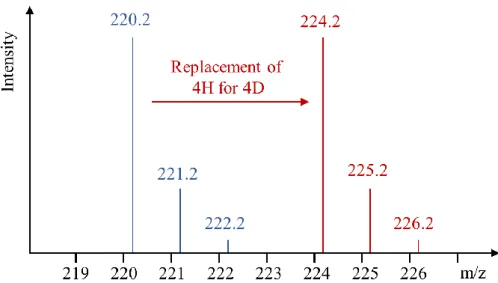
The application of deuterium labelled molecules described herein is based on the fact that the replacement of a hydrogen for a deuterium atom endows an organic molecule with a mass label and generates a shift within its mass spectrum but does not significantly change the chemical properties of the molecule. In this manner, the unlabelled compound and the deuterium labelled compound have the same retention times in liquid- and gas chromatography, but they can be still distinguished through mass-spectrometry (MS) (figure
3).
Figure 3. Mass shift achieved when four hydrogen atoms are replaced by four deuterium
atoms in a hypothetical organic molecule.
Thus, the deuteration of a molecule of interest can generate an internal standard that paves the way to many analytical tools through liquid chromatography (LC) and gas chromatography (GC) coupled to mass spectrometry (LC-MS/MS, GC-MS/MS). In order to avoid an overlap of the MS pattern of the natural isotopomer and isotopologue distribution of the unlabelled analyte with the MS pattern of the internal MS-standard, in the ideal case, the deuterated internal standard is supposed to accommodate at least three deuterium atoms and to contain less than 0.5% of unlabelled starting material.9 Isotopologues are mass variants of the same molecule displaying different amounts of isotopes in their chemical structures (figure 4, top). Chemical structures of an isotopologue with different isotope substitution patterns and the same molar masses, are called isotopomers (figure 4, bottom).
Figure 4. Different isotopologues and isotopomers of 2,5-diphenyloxazole as an example.
A deutero-analogue that is used in this kind of application is referred to as a stable isotopically labelled internal standard (SILS), because deuterium is a stable isotope. The incorporation of other stable isotopes like 13C, 15N and 18O into organic molecules very often proceeds through synthetic pathways and usually not over isotope exchange reactions as for hydrogen isotopes (see chapter I.3). Owing to the availability of more convenient methods for the incorporation of several deuterium atoms per molecule which brings about higher time- and cost efficiency, deuterium can be largely preferred over 13C, 15N and 18O in some of the following applications.
1.1.3 SILSs for metabolism studies of drugs
The first applications of deuterium in metabolism studies are even longer ago than the invention of nuclear magnetic resonance (NMR).10 Deuterated analogues of drug development candidates are predominantly used by pharmaceutical companies for monitoring the fate of the drug compound issued and its metabolism inside an organism. In the early stages of clinical studies in drug development processes, it is crucial to gain knowledge about the structures, pharmacokinetics and toxicological profiles of every drug metabolite. To this end, usually 1:1 mixtures are prepared from the unlabelled analyte and the deuterated analogue.
After being applied to the body and isolated from a biological liquid (blood plasma, urine etc.) every downstream metabolite stemming from the metabolized drug would show again the characteristic 1:1 pattern in MS-analysis. In this manner, every drug metabolite can be isolated through HPLC and identified, e.g. through additional isotopic labelling (13C labelling for subsequent 13C-NMR analysis).3
1.1.4 SILSs in metabolomics
The term metabolomics defines the study and analysis of the metabolic phenotype of a biological system. The metabolic phenotype refers to the entirety of all metabolites and their quantities in a biological system or sample.11 Metabolomic studies are subdivided into three main branches. Firstly, the identification and characterization of all metabolites of an organism. Secondly, their quantification and thirdly, the investigation of their pathways inside the organism, in other words, the metabolite flux analysis. The so called untargeted or nonbiased approach in metabolomics is used to determine and to characterize the ensemble of all present metabolites in the sample. The difference between metabolism studies (I.1.1.3) and untargeted metabolomics is that the latter require the pool of all metabolites which are isotopically labelled, not just the compound of interest. Thus, the internal standard in untargeted metabolomics represents usually a mixture of hundreds of different deuterated metabolites to exclude background ions like sample contaminations which are visible in MS-spectra but do not result from metabolic processes. Such mixtures can be prepared e.g. by supplying [13C6]-glucose to an organism as a feedstock of 13C atoms. However, the
determination of the full metabolic phenotype also requires thorough quantifications of every metabolite. This kind of analysis can be disturbed by several factors. On the one hand, highly abundant metabolites could saturate the detector of the mass spectrometer, making the measurement of high metabolite amounts impossible. On the other hand, metabolite concentrations could deviate due to matrix effects of biological probes. Matrix effects occur in mixtures of complex composition and they can affect the stability, binding behavior and other properties of a certain compound which is present in the given mixture. To overcome those effects and to solve these problems, internal standardization through isotopically labelled analogues is effectuated to generate calibration curves for the detector response. For this analytical step, a SILS of the metabolite of interest is needed to be isolated and pure. Hence, it is indispensable to have many alternative methods available for the preparation of
stable isotopically labelled metabolites, also because of the high number of possible metabolites and their structural diversity. The metabolite flux analysis is rather achieved through 13C-labelling. Deuterium plays a minor role in this context, because it is known to have a potential impact on metabolic pathways, as described for “Heavy Drugs” in section
I.1.1.1. Published works using deuterated molecules for this purpose are still known.
Metabolic fluxes in potato tubers, for example, could be elucidated by incubating tuber slices with deuterated phenylalanine and subsequent LC-MS analysis.12
Apart from drug development, metabolism studies and the investigation of other cellular processes, deuterium is applied in many other disciplines, which cannot be described in detail here. Deuterated analogues are also used in material science13, for the elucidation of mechanistic pathways in chemical synthesis14 and different imaging techniques like deuterium metabolic imaging (DMI) by means of magnetic resonance spectroscopic imaging (MRSI).15 The stability of 11C-labelled radiotracers for positron emission tomography (PET) towards metabolism could be also increased by deuteration.16
1.2 Tritium and its applications
The radioactive isotope tritium is produced in nuclear reactors through neutron irradiation of compounds containing high percentages of lithium-6, e.g. lithium fluoride or lithium alloys as Li–Al and Li–Mg. One part is formed as 3H2 (T2) gas and the other part is retained in the solid
state which can be recovered chemically. Since tritium is a β-emitter, the maximum penetration depth of the radiation in air is 6mm and in glass or concrete 2µm.17 For this reason, tritium containing material can be handled in usual glass ware without further risks of irradiation, if all safety rules are respected. The incorporation of tritium into organic molecules is performed in order to obtain tracer molecules and radioligands used in diverse life-science applications. As it could be seen in the precedent sections, light can be already shed on many aspects related to metabolism with deuterium labelled compounds in hand, but the detection of a radioactive compound unambiguously confirms with higher precision that it must be a metabolite of the tritium labelled compound which was applied initially. Radioactive detection also ensures that no relevant compound is missed during analysis of a mixture. Hence, drug metabolism studies are rather carried out through radioactive isotope labelling of the drug candidate. Indeed, when candidates of interest are evaluated within drug development processes, tritiated analogues thereof are constantly demanded for absorption,
distribution, metabolism and excretion (ADME) studies. The general preference of 3H over
14
C can be explained in the lower costs and more rapid procedures for the preparation of tritium labelled compounds. However, the common risk of tritium and deuterium labels is that they can be lost more easily then 13C- or 14C labels when attached to a position being sensitive to metabolic degradation. In vitro, tritium labelled drug development candidates or reference compounds, i.e. well-characterized benchmark compounds, are mainly used for radio ligand binding assays. Although there is a big variety of strategies using different tags, the general objective is the determination of the affinity of a compound to a biological target, which is very often a protein. In a typical example of an in vitro competitive binding assay, a tritium labelled reference ligand [3H]-astemizole, the membrane suspension of a cell that was transfected before with the membrane protein of interest (figure 5, vial 1) and the analyte, a small-molecule drug as potential inhibitor of the membrane protein, were incubated together (figure 5, vial 2). After filtration and washing of the mixture, the value of the scintillation counter reflected the amount of bound radioligand relative to the amount of bound analyte (figure 5, vial 3).18
Figure 5. General principle of a competitive radioligand binding assay
Tritium labelled compounds also allow to monitor a compounds distribution and clearance from a body and single organs, which are additional features and probably the main objectives of ADME studies. One example is quantitative whole body audioradiography (QWBA) that visualizes the accumulation behavior of a tritiated drug candidate in the body of small animals. This allows to calculate and to estimate the maximum dose of the radioactively
labelled drug candidate that can be applied to a human test candidate in ongoing clinical ADME studies.
1.3 Analysis of molecules labelled by hydrogen isotopes
This section is going to explain how to determine whether the structure of an organic molecule contains a certain isotope (2H or 3H). Further, the question is addressed how to figure out at which position of its structure the isotope is situated. The quantification of the isotope labelling, in other words, the determination of the isotopic enrichment, will be also addressed in relation with the presented analytical methods. The isotopic enrichment is the percentage of molecules present in a given mixture of isotopologues and isotopomers which incorporates the isotope of interest at a certain position of the chemical structure. As chemical differences between hydrogenated and deuterated or tritiated small molecules are usually not measurable under clinical conditions, a chromatographic separation and isolation of every isotopologue and isotopomer of the same molecule is not possible. For this reason, isotope chemistry requires analytical methods being able to unambiguously indicate the presence of an atom and to quantify its abundance with high accuracy.
1.3.1 Mass spectrometry
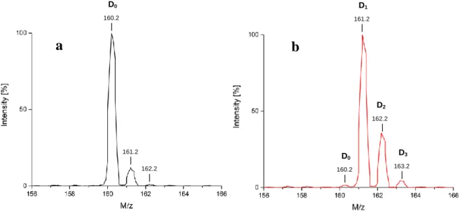
Mass spectrometric analysis is the most reliant method to confirm the presence of compounds in complex mixtures and to determine their isotopic composition as already mentioned at the beginning of section I.1.1.2 with figure 3. The principle of mass spectrometry is to ionize the molecules in the probe and to bring their ions into the gas phase, either by electrospray ionization (ESI) or matrix assisted laser desorption ionization (MALDI). For the ESI process the analyte solution is conducted through a metallic capillary and at its tip, an electrical field is applied that leads to an ionization of the molecules, either to positively charged species by a cation uptake (H+, Na+ etc.) or to negatively charged species by proton releases. The ions migrate then to the counter electrode at the opposite of the capillary outlet. In MALDI, the analyte is embedded into an organic matrix together with inorganic salts. Molecules are excited and released for example by a nitrogen laser. Collisions in the gas phase lead to the formation of ions. Ions in the gas phase can be then accelerated by the application of an electrical field.19 The ion separation and analysis by their mass to charge ratio (m/z) can
proceed on different ways. Quadrupole mass spectrometers conduct the ion beam through an oscillating voltage that is applied between four parallel metallic rods. Just ions with a defined m/z can pass the quadrupole at a given voltage. Thus, by tuning the quadrupole voltage, the whole m/z range of the analyte can be monitored. The time of flight (TOF) mass spectrometer gives the same initial energy to all ions by accelerating them over a short distance. Then, the time they need to cross a defined way is measured by a detector at the end. The higher the mass of the ion, the longer it needs to fly to the detector. Given that isotope incorporations always result in mass changes, mass spectrometry is used as the most current method in isotope chemistry because it is able to measure even the tiniest mass shifts. Consequently, isotopologue mixtures can be thoroughly analyzed, since isotopologues also manifest separation on MS spectra. On this path, the measurement of relative isotopic abundances (RIS) becomes possible, for example of naturally occurring isotopologues or after isotope labelling experiments. In a hydrogen isotope labelling experiment, the type and the quantity of hydrogen isotopes incorporated on the same molecule (+ ~1g/mol for the replacement of one hydrogen for one deuterium or + ~2g/mol for the replacement of one hydrogen for one tritium) can be determined by computer-assisted methods from the recorded MS-spectrum, given that the different intensities or integrals of the mass peaks reflect the ratio of isotopologues in a mixture. To this end, the isotopologue mass peaks are integrated before and after the deuterium incorporation and the contribution of the natural isotope abundance is subtracted from the integrals of the labelled molecule. An example is given in figure 6 by ESI-MS. In order to determine the amount of D1-isotopologue after the deuteration of a
theoretical molecule (figure 6b, red spectrum), the integration value of the peak that appears in spectrum a at the same mass shift (161.2) needs to be subtracted from the integration value of the D1-isotopologue in spectrum b. The same procedure is repeated for every isotopologue
peak of the labelled material. The D0 peak in spectrum b gives the amount of unlabelled
starting material left after the deuteration experiment. The total isotope incorporation on the whole molecule is obtained by the sum of all deuterated isotopologue amounts relative to the amount of unlabelled starting material left.
Figure 6. (a) Natural isotope pattern of a starting material (b) ESI-mass spectrum of the same
molecule after deuterium incorporation
High-resolution mass spectrometry even allows differentiating other types of stable isotopes (2H, 13C, 15N, 18O). If a molecule contains different types of stable isotopes, higher mass isotopologues can be split into several interfering mass peaks. The slight shifts between different isotopes can be interpreted by the different gaps in mass, e.g. between protium and deuterium (M(2H) - M(1H) = 1.0063g/mol) and 12C and 13C (M(13C) - M(12C) = 1.0031g/mol). Thus, the peak of an isotopologue possessing just one deuterium would be shifted by 0.0032g/mol compared to the peak of an isotopologue possessing just one 13C. These isotopic distances are related to the different sums of protons and neutrons in chemical elements. The high-resolution potential of this technique resolves interferences between mass peaks, it indicates whether a mass shift results from the one or the other isotope and permits computer-assisted quantifications, which is also relevant for environmental geochemistry, earth- and planetary sciences.20
1.3.2 NMR spectroscopy
NMR spectroscopy provides information about the nuclei present in an organic molecule, their ratios in the molecular framework and their electronic situations. An NMR spectrometer consists of a magnet that can generate a strong and homogeneous magnetic field and a source of electromagnetic radiation from the radiofrequency range. When an analyte is placed into
the magnetic field, atomic nuclei with a nuclear spin I = , as hydrogen for example, form two
different energetic levels mI = and with the transition ΔE = γħB0 between both (γ is
D1 161.2 | D2 162.2 | D3 163.2 | 161.2 | 162.2 | D0 160.2 |
a
b
D0 160.2 |the gyromagnetic ratio, ħ = with h as the Planck constant, B0 is the strength of the magnetic field, figure 7). At this stage, the nuclei are able to absorb electromagnetic radiation to undergo excitation from one level to the other if the condition hνL = γħB0 is fulfilled. The frequency νL that is needed to excite the nucleus is called Larmor-frequency.21
Figure 7. Split of the energetic levels of a nucleus with the spin I = in a magnetic field Further, the applied magnetic field interacts with the electrons of every atom and induces an additional little magnetic field that contributes to the local magnetic field at the nucleus. As a result, the local magnetic field at the nucleus of every atom is never equal to the applied magnetic field. Thus, if B0 is replaced for Blocal, the condition for resonance turns into hνL =
γħBlocal and the Larmor-frequency depends now on the strength of the local magnetic field which is different for every nucleus of the molecule, regulated by the electronic situation. For this reason, same atoms that are situated at different positions of a molecule usually have different Larmor-frequencies. When a measured resonance is converted to the NMR spectrum by a “Fourier transformation”, the chemical shift of every nucleus can be then related to its electronic situation. The less a nucleus is shielded by its electrons, the more downfield the NMR signal appears in the spectrum. However, exact attribution of NMR signals to corresponding nuclei usually succeeds through coupling effects. Coupling, the spreading of signals into multiplets, occurs because magnetic moments of neighboring nuclei interact. The formula to calculate the multiplicity is 2nI+1, where I is the spin and n the number of equivalent nuclei the considered nucleus is coupling with. 1H-NMR spectroscopy is used in hydrogen isotope labelling in order to reveal the exact positions of the incorporated isotope in
the structure of a molecule, as demonstrated on 1-phenyl-1H-1,2,4-triazole in figure 8. By comparing the spectra of the non-labelled protio-analogue (figure 8, top) with the deuterium labelled molecule (figure 8, bottom), we will see that integration values diminish for the signals where hydrogen is exchanged for deuterium (figure 8 bottom, positions 1, 2 and 3), because deuterium does not resonate at the frequency of 1H-NMR. Precise isotopic enrichment values of a certain position can be then derived from the integration values (figure
8, square brackets). Certain positions can be also assigned by considering the change in
multiplicity when hydrogen is exchanged for deuterium on neighboring positions. Since the two 3-positions are deuterated to 80%, the multiplicity of the 4-positions changes from a doublet of a doublet (dd) to a doublet (d) as it can be seen in figure 8 when both 1H-NMR spectra are compared against each other.
Figure 8. 1H-NMR spectra of 1,2,4-triazole (top) and deuterated 1-phenyl-1H-1,2,4-triazole (bottom) with signal assignment, chemical shifts are given in ppm
Proton decoupled 13C-NMR (13C{1H}-NMR) also helps to confirm the deuteration of a position because the multiplicity of a carbon signal where deuteration takes place changes
from a singlet into a triplet, due to the spin of deuterium (ID = 1). 2H- and 3H-NMR analysis
stay the most indispensable tools to thoroughly determine the positions of hydrogen isotopes in a molecule. However, the deuteron has a 6.15 times lower gyromagnetic ratio than the proton which diminishes the resolution in 2H-NMR. For this reason, it is rather common to use high-field NMR instruments and 2H selective cryogenic probes to obtain qualitative 2 H-NMR spectra. Additionally, several other technical advances turned 2H-NMR into a very sensitive analytical method that is even able to detect the natural abundance of deuterium. The high sensitivity makes it a very convenient method for hydrogen isotope labelling experiments because every deuterated position in an organic molecule can be thoroughly determined. Further, 2H-NMR is an excellent purity control as it shows the presence of small amounts of deuterated side-products which is not an easy task for other methods.22 The triton is related to as the most sensitive nucleus for NMR-analysis due to its high gyromagnetic ratio. Additionally, the extremely low natural abundance of tritium leads to an efficient suppression of background signals which facilitates the analysis of 3H-NMR spectra.23 In order to diminish the fine structure of 2H- and 3H-NMR spectra, it is important to decouple the protons of the molecule from the 2H- and 3H-nuclei and to generate proton-decoupled deuterium- (2H-{1H}) NMR or proton-decoupled tritium (3H-{1H}) NMR spectra. In the following a comparative overview of the properties of the hydrogen isotope nuclei is illustrated (table 1).
1
H 2H 3H
Usual notation H D T
Radioactivity No No Yes (β--emitter)
Half-life (Days) NA NA 4540 Natural abundance (%) 99.985 0.015 10-18
Spin quantum number
(no unit) 1/2 1 1/2 Gyromagnetic ratio γ (MHz.T-1) 42.576 6.536 45.403 Quadrupolar moment (10-24 cm2) 0 +2.87 10 -3 0 Larmor frequency at 14.09 T (MHz) 600.00 92.10 639.98
Relative sensibility(a) (no unit) 1.00 9.65 10-3 1.21 Absolute sensibility(b) (no unit) 1.00 1.45 10-6 -
Typical Chemical shift range
(ppm) 0 to 20 0 to 20 0 to 20 Typical T1 range (s) 0.1 to 20 0.1 to 10 0.1 to 10 Typical T2 range (s) 0.1 to 20 0.1 to 10 0.1 to 10
Typical J(H,X) scalar coupling range (Hz)
0 to 20 0 to 3 (c) 0 to 22 (d)
NOE effect by 1H decoupling Yes Negligible Yes
(a) Value at constant magnetic field or equal number of nuclei (b) Product of the relative sensitivity and natural abundance (c) J(D,H) = J(H,H) × (D /H)
(d) J(T,H) = J(H,H) × (T /H)
Table 1. Comparison of the hydrogen, deuterium and tritium nucleus23
Moreover, due to the radioactive decay of tritium, molecules which contain tritium atoms in their structure are analyzed by scintillation counting. The measured parameter gives the specific activity [Ci/mg] or the molar activity [Ci/mmol] that indicates the amount of tritium atoms present in the structure of a purified molecule.
2.
N
-Based heterocycles: promising bioactive targets for
the introduction of deuterium and tritium
The following chapter demonstrates a manifold of important targets for the incorporation of hydrogen isotopes. As we will see, aromatic heterocycles containing nitrogen atoms in their structures (N-heterocycles) appear to be highly relevant compounds or structural patterns in many fields of our life as pharma- and food industry. Heterocyclic scaffolds are involved as building blocks in the majority of commercial drugs, with nitrogen containing heterocycles being the most popular among them.24 59% of FDA approved drugs contain at least one nitrogen-based heterocycle.25 For every compound of biological or technological relevance, it is favorable to have available methods that allow labelling by deuterium and tritium. For this
purpose, the most relevant N-heterocyclic scaffolds for this work will be classified and the nomenclature of their rings will be explained. Further, light will be shed on their chemical properties and on the advantages or drawbacks to incorporate hydrogen isotopes on certain heterocyclic sites. Finally, this chapter will analyze in more detail the presence of N-heterocyclic scaffolds in Active Pharmaceutical Ingredients (APIs) and the motivation of drug discovery and development to introduce N-heterocyclic cores into these agents.
2.1 Nomenclature and numbering of heterocyclic rings
The most relevant scaffolds for the follow-up of this work are introduced in this section. The focus lies on nitrogen-containing five-membered aromatic heterocycles and on structural analogues thereof with attached benzene rings, which are going to be called “benzo-derivatives”. Benzimidazole, for example, is a benzo-derivative of imidazole and indole a benzo-derivative of pyrrole (figure 9). The numbering starts at the heteroatom that has the highest atomic number in the periodic table, e.g. in oxazole at the oxygen. In imidazole, the N–H has a higher priority than the nitrogen atom without hydrogen. An exception is carbazole which is not considered to be a classical N-heterocycle and numbering is started at the attached benzene ring next to the N–H moiety. The numbering continues towards the next heteroatom that is localized closest to the first one. For this reason, the C2-atom lies between
the oxygen and the nitrogen atom in oxazoles and benzoxazoles. Angular atoms are usually referred to with the number of the precedent position and an “a” or “b” is added (figure 9). Just angular carbon atoms of nucleobases get own numbers, but this is due to historical reasons.26
Figure 9. Nitrogen containing heterocyclic scaffolds (N-heterocycles) and the numbering of
their positions
Sometimes the labels “α”, “β” or “γ” are used to indicate a position on a heterocycle relative to a heteroatom. In this context, “α”, “β” or “γ” would refer to the distance of the considered position from the heteroatom. If we follow this system, the C2 atom of benzimidazole is the
neighboring atom of the two nitrogens, thus, it would be the α position. The C4 and C7 atoms
would be the β positions relative to the two nitrogen atoms. Positions situated on the third carbon with respect to a heteroatom are referred to as γ positions. The ortho positions of the phenyl ring on 2-phenyl-benzimidazole, for example, are γ positions (figure 10).
Figure 10. α-, β- and γ positions on benzimidazole and 2-phenyl-benzimidazole as example
Triazoles are named by the arrangement of their nitrogen atoms in the 5-membered heterocycle. This can be a 1,2,3- or a 1,2,4-constellation. In addition, the nomenclature has to
contain the position of the nitrogen atom that carries the proton because tautomerization takes place on both isomers (1,2,3- and 1,2,4-triazole) that leads to tautomers which are chemically different (figure 11).27
Figure 11. Tautomerism of 1,2,3- and 1,2,4-triazole
Analogous to triazoles, the hydrogen of the N-H moiety in imidazole is also exchangeable and migrates in solution by tautomerism from one to the other nitrogen atom. However, this proton migration yields two equivalent tautomers, thus, the protons on the C4 and C5 positions
of imidazole also become chemically equivalent and give one signal in 1H-NMR. The same goes for benzimidazole (one signal for C4–H and C7–H, another signal for C5–H and C6–H).
2.2 Basicity/acidity of N-Heterocycles
Every position in a N-heterocycle’s structure displays a certain acidity, say, a tendency to be deprotonated or to exchange the proton in the presence of a proton acceptor. Acidity depends on several factors, but if we first just have a look at unsubstituted or simply substituted N-heterocycles, it is mostly the electronic situation of the considered position that defines the
pKa. Generally speaking, sites next to a heteroatom or between two heteroatoms, i.e. in the α position of heteroatoms, display the highest acidity from every other position in the ring, reasoned in the higher electronegativity and electron-withdrawing effect of the heteroatoms (figure 12).28
Figure 12. pKa values of some acidic N-heterocyclic protons measured in THF
For the upcoming work, the provided pKa values by Fraser et al. in figure 12 are just benchmarks because they do not take into account further substituent effects on the conjugated aromatic rings. Consequently, they do not replace test experiments at higher temperatures by adding deuterated solvents (D2O, CD3OD), bases, acids etc. to estimate the
acidic or basic character of a given N-heterocyclic derivative. The isotopic enrichment within such a test experiment, will indicate the positions on the N-heterocycle where also back-exchange can be theoretically expected in the presence of proton acceptors, even after successful introduction of the isotope label, e.g. by one of the methods that are going to be shown in the next chapter (I.3). Consequently, targeting N-containing heterocyclic moieties for the chemical incorporation of hydrogen isotopes bears the risk to lose a label that has been introduced over cost and time demanding chemical transformations. In the case of a tritium back-exchange, the impediment would be even twofold. On the one hand, undoubtedly, the loss of the tritium label from the labelled candidate is everything else than encouraging, and on the other hand, the formation of a potentially volatile and radioactive solvent represents an additional safety issue. Apart from that, it can be in so far reasonable to label N-heterocycles by hydrogen isotopes as we consider that the majority of N-heterocyclic sites exhibits practically no acidity, many of them are even known to be relatively stable towards metabolism. Last but not least, it is mainly due to the vast presence and application of N-heterocycles in pharmaceutical-, agrochemical- and material sciences that methodological research for the incorporation of hydrogen isotopes is needed for this group of compounds.
2.3 Implementation as therapeutics, in agricultural chemistry & material sciences
Among all other fields of application, N-heterocyclic scaffolds are mostly represented in bioactive molecules. The latter comprise natural products like nucleobases, amino acids,
hormones, cofactors, poisons etc. but also synthetic substances like drugs and agrochemicals. Since the structures of numerous natural bioactive molecules are built up of N-heterocycles, drug discovery often aims at mimicking a natural structure by preserving the same heterocyclic core and endowing it with a different substitution pattern to achieve or to enhance the desired effects. A medicinal chemist could call such a bioactive natural product the “lead structure”. The term “lead structure” refers to a molecule that has attracted interest because of its promising effects and that serves as template in the ongoing drug development and optimization process. Popular examples for this strategy of drug design are nucleoside analogues, mostly used as anti-tumor agents (cytostatics).29 An example, for copying the imidazole scaffold of a natural neurotransmitter into a drug´s structure that is supposed to have a higher affinity to the same receptor, is demonstrated in figure 13. The design of the drug cimetidine, an antagonist of the H2 receptor, was based on the structure of the natural
ligand histamine.30
Figure 13. Structures of histamine, the natural ligand of the H1-4 receptors (left), and the H2
antagonist cimetidine (right)
Another common strategy in drug discovery and development is the substitution of functionalities in a lead structure by heterocycles to form bioisosters, also called biomimetics, which is probably the broadest application of the shown N-heterocyclic scaffolds in section
I.2.2 for drug development, because some of them such as triazoles are mostly absent in
natural molecules. The objective of using a bioisoster is to achieve either a stronger interaction between the drug and the desired target in the body or more convenient pharmacological drug properties.5 In most cases the introduction of an aromatic N-heterocyclic scaffold in the structure of a drug development candidate is considered to establish hydrogen bonds, more efficient metal ion complexation or π-interactions in the binding pocket of an enzyme. This can be achieved by presenting conformationally restricted proton donors or -acceptors, aromatics for π-interactions or chelating groups for metal complexation. Indeed, each of these properties is accommodated by one or several N-heterocyclic scaffolds. Figure 14 shows an example for the replacement of a cis-olefin by an
oxazole during a development process of inhibitors of ADP- and collagen induced blood platelet aggregation.5 Indeed, it was crucial for the drug´s efficiency to introduce a central oxazole. This modification furnished BMY-45778, the compound in middle with a much lower EC50 value, compared to the olefin on the left (figure 14). EC50 is a pharmacological
parameter that indicates a ligand concentration at which 50% of the expected effect is observed. Based on several data, the oxazole unit of BMY-45778 in the red box is most probably involved in a decisive hydrogen bonding with the PGI2 receptor. Further, this
hypothesis is reinforced by the EC50 of the isomeric compound on the right (figure 14). The
drug candidate loses its efficiency almost completely as soon as the nitrogen atom of the central oxazole, being the stronger hydrogen bond acceptor inside the 5-membered heterocycle, is exposed to the opposite side (see the trend of EC50 values, figure 14).
Figure 14. EC50 dependence on the C – C double bond - oxazole substitution on PGI2
receptor ligands.
In the following, the interest in N-heterocycles, which are relevant for this work, is going to be explained and exemplified by illustrating popular bioactive compounds that accommodate the respective N-heterocyclic unit in their structure. As already alluded with figure 14, the oxazole unit is utilized as a versatile hydrogen bond acceptor in drug development, because it has two different heteroatoms for doing so. Further, the C2-H position of oxazole is able to
establish weak interactions by acting as H-bond donor.5 Since many oxazole-based drugs are well-established on the market,31 oxazole is considered to be a perspective scaffold for the discovery of new drugs. The work with oxazoles is an emerging field given the wide range of newly synthesized aryl derivatives which manifested promising potency as anti-tuberculosis and patented anti-cancer agents.32 Consequently, an upcoming request for the synthesis of isotopically labelled analogues for ADME studies can be expected.33 Moreover, the oxazole
core is accommodated in the structures of natural alkaloids, e.g. pimprinine, pimprinoles A-C and many others34. Pimprinine in particular has gained attention with regard to its important anticonvulsant35 and antiviral activity (left, figure 15).36
Figure 15. Examples of an oxazole-based natural product (left) a commercial drug (middle)
and a patented drug development candidate (right).
The Imidazole scaffold can play the role of a hydrogen bond donor and a hydrogen bond acceptor at the same time within drug design. Apart from cimetidine in figure 13, imidazole is a widespread nucleus in the structures of many other commercial drugs as it can be seen in
figure 16, e.g. in drugs for the treatment of osteoporosis (zoledronic acid)37, spinocerebellar degeneration (taltirelin);38 but also in sedative-analgesic agents (midazolam, dexmedetomidine)39 and antifungal drugs (bifonazole).40 A few molecules of the latter do not even display complex functionalization, like bifonazole or the α2 agonist dexmedetomidine
(figure 16).
Figure 16. Marketed imidazole-based bioactive molecules
1,2,4-triazoles are also prominent structural motives in many fields of life as medicine and agrochemistry. Some reviews present a synopsis of their recurrence in several antifungal drugs, fungicides, pesticides and sedative agents.41 Additionally, the 1,2,4-triazole group can be also found in anti-tumor-42 and anti-migraine agents (figure 17).43 Due to the frequent occurrence of the 1,2,4-triazole scaffold in all those types of molecules, there is an obvious
and high interest in the preparation of hydrogen isotope labelled analogues of 1,2,4-triazole containing bioactive molecules.
Figure 17. 1H-1,2,4-triazole-based antifungal drug (first from left), -fungicide (second from
left), -anti-tumor- (third compound) and anti-migraine agent (fourth compound)
The popularity of N-heterocyclic benzoderivatives especially refers to their implementation as bioisosteres in drug design, reasoned in their low basicity and capability to form hydrogen bonds at the same time. This is manifested in the high ranking of benzimidazole derivatives in the top 25 of the most frequent nitrogen heterocyclic drugs approved by the FDA. The benzimidazole motif contributes to the affinity of drugs to their target by acting as a bioisostere for phenols, catechols, amidines and guanidines.5 Benzoxazoles can serve as conformationally restricted biomimetics for N-aryl amides. The H1-antihistamine astemizole
is representative for benzimidazole-based drugs (figure 18). Although it was withdrawn from the market in 1997,44 astemizole and tritium labelled astemizole stayed important benchmark compounds for ADME studies, as shown in chapter I.1.2. Two benzoxazole-based drug examples are tafamidis, used for the treatment of transthyretin amyloidosis, and benoxaprofen, an anti-inflammatory drug which was also withdrawn from the market due to its hepatotoxicity (figure 18). Deuterated benoxaprofen in particular could be even applied by a pharmaceutical company as an internal standard for metabolism studies in man.45