HAL Id: tel-01692151
https://tel.archives-ouvertes.fr/tel-01692151
Submitted on 24 Jan 2018HAL is a multi-disciplinary open access archive for the deposit and dissemination of sci-entific research documents, whether they are pub-lished or not. The documents may come from teaching and research institutions in France or abroad, or from public or private research centers.
L’archive ouverte pluridisciplinaire HAL, est destinée au dépôt et à la diffusion de documents scientifiques de niveau recherche, publiés ou non, émanant des établissements d’enseignement et de recherche français ou étrangers, des laboratoires publics ou privés.
Evaluation of voxel-based analysis in stroke using
multiparametric MR imaging
Rui He
To cite this version:
Rui He. Evaluation of voxel-based analysis in stroke using multiparametric MR imaging. Human health and pathology. Université Grenoble Alpes, 2016. English. �NNT : 2016GREAS026�. �tel-01692151�
THÈSE
Pour obtenir le grade de
DOCTEUR DE LA COMMUNAUTÉ UNIVERSITÉ
GRENOBLE ALPES
Spécialité : Biotechnologie, Instrumentation, Signal et Imagerie pour la Biologie, la Médecine et l’Environnement (BIS)
Arrêté ministériel : 7 août 2006
Présentée par
Rui HE
Thèse dirigée par Alexandre KRAINIK et codirigée par Emmanuel BARBIER
préparée au sein de l’Institut des Neurosciences de Grenoble dans l'École Doctorale Ingénierie pour la Santé, la Cognition et l’Environnement
Évaluation d'une analyse voxel à voxel dans
l'accident vasculaire cérébral à partir d'images
IRM multiparamétriques
Thèse soutenue publiquement le 30 novembre 2016 à Grenoble devant le jury composé de :
M. Thomas TOURDIAS
Professeur CHU de Bordeaux Rapporteur / Président du jury M. Samuel VALABLE
Chargé de recherche CNRS Rapporteur M. Mathieu HATT
Chargé de recherche INSERM U1101 Examinateur M. Benjamin LEMASSON
Chargé de recherche INSERM U1216 Examinateur M. Alexandre KRAINIK
Professeur CHU Grenoble Alpes Directeur de thèse M. Emmanuel BARBIER
Acknowledgements
I would like to express my sincerest gratitude to my supervisors, Prof. Alexandre Krainik and Dr. Emmanuel Barbier. They have given their time generously, been a great sources of guidance, support and encouragement. They have led me into multidisciplinary environment, taught me principles of research, and how to carry out new ideas. In the preparation of the thesis, they have spent much time reading through each draft and provided me brilliant advice. Under their extraordinary supervision, the progress in the manuscript writing is obvious. Without their patient instruction, insightful criticism and expert comments, the completion of this thesis would not have been possible.
Many thanks go to Dr. Benjamin Lemasson. He has supervised me since the project started and gave me invaluable help. I gained my experience in operation when I followed him in the animal experiments. Furthermore, I do appreciate that the technical support such as software package update and analytical method improvement he provided kindly. For the revision of my thesis and article, he has also put considerable time and effort into them.
Special thanks should go to Dr. Olivier Detante and Dr. Anaïck Moisan for their direct help in the research of stroke, both clinical and preclinical, and also for their valuable comments on my thesis and article. The experiment data and clinical information they provided were crucial to continue my research.
I gratefully acknowledge the help of Dr. Julien Bouvier and Dr. Duc Ha Hoang, who have offered me useful suggestions in my academic studies.
I would like to thank Dr. Olivier Heck for his help in the analysis of clinical data and the collection of typical cases for my thesis.
I am also deeply indebted to all the other people in our team of Institut des Neurosciences (GIN) for their direct and indirect help.
My gratitude also extends to Prof. Jean-François LeBas (Centre Hospitalier Universitaire Grenoble Alpes) and Prof. Daniel Plusquellec (UMR CNRS 6226, Institut des Sciences Chimiques de Rennes), for their advisory opinions and assistance before I came to follow my PhD study. I would like to acknowledge my indebtedness to Prof. Ming Zhu (Shanghai Children’s Medical Center), for his support and help in the preparation of all the materials for the PhD application.
Finally, I am indebted to my parents for their continuous support and encouragement during my stay in France.
Declaration
I declare that this thesis was composed by myself, that the work contained herein is my own except where explicitly stated otherwise in the content, and that the achievement in research has not been submitted for any other degree or professional qualification except as specified.
Table of contents
1 Introduction of stroke ... 1
1.1 Definition ... 1
1.2 Cause of stroke... 1
1.3 Consequence and symptoms of ischemic stroke... 1
1.4 Etiology of ischemic stroke ... 2
1.4.1 Cerebrovascular anatomy ... 2
1.4.1.1 Cerebrovascular anatomy of human ... 2
1.4.1.2 Cerebrovascular anatomy of the rat ... 3
1.4.2 Subtype of ischemic stroke ... 4
1.4.2.1 Subtype classification methods of ischemic stroke ... 4
1.4.2.1.1 TOAST ... 5
1.4.2.1.2 ASCO ... 5
1.4.2.1.3 CCS ... 5
1.4.2.2 TIA ... 6
1.4.3 Infarct patterns of ischemic stroke ... 6
1.5 Evolution of ischemic stroke ... 7
1.5.1 Phase of ischemic stroke ... 7
1.5.2 Pathophysiology of ischemic stroke ... 8
1.5.2.1 The neurovascular unit ... 8
1.5.2.2 Kinetics of ischemia ... 9 1.5.2.3 Microcirculation ... 11 1.5.2.4 Hypoxia ... 11 1.5.2.5 Excitotoxicity... 11 1.5.2.6 Peri-infarct depolarizations ... 11 1.5.2.7 Calcium toxicity... 12 1.5.2.8 Inflammation ... 13 1.5.2.9 Apoptosis ... 13
1.5.2.10 Edema and permeability change of blood-brain barrier .... 14
1.6 From emergent to preventive strategies for ischemic stroke ... 16
1.6.1 Target of ischemic stroke therapies ... 16
1.6.2 Demand of development in stroke therapies ... 17
1.6.3 Thrombolysis therapy ... 17
1.6.4 Endovascular therapy ... 18
1.6.4.1 Intraarterial thrombolysis and correlative clinical trials ... 19
1.6.4.2 Endovascular mechanical approaches and correlative clinical trials ...20
1.6.6 Development space and prospect of regenerative stroke therapy ... 23
1.6.7 Potential of biotherapies for post-stroke plasticity and neural repair... ... 25
1.6.8 Cell sources and cell therapy products ... 26
1.6.8.1 Cell types selection ... 26
1.6.8.2 The ideal route for cell delivery... 27
1.6.8.3 Comprehensive evaluation of cell therapy for stroke ... 27
2 Magnetic resonance imaging in ischemic stroke ... 29
2.1 Clinical standard MRI sequences for acute ischemic stroke ... 29
2.1.1 Fluid attenuated inversion recovery imaging ... 31
2.1.2 Three-dimensional time-of-flight magnetic resonance angiography... ... 31
2.1.3 Two-dimensional gradient recalled echo (T2*)- weighted imaging 33 2.1.4 Diffusion associated imaging approaches... 34
2.1.4.1 Diffusion-weighted imaging and apparent diffusion coefficient...34
2.1.4.2 Diffusion-based structural connectivity ... 37
2.1.5 Perfusion imaging ... 39
2.1.5.1 The fundamental of dynamic susceptibility contrast MRI perfusion technique ... 40
2.1.5.2 Blood supply relative perfusion parameters ... 42
2.1.5.3 Arrival time relative perfusion parameters ... 43
2.2 Advanced MRI techniques for clinical ischemic stroke... 49
2.2.1 Functional connectivity MRI application after ischemic stroke ... 49
2.2.2 Cerebral vasoreactivity in ischemic stroke ... 52
2.3 Multiparametric MRI application in ischemic stroke ... 54
2.3.1 Mismatch of diffusion-weighted imaging / perfusion-weighted imaging...54
2.3.2 Multiparametric routine clinical MRI selections ... 58
3 Overview of methods for MR imaging analysis ... 63
3.1 Region of interest ... 63
3.2 Histogram analysis ... 64
3.3 Texture analysis ... 65
3.4 Voxel-based analysis ... 67
3.4.1 Principal of the parametric response map (PRM): a voxel-wise methodology ... 69
3.4.2 Applications of parametric response map in other diseases ... 72
4 Challenges and fundamental objective ... 75
5 Evaluation of parametric response map to assess cell therapy effects after experimental stroke ... 79
5.1 Brief abstract ... 79
5.2 Introduction ... 79
5.3.1 Transient Middle Cerebral Artery occlusion (MCAo) model ... 81
5.3.2 Experimental groups and intravenous administration of hMSCs ... 82
5.3.3 MRI experiments: acquisition ... 82
5.3.3.1 MRI experiments: data processing ... 82
5.3.3.2 Data analysis ... 83
5.3.3.2.1 Regions of interest (ROIs) definition ... 83
5.3.3.2.2 Whole-lesion analysis ... 83
5.3.3.2.3 Parametric response map (PRM) analysis ... 83
5.3.3.2.4 Statistical analysis ... 84
5.4 Results ... 84
5.4.1 Evolution of lesion volume ... 84
5.4.2 Evolution of ADC... 84
5.4.3 Evolution of CBV... 85
5.4.4 Evolution of VSI ... 86
5.5 Discussion ... 87
5.6 Conclusion ... 88
5.7 Acknowledgements and sources of funding ... 88
5.8 Disclosure Statement ... 88
6 Correlating clinical outcome with voxel-based quantitative multiparametric MRI analysis in chronic ischemic stroke ...101
6.1 Brief abstract ...101
6.2 Introduction ...101
6.3 Subjects and methods ...102
6.3.1 Study population and study design...102
6.3.2 Clinical assessment...103
6.3.3 MRI assessment ...104
6.3.4 Data processing ...104
6.3.4.1 Diffusion data processing ...105
6.3.4.2 Perfusion data processing ...105
6.3.4.3 Image registration ...106
6.3.4.4 Regions of interest...106
6.3.5 Imaging analysis ...107
6.3.5.1 Whole-lesion analysis ...107
6.3.5.2 Parametric response map (PRM) analysis ...107
6.3.6 Statistical analysis ...107
6.4 Results ...108
6.4.1 Clinical outcome ...108
6.4.2 Imaging data ...109
6.4.2.1 Evolution of lesion volume ...109
6.4.2.2 Evolution of ADC ...109
6.4.2.3 Evolution of perfusion parameters ...109
6.4.3 Correlation between imaging and clinical data ... 111
6.6 Conclusion ... 118
6.7 Acknowledgements ... 118
6.8 Disclosure statement ... 118
7 General conclusion and perspectives ...135
7.1 General conclusion ...135
7.1.1 The assessment of stem cell therapeutic effects in preclinical application ...135
7.1.2 The prediction of outcome in clinical application ...136
7.2 Perspectives ...137
8 MRI segmentation methods (Appendix 1) ...143
8.1 Manual segmentation ...144 8.2 Automatic segmentation ...146 8.2.1 Intensity-based methods ...147 8.2.1.1 Thresholding ...147 8.2.1.2 Region growing ...148 8.2.1.3 Edge detection ...149 8.2.2 Atlas-based methods ...150
8.2.2.1 Probabilistic atlas segmentation ...150
8.2.2.2 Label propagation ...151
8.2.2.3 Multi-atlas propagation ...152
8.2.2.4 Classification ...153
8.2.2.5 Clustering ...155
8.2.3 Artificial neural network...157
8.2.4 Surface-based methods ...159
8.2.5 Hybrid segmentation methods ...161
8.2.6 Other methods ...165 List of figures...169 List of tables...173 Summary...174 NIHSS (Appendix 2)...179 mRS (Appendix 3)...185 Poster (Appendix 4)...189 CV (Appendix 5)...193 References...199
List of abbreviations
kNN - k-nearest-neighbor
AC - anterior commissure
ADC - apparent diffusion coefficient
AIF - arterial input function
AIS - acute ischemic stroke
Ang - angiopoietin
ANN - artificial neural network
ASL - arterial spin labeling
ASM - angular second moment
ATPase - adenosine triphosphatase
AUC - area under curve
BBB - blood-brain barrier
BCL - B-cell lymphoma
BDNF - brain-derived neurotrophic factor
BOLD - blood oxygenation level dependent
BP - blood pressure
CBF - cerebral blood flow
CBV - cerebral blood volume
CCS - Causative Classification System
CDP - crossed-diameter product
CE-MRA - contrast-enhanced magnetic resonance angiography
CI - confidence interval
CMRO2 - cerebral metabolic rate of oxygen
COPD - chronic obstructive pulmonary disease
CoW - Circle of Willis
COX2 - cyclooxygenase 2
CSF - cerebrospinal fluid
CT - computed tomography
CTA - computed tomography angiography
CVR - cerebral vasoreactivity[1], cerebrovascular reserve[2]
DEFUSE - Diffusion and Perfusion Imaging Evaluation for Understanding Stroke Evolution
DIAS - Desmoteplase In Acute Ischemic Stroke
DSA - digital subtraction angiography
DSC - dynamic susceptibility contrast
DTI - diffusion tensor imaging
DWI - diffusion-weighted imaging
EM - expectation-maximization
ENT - entropy
EP - endothelial progenitors
EPC - endothelial progenitor cell
EPITHET - Echoplanar Imaging Thrombolytic Evaluation Trial
EPO - erythropoietin
ET - endovascular treatment
FA - fractional anisotropy
FCM - fuzzy C-means
fDM - functional diffusion map
FLAIR - fluid attenuated inversion recovery
fMRI - functional magnetic resonance imaging
FOV - field of view
fSAD - functional small airway disease
FWHM - full width at half maximum
GBM - glioblastoma
GCSF - granulocyte-colony stimulating factor
Gd-DOTA - Gadolinium 1,4,7,10-tetraazacyclododecane-1,4,7,10-tetraacetic acid
GF - growth factor
GLCM - grey-level co-occurrence matrix
GluR2 - glutamate receptor 2
GM - grey matter
GMM - Gaussian mixture model
GRE - gradient recalled echo
HIE - hypoxic-ischemic encephalopathy
hMSC - human mesenchymal stem cell
HNSCC - head and neck squamous cell carcinoma
HSC - hematopoietic stem cell
HSP - heat-shock protein
HT - hemorrhagic transformation
HVS - hyperintense vessels sign
IA - intraarterial
IAT - intraarterial treatment
IC - intracerebral[1], infarct core[2]
ICA - internal carotid artery
ICH - intracerebral hemorrhage
ICP - intracranial pressure
IL - interleukin
IMS-2 - Interventional Management of Stroke phase 2
iNOS - inducible nitric oxide synthase
IP3 - inositol tri-phosphate
IV - intravenous
LoG - Laplacian-of-Gaussian
LP - label propagation
LRP - lipoprotein receptor-related protein
LVQ - learning vector quantization
MA - multi-atlas propagation
MCA - middle cerebral artery
MCAo - middle cerebral artery occlusion
MCI - malignant cerebral infarction
MD - mean diffusivity
MEG - magnetoencephalography
MEMa - Minimum Essential Medium with a modification
MGESE - multi-gradient echo and spin echo
MNC - mononuclear cell
mPRM - multiparametric respons map
MR CLEAN - Multicenter Randomized Clinical trial of Endovascular treatment for Acute ischemic stroke in the Netherlands
MR RESCUE - Mechanical Retrieval and Recanalization of Stroke Clots Using Embolectomy
MRA - magnetic resonance angiography
MRF - Markov random field
MRI - magnetic resonance imaging
mRS - modified Rankin Scale
MSC - mesenchymal stem cell
MTT - mean transit time
NAC - neoadjuvant chemotherapy
NAWM - normal appearing white matter
NECT - non-enhanced computed tomography
NIHSS - National Institutes of Health Stroke Scale
NO - nitric oxide
NOS - nitric oxide synthase
NSC - neural stem cell
NSOPT - nonsurgical organ preservation therapy
NVU - neurovascular unit
OEF - oxygen extraction fraction
decomposition
PA - probabilistic atlas
PBS - phosphate-buffered saline
PC - posterior commissure
PD - progressive disease
PDGF - platelet-derived growth factor
PDGFR-a - platelet-derived growth factor receptor a
PET - positron emission tomography
PICH - primary intracerebral hematoma
pMCAo - permanent middle cerebral artery occlusion
PP - pseudoprogression
PR - partial response
PRM - parametric response map
PROACT - Prourokinase for Acute Cerebral Thromboembolism
PTS - prothrombotic state
PV - partial volume
PWI - perfusion-weighted imaging
rCBF - regional cerebral blood flow
rCBV - regional cerebral blood volume
RECIST - Response Evaluation Criteria in Solid Tumors
RESTORE - reperfusion therapy in unclear-onset stroke based on MRI evaluation
RF - radiofrequency
ROC - receiver operating characteristic
ROI - region of interest
ROS - reactive oxygen species
rt-PA - recombinant tissue plasminogen activator
SAH - subarachnoid hemorrhage
SD - stable disease[1], standard deviation[2]
SEM - standard error mean
SI - signal intensity
SIAS - severe intracranial arterial stenosis
SOM - self-organizing map
SRI - serotonin reuptake inhibitor
STAIR - Stroke Therapy Academic Industry Roundtable
SVZ - subventricular zone
SWIFT PRIME - Solitaire with the Intention for Thrombectomy as Primary Endovascular Treatment
T - Tesla
T2WI - T2-weighted imaging
TA - texture analysis
TAR - tissue-at-risk
TBSS - track-based spatial statistics
TGF-β1 - transforming growth factor-β1
TIA - transient ischemic attack
Tmax - time-to-maximum
tMCAo - transient middle cerebral artery occlusion
TOAST - Trial of Org 10172 in Acute Stroke Treatment
TOF - time-of-flight
t-PA - tissue plasminogen activator
TTP - time-to-peak
USPIO - ultrasmall superparamagnetic iron oxide particles
VBM - voxel-based morphometry
VEGF - vascular endothelial growth factor
VOI - volume-of-interest
VSI - vessel size index
WHASA - white matter hyperintensities automated segmentation algorithm
WM - white matter
Evaluation of voxel-based analysis in stroke using
multiparametric MR imaging
Summary:
Stroke is the leading cause of disability in adults. Beyond the narrow time window and possible risks of thrombolysis and mechanical thrombectomy, cell-therapies have strong potential. Reports showed that transplanted stem cells can enhance functional recovery after ischemic stroke in rodent models.
To assess the mechanisms underlying the cell-therapy benefit after stroke, imaging is necessary. Multiparametric magnetic resonance imaging (MRI), including diffusion-weighted imaging (DWI) and perfusion-weighted imaging (PWI), has become the gold standard to evaluate stroke characteristics. MRI also plays an important role in the monitoring of cerebral tissue following stroke from the acute to the chronic phase. However, the spatial heterogeneity of each stroke lesion and its dynamic reorganization over time, which may be related to the effect of a therapy, remain a challenge for traditional image analysis techniques. To evaluate the effect of new therapeutic strategies, spatial and temporal lesion heterogeneities need to be more accurately characterized and quantified.
The current image analysis techniques, based on mean values obtained from regions of interest (ROIs), hide the intralesional heterogeneity. Histogram-based techniques provide an evaluation of lesion heterogeneity but fail to yield spatial information. The parametric response map (PRM) is an alternative, voxel-based analysis technique, which has been established in oncology as a promising tool to better investigate parametric changes over time at the voxel level which concern the therapeutic response or prognosis of disease.
The PhD project was divided into two parts: a preclinical and a clinical study. The goal of the first study was to evaluate the PRM analysis using MRI data collected after the intravenous injection of human mesenchymal stem cells (hMSCs) in an experimental stroke model. The apparent diffusion coefficient (ADC), cerebral blood volume (CBV) and vessel size index (VSI) were mapped using 7T MRI. Two analytic procedures, the standard whole-lesion approach and the PRM, were performed on data collected at 4 time points in transient middle cerebral artery occlusion (MCAo) models treated with either hMSC or vehicle and in sham animals. During the second PhD project, 6 MR parametric maps (diffusion and perfusion maps) were collected in 30 stroke patients (PHRC NCT00875654). MRI data, analyzed with both a classic mean value and a PRM approaches, were correlated with the evaluation of functional recovery after stroke measured with the National Institutes of Health Stroke Scale (NIHSS) and the modified Rankin Scale (mRS) at 4 time points.
In both studies, PRM analysis of MR parametric maps reveals fine changes of the lesion induced by a cell therapy (preclinical study) and correlate with long-term prognosis (clinical study).
therapeutic efficacy and of prognostic biomarker of stroke patients.
Key words: magnetic resonance imaging, brain, diffusion, perfusion, parametric response map, cerebral ischemia, cell therapy, mesenchymal stem cells, prognostic biomarker
Évaluation d'une analyse voxel à voxel dans l'accident
vasculaire cérébral à partir d'images IRM multiparamétriques
Résumé:
L'accident vasculaire cérébral (AVC) est la principale cause de handicap acquis chez l'adulte. Au-delà de l'étroite fenêtre thérapeutique et des risques éventuels de la thrombolyse et de la thrombectomie mécanique, la thérapie cellulaire par cellules souches présente un fort potentiel. Plusieurs études ont montré que les cellules souches transplantées peuvent améliorer la récupération fonctionnelle après un AVC sur des modèles de rongeurs.
L’imagerie multiparamétrique par résonance magnétique (IRM), qui inclue l'imagerie de diffusion et de perfusion, est aujourd’hui le protocole standard pour caractériser l'AVC. L'imagerie permet également de suivre in vivo les mécanismes sous-jacents de la thérapie cellulaire après un AVC de la phase aigüe à la phase chronique. Cependant, la quantification de l'hétérogénéité spatiale des lésions, clairement visible par IRM, reste un défi à l'heure actuelle. En effet, les techniques d'analyses d'images utilisées en routine sont basées sur le calcul des valeurs moyennes à partir de régions d'intérêts (ROI). Cette technique par ROI ne peut pas refléter l'hétérogénéité intra-lésionnelle. C'est pourquoi, de nouvelles stratégies d'analyses d'images doivent être développées et évaluées afin de quantifier l'hétérogénéité des lésions ischémiques mais aussi pour suivre l'évolution de cette hétérogénéité au cours du temps. Des approches utilisant des analyses par histogramme permettent d'évaluer l'hétérogénéité des lésions mais perdent l'information spatiale. Une alternative est l'utilisation d'une analyse d'image à l'échelle du voxel appelée "Parametric Response Map (PRM)". Cet outil a été décrit comme plus sensible que l'analyse par ROI dans le pronostic mais aussi dans le suivi thérapeutique chez des patients porteurs de tumeurs cérébrales ou encore atteints d'hémorragies cérébrales.
Mon projet de thèse est divisé en deux parties: une étude préclinique chez le rat et une étude clinique (PHRC NCT00875654). La première partie de ma thèse vise à évaluer les changements physiopathologiques mesurés par l'IRM après un traitement par cellules souches mésenchymateuses humaines (CSMh) sur un modèle d'AVC chez le rat. Des animaux présentant une occlusion transitoire de l'artère cérébrale moyenne (oACM) ou non (sham) ont été traités ou non par une injection de CSMh. Au cours de cette étude, différents paramètres IRM ont été cartographiés en utilisant une IRM 7T (4 temps d'imagerie): le coefficient apparent de diffusion (ADC), le volume sanguin cérébral (CBV) et l'indice de taille des vaisseaux (VSI). Les cartes d'ADC, CBV et VSI ont été analysées en utilisant l'approche classique par ROI mais aussi par PRM. L'objectif de cette étude était de déterminer si l'analyse par PRM était capable de détecter plus précocement l'effet des CSMh que l'analyse par ROI. Durant la seconde partie de ma thèse, 6 paramètres IRM (imagerie de diffusion et de perfusion) ont été acquis chez 30 patients AVC. Les données IRM, analysées par valeur moyenne classique et par PRM, ont été corrélées avec des évaluations de la récupération fonctionnelle : le score NIHSS (National Institutes of Health Stroke Score) et l'échelle de Rankin modifiée (mRS) mesurés à différents temps
post-ischémie. L’analyse PRM des cartes paramétriques IRM révèle des changements fins de la lésion et corrèle avec le pronostic à long terme après l’ischémie.
En conclusion, la PRM pourrait être utilisée comme biomarqueur d’efficacité thérapeutique (combinaison d’images IRM et d’outils innovants d’analyse d'images) et comme biomarqueur pronostique des patients AVC.
Mots clés: imagerie résonance magnétique, cerveau, diffusion, perfusion, carte paramétrique de réponse, ischémie cérébrale, thérapie cellulaire, cellules souches mésenchymateuses, biomarqueur pronostique
1
Chapter 1
Introduction of stroke
1.1 Definition
“Stroke” is termed to describe damage to the central nervous system such as brain and spinal cord due to abnormalities in its blood supply (ischemia) or bleeding. It is classified as ischemic or hemorrhagic. By far, the most common subtype of stroke is ischemic also called cerebral infarction [Mozaffarian et al. 2016]. Ischemic stroke results from a transient or permanent insufficient or interrupted flow of cerebral blood flow that is restricted to the territory of a brain artery. The reduction in flow is, in most cases, typically caused by the occlusion of a cerebral artery either by an embolus or by local thrombosis [Taylor et al. 1996].
1.2 Cause of stroke
Ischemic and hemorrhagic are the two main types of stroke. The former one is far more common. Ischemic stroke accounts for around 80% of strokes, and its origin is divided into cardiogenic, arterosclerotic, lacunar, hemodynamic or cryptogenic source [Adams et al. 1993]. Another 15% of strokes are related to disruption of a cerebral artery resulting in intracerebral hemorrhage (ICH). Hypertension is the main source for ICH with other reasons such as specific blood vessel abnormalities [Rathore et al. 2013]. The acute ischemic stroke (AIS) may also be complicated by a hemorrhagic transformation notably by the use of anticoagulation [Marsh et al. 2016]. In this thesis, we will focus on ischemic stroke.
1.3 Consequence and symptoms of ischemic stroke
Ischemic stroke caused by a sudden artery occlusion is one of the leading causes of mortality and long-term disability worldwide. Statistics from the American Heart Association estimate an average of one stroke occurrence every 40 seconds in United States, amounting to approximately 795,000 people experiencing new or recurrent strokes per year. From the micro-aspect, if an appropriate therapy is not given, it is calculated that about 1.8 million neurons are lost every minute [Saver et al. 2006, Mijajlovic et al. 2014]. In view of the widespread public health impact of stroke and its profound impact on patients, stroke research has remained in the forefront [Go et al. 2014].2
The most common symptom of a stroke is sudden weakness or numbness of the face, arm or leg, usually on one side of the body. Other symptoms include aphasia, visual disorders, dizziness, impaired consciousness and different extent of headache. However, stroke can be occult or with slight symptoms which are not seriously taken for clinical observation.
1.4 Etiology of ischemic stroke
1.4.1 Cerebrovascular anatomy
A good working knowledge of the brain anatomy and vascular systems is crucial.
1.4.1.1 Cerebrovascular anatomy of human
Arteries supplying blood to the brain are all branches of the large brachiocephalic arteries that stem from the aorta. The common carotid arteries on either side ascend the anterior neck and bifurcate around the level of the angle of the mandible, into the internal and external carotid arteries [Wholey et
al. 1997]. The internal carotids enter the skull through the intracranial branches
and supply the anterior 2 / 3 of the brain, which are more commonly involved in human cerebrovascular diseases. The internal carotid arteries and their tributaries constitute the anterior circulation [Krishnaswamy et al. 2010].
The posterior circulation is composed of the left and right vertebral arteries, which are branches of the subclavian arteries arising from the aorta [Krishnaswamy et al. 2010]. The vertebral arteries on both sides merge and form the basilar artery, which then ascends and bifurcates into left and right posterior cerebral arteries. The vertebral arteries, the basilar artery and their branches constitute the so-called vertebrobasilar system [Cereda et al. 2012].
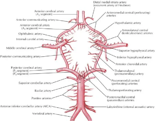
At the base of the skull, shortly after the large arteries enter the cranial cavity, the major arteries join to form anastomoses, the Circle of Willis (CoW), formed by arteries which communicate between the anterior and posterior systems and between the left and right side of the brain [Cereda et al. 2012] (Fig.1.1).
3
Figure 1.1: Main structure of the cerebrovascular anatomy and the CoW (from Atlas of
Human Anatomy Sixth Edition by Dr. Frank H. Netter)
The bilateral anterior, middle and posterior cerebral arteries are the main supplying arteries of the brain which interconnected by two major collateral systems [Liebeskind et al. 2003]. Under clinical conditions, the individual variability of the anastomoses which interconnect these arteries is responsible for the vascular occlusion which may result in a wide range of volumes from small ischemic lesions located in the central region to large infarcts involving the entire vascular supplying territory.
1.4.1.2 Cerebrovascular anatomy of the rat
Human ischemic stroke usually results from middle cerebral artery occlusion (MCAo), so animal models which occlude the MCA are closest to the clinical prospect [Longa et al. 1989, Mhairi Macrae et al. 1992]. Different animal models for experimental stroke research have been developed with a number of additional techniques proposed [Kawamura et al. 1994, Ma et al. 2006]. Models based on different species may have slightly different vascular brain anatomy which impacts the feasibility of operation [Scremin et al. 1982,
Maeda et al. 1998]. Currently, the rodent MCAo models are the commonest in
experimental stroke studies.
The arterial blood supply of the rat brain is relatively similar with the CoW in human but without the anterior communicating artery [Longa et al. 1989]
4 (Fig.1.2).
Figure 1.2: Diagram of cerebrovascular anatomy in rats illustrates extracranial and
intracranial vascular relations exploited in the method of reversible MCAo. Vessel size is disproportionately enlarged for clarity [Longa et al. 1989]. Cervical dissection of common, external and internal carotid arteries with their branches is shown in red (experiment method mentioned in Chapter 5).
1.4.2 Subtype of ischemic stroke
Post-ischemic stroke outcomes including disability, fatality and recurrence differ according to subtypes defined by stroke mechanisms [Lovett et al. 2004]. Identification of the underlying cause of stroke is an important element of daily clinical practice including treatment and prognosis evaluation for individual patients. Accurate and reproducible assignment of the likely mechanism of ischemic stroke is also important in clinical trials investigating benefit in specific patient groups [Kolominsky-Rabas et al. 2001]. The potential etiologies of the stroke should be taken into consideration for the selection of therapeutic strategies, the assessment of evolution and the prediction of prognosis besides the basic information such as the size and site of the ischemic lesion.
1.4.2.1 Subtype classification methods of ischemic stroke
5
various etiologies often result in different clinical presentations and characteristic appearance of the lesion on imaging. Several subclassification schemes for ischemic strokes have been proposed [Chen et al. 2012], including the Harvard Stroke Registry (1978) [Mohr et al. 1978], Trial of Org 10172 in Acute Stroke Treatment (TOAST) (1992) [Adams et al. 1993], Oxfordshire Community Stroke Project (1993) [Lindley et al. 1993], Causative Classification System (CCS) of stroke [Ay et al. 2005], A-S-C-O (phenotypic) classification (2009) [Marnane et al. 2010] and Chinese Ischemic Stroke Subclassification (2011) [Gao et al. 2011].
1.4.2.1.1 TOAST
The TOAST classification is the most widely used and denotes five subtypes of ischemic stroke: 1) large-artery atherosclerosis, 2) cardioembolism, 3) small-vessel occlusion, 4) stroke of other determined etiology, and 5) stroke of undetermined etiologies which includes cases invoking more than one primary mechanism. Differentiation of this classification are based on clinical features, computed tomography (CT) / magnetic resonance imaging (MRI) data, cardiac imaging, duplex scanning of extracranial arteries, arteriography and laboratory tests for a prothrombotic state (PTS) [Adams et al. 1993].
The TOAST system promotes better understanding of different potentially contributory mechanisms in growing stroke patients [Lee et al. 2000].
1.4.2.1.2 ASCO
The phenotype-based classification is defined by A-S-C-O: “A” for atherosclerosis, “S” for small vessel disease, “C” for cardiac source and “O” for other cause. Each of the four phenotypes is graded “1” to “3”. “1” for definitely a potential cause of the index stroke, “2” for causality uncertain and “3” for unlikely a direct cause of the index stroke. When the disease is completely absent, the grade is “0”. When grading is impossible due to insufficient definition caused by uncertain elements, the grade is “9” [Amarenco et al. 2009].
This phenotypic classification method also improves the capability to identify the most likely cause where multiple potential mechanisms are explored [Ay et al. 2007, Amarenco et al. 2009].
1.4.2.1.3 CCS
The Causative Classification System (CCS) scheme also assigns objects into 5 categories according to the mechanisms: cardio-aortic, large artery atherosclerosis, small artery occlusion, other cause and undetermined. However, compared with TOAST, the CCS assigns the most likely subtype based on updated estimates of stroke risk associated with specific cardiac and vascular pathologies, clinical features or imaging parameters known to be more commonly associated with particular stroke mechanisms [Marnane et al.
6 2010].
Results from previous studies illustrate that the feasibility of a harmonized combined classification system should be improved for clinical stroke research based on the specific strength of each single classification system [Weimar et
al. 2016]. 1.4.2.2 TIA
The transient ischemic attack (TIA), also as known as “mini stroke”, is a special type of stroke. It means a transient episode of neurological dysfunction caused by the blood flow interruption for a short period of time that tends to resolve itself quickly without acute infarction, usually within 10–20 minutes and less than 24 h before disappearing [Easton et al. 2009]. TIA occurs with a high rate, nearly 7.5 million cases worldwide each year. Although TIA is considered as benign by the public, it is still a strong warning sign of an ischemic stroke and should not be ignored to prevent a more serious attack [Giles et al. 2007,
Merwick et al. 2010].
TIA is also associated with high risk of recurrent ischemic events. According to the guidelines, it recommends that patients with TIA should undergo neuroimaging evaluation within 24 h of symptom onset, by CT or MRI, preferably by MRI, especially diffusion-weighted imaging (DWI, technique mentioned in Chapter 2) because it is more sensitive than CT. However, the strength of CT is its availability and ability to quickly exclude ICH. Generally, a follow-up MRI should be performed after the emergent CT because of its superiority in identifying cerebral infarction. A lack of evidence of infarction on MRI in patients who have symptoms consistent with cerebral ischemia distinguishes TIA from minor stroke. The noninvasive imaging approaches of the intracranial and cervical arteries such as CT or MR angiography, or transcranial Doppler should also be performed within 48 h [Simmons et al. 2012]. Refinement in brain imaging and better understanding of the time course of TIA-related lesions may lead to the further potential of the MRI signature associated with transient ischemia [Olivot et al. 2011,
Souillard-Scemama et al. 2015].
1.4.3 Infarct patterns of ischemic stroke
Infarct patterns (especially following MCAo) can be categorized into 3 basic patterns based on DWI features, including scattered, territorial and lenticulostriatal. A territorial pattern defined as wedge-shaped infarctions involving the cortex and subcortex with clearly noticeable margins, while a lenticulostriatal pattern represents an infarction restricted only in MCA perforator artery territory. A scattered pattern is defined as infarct patterns that are not describable by the previous two patterns regardless of lesion location or infarction size. The extent of this pattern includes the cortex, subcortex of
7
the MCA territory and deep structures like internal capsule and basal ganglia [Lee et al. 2014] (Fig.1.3).
Figure 1.3: Three basic infarct patterns of acute ischemic stroke: (A) scattered, (B)
territorial and (C) lenticulostriatal by DWI. Infarct lesion of each pattern: white arrow [Lee et al. 2014].
With early identification of DWI patterns a consideration to the stroke cause may be provided [Wessels et al. 2006, Bang et al. 2009b]. Emboli originating from the cardiac chamber have a high chance of becoming large, leading to a sudden major artery occlusion and a larger infarct [Arboix et al. 2010]. Furthermore, stroke caused by an embolus arising from the heart or aortic arch or recurrent emboli tends to occur in more than one arterial territory, often favors the anterior circulation and are often bilateral by neuroimaging [Wessels et al. 2006, Chung et al. 2010]. The criteria including imaging evidences indicating spontaneous recanalization and the absence of preceding atherosclerosis or other arteriopathy, along with proven cardioembolic sources can be applied to distinguish embolic occlusion from thrombotic occlusion [Lee et al. 2014].
1.5 Evolution of ischemic stroke
1.5.1 Phase of ischemic stroke
Temporal evolution of ischemic stroke is typically categorized into hyperacute (0-6 h), acute (6-24 h), subacute (24 h to approximately 2 weeks) and chronic phase of stroke (> 2 weeks old) [Gonzalez et al. 2002, Kloska et
al. 2010]. During the acute phase (6-24 h), the ischemia-induced energy failure
and the terminal depolarization of cell membranes result in the consequent tissue injury which established within a few minutes after the onset of ischemia. During the subsequent subacute phase (24 h-2 weeks), the largest increment of infarct volume is the expansion of the infarct core into the penumbra until, after another 4-6 h, it becomes congruent with the peri-infarct area. Finally, a delayed phase of injury evolves which may last for several days or even weeks. During chronic phase (> 2 weeks), secondary phenomena such as vasogenic
8
edema, inflammation and possibly cell death may contribute to a further progression of injury. By using multiparametric imaging techniques, evidence could be provided that shortly after occlusion of the MCA, the penumbra is approximately of the same size as the infarct core [Hata et al. 2000]. After 3 h more than 50% and between 6 and 8 h almost all of the penumbra has disappeared and is now part of the irreversibly damaged infarct core (details mentioned in 1.5.2.6).
1.5.2 Pathophysiology of ischemic stroke
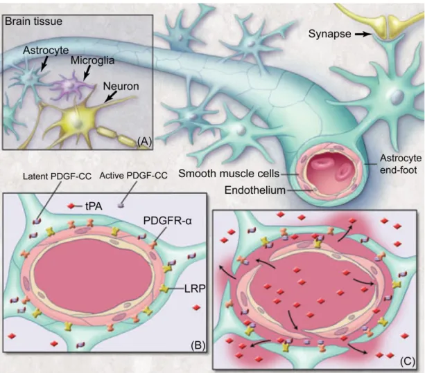
1.5.2.1 The glial neurovascular unitThe control and modulation of regional cerebral blood flow depend on neurovascular coupling without the condition of ischemic injury [Zonta et al. 2003, Koehler et al. 2009]. Microvessel responses reflect the presence of neuronal activation, requiring the function of intact neurons. Previous studies reported that signal transmissions might be directed from microvessels to the neurons which based on the alterations in basal lamina matrix, protease generation, endothelial cell activation and glia astrocyte-endothelial cell adhesion, in view of the proximity of microvascular endothelial cells to the circumferential astrocyte end-feet [del Zoppo et al. 2003, del Zoppo et al. 2006]. This coupling is accomplished by a group of cells close to each other, termed neurovascular unit (NVU), which composed by neurons, astrocytes, endothelial cells of blood-brain barrier (BBB), myocytes, pericytes and extracellular matrix components [Muoio et al. 2014]. These cells all work together in a coordinated way through their intimate anatomical and chemical relationship, detect the needs of neuronal supply, trigger necessary response for such demands and regulate the extracellular environment of brain parenchyma [Su et al. 2009] (Fig.1.4).
9
Figure 1.4: Neurovascular unit (NVU): (A) components of the NVU, (B) precapillary
arteriole with intact NVU and (C) response of the NVU to focal cerebral ischemia. Endogenous t-PA bound to LRP activates latent PDGF-CC, and active PDGF-CC binds to PDGFR-ɑ. Thrombolytic t-PA in the blood can cross a compromised BBB and active additional PDGF-CC, which exacerbates loss of BBB integrity. Tissue plasminogen activator, t-PA; lipoprotein receptor-related protein, LRP; platelet-derived growth factor CC, PDGF-CC; platelet-derived growth factor receptor ɑ, PDGFR-ɑ, adapted from [Su et al. 2009].
Whereas maintenance or timely re-establishment of flow reduces ischemic injury of both tissue and neuron, protection of neuron function in human has not prevented the evolution of injury despite the inherent mechanisms of neurovascular coupling. However, occlusion of cerebral blood flow rapidly identifies regions of neuron-vascular vulnerability within the vascular territory-at-risk which coalesce to turn into the mature ischemic lesion. So far, the failure of neuroprotective agents efficacy in clinical trials for the detectable tissue salvage could be explained by the decompensation of the NVU components due to their self-maladjustment [del Zoppo et al. 2010].
10
Oxidative glucose metabolism and brain energy state recover rapidly throughout the MCA territory, even after occlusion of as long as 1 h. After more than 3-6 h occlusion, recovery fails in the peripheral parts of the vascular territory and the volume of ischemia approaches that of permanent size [Kita
et al. 1995]. Compared with energy metabolism, recovery of protein synthesis
is much slower, which depends on both the duration of ischemia and the residual blood flow rate. After longer ischemia times, recovery fails in a gradually expanding core region until, after about 2 h of vascular occlusion, recovery disappears [Hermann et al. 2001].
Besides the imbalance of cerebral hemodynamics and oxygen / nutrients triggered by the ischemia, cellular disturbances may also contribute to the ischemia progression that cannot be explained by the single influence of impairment of blood flow or energy metabolism. The relevance of complex molecular mechanisms such as excitotoxicity, peri-infarct depolarizations, inflammation and apoptosis to delayed mechanisms of damage within the peri-infarct zone or penumbra which leads to permanent ischemic injury should be taken into consideration [Hossmann et al. 2006, Durukan et al. 2007] (Fig.1.5).
Figure 1.5: Schematic representation of molecular ischemic injury pathways leading
to cell death. Multiple approaches are provided for the amelioration of both necrotic and apoptotic tissue injury by blocking these pathways at numerous sites [Hossmann
11
1.5.2.3 Microcirculation
In focal ischemia, microcirculation is progressively disturbed. This disturbance is caused by at least three different pathophysiological mechanisms: the adhesion of white blood cells to the vessel wall; the increase in the blood viscosity due to the aggregation of blood corpuscles, and the compression of the capillaries by swollen astroglia [Vogel et al. 1999].
1.5.2.4 Hypoxia
Once the resistance vessels are completely dilated, both autoregulation and CO2 reactivity are abolished. Blood flow follows passively the fluctuations
of the systemic blood pressure. The abolishment of CO2 reactivity can also
result in an uncoupling from metabolic activity which leads to the phenomenon that, during peri-infarct depolarization (details mentioned in 1.5.2.6), the cerebral blood flow is dissociated from the metabolism. Therefore, the loss of CO2 reactivity is a serious predictor of impending brain ischemia that requires
immediate therapeutic interventions [Markus et al. 2001].
1.5.2.5 Excitotoxicity
The main excitotoxicity mechanism is the high concentration of glutamate which results in primary neuronal necrosis. However, following pharmacological inhibition of ionotropic glutamate receptors, an apoptotic injury mechanism develops that may prevail under certain pathophysiological conditions [Kohara et al. 2008].
Evidence indicates that activation of glutamate receptors, through the attendant failure of ion homeostasis and increase in intracellular calcium ions concentration, is a major factor involved in initiating cell death. Although blocking glutamate receptors protects against excitotoxicity, it still has serious side-effects, such as psychotomimesis, respiratory depression or cardiovascular dysregulation [Chuang et al. 2011].
Excitotoxicity is well established as an important trigger and executioner of tissue damage in focal cerebral ischemia. Excitotoxic mechanisms can not only cause acute cell death (necrosis) but also initiate molecular events that lead to delayed cell death (apoptosis) [Hossmann et al. 1996a].
1.5.2.6 Peri-infarct depolarizations
In the core region of the affected brain tissue, cells can undergo an irreversible anoxic depolarization and without repolarization. The core area is surrounded by tissue in which energy failure leads to electric silence with function loss, but because of residual perfusion in this region, vascular, cellular and molecular processes may lead to a certain extent of rescue for the recovery of function against the ischemic core. In focal ischemia, the blood flow range corresponds to a crescent-shaped region intercalated between the necrotic and normal brain tissue, it has been termed “penumbra” in analogy to
12
the partly illuminated area around the complete shadow of the moon in full eclipse [Manning et al. 2014]. This concept was further developed that cortical spreading depolarizations propagate the expansion of the ischemic core into the penumbra, making them a potential therapeutic target [Hossmann et al. 1996b]. Until recently, by using invasive electrophysiological recordings, spreading depolarizations of electric activity was shown in ischemic stroke of human beings, which in turn enlarged focal ischemic regions to the final size [Drenckhahn et al. 2012, Woitzik et al. 2013]. As the number of depolarizations increases, the infarcts grow larger [Mies et al. 1993]. In rat models, the drug MK-801 which can reduce the number of depolarizations was proved to decrease the infarct size [Iijima et al. 1992].
Cells can repolarize at the expense of additional energy consumption in penumbral regions with preserved perfusion. The same cells can depolarize repetitively, which termed “peri-infarct depolarizations”, in response to increasing glutamate or potassium ion levels, or both, by accumulating in the extracellular space [Hossmann et al. 1996b]. Peri-infarct depolarizations have been demonstrated in several animal models. They occur with a frequency of several events per hour and can be recorded for at least 6 to 8 h. That is why the penumbra is also defined as time-limited “fundamental reversibility”, which means the reduction in blood flow and the fundamental viability of the ischemic tissue. The blood supply is constrained in this region but with energy metabolism preserved. A direct consequence of the metabolic disturbances associated with focal ischemia is the rise of cell osmolality which causes a shift of water from the extracellular into the intracellular compartment. The resulting decline in the fluid volume of the extracellular space may be detected by measurement of electrical impedance or by DWI, both of which are sensitive to cell volume changes. A precise demarcation by tissue segmentation based on regional measures using confidence intervals (CIs) and graphic representation of ADC map could be obtained to provide additional quantitative information on the evaluation of penumbra and options of treatment [Lopez-Mejia et al. 2015].
1.5.2.7 Calcium toxicity
Following anoxic depolarization, the calcium concentration gradients break down, leading to a sharp rise of calcium ion activity in the cytoplasm and its decline in the endoplasmic reticulum. The accumulation of calcium in the cytoplasm results in the activation of catabolic enzymes and mitochondrial disturbances, and the fall of calcium in the endoplasmic reticulum evokes a stress response, which mediates a great number of endoplasmic reticulum-dependent functional disturbances. The resulting dysfunction of the endoplasmic reticulum exhibits correlation with various cell biological abnormalities such as misfoldings of proteins, expression of stress proteins and disturbances of global protein synthesis [Paschen et al. 2003].
13
1.5.2.8 Inflammation
Brain infarcts evoke a strong inflammatory response which is thought to contribute to the progression of ischemic brain injury [del Zoppo et al. 2000]. Post-ischemic inflammation could contribute to ischemic damage by many mechanisms. The pro-inflammatory cytokines are massively upregulated both during permanent and after transient focal ischemia. Whereas microvascular obstruction by neutrophils can lead to a worse situation of ischemia, production of toxic mediators by activated inflammatory cells and injured neurons also has important consequences [del Zoppo et al. 1991].
In animal models, as well as human patients with ischemic stroke, infiltrating neutrophils produce inducible nitric oxide synthase (iNOS), an enzyme that produces toxic amounts of nitric oxide (NO) [Forster et al. 1999]. In endothelial cells, the generation of NO leads to vascular dilation, an improvement of blood flow and the alleviation of hypoxic injury, whereas in neurons it contributes to glutamate excitotoxicity and leads to free radical-induced injury by formation of peroxynitrite. The pathogenic potential of NO produced by iNOS is underscored by the observations that pharmacological inhibition of iNOS reduces ischemic brain injury and that iNOS null mice have a reduction in ischemic damage [Iadecola et al. 1997]. The fact that protection exerted by iNOS inhibition or gene deletion is based on the mechanism that is consistent with the hypothesis that ischemic injury evolves over several days [Dereski et al. 1993, Baird et al. 1997]. In addition, ischemic neurons express cyclooxygenase 2 (COX2), an enzyme that mediates ischemic injury by producing superoxide and toxic prostanoids. It is also strongly upregulated in the penumbra and can be detected in neutrophils, vascular cells and neurons [Nogawa et al. 1997, Bidmon et al. 2000].
1.5.2.9 Apoptosis
The reactive oxygen species (ROS) produce peroxidative injury of plasma membranes and intracellular organelles. Different causes like excessive glutamate receptor activation, calcium ions overload, oxygen radicals or by mitochondrial and deoxyribonucleic acid (DNA) damage can bring about compromise to death of cells by necrosis or apoptosis. Secondary consequences of free radical reactions are the release of biologically active free fatty acids, the induction of endoplasmic reticulum stress, the induction of mitochondrial disturbances and the fragmentation of DNA [Mitsios et al. 2007]. Necrosis is the predominant mechanism that follows acute, permanent vascular occlusion, whereas in milder injury, cell suicide becomes unmasked and death resembles apoptosis, particularly within the penumbral region. The concurrence of an increased cytosolic calcium activity with the generation of reactive oxygen species causes the increase in permeability of the inner mitochondrial membrane, which leads to delayed apoptosis. This outcome may be induced by impairment of the energy state and thus enhance molecular injury pathways [Hossmann et al. 2006]. It partly depends on the
14
nature and intensity of the stimulus, the type of cell, and the stage it has reached in its life-cycle or development [Leist et al. 1998].
The genes for caspases as well as genes that suppress or augment cell death are expressed at higher levels and activated in both the early and late stages of ischemia, and genetic manipulations or drugs that block caspase family members or enhance the actions of suppressed gene confer resistance to ischemic injury [Thornberry et al. 1998]. Caspases are aspartate-specific cysteine proteases and exist as zymogens in cells, while caspase 1 and 3 play a pivotal role in ischemia-mediated apoptosis [Sairanen et al. 2009].
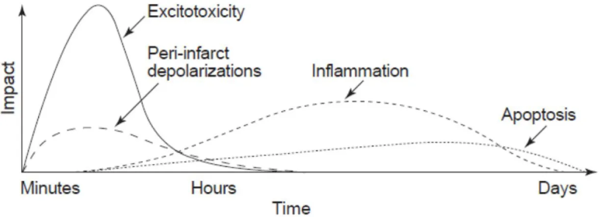
From the beginning of recirculation to the cascading impacts, the ischemic lesion evolves inversely by different mechanisms following the duration of ischemic injury (Fig.1.6).
Figure 1.6: The evolution and putative impact of each element of the cascade in focal
cerebral ischemia [Dirnagl et al. 1999].
1.5.2.10 Edema and permeability change of blood-brain barrier
An important modulator of focal ischemia is brain edema which can be differentiated into two phases: an early cytotoxic type of edema, followed by a late vasogenic type of edema after a period [Sakoh et al. 2003]. The cytotoxic type of edema is threshold dependent. The anoxic depolarization and equilibration of ion gradients across the cell membranes enhance intracellular osmolality, increase the intracellular uptake of sodium and cause cell swelling [Marmarou et al. 2007]. After 4-6 h from ischemia onset, the BBB breaks down and serum proteins start to leak from the blood into the brain parenchyma with the manifestation of necrosis [Gerriets et al. 2009]. This disturbance initiates a vasogenic type of edema which further increases the water content of the tissue. Vasogenic edema reaches its peak at 1-2 days after the onset of ischemia. Under clinical conditions, the malignant cerebral infarction (MCI) form, which terms a large MCA infarction, with or without involvement of the ipsilateral anterior and posterior cerebral artery territories, is the by far most dangerous complication and an indication for decompressive craniectomy due to the elevated intracranial pressure (ICP) or brain herniation [Walz et al. 2002,
15
the extracellular compartment influenced by multiple factors [Juenemann et al. 2015]. It secondarily reverses the decreased extracellular space and could explain the pseudonormalization of the signal intensity observed in diffusion-weighted MRI [Lansberg et al. 2001].
Inhibition of aquaporin water conductance or sodium exchange across sodium channels may reduce the formation or severity of ischemic brain edema [Badaut et al. 2011, Welling et al. 2011]. The gradient of osmotic and ionic concentrations built up during ischemia is regarded as the driving force and key factor for the generation of edema after stroke. However, aquaporin channels may modulate the speed of edema generation, but cannot change the final extent of water accumulation in brain tissue [O'Donnell et al. 2004].
All these potential mechanisms mentioned before will more or less affect the formation and dynamic changes of penumbra, which is the premise of AIS treatment. The fate of the penumbra depends on the extent of reperfusion in the ischemic area. If persistent artery occlusion exists, the infarct will grow constantly and progressively replace the penumbra, as an irreversibly damaged lesion. Under the condition of either spontaneous or post-treatment recanalization, the penumbra will be reversible within the limited time frame (Fig.1.7). A better understanding of penumbra development which converts from microcosmic to macrocosmic level may help to define the time window for clinical therapy and the selection criteria of patients by imaging evidence with the consideration of stroke symptoms.
16
Figure 1.7: Stroke symptoms are reflections of the post-ischemic functional deficit in
the early stage, but not necessarily of the structural lesion volume. Regression of the function occurs in some spontaneous or post-treatment reperfused areas while the structural lesion grows. From the onset of focal ischemia, the core and salvageable tissue are dynamic based on different biochemical mechanisms, adapted from [Dirnagl et al. 1999].
1.6 From emergent to preventive strategies for
ischemic stroke
1.6.1 Target of ischemic stroke therapies
The fundamental objective of stroke treatment is to enable rapid reperfusion for maximal penumbral tissue salvation, which is also the main target of putative neuroprotection therapies [Ebinger et al. 2009] (details mentioned in 1.6.5). It is the conceptual basis not only for the progressive evolution of ischemia, but for the therapeutic reversal of the acute neurological symptomatology arising from ischemic stroke as well [Fisher et al. 2004,
17
1.6.2 Demand of development in stroke therapies
During the past decades, most major stroke trials developed worldwide have failed, including the following:
(1) Large clinical trials of secondary prevention, such as the Warfarin-Aspirin Symptomatic Intracranial Disease Study, the Management of Atherothrombosis with Clopidogrel in High-Risk Patients with Recent Transient Ischemic Attack or Ischemic Stroke, and Clopidogrel for High Atherothrombotic Risk and Ischemic Stabilization, Management and Avoidance studies [Chimowitz et al. 2005, Bhatt et al. 2006];
(2) Recent randomized trials of new thrombolysis agents, such as the Desmoteplase In Acute Ischemic Stroke phase 2 (DIAS-2) and Abciximab in Emergent Stroke Treatment trials (phase 2) [Adams et al. 2008];
(3) Neuroprotection Therapy like the Stroke Therapy Academic Industry Roundtable (STAIR) criteria-guided Neuroprotection Trial (NXY-059) [Shuaib
et al. 2007].
These results indicate the urgent need for measuring the heterogeneity and individualization of patients’ condition due to the complex mechanisms of ischemic stroke. Beyond this situation, alternatives or combination of the traditional therapeutic approaches leading to a high level of specialized care provided by an interdisciplinary team is required.
1.6.3 Thrombolysis therapy
Nowadays, the clinical treatment of the AIS includes two possible methods of therapy: medication, mainly IV thrombolysis and endovascular therapy (details mentioned in 1.6.4). Prompt reestablishment of effective blood flow with lysis of an occlusive thrombus is the goal of thrombolysis. Thrombolytic agents, including streptokinase, urokinase or recombinant tissue plasminogen activator (rt-PA), convert the proenzyme plasminogen to the enzyme active in thrombin lysis, plasmin [Dirks et al. 2012]. The rt-PA, which is fibrin specific and activating only thrombin-bound plasminogen, is the most favorable choice for thrombolytic therapy [Ehlers et al. 2007].
Substantial evidence suggests that efficacy of IV rt-PA in the first 4.5 h from onset of symptoms is higher compared with the efficacy outside the therapeutic window, but with increased risk of hemorrhagic complications as well [Hacke et al. 2008, Shobha et al. 2011]. However, the procedure of IV thrombolysis has a low percentage restoration of cerebral arteries patency so that is characterized by relatively low efficiency [Saqqur et al. 2007, Bhatia et
al. 2010].
To balance the benefits of extended therapeutic time window and the risks of subsequent ischemia-reperfusion injury, it is important to select appropriate patients based on an assessment of individual risks and potential benefits for
18 thrombolysis [Khatri et al. 2012].
In contrast to rt-PA induced recanalization, and probably also to spontaneous recanalization in humans, both of which allow gradual restoration of blood flow to the ischemic area, mechanical opening of an occlusion results in abrupt reperfusion, and thus the mechanical thrombectomy may be the only clinical selection (details mentioned in 1.6.4.2). It may be speculated that this type of ischemia and then reperfusion produces a bias in post-ischemic signaling toward reperfusion-induced secondary mechanisms of injury, which may be amenable to pharmacological intervention even after relatively long time frames [Hossmann et al. 2012]. Results from previous studies provided a huge amount of information on the concerned intervention after stroke. However, some of these results were with low predictive value due to the existed bias which caused by the lack of control on randomization, preset criteria for inclusion and exclusion, blinding and universality of experimental units [Dirnagl et al. 2009].
1.6.4 Endovascular therapy
Recanalization of occluded artery is strongly associated with improved functional recovery and reduced mortality in ischemic stroke [Rha et al. 2007]. Alternative recanalizing approaches, such as endovascular treatment (ET), have been employed for many years and have progressively gained favor profited from the possibility of using a multimodal approach and to the evolving imaging and delivery technologies that have increased safety, efficiency and frequency of recanalization [Ciccone et al. 2014].
ET can be broadly divided into the chemical dissolution of clots with intraarterially delivered thrombolytic agents, and clot retrieval / thrombectomy with mechanical devices. The main forms of endovascular approaches for AIS are illustrated [Asadi et al. 2015] (Fig.1.8). The move to acute intervention for rapid thrombus lysis in the cerebral arterial circuit was highly relevant to the progress by multiple approaches [Jeong H. S. et al. 2014, Zhang et al. 2015].
Figure 1.8: Diagrammatic sketches of main endovascular approaches in clinical trials,
adapted from [Asadi et al. 2015]. The high efficiency of penumbra aspiration system has been confirmed in a Penumbra Pivotal Stroke Trial study for AIS patients within 8 h of symptom onset [Penumbra Pivotal Stroke Trial et al. 2009]. MERCI is a corkscrew shape device with helical Nitinol loops specifically designed and tested for