An enzyme-linked
immunosorbent
assay for diagnostic
detection
of Taenia
saginata
copro-antigens
in humans
P. Deplazes’, J. Eckert’, Z. S. Pawlowski’, L. Machow&* and B. Gottstein’ ‘Institute of Parasitology,
University of Ziirich, Winterthurerstrasse 266a, CH-8057 Ziirich, Switzerland; ‘Clinic of Parasitic and Tropical Diseases, Academy of Medicine, Poznan, Przybyszwewskiego 49, 60-355 Poznan, Poland
Abstract
An immunodiagnostic sandwich enzyme-linked im- munosorbent assav (ELISA) was develowd for the detection of soluble Taeniu saginatu antigens in stool samples (copro-antigens) of infected humans, using affinity-purified polyclonal antibodies obtained from rabbits hyperimmunized with excretory/secretory antigens derived from T. saginata maintained in vitro. Investigation of operating characteristics showed very low cross-reactivity with crude antigens from hel- minths other than Taenia, including Dipylidinm cani- num and Diphyllobothrium latnm. The specificity of the assay was 95% when testing stool samples from 100 persons who were either infected with Ascaris lumbri- coides, Trichuris trichiura, hookworms, Enterobius vemncularis or Hymenolepis nanu, or who had no intestinal hehninthosis detected. Analysis of diagnos- tic sensitivity demonstrated that in 85% of 34 samples from 23 untreated persons with intestinal T. saginatu infection (selected by previous proglottid and/or egg detection) copro-antigens were detected by the T. saginata ELISA. In the same samples, Taenia eggs were detected in 62%. Only 41% of the samples reacted positively in a heterologous T. hydutigenu ELISA. Post-treatment control revealed a high con- centration of T. suginatu copro-antigens for l-4 d after administration of niclosamide or praxiquantel, and negative values 9-17 d after treatment. The Tueniu copro-antigens remained detectable by ELISA even after storage of untreated faeces at 25°C for at least 5 d.
Introduction
Infections of humans with Tueniu suginatu or T. solium are of medical and veterinary importance as the larval forms of these two species cause cysticercosis in cattle or in pigs and man, respectively. Efficient control of these zoonotic infections is hampered by many factors, including unsatisfactory methods of diagnosing intestinal Tuenia infections in man; it is almost impossible to diagnose early infection before proglottids and/or eggs start to be excreted. During patency proglottids and eggs are irregularly excreted and the available techniques for faecal examination have a relatively low sensitivity. Although the perianal swab method for egg detection gives somewhat better results, the sensitivity is variable and estimated to be between 35% and 85% (HALL et al., 1981; KIRICHEK et al., 1986). Therefore, new diagnostic tools with a higher diagnostic sensitivity are needed for detecting Address for correspondence: Dr P. Deplanes, Institute of Parasitology, University of Ziirich, Winterthurerstrasse 266a, CH-8057 Ziirich, Switzerland.
the infection in individuals, for assessing efficacy of chemotherapy and for screening larger populations i;;~ 1985; CRUZ et al., 1989; PAWLOWSKI, The diagnostic detection of parasite antigens in stool samples (copro-antigens) by enzyme-linked im- munosorbent assay (ELISA) was initially applied for the diagnosis of Giardia lamb& and Entamoeba histolytica infections in humans (GRUNDY, 1982; UN- GAR et al., 1984, 1985; BAUMANN & GOTTSTEIN, 1987). MACHNICKA & KRAWCZUK (1988) and ALLAN & CRAIG (1989) demonstrated the detection of Hymen- olepis-specific antigens in faeces of rats infected with H. diminutu. For the detection of Taenia copro- antigens, a sandwich ELISA using affinity-purified polyclonal rabbit antibodies directed against excre- tory/secretory (E/S) antigens from adult T. hydatigenu was developed (DEPLAZES et al., 1990). This test allowed the diagnosis of prepatent and patent infec- tions in 6 dogs with experimental T. hydutigenu infection. The test exhibited a high degree of specific- ity at the genus level. Recently, ALLAN et al. (1991) described two ELISAs for the detection of T. solium and T. suginara antigen in faeces of golden hamsters infected with T. solium. Both tests, using immunoglo- bulin fractions directed against somatic parasite anti- gens, also recognized Tuenia-specific antigens in stool samples of 11 persons infected with T. suginatu or T. solium .
In this paper we describe an ELISA detecting T. suginatu copro-antigens using affinity-purified anti- bodies against T. saginatu E/S-antigens.
Materials and Methods Stool samples
Stool samples were collected from the following groups of persons.
Group I. 23 persons (17 from Poland and 6 from Switzerland) infected with T. suginuru; 34 samples were collected before specific chemotherapy and 16 samples 1-17 d after therapy with niclosamide or praxiquantel (for details see the Table). The patients were selected on the basis of proglottid excretion reported in their history (shortly before sampling stool specimens). Specific identification of proglottids relied upon routine parasitological criteria.
Group 2. A negative control group of 40 healthy persons (20 from Poland and 20 from Switzerland), not infected with T. suginuta (contirmed by no report of proglottid excretion and by routine stool examina- tion with sedimentation/flotation technique).
Group 3. 100 stool samples (40 from Swiss persons and 60 from immigrants) submitted to our Institute by physicians for routine coproscopical examination.
392
Fresh stool samples were collected (groups 1 and 2) and immediately mixed in a 1:2 ratio with phosphate- buffered saline (PBS) containing 0.04% NaNs, 0.05% bovine haemoglobin (Fiuka) and 0.3% Tween 20*. These samnles were stored at -20°C for 1 to 4 weeks until further processing. The thawed suspensions were ultrasonicated for 30 set (40 W) and subse- quently sedimented at 3000 g for 10 min before use in the ELISA. One of these negative samples (desig- nated as control ‘stool-in-buffer suspension,’ SBS) was subsequently used for analysis of the sensitivity of the method in recovery experiments (briefly referred to as ‘sensitivity’). Stool samples of group 3 were suspended in the buffer solution described above on arrival at the Institute (after l-3 d in transit without any preservation or buffer solution). All supernatants were frozen at -20°C.
Coproscopical examination
The coproscopical examination was performed by an established sedimentation/flotation technique (BOCH & SUPPERER, 1983) on the sediments of the same samples (2 g) using zinc chloride (specific gravity 1.3) as flotation medium. Preliminary experi- ments had shown that the embryophore of Taenia eggs in SBS was not destroyed by ultrasound treat- ment (see above).
Antigens
T. saginata EIS antigens. Part of a living T. saginata strobila with gravid proglottids and without scolex (spontaneously excreted from a patient) was washed with PBS and maintained at 37°C with air as gas phase in a 75 cm* tissue culture flask (Corning Glass Works, no. 25110) containing 200 ml serum-free Eagle’s Minimal Essential Medium (EMEM) (Gibco. no-072 1100) with D-glucose (4 mgjml), ge&amycin (200 pg/ml) and Fungizone@ (250 r&ml), pH 7.2. The medium was replaced after 4, 10, 18 and 28 h of incubation, then every day up to day 10. The collected batches of tissue culture medium (TCM) (from days 3 to 6) were stored at -20°C until further processing. The viability of the strobila was judged by its motility. The sterility of the cultures was tested on day 3 of maintenance according to standard bacter- iological procedures.
E/S antigens were dialysed and concentrated from the collected TCM to 0.7 mg protein per ml using an Amicon@ untrafiltration unit, a YM-10 membrane and PBS. All protein concentrations were assessed by a Bio-Rad* nrotein assav with bovine albumin as standard. The T. saginata E/S antigens were used in the antigen-detecting ELISA (= T. saginata ELISA) as described below (see ‘anti-T. saginata E/S hvperim-
munoglobulins’). __
The following antigens were used for the deter- mination of specificity.
Heteroloaous helminth somatic and metabolic antiwns. Somatic and metabolic antigens prepared from the adult stage of various helminth species were used for the evaluation of the specificity of the test system. The various helminth species as well as the host origins are listed in Fig. 2.
Non-helminth antigens. Crude extracts were obtained from Escherichia cob strain Y 1089. from Entamoeba histolytica strain HK9 (ICN Medica Di- agnostic Products, Covina, California, lot no. 4729)
and from trophozoites of Giardia lamblia (Swiss bovine strain) (ROHRER et al., 1987). Mammalian antigens included bovine and human serum diluted l:SO, cow’s milk diluted 1:2 with PBS-Tween to@, and muscle extracts of calf, chicken, and swine.
All freshly obtained materials were processed as described by DEPLAZES et al. (1990).
Anti-T. saginata hyperimmunoglobulins
A rabbit was hyperimmunized as described by BAUMANN & GOTTSTEIN (19871, but usinn T. saainata E/S antigens for immunization. All other steps, including the coupling of T. saginata EIS antigens to CNBr-activated Sepharose@ 4B, the purification of antibodies by affinity chromatography, the prepara- tion of rabbit anti-T. saginata E/S conjugates (alkaline phosphatase), and the preparation of normal rabbit immunoglobulin G (IgG) for control reactions were done according to the procedures described by BAUMANN & GOTTSTEIN (1987).
Enzyme-linked immutwsorbent assay
A sandwich ELISA was devised for the detection of T. saginata antigens in human stool samples. Techni- ques and concepts were exactly as previously elabo- rated for the T. hydatigena copro-antigen test (DE- PLAZES et al., 1990), and can be summarized briefly as follows. (i) Affinity-purification of T. saginata copro- antigen-specific catching (solid phase) antibodies (from hyperimmunized rabbits), in parallel with inclusion of irrelevant rabbit catching (solid phase) IgG as a control; (ii) antibody-copro-antigen immune reaction; (iii) visualization of immune reaction bv using affinity~purified antibody (liquid phase) as in (3 but labelled with alkaline nhosnhatase and the corres- ponding chromogenic substrate solution. The results are expressed as the absorbence at 405 nm (bsnm) and controlled with reference to standard uositive and negative samples run in triplicate. When the results obtained with control rabbit IPG amounted to >30% of a positive A405nm value of thi specific IgG reaction, the sample run in question was discarded. This T. saginatu ELISA was evaluated as described below; the previously described T. hydatigena ELISA was used for comparison.
For the assessment of potential cross-reactivity and non-specific reactions in the ELISA, the antigens of various helminths, urotozoa, bacteria and mammalian control tissues (Fig: 2) were tested at a concentration of 10 ue nrotein ner ml of the control SBS: human and b&&h serum’ were used diluted 1:50 and cow’s milk 1:2. The procedure for the evaluation of the assays was carried out as listed in the following experimental design.
(i) Determination of a significant threshold in the ELISA, discriminating between positive (= T. sugina- ta ant&ens detectable) and negative (no T. saainatu antigen detectable) in both systems. ’
(ii) Determination of the sensitivitv of the T. sa&ata ELISA and the T. hydatigenh ELISA in recovery experiments using stool samples diluted 1:2 and known protein concentrations of T. saginata metabolic E/S antigens.
(iii) Determination of the snecificitv of the T. sa&natu ELISA using SBS containing known protein concentrations of somatic or metabolic antigens
obtained from various heterologous parasite species, bacteria, or mammalian control tissues.
(iv) Definition of diagnostic sensitivity and specific- ity, etc., by testing stool samples from persons infected with T. s&aura before and after specific chemotherapy, from persons with other hehninth infections, or from parasite-free persons.
(v) Examination of the potential stability of copro- antigens in stool samples under different storage conditions.
Results
Sensitivity of the ELISA
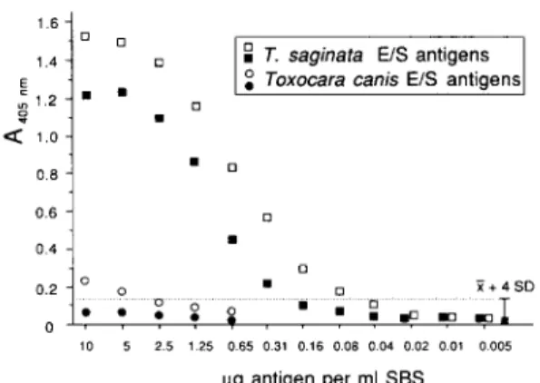
In order to determine the sensitivity of the ELISA, T. saginata E/S antigens were mixed with negative SBS and then diluted in serial 2-fold dilutions with the same SBS. These samples were tested by ELISA using anti-T. hydatigenu and anti-T. saginata-anti- bodies (T. hydatigena and T. saginata ELISA, respec- tively) (Fig. 1). The threshold (cut-off point) for both assays for discriminating a negative from a positive reaction was determined by calculating the mean AM5,,,,, value (Z) of the 40 stool specimens from the negative control group 2 plus 4 standard deviations (SD). According to this calculation, both test systems showed the same background reactions. The T. hydutigena ELISA (DEPLAZES et al., 1990) detected at
least 0.2 pg of T. saginatu E/S antigens per ml of diluted (1:2) SBS; the newly developed T. suginatu ELISA had a higher sensitivity, detecting a lower limit of 0.07 pg, equivalent to 140 &ml of stool. All control reactions with normal rabbit IgG were very low (AMsnm<0.04).
Specificity of the ELISA
The results of the specificity evaluation with defined protein concentrations in control SBS are shown in Fig. 2. The antibody reactivities with all non-hehninth antigens were negligible compared to the Tueniu antigens tested. Toxocaru canis E/S anti- gens from adult stages showed cross-reactivity at high antigen concentrations (l&2.5 pg/ml of stool sample) in the T. sugi~za ELISA (Fig. 1) but not in the T. hydutigena ELISA. All antigens of the other investi- gated helminth species showed no significant reaction in the assay.
Diagnostic sensitivity and specificity of the ELISA In the T. saginatu ELISA (Fig. 3), the range of negative reactions was determined by testing 40 stool samples from a hehninth-free group (group 2). The bsnm values obtained in the control reactions for both assays with purified irrelevant rabbit control IgG (mean AMsnrn value=0*02, range 0*01-0.04) were subtracted from the values obtained with rabbit anti-T. hydatigena E/S and anti-T. saginata E/S antigens, respectively. The mean values of these corrected data plus 4 SD amounted to &s-=0.14 (Y=O*O27, SD=0.027) and Ah05,,=0.14 (‘Z=O.O2!, SD=0*028), respectively, and were arbitrarily consl- dered as the cut-off limit between specific antigen- detecting reactions and background reactions.
Twenty-three specimens from 23 persons infected with T. suginatu were collected before treatment (only the initial sample from each person was used for the present study), examined for copro-antigens with the two ELISAs mentioned above, and investigated in
Fig. 1. Titration of (homologous) Tamia saginafa and control Toxocara canis excretory/secretory (E/S) antigens artificially diluted in human control stool-in-buffer solution (SBS). Testing was carried out with T. sagi~ta (0,O) and T. hydatigena ELISA (B, 0). The values obtained in the control reaction with purified irrelevant rabbit control IgG were subtracted from the values obtained with specific rabbit anti-T. raginnra- or anti-T. hydutigena E/S antigen Ig. The cut-off point (---) was determined in both systems by calculating the mean absorbance at 405 nm of 40 negative control samples plus four standard deviations.
Excretory/secretory antigens (E/SAG)
or somatic antigens (SAG) 0 A 405 “n. 10
Taenia saginata E/SAG (man) T. saginata SAG
T. hydatigena E/SAG (dog) Echinococcus granulosus SAG (dog) Dipylidium caninum SAG (man) Diphyllobofhrium /aturn S-AG (man) Toxocara canis E/S-AG (dog) Ascaris lumbricoides SAG (man) Trichuris ovis SAG (goat) Schistosoma sp. S-AG (man)
Fasciola hepafica S-AG (sheep) non helminths control antigens control stool-in-buffer suspension stool samples of 40 control
persons negative for helminths i + 4 SD ~W-011 PO’“,
,
Fig. 2. Specificity evaluation of the Taenia sa&zca ELISA with ldpg antigen f&m helminth and non-helm& (somatic antigens from Giardia kmblia, Entamoeba hirtolytica, Escherichia coli, calf, swine, chicken and human muscle; 150 diluted calf and human serum, and 1:2 diluted cow’s milk) per ml of control stool-in-buffer suspension. The host origin of the antigens is given in parentheses. Aws,,=absorbance at 405 nm.
parallel by a sedimentation/flotation technique for Tueniu eggs. Tuenia eggs were detected in 17 samples (74%); copro-antigens were detected by T. saginata ELISA in 20 samples (87%) (Fig. 3). One of the 3 samples was negative for copro-antigen detection, although a few Taenia eggs had been found by routine examination. In the same group of 23 specimens, 13 (57%) samples were positive in the T. hydutigena ELISA (data not shown).
Samples from group 3 were also examined for eggs and copro-antigens with the T. suginata ELISA (Fig.
3). Three of 40 samples from Swiss persons were positive in the T. saginata ELISA, and Taenia eggs were found in one of them. The other samples were free of helminth eggs. Three of 60 samples from immigrants (4 infected with Hymenolepis nana, 2 with Enterobius vermicularis, 4 with Trichuris trichiura? 11 with hookworms, 5 with hookworms and T. trichtura, 2 with Ascaris lumbricoides and T. trichiura and one with A. lumbricoides and hookworms) gave positive reactions for copro-antigens in the T. saginata ELISA (Fig. 3). In 2 of these cases, helminth infection could not be detected; the third case had an infection with A. lumbricoides and T. trichiura. The diagnostic specificity of the T. saginata ELISA was thus 95% (5
.
0 no Taenia eggs
0
A B ai P
negative controls Fig. 3. Diagnostic sensitivity and specificity of the Taenio raginata ELISA with the following stool samples (with multiple samples, only the first was included): (A) from 23 persons with proven T. saginata infection (group 1); (B) from 100 persons (group 3) with the following species of eggs detected at routine coproscopical examination: T.
saginata (n=l), Hpwmlqis mma (n=4), Emmobius vtwnicularis
(n=4), Trichuris oichiura (n=4), hookworms (n= 11) and mixed infections with hookworms, T. trichiura and Ascaris lumbricoides
(n=8). The cut-off point (---) was determined by calculating the mean absorbance at 405 nm (A,,,,) of 20 Swiss (CH) and 20 Polish (P) negative control samples (group 2) plus four standard deviations.
positive results without proven T. saginata infection among 100 samples originating from different per- sons).
Reproducibility of test results and antigen detection after treatment
The diagnostic reproducibility of a positive result was tested by examination of l-3 samples per infected person before treatment (Table). The data show an acceptable degree of reproducibility in antigen detec- tion. Considering all data, including those from a single examination and from repeated examinations, the following diagnostic sensitivities were calculated: 62% for Taenia egg detection; 85% for copro-antigen detection by T. saginata ELISA; and 41% by T. hydatigena ELISA. T. saginata ELISA was positive with 9 of 13 samples (69%) from infected persons without egg detection, and with T. hydatigena ELISA copro-antigens were detected in only one sample. Eleven samples from 11 persons were all positive in the T. saginata ELISA during l-4 d after treatment, whereas no copro-antigen was detected in any samples 9-17 d after treatment (Table).
Storage conditions of samples
The stability of the copro-antigens in stool samples (diluted 1:2 with buffer solution) was tested with 4 positive specimens. After 5 d storage at room temper- ature, all samples were still positive. The mean value of ANY,,,,, decreased by 9%. Four negative specimens, either fixed in buffer or without buffer solution, remained negative after the same exposure time.
Discussion
Man is the only natural final host for both T. saginata and T. solium. T. saginata, a relatively harmless intestinal parasite of humans, causes in its metacestode form bovine cysticercosis which induces considerable economic losses throughout the world. T. solium, inhabiting the intestine of humans, also has a low pathogenicity in its adult stage, but may cause cysticercosis in pigs as natural intermediate hosts and in humans as accidental hosts. Among various forms of clinical cysticercosis of man, neurocysticercosis represents. the most serious form and is potentially fatal.
Table. Comparison of Taenia egg versus copro-antigen detection in stool samples of persons with T. saginata infection
No. of samples with positive result No. of No. of
Pesons with T. saginara infection persons samples Before treatment
Examination with one sample (Switzerland)
8 8
Examination with one sample (Poland)
Exarn+a$on with 2 samples (Poland) 10
~ounauon wnh 3 samples (Poland) ;
23 3:
After treatment
1-l d after 11 11
9-7 d after treatment 5 5
‘2 persons with both negative egg and copro-antigen results.
bathe 3 samples corresponded to 2 persons.
‘All positive reactions were positive in the T. saginata ELISA.
*nd=not done.
Taenia egg
Copro-antigen Copro-antigen
(T. saginata
detection ELISA) CT. Eh{;;“ym
4 6 : 6” Ea ; 2:‘(62%) 2; (85%) 14 (41%) 10 11 (100%) nd* 0 0 nd*
At present, the only available practical ways to control these two zoonotic parasites are (i) the detection and treatment of carriers and (ii) socio- hygienic measures to prevent infection of man and intermediate host animals.
Immunization of intermediate host animals appears potentially feasible but vaccines are not yet available (MITCHELL, 1989). An indispensable prerequisite for the control measures listed above and for treatment of individual patients is efficient and correct diagnosis. This is at present based on the detection of proglottids and/or eggs by faecal examination or on egg detection by perianal swab. These methods are not very sensitive (see the Introduction) and, furthermore, have the disadvantage that parasites during prepaten- cy, or during patency but lacking proglottid or egg excretion, cannot be readily diagnosed. An alternative immunological approach is the detection of antibodies in serum samples from persons serving as definitive hosts. FLENTJE & PADELT (1981), using the indirect immunofluorkcent antibody test;.found a diagnostic sensitivity of 56% in 200 persons infected with T. saginata. In general, and with respect to other cestode and host species (GASSER~~ al., 1988; HEATH et al.,
1985; JENKINS & RICKARD, 1985, 1986a, 1986b),
antibody detection did not allow individual diagnosis of infected definitive hosts, but it matched and reflected the parasite prevalence in populations CTENKIN~ et al.. 1990: G~TT~TEIN et al.. 1991).
\” The present iaper dkmonstrates that, oierall, k5% of 34 samples from 23 untreated persons with intestinal T. saginata infection contained copro- antigens detectable by a sandwich ELBA using affinity-purified polyclonal antibodies. These anti- bodies had been generated in rabbits by hyperimmu- nization with E/S antigens released in vitro by adult T. saginata. In the same group of patients, Taenia eggs were found in only 62% of the cases at a single examination. With the heterologous T. hydatigena ELISA, copro-antigens were detected in only 41% of the same samples.
There are still unknown features to be investi- gated-the diagnostic sensitivity/cross-reactivity inde- pendence of parasite species (i) used for generating hyperimmune antibodies or (ii) targeted in the sandwich ELISA for diagnosis, which may also be dependent upon the stage of infection (e.g. patent or not) and other criteria. ALLAN et al. (1991) used a T. sagimzra copro-antigen ELISA and observed cross reactions with stool specimens from human patients infected with intestinal T. solium (with egg excretion), whereas the same infection in hamsters, but without egg excretion, gave negative test results. In general, we found that stool specimens with large numbers of T. saginata eggs had very high activity in the copro-antigen ELISA (diagnostic sensitivity 95%; some specimens were still positive when diluted to 1:512; data not shown). Even with diagnostically interesting samples from patients with proven taeniasis but without detectable egg excretion, we obtained a diagnostic sensitivity of 69%. These data indicate the need to include samples from infected patients without egg excretion in trials, which more reliablv reflects the diagnostic sensitivitv under routin; field conditions. W< conclude that asiessment of diagnostic sensitivitv of the couro-antigen ELISA tested-exclusively on infected patients excreting eggs might be too optimistic.
The specificity of the present T. saginata copro- antigen ELISA was high: 95% when testing faecal samples from persons infected with Hymenolepis nana or with various nematode species. Cross-reactivity occurred with T. hydatigena antigen and with faecal samoles from does infected with T. hvdatimnu (data not -shown), but this infection does- not-occur in humans. The specificity of the present test thus operated at the genus level; we are currently assessing its potential for diagnosing carriers of adult T. s&m.
After having previously demonstrated that T. hydatigena metabolic copro-antigens were detectable during the prepatent period in experimentally in- fected dogs (DEPLAZES et al., 1990), we have subse- quently shown in the present study that the same applies to T. saginata copro-antigens in persons without parasite egg excretion. We are now addres- sing the problem of specificity by (i) identification and characterization of parasite epitopes of copro-antigens and (ii) generation of monoclonal antibodies against these epitopes for the development of species-specific tests. In this context we have to mention another option for the differential diagnosis of Taenia species, the use of deoxyribonucleic acid (DNA) probes and the polymerase chain reaction (HARRISON et al., 1990; GOTTSTEIN et al., 1991 and in press), which, howev- er, requires proglottids or parasite eggs as a source of the diagnostic DNA template. All the new immunolo- gical or molecular biological tools discussed above clearly have the potential of being further developed to provide test kits applicable under field conditions and may prove very useful as diagnostic tools in the near future.
Acknowledgements
We thank Mrs J. S. Skaggs and I. Tanner for technical assistance, and Dr K. Wolff and F. Azzilonna for providing routine diagnostic samples. The studies were partly sup- ported by a grant from the Parasitic Diseases Programme, World Health Organization.
References
Allan, J. C. & Craig, P. S. (1989). Coproantigens in gut tapeworm infections: Hymenolepis diminuta in rats. Para- sitology Research, 76, 68-73.
Allan, J. C., Avila, G., Garcia Nova& J., Flisser, A. & Craig, P. S. (1991). Immunodiagnosis of taeniasis by coproan- tigen detection. Parasitology, in press.
Baumann, D. & Gottstein, B. (1987). A double-antibody sandwich ELISA for the detection of Entamoeba histolyti- ca antigen in stool samples of humans. Tropical Medicine and Parasitology, 38, 81-85.
Both, J. & Supperer, R. (1983). Vewritziinnedizinische Parasiwlogie, 3th edition. Berlin: Parey.
Cruz, M., Davis, A., Dixon, H., Pawlowski, Z. S. & Proano, J. (1989). Operational studies on the control of Taenia solium taeniasislcysticercosis in Ecuador. Bulletin of the World Health Organization, 67, 401407. Deplazes, P., Gottstein, B., Stingelin, Y. & Eckert, J.
(1990). Detection of Taenia hydatigena coproantigens by ELISA in dogs. Veterinay Parasitology, 36, 91-103. Flentje, V. B. & Padelt, H. (1981). Wert einer serologischen
Diagnostik der Taenia saginata infestation des Mens- then. Angewandte Parasiwlogie, 22? 65-68.
Flisser, A. (1985). Cysticercosis: a major threat to human health and livestock production. Food Technology, 39, 61-64.
Gasser, R. B., Lightowlers, M. W., Obendorf, D. L.,
Jenkinq, D. J. & Rickard, M. D. (1988). Evaluation of a serological test system for the diagnosis of natural Echinococcus pranulosus infection in dogs using E. .vanu- losus protoscolex and oncosphere aniigens.-Ausiralian Veterinary 3ouma1, 65, 369-373.
Gottstein, B. & Mowatt, M. R. (1991). Sequencing and characterization of an E. multilocularis DNA urobe and its use in the polymerase chain reaction. M&cular and Biochemical Parasitology, 44, 18%194.
Gottstein, B., Deplaxes, I’:, Tanner, I. & Skaggs, J. S. (1991). Diagnostic identification of Taenia saginara with the polymerase chain reaction. Transactions of the Royal Society of Tropical Medicine and Hygiene, 85, 248-249.
Grundy, M. S. (1982). Preliminary observations using a
multi-layer ELISA method for the detection of En-
zamoeba histolytica trophozoite antigen in stool samples. Transactions of the Royal Society of Tropical Medicine and Hygiene, 76, 396-400.
Hall, A., Latham, M. C., Crompton? D. W. T. &
Stephenson, L. S. (1981). Taenia sagtnata (Cestoda) in western Kenya: the reliability of faecal examinations in diagnosis. Parasitology, 83, 91-101.
Harrison, L. J. S., Delgado, J. & Parkhouse, R. M. E. (1990). Differential diagnosis of Taenia saginata and
Taenia solium with DNA probes. Parasitology, 100,
459-461.
Heath, D. D., Lawrence, S. B., Glennie, A. & Twaalhoven, H. (1985). The use of excretory and secretory antigens of the scolex of Taenia or~is for the serodiagnosis of infections in dogs. 3ournal of Parasitology, 71, 192-199. Jenkins, D. J. & Rickard, M. D. (1985). Specific antibody response to Taenia hydatigena, Taenia pisiformis and Echinococcus granulosus infectton in dogs. Australian Veterinary Journal, 62, 72-78.
Jenkins, D. J. & Rickard, M. D. (1986a). Specificity of
scolex and oncosphere antigens for the serological
diagnosis of taeniid cestode infections in dogs. Australian Veterinary Journal, 63, 40-42.
Jenkins, D. J. & Rickard, M. D. (1986b). Specific antibody responses in dogs experimentally infected with Echino- coccus granulosus. American 3oumal of Tropical Medicine and Hygiene, 35, 345-349.
Jenkins, D. J., Gasser, R. B., Zeyhle, E., Romig, T. & Macpherson, C. N. L. (1990). Assessment of a serologic- al test for the detection of Echinococcus granulosus infection in dogs in Kenya. Acta Tropica, 47, 245-248. Kirichek, V. S., Nikitin, A. S., Frolova, A. A. & Yarotskii, L. S. (1986 2.. Some aspects of the biology of the northern strain of aema sagmata. Meditsinskaia Parazitologiia i Paran’tarnye Bole&, 6, 37-39.
Machnicka, B. & Krawczuk, S. (1988). Hymenolepis diminu- ta antigen: detection in faeces of rats. Bulletin of the
~;~~06Academy of Sctences, Baological Scwnces, 36,
Mitchell, G. F. (1989). Problems specific to parasite
vaccmes. Parasitology, 98, S19-S28.
Pawlowski, S. P. (1990). Cestodiases: taeniasis, cysticer- cosis, diphyllobothriasis, hymenolepiasis, and others. In: Tropical and Geographical Medicine, Warren, K. S. & Mahmoud, A. A. F. (editors), 2nd edition. New York: McGraw-Hill, pp. 490-504.
Rohrer, L., Winterhalter,, K. H., Eckert, J. & Kohler, P. (1987). Killing of Gtardia lamblia by human milk is mediated by unsaturated fatty acids. Antimicrobial Agents and Chemotherapy, 30, 254-257.
Ungar, B. L. P., Yolken, R. H., Nash, T. E. & Quinn, T. C. (1984). Enzyme-linked immunosorbent assay for the detection of Giardia lamblia in fecal specimens. Journal of Infectious Diseases, 149, 9&97.
Ungar, B. L. P., Yolken, R. H. & Quinn, T. C. (1985). Use of a monoclonal antibody in an enzyme immunoassay for the detection of Entamoeba histolytica in fecal specimens. t6~4;~n 3oumal of Tropical Medicine and Hygiene, 34,
Received 14 December 1990; accepted for publication 1.5